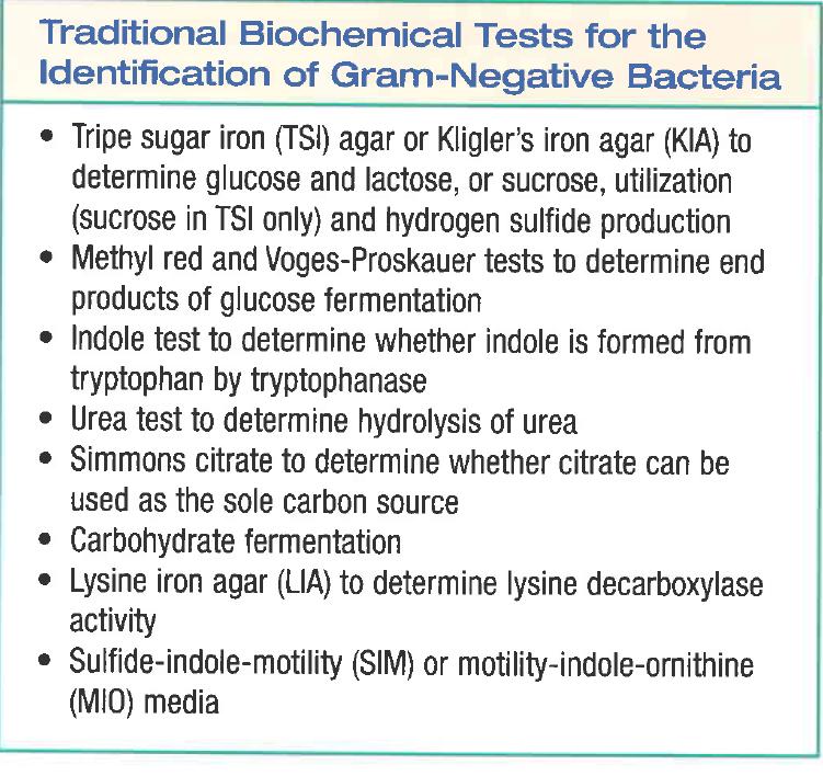
Principles of biochemical tests commonly used in the identification of gram-negative bacteria
|
|
|
- Juliana Lambert
- 6 years ago
- Views:
Transcription
1 Dr. Khoramrooz 1 In the name of God Department Of Microbiology Yasouj University of Medical Science Principles of biochemical tests commonly used in the identification of gram-negative bacteria By: Dr. S. S. Khoramrooz, Ph.D. Department of Microbiology, Faculty of Medicine, Yasuj University of Medical Sciences, Yasuj, Iran
2 Introduction Historically, the identification of bacteria based on: Colony morphology, Gram staining, Biochemical testing. Based on the phenotype of microorganisms This method required a lot of time and lots of space in incubators. Over time, biochemical tests were miniaturized and multitest systems were developed Resulting in time and space savings. More recently, automated systems have been developed Dr. Khoramrooz 2
3 Clinical microbiology entered the next generation of test systems for the identification of bacteria: Molecular biology assays. Based on nucleic acid sequences, are highly sensitive, specific, and rapid, thereby providing accurate results in a few hours or less. More accurate than examining the phenotype. In this lecture discuss the principles of biochemical tests commonly used in the identification of gram-negative bacteria. Dr. Khoramrooz 3
4 Characters of Enterobacteriaceae All Enterobacteriaceae Gram-negative rods Ferment glucose with acid production Reduce nitrates into nitrites Oxidase negative Facultative anaerobic Motile except Shigella and Klebsiella Non-capsulated except Klebsiella Non-fastidious Grow on bile containing media (MacConkey agar) Dr. Khoramrooz 4
5 Classification of Enterobacteriaceae Enterobacteriaceae Lactose fermenters E. coli, Citrobacter, Klebsiella, Enterobacter Non-lactose fermenter Salmonell, Shigella Proteus, Yersinia There are several selective and differential media used to isolate distinguishes between LF & LNF The most important media are: MacConkey agar Eosin Methylene Blue (EMB) agar Salmonella Shigella (SS) agar In addition to Kiligler Iron agar (KIA) Dr. Khoramrooz 5
6 Tests To Know Case Study Tests Indole Methyl Red/Voges Proskauer Citrate H 2 S production in SIM Urea hydrolysis Motility Lactose fermentation Glucose fermentation & gas production Decarboxylation of amino acis Fermentation of sugars Reaction on selective media Dr. Khoramrooz 6
7 Dr. Khoramrooz 7
8 Selection of Primary Isolation Media Used to recover the significant species of bacteria from specimens that may harbor a mixture of microorganisms. Microbiologists must know the composition of each formula and the purpose and relative concentration of each chemical or compound that is included. Not sufficient to know that bile salts are included in selective media In SS agar contains about 5 times the concentration of bile salts compared with MacConkey agar and is more inhibitory to E.coli and more selective for the recovery of Salmonella species from stool cultures. Dr. Khoramrooz 8
9 For the recovery of the Enterobacteriaceae from clinical, three general types of media are available: 1) nonselective media for primary isolation (e.g., blood agar); 2) Selective or differential agars (e.g., MacConkey and Hektoen enteric agars); 3) enrichment broths. Dr. Khoramrooz 9
10 Selective Isolation Media MacConkey and EMB agars are only moderately inhibitory and are designed primarily to prevent growth of gram positive bacteria from mixed cultures. Dr. Khoramrooz 10


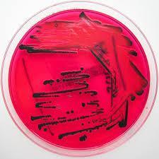
11 Growth of Enterobacteriaceae on MacConkey agar Colorless colonies Pink colonies Uninoculated plate Lactose non feremters Lactose feremters Salmonella, Shigella, E. coli, Citrobacter Proteus Klebsiella, Enterobacter Dr. Khoramrooz 11
12 MacConkey Agars Intended Use MacConkey agars are slightly selective and differential plating media mainly used for the detection and isolation of gram-negative organisms from clinical, dairy, food, water, pharmaceutical, cosmetic, and other industrial sources. MacConkey Agar is used for isolating and differentiating lactose-fermenting from lactose-nonfermenting gramnegative enteric bacilli. MacConkey Agar Base is used with added carbohydrate in differentiating coliforms based on fermentation reactions. Dr. S. S. Khoramrooz 12
13 MacConkey Agar without Crystal Violet is used for isolating and differentiating enteric microorganisms while permitting growth of staphylococci and enterococci. The medium can be used also to separate Mycobacterium fortuitum and M. chelonae from other rapidly growing mycobacteria. MacConkey Agar without Crystal Violet or Salt and MacConkey Agar without Salt are used for isolating and differentiating gram-negative bacilli while suppressing the swarming of most Proteus species. Dr. S. S. Khoramrooz 13
14 Principles of the Procedure Lactose is a fermentable carbohydrate. When lactose is fermented, a local ph drop around the colony cause a color change in the ph indicator (neutral red) and bile precipitation. Dr. S. S. Khoramrooz 14
15 Bile salts, bile salts no. 3, oxgall and crystal violet are selective agents that inhibit growth of gram-positive organisms. Magnesium sulfate is a source of divalent cations. Agar is the solidifying agent. Dr. S. S. Khoramrooz 15
16 Dr. S. S. Khoramrooz 16
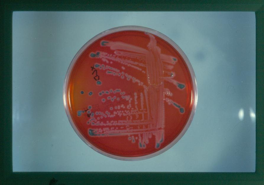
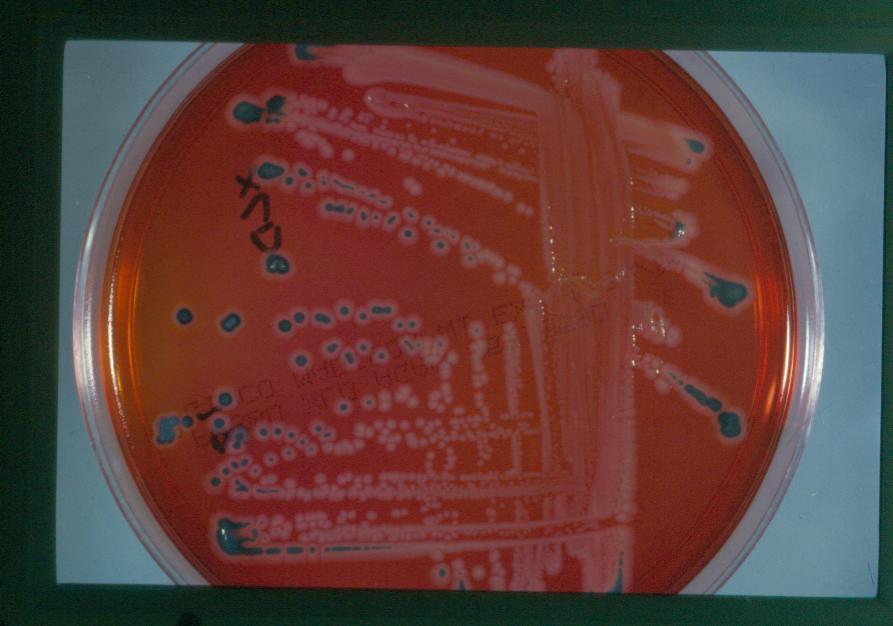
17 Expected Results Lactose-fermenting organisms grow as pink to brick-red colonies with or without a zone of precipitated bile. Lactose-nonfermenting organisms grow as colorless or clear colonies. Swarming by Proteus spp. is reduced on MacConkey agars without salt. Dr. S. S. Khoramrooz 17
18 Dr. S. S. Khoramrooz 18
19 Limitations of the Procedure 1. Although MacConkey media are selective primarily for gram-negative enteric bacilli, biochemical and, if indicated, serological testing using pure cultures are recommended for complete identification. 2. Incubation of MacConkey Agar plates under increased CO2 has been reported to reduce the growth and recovery of a number of strains of gram-negative bacilli. Dr. S. S. Khoramrooz 19
20 Intended Use Eosin Methylene Blue Agar, Levine is a slightly selective and differential plating medium for the isolation of gramnegative enteric bacteria. Principles of the Procedure The eosin Y and methylene blue dyes in Levine EMB Agar render the medium slightly selective in that they inhibit gram- positive bacteria to a limited degree. Dr. S. S. Khoramrooz 20
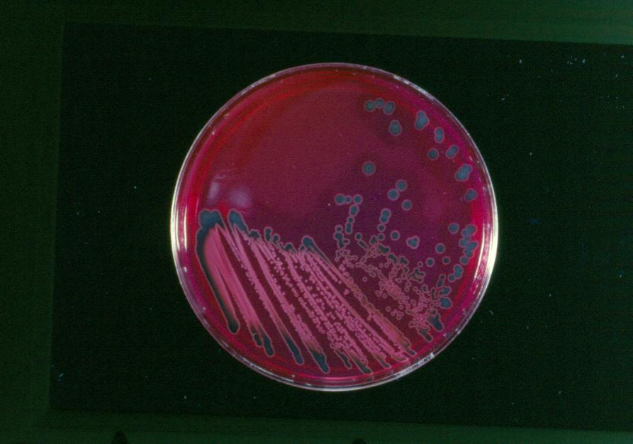
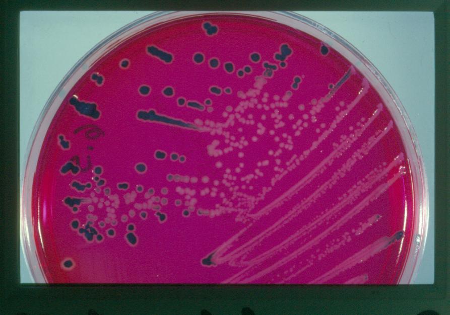
21 These dyes also play a role in differentiating between lactose fermenters and lactose nonfermenters due to the presence or absence of dye uptake in the bacterial colonies. Coliforms, as lactose-fermenting organisms, are visualized as blue-black colonies, whereas colonies of Salmonella and Shigella, as lactose nonfermenters, appear colorless, transparent or amber. Some gram-positive bacteria, such as fecal streptococci, staphylococci and yeasts, will grow on this medium and usually form pinpoint colonies. Dr. S. S. Khoramrooz 21

22 Dr. S. S. Khoramrooz 22
23 Expected Results Typical colonial morphology on Eosin Methylene Blue Agar, Levine is as follows: Dr. S. S. Khoramrooz 23

24 Dr. S. S. Khoramrooz 24
25 Highly Selective Isolation Media Used Primarily for Gastrointestinal Specimens Media are made highly selective by the addition of a variety of inhibitors to their formulas, generally in higher concentrations than in MacConkey and EMB agars. These media are used primarily to inhibit the growth of E. coli and other "coli forms," but they allow Salmonella and Shigella species to grow out from stool specimens The most commonly used are Salmonella-Shigella (SS) agar, xylose lysine deoxycholate (XLD) agar, and Hektoen enteric (HE) agar. Dr. Khoramrooz 25
26 Intended Use XL (Xylose Lysine) Agar Base is used for the isolation and differentiation of enteric pathogens and, when supplemented with appropriate additives, as a base for selective enteric media. XLD Agar is the complete Xylose Lysine Desoxycholate Agar, a moderately selective medium recommended for isolation and differentiation of enteric pathogens, especially Shigella species. Dr. S. S. Khoramrooz 26
27 Principles of the Procedure Xylose is incorporated into the medium because it is fermented by practically all enterics except for the shigellae. This property enables the differentiation of Shigella species. Lysine is included to enable the Salmonella group to be differentiated from the nonpathogens. Without lysine, salmonellae rapidly would ferment the xylose and be indistinguishable from nonpathogenic species. Dr. S. S. Khoramrooz 27
28 After the salmonellae exhaust the supply of xylose, the lysine is attacked via the enzyme lysine decarboxylase, with reversion to an alkaline ph, which mimics the Shigella reaction. To prevent similar reversion by lysine-positive coliforms, lactose and sucrose (saccharose) are added to produce acid in excess. Degradation of xylose, lactose and sucrose generates acid products, which in the presence of the ph indicator phenol red, causes a color change in the medium from red to yellow. Dr. S. S. Khoramrooz 28
29 To add to the differentiating ability of the formulation, an H2S indicator system, consisting of sodium thiosulfate and ferric ammonium citrate, is included for the visualization of the hydrogen sulfide produced, resulting in the formation of colonies with black centers. The nonpathogenic H2S producers do not decarboxylate lysine; therefore, the acid reaction produced by them prevents the blackening of the colonies. Dr. S. S. Khoramrooz 29
30 XLD Agar is both a selective and differential medium. It utilizes sodium desoxycholate as the selective agent and, therefore, it is inhibitory to grampositive microorganisms. Dr. S. S. Khoramrooz 30
31 Dr. S. S. Khoramrooz 31

32 Dr. S. S. Khoramrooz 32
33 Expected Results Degradation of xylose, lactose and sucrose generates acid products, causing a color change in the medium from red to yellow. Hydrogen sulfide production under alkaline conditions causes colonies to develop black centers. This reaction is inhibited by the acid conditions that accompany carbohydrate fermentation. Lysine decarboxylation in the absence of lactose and sucrose fermentation causes reversion to an alkaline condition and the color of the medium changes back to red. Typical colonial morphology and reactions on XLD Agar are as follows: Dr. S. S. Khoramrooz 33
34 Dr. S. S. Khoramrooz 34

35 Dr. S. S. Khoramrooz 35

36 Dr. S. S. Khoramrooz 36
37 Limitations of the Procedure 1. Red, false-positive colonies may occur with some Proteus and Pseudomonas species. 2. Incubation in excess of 48 hours may lead to falsepositive results. 3. S. Paratyphi A, S. Choleraesuis, S. pullorum and S. gallinarum may form red colonies without black centers, thus resembling Shigella species. 4. Some Proteus strains will give black-centered colonies on XLD Agar. Dr. S. S. Khoramrooz 37
38 Dr. Khoramrooz 41
39 Dr. Khoramrooz 42
40 Dr. Khoramrooz 43
41 Dr. Khoramrooz 44
42 Intended Use Hektoen Enteric (HE) Agar is a moderately selective medium used in qualitative procedures for the isolation and cultivation of gram-negative enteric microorganisms, especially Shigella, from a variety of clinical and nonclinical specimens. Dr. S. S. Khoramrooz 45
43 Principles of the Procedure The selective nature of Hektoen Enteric Agar is due to the incorporation of bile salts in the formulation. These substances inhibit gram-positive organisms but also can be toxic for some gram-negative strains. Dr. S. S. Khoramrooz 46
44 This medium contains three carbohydrates, lactose, sucrose (saccharose) and salicin, for optimal differentiation of enteric pathogens The lactose concentration is higher than in many other media used for enterics in order to aid in the visualization of enteric pathogens and minimize the problem of delayed lactose fermentation. Ferric ammonium citrate and sodium thiosulfate in the medium enable the detection of hydrogen sulfide production. The indicator system, consisting of acid fuchsin and bromthymol blue, has a lower toxicity than that of many other enteric media, resulting in improved recovery of enteric pathogens. Dr. S. S. Khoramrooz 47
45 Procedure A nonselective medium should also be streaked to increase the chance of recovery when the population of gram-negative organisms is low and to provide an indication of other organisms present in the specimen. Incubate plates, protected from light, at 35 ± 2 C for hours. Dr. S. S. Khoramrooz 48

46 DO NOT AUTOCLAVE. Dr. S. S. Khoramrooz 49

47 Dr. S. S. Khoramrooz 50

48 Dr. S. S. Khoramrooz 51

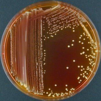
49 Salmonella mixed with normal fecal flora. Dr. S. S. Khoramrooz 52

50 Dr. S. S. Khoramrooz 53
51 Hektoen Enteric (HE) Agar: Composition Peptone 1.2% Yeast extract 0.3% Bile salts 0.9% Lactose 1.2% Sucrose 1.2% Salicin 0.2% Sodium chloride 0.5% Ferric ammonium citrate Acid fuchsin Thymol blue Agar 1.4% ph = 7.6 Dr. Khoramrooz 54
52 HE Agar: Growth of Enteric Pathogens and Commensals High bile salt concentration inhibits growth of grampositive and gram-negative intestinal commensals, and thereby selects for pathogenic Salmonella (bileresistant growth) present in fecal specimens. Salmonella species as non-lactose and non-sucrose fermenters that produce H2S form colorless colonies with black centers. Shigella species (non-lactose and non-sucrose fermenters, no H2S production) form colorless colonies. Lactose and sucrose fermenters (E. coli, K. pneumoniae) form orange to yellow colonies due to acid Dr. Khoramrooz production. 55
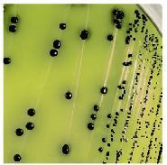
53 Intended Use Bismuth Sulfite Agar is a highly selective medium used for isolating Salmonella spp., particularly Salmonella Typhi, from food and clinical specimens. Dr. S. S. Khoramrooz 56
54 Principles of the Procedure Dextrose is an energy source. Bismuth sulfite indicator and brilliant green are complementary in inhibiting gram-positive bacteria and members of the coliform group, while allowing Salmonella to grow luxuriantly. Ferrous sulfate is included for detection of H2S production. When H2S is present, the iron in the formula is precipitated, giving positive cultures the characteristic brown to black color with metallic sheen. Dr. S. S. Khoramrooz 57
55 Dr. S. S. Khoramrooz 58
56 For isolation of Salmonella spp. from clinical specimens, inoculate fecal specimens and rectal swabs onto a small area of one quadrant of the Bismuth Sulfite Agar plate and streak for isolation. This will permit the development of discrete colonies. Incubate plates at 35 C. Examine at 24 hours and again at 48 hours for colonies resembling Salmonella spp. Dr. S. S. Khoramrooz 59
57 Expected results The typical discrete S. Typhi surface colony is black and surrounded by a black or brownish-black zone which may be several times the size of the colony. By reflected light, preferably daylight, this zone exhibits a distinctly characteristic metallic sheen. Plates heavily seeded with S. Typhi may not show this reaction except near the margin of the mass inoculation. Dr. S. S. Khoramrooz 60
58 In these heavy growth areas, this organism frequently appears as small light green colonies. This fact emphasizes the importance of inoculating plates so that some areas are sparsely populated with discrete S. Typhi colonies. Other strains of Salmonella produce black to green colonies with little or no darkening of the surrounding medium. Dr. S. S. Khoramrooz 61

59 Dr. S. S. Khoramrooz 62
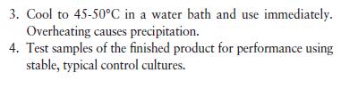
60 Heat with frequent agitation and boil for 1 minute to completely dissolve the powder. DO NOT AUTOCLAVE. Use the medium the same day it is prepared. Dr. S. S. Khoramrooz 63
61 Dr. Khoramrooz 64
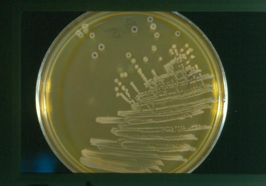
62 Intended Use Brilliant Green Agar is a highly selective medium for the isolation of Salmonella other than S. Typhi from feces and other materials. Principles of the Procedure Brilliant green dye inhibits gram-positive bacteria and a majority of gram-negative bacilli. Phenol red serves as a ph indicator and yields a yellow color as a result of acid production in the fermentation of the lactose and/or sucrose in the medium. Dr. S. S. Khoramrooz 65
63 Dr. S. S. Khoramrooz 66
64 Procedure A less selective medium and a nonselective medium should also be streaked to increase the chance of recovery when the population of gram-negative organisms is low and to provide an indication of other organisms present in the specimen. Incubate plates, protected from light, at 35 ± 2 C for hours. If negative after 24 hours, reincubate an additional 24 hours. Dr. S. S. Khoramrooz 67


65 Salmonella on BPLS Agar. The colonies are red because the bacterium does not ferment lactose or sucrose. Escherichia coli on BPLS Agar. The colonies are yellow due to the low ph which is caused by the production of acid during fermentation of lactose and/or sucrose. Dr. S. S. Khoramrooz 68

66 Dr. S. S. Khoramrooz 69
67 Differential Identification Characteristics Dr. Khoramrooz 70
68 Carbohydrate utilization Lactose degradation; Differentiate those (lactose fermenter LF]) and those that are nonlactose fermenters (NLFs). Examples of sugars used to differentiate bacteria include lactose, maltose, rhamnose, sucrose, raffinose, and arabinose Bacteria can utilize carbohydrates: oxidation (aerobically), fermentatively (anaerobically),or both. Bacteriat hat can grow either aerobically or anaerobically are called facultative anaerobes. Some bacteria are asacchrolytic. They do not use any carbohydrate; instead they use other organic molecules for energy and carbon sources. Dr. Khoramrooz 71
69 Oxidation/Fermentation (O/F) Test Principle : To determine the ability of bacteria to breakdown glucose oxidative or fermentative O/F medium ( Hugh and Leifson Medium) is formulated to detect weak acids produced from saccharolytic M.O. O/F medium contains Sugar (glucose 1%) Low percentage of Agar and Peptone ph indicator (Bromothymol blue) Alkaline Blue Neutral Green Acidic Yellow Dr. Khoramrooz 72
70 Intended Use OF (Oxidation Fermentation) media are used for the determination of oxidative and fermentative metabolism of carbohydrates by gram-negative rods on the basis of acid reaction in either the open or closed system. Summary and Explanation OF Medium was developed by Hugh and Leifson who described the taxonomic significance of fermentative versus oxidative metabolism of carbohydrates by gram-negative bacteria. Dr. S. S. Khoramrooz 73
71 They showed that when an organism is inoculated into two tubes of OF Basal Medium containing a carbohydrate and the medium in one of the tubes is covered with melted petrolatum prior to incubation, the patterns of metabolism are of differential significance. Oxidative organisms only produce an acid reaction in the open tube with little or no growth and no acid formation in the covered tube. Fermentative organisms will produce an acid reaction in both types of tubes. Changes in the covered agar are considered to be due to true fermentation, while changes in the open tubes are due to oxidative utilization of the carbohydrate present. If the carbohydrate is not utilized by either method, there is no acid production in either tube. Dr. S. S. Khoramrooz 74
72 Principles of the Procedure Dextrose is the most important carbohydrate for use in OF Basal Medium; however, certain organisms may metabolize other carbohydrates even if they are unable to utilize dextrose. Prepared tubed media containing arabinose, dextrose, dulcitol, fructose, galactose, lactose, maltose, mannose, raffinose, rhamnose, salicin, sorbitol, sucrose and xylose are provided. Dr. S. S. Khoramrooz 75
73 Dr. S. S. Khoramrooz 76
74 Procedure Inoculate a pair of OF tubes of each carbohydrate used with each organism being tested. The tubes should be stabbed to approximately 1/4 inch from the bottom using an inoculating needle and a light inoculum. Overlay one tube of each pair with sterile mineral oil. Incubate tubes at 35 ± 2 C in an aerobic atmosphere for 48 hours. Do not discard as negative until after 4 days of incubation. Dr. S. S. Khoramrooz 77
75 Expected Results Record results as acid (A) or alkaline/no change ( ). Also record whether or not the organism is motile as evidenced by the appearance of growth away from the line of inoculation. Typical reaction patterns are as follows. Dr. S. S. Khoramrooz 78
76 Limitations of the Procedure 1. The acid reaction produced by oxidative organisms is apparent at the surface and gradually spreads throughout the medium. If the oxidation is weak or slow, however, an initial alkaline reaction at the surface of the open tube may persist for several days and eventually convert to an acid reaction. 2. If an organism is unable to grow on OF Basal Medium, Cowan recommends adding either 2% serum or 0.1% yeast extract to each carbohydrate tube. Dr. S. S. Khoramrooz 79

77 Dr. S. S. Khoramrooz 80

78 Dr. S. S. Khoramrooz 82
79 Triple Sugar lron Agar Kligler Iron Agar Lactose Fermentation Glucose fermentation Gas Production (H2 & CO2 ) H2S Production Dr. Khoramrooz 83


80 TSI and KIA glucose lactose Ferrous sulfate ph indicator: phenol red Proteins Dr. Khoramrooz 84
81 Dr. Khoramrooz 85
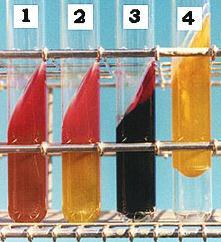
82 Intended Use Triple Sugar Iron Agar (TSI Agar) is used for the differentiation of gram-negative enteric bacilli based on carbohydrate fermentation and the production of hydrogen sulfide. Principles of the Procedure TSI Agar contains three sugars (dextrose, lactose and sucrose), Phenol red for detecting carbohydrate fermentation Dr. S. S. Khoramrooz 86
83 Ferrous ammonium sulfate for detection of hydrogen sulfide production (indicated by blackening in the butt of the tube). Carbohydrate fermentation is indicated by the production of gas and a change in the color of the ph indicator from red to yellow. To facilitate the detection of organisms that only ferment dextrose, the dextrose concentration is one-tenth the concentration of lactose or sucrose. The small amount of acid produced in the slant of the tube during dextrose fermentation oxidizes rapidly, causing the medium to remain red or revert to an alkaline ph. In contrast, the acid reaction (yellow) is maintained in the butt of the tube because it is under lower oxygen tension. Dr. S. S. Khoramrooz 87
84 After depletion of the limited dextrose, organisms able to do so will begin to utilize the lactose or sucrose. To enhance the alkaline condition of the slant, free exchange of air must be permitted by closing the tube cap loosely. If the tube is tightly closed, an acid reaction (caused solely by dextrose fermentation) will also involve the slant. Dr. S. S. Khoramrooz 88
85 Dr. S. S. Khoramrooz 89
86 Procedure To inoculate, carefully touch only the center of an isolated colony on an enteric plated medium with a cool, sterile needle, stab into the medium in the butt of the tube, and then streak back and forth along the surface of the slant. Several colonies from each primary plate should be studied separately, since mixed infections may occur. Dr. S. S. Khoramrooz 90
87 Incubate with caps loosened at 35 C and examine after hours for carbohydrate fermentation, gas production and hydrogen sulfide production. Any combination of these reactions may be observed. Do not incubate longer than 24 hours because the acid reaction in the slant of lactose and sucrose fermenters may revert to an alkaline reaction. Dr. S. S. Khoramrooz 91

88 Dr. S. S. Khoramrooz 92
89 Expected Results Carbohydrate fermentation is indicated by a yellow coloration of the medium. If the medium in the butt of the tube becomes yellow (acidic), but the medium in the slant becomes red (alkaline), the organism being tested only ferments dextrose (glucose). A yellow (acidic) color in the slant and butt indicates that the organism being tested ferments dextrose, lactose and/or sucrose. A red (alkaline) color in the slant and butt indicates that the organism being tested is a nonfermenter. Dr. S. S. Khoramrooz 93
90 Hydrogen sulfide production results in a black precipitate in the butt of the tube. Gas production is indicated by splitting and cracking of the medium. Dr. S. S. Khoramrooz 94
91 1. Hydrogen sulfide production may be evident on Kligler Iron Agar but negative on Triple Sugar Iron Agar. Studies by Bulmash and Fulton showed that the utilization of sucrose could suppress the enzymatic mechanisms responsible for H2S production. Padron and Dockstader8 found that not all H2S-positive Salmonella are positive on TSI. 2. Sucrose is added to TSI to eliminate some sucrose-fermenting lactose-nonfermenters such as Proteus and Citrobacter spp. 3. Further biochemical tests and serological typing must be performed for definite identification and confirmation of organisms. Dr. S. S. Khoramrooz 95
92 4. Do not use an inoculating loop to inoculate a tube of Triple Sugar Iron Agar. While stabbing the butt, mechanical splitting of the medium occurs, causing a false positive result for gas production. 5. A pure culture is essential when inoculating Triple Sugar Iron Agar. If inoculated with a mixed culture, irregular observations may occur. 6. Tubes should be incubated with caps loosened. This allows a free exchange of air, which is necessary to enhance the alkaline condition on the slant. Dr. S. S. Khoramrooz 96
93 Red/Red Alkaline /Alkaline K/K Lactose -/Glucose Yellow/Yellow Acid/Acid A/A Lactose +/Glucose + Red/Yellow Alkaline/Acid K/A Lactose -/Glucose + Gas - H2S - Red/Yellow Alkaline/Acid K/A Lactose -/Glucose + Gas + H2S - Red/Yellow Alkaline/Acid K/A Lactose -/Glucose + Gas - H2S + Red/Black Alkaline/Acid K/A Lactose -/Glucose + Gas + H2S + Dr. Khoramrooz 97
94 Result Reaction on KIA Example Butt color Slant color H 2 S Result Red Red Negative Alk/Alk/- (No action on sugars) Non fermenter e.g. Pseudomonas Yellow Red Negative A/Alk/- (Glucose fermented without H 2 S) LNF e.g. Shigella Yellow Red Positive black in butt A/Alk/+ (Glucose fermented with H 2 S) LNF e.g. Salmonella & Proteus Yellow Yellow Negative A/A/- (three sugars are fermented) Dr. Khoramrooz LF e.g. E. coli, Klebsiella, Enterobacter 98
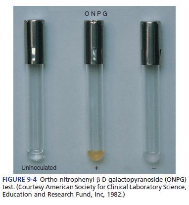
95 Ortho- Nitrophenyl - β D- galactopyranosidetest Organisms that are delayed lactose-fermenters appear as nonfermenting colonies. The o-nitrophenyl β D- galactopyranoside (ONPG) and the p- nitrophenyl-b-d-galactopyranoside (PNPG) tests determine whether the organism is a delayed lactose fermenter (one that lacks the enzyme β-galactoside permease but possesses β- galactosidase) or a true nonlactose fermenter (NLF). The test can be performed by making a heavy suspension of bacteria in sterile saline and adding commercially prepared ONPG disks or tablets. The suspension is incubated at 37' C, and positive results can generally be seen within 6 hours. Dr. Khoramrooz 99
96 Dr. Khoramrooz 100
97 IMViC Test Indole, Methyl Red, Voges-Prosakaur, Citrate (IMViC) Tests: The following four tests comprise a series of important determinations that are collectively called the IMViC series of reactions The IMViC series of reactions allows for the differentiation of the various members of Enterobacteriaceae. Dr. Khoramrooz 101
98 Principle IMViC: Indole test Certain microorganisms can metabolize tryptophan by tryptophanase The enzymatic degradation leads to the formation of pyruvic acid, indole and ammonia The presence of indole is detected by addition of Kovac's reagent. Tryptophanase Tryptophane amino acids Indole + Pyurvic acid + NH3 Kovac s Reagent Red color Dr. Khoramrooz in upper organic layer` 102
 from Klebsiella (-). Dr. Khoramrooz 103")
99 IMViC: Indole test Result: A bright pink color in the top layer indicates the presence of indole The absence of color means that indole was not produced i.e. indole is negative Negative test e.g. Klebsiella Positive test e.g. E. coli Special Features: Used in the differentiation of genera and species. e.g. E. coli (+) from Klebsiella (-). Dr. Khoramrooz 103
100 Intended Use SIM Medium is used to differentiate enteric bacilli on the basis of sulfide production,indole formation and motility. Dr. S. S. Khoramrooz 104
101 Summary and Explanation Hydrogen sulfide production, indole formation and motility are distinguishing characteristics which aid in the identification of the Enterobacteriaceae, especially Salmonella and Shigella. SIM Medium, therefore,is useful in the process of identification of enteric pathogens. Dr. S. S. Khoramrooz 105
102 Principles of the Procedure Sodium thiosulfate and ferrous ammonium sulfate are indicators of hydrogen sulfide production. The ferrous ammonium sulfate reacts with H2S gas to produce ferrous sulfide,a black precipitate. The casein peptone is rich in tryptophan,which is attacked by certain microorganisms resulting in the production of indole. The indole is detected by the addition of chemical reagents following the incubation period. Dr. S. S. Khoramrooz 106
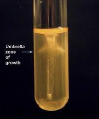
103 Motility detection is possible due to the semisolid nature of the medium. Growth radiating out from the central stab line indicates that the test organism is motile. Dr. S. S. Khoramrooz 107
104 Dr. S. S. Khoramrooz 108
105 Procedure Loosen caps, boil and cool before use. Using growth from a pure culture, stab an inoculating needle two-thirds of the distance to the bottom in the center of the tube. Incubate tubes with loosened caps for18-24 hours at 35±2 C in anaerobic atmosphere. Dr. S. S. Khoramrooz 109


106 Dr. S. S. Khoramrooz 110
107 IMViC test Methyl Red-Voges Proskauer (MR-VP) Tests Principle Glucose Acidic pathway Or Neutral pathway Mixed acids ph less than 4.4 Methyl Red indicator Red color MR positive E. coli Acety methyl carbinol (ACETOIN) VP positive Klebsiella Dr. Khoramrooz solution A solution B Pink color 111
108 Butylene Glycol Pathway of Glucose Fermentation In the butylene glycol pathway pyruvic acid to acetoin and butylene glycol. Acetoin and butylene glycol are detected by oxidation to diacteyl at an alkaline ph. Addition of -naphthol which forms a red-colored complex with diacetyl. Important biochemical property used for the identification of Klebsiella, Enterobacter, and Serratia. Dr. Khoramrooz 112
109 IMViC test: MRVP test Method Inoculate the tested organism into MRVP broth Incubate the tubes at 37 C for 24 hours For methyl red: Add 6-8 drops of methyl red reagent. For Voges-Proskauer: Add 12 drops of solution A ( -naphthol), mix, 4 drops of Solution B (40% KOH), mix Dr. Khoramrooz 113
")
 Yellow or orange: Negative MR (Klebsiella) Dr.")
110 Results IMViC test: MR/VP test Methyl Red test Voges-Proskauer test Pink: Positive VP (Klebsiella) Red: Positive MR (E. coli) No pink: Negative VP (E. coli) Yellow or orange: Negative MR (Klebsiella) Dr. Khoramrooz 114
 are used for the differentiation of bacteria by means of the methyl red and Voges-Proskauer reactions. Dr. S.")
111 Intended Use MR-VP Medium and MR-VP Broth (Methyl Red- Voges Proskauer Medium/Broth, also known as Buffered Peptone- Glucose Broth) are used for the differentiation of bacteria by means of the methyl red and Voges-Proskauer reactions. Dr. S. S. Khoramrooz 115
112 Principles of the Procedure Methyl red-positive organisms produce high levels of acid during fermentation of dextrose, overcome the phosphate buffer system and produce a red color upon the addition of the methyl red ph indicator. In the Voges-Proskauer test, the red color produced by the addition of potassium hydroxide to cultures of certain microbial species is due to the ability of the organisms to produce a neutral end product, acetoin (acetylmethylcarbinol), from the fermentation of dextrose. The acetoin is oxidized in the presence of oxygen and alkali to produce a red color. This is a positive Voges-Proskauer reaction. Dr. S. S. Khoramrooz 116
113 Dr. S. S. Khoramrooz 117
114 Procedure Using a light inoculum, inoculate tubes of MR-VP media with 18- to 24-hour pure cultures. Incubate tubes aerobically at 35 2 C for a minimum of 48 hours but preferably for 5 days. Prepare the methyl red indicator: 0.1 g of methyl red in 300 ml of 95% ethyl alcohol. Add sufficient purified water to make 500 ml. After the appropriate incubation period, aseptically remove aliquots (1 ml for the VP test) of the medium and conduct the following tests: Dr. S. S. Khoramrooz 118
115 1. Methyl Red Test: Add 5 drops of methyl red indicator to an aliquot of the broth. Interpret the color result immediately. 2. Voges-Proskauer Test: Empty the contents (15 drops) from the reagent A dropper 5 drops from the reagent B dropper into 1 ml of broth culture. Shake well after the addition of each reagent to aerate the sample. Dr. S. S. Khoramrooz 119
116 Expected Results 1. Methyl Red Test a. Positive red color at surface of the medium. b. Negative yellow color at surface of the medium. 2. Voges-Proskauer Test A positive reaction is indicated by the development of a distinct red color which occurs within 5 minutes. Certain species within Enterobacteriaceae genera may react differently or give variable results. Consult appropriate texts for reactions of specific species. Dr. S. S. Khoramrooz 120
117 Limitations of the Procedure 1. Results of the MR and VP tests need to be used in conjunction with other biochemical tests to differentiate genus and species within the Enterobacteriaceae. 2. A precipitate may form in the potassium hydroxide reagent solution. This precipitate has not been shown to reduce the effectiveness of the reagent. Dr. S. S. Khoramrooz 121
118 3. Most members of the family Enterobacteriaceae give either a positive MR test or a positive VP test. However, certain organisms such as Hafnia alvei and Proteus mirabilis may give a positive result for both tests. 4. Incubation time for the Methyl Red test cannot be shortened by increasing the dextrose concentration in the medium or by heavily inoculating the broth. 5. Incubate MR-negative tests for more than 48 hours and test again. Dr. S. S. Khoramrooz 122
119 6. Read the VP test at 48 hours. Increased incubation may produce acid conditions in the broth that will interfere with reading the results. 7. VP reagents must be added in the order and the amounts specified or a weak-positive or false-negative reaction may occur. A weak-positive reaction may be masked by a copper-like color which may form due to the reaction of KOH and α-naphthol. 8. Read the VP test within 1 hour of adding the reagents. The KOH and α-naphthol may react to form a copper-like color, causing a potential false-positive interpretation. 9. Due to the possible presence of acetoin, diacetyl or related substances in certain raw materials, the use of media low in these substances (such as MR-VP media) is recommended for this test. Dr. S. S. Khoramrooz 123

120 Dr. S. S. Khoramrooz 124
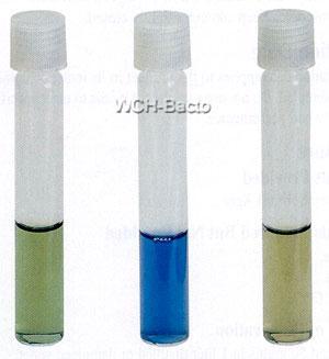
121 Citrate Utilization Test Principle: Citrate Pyruvate CO2 + Na + H2O Na 2 CO 3 Simmone s Citrate media Contains Citrate as a sole of C source Alkaline, ph Positive test Blue colour Bromothymol blue Positive test: Klebsiella, Enterobacter, Citrobacter Dr. Khoramrooz Negative test: E. coli 125


122 Citrate Utilization Test Method Streak a Simmon's Citrate agar slant with the organism Incubate at 37 C for 24 hours. Dr. Khoramrooz 126
123 Result Examine for growth (+) Citrate Utilization Test Growth on the medium is accompanied by a rise in ph to change the medium from its initial green color to deep blue Positive Klebsiella, Enterobacter Dr. Khoramrooz Negative E. coli 127
124 Intended Use Simmons Citrate Agar is used for the differentiation of gram negative bacteria on the basis of citrate utilization. Principles of the Procedure Organisms able to utilize ammonium dihydrogen phosphate and sodium citrate as the sole sources of nitrogen and carbon, respectively, will grow on this medium and produce an alkaline reaction as evidenced by a change in the color of the bromthymol blue indicator from green (neutral) to blue (alkaline). Dr. S. S. Khoramrooz 128
125
126 Procedure Inoculate slants with growth from a pure culture using a light inoculum. Incubate all tubes for 4 days at 35 ± 2 C in an aerobic atmosphere. Expected Results A positive reaction is indicated by growth with an intense blue color in the slant. A negative reaction is evidenced by no growth to trace growth with no change in color (medium remains dark green). Dr. S. S. Khoramrooz 130

127 Dr. S. S. Khoramrooz 131

128 Dr. S. S. Khoramrooz 132
129 Dr. S. S. Khoramrooz 133
130 Amino Acid Decarboxylation Enterobacteriaceae contain decarboxylases with substrate specificity for amino acids, and are detected using Moeller decarboxylase broth overlayed with mineral oil for anaerobiosis. Moeller broth contains: glucose for fermentation, peptone and beef extract, amino acid, pyridoxal, ph indicator bromcresol purple. Dr. Khoramrooz 134
131 Amino Acid Decarboxylation If an Enterobacteriaceae contains amino acid decarboxylase, amines produced by decarboxylase action cause an alkaline ph, and bromcresol purple turns purple. Lysine, ornithine, and arginine are utilized. A base broth without amino acid is included in which glucose fermentation acidifies the broth, turning the bromcresol purple yellow. Dr. Khoramrooz 135
132 Amino Acid Decarboxylation 1 Lysine Cadaverine Ornithine Putrescine Arginine Citrulline Ornithine Putrescine 1 Conversion of arginine to citrulline is a dihydrolase reaction Dr. Khoramrooz 136
133 Amino Acid Decarboxylation Decarboxylation patterns are essential for the genus identification of Klebsiella, Enterobacter, Escherichia, and Salmonella. Decarboxylation patterns are also essential for the species identification of Enterobacter aerogenes, Enterobacter cloacae, Proteus mirabilis, and Shigella sonnei. Dr. Khoramrooz 137
134 Amino Acid Decarboxylation Lys Orn Arg Klebsiella + Enterobacter +/ + +/ Escherichia + +/ /+ Salmonella Dr. Khoramrooz 138
135 Amino Acid Decarboxylation Lys Orn Arg E. aerogenes + + E. cloacae + + P. mirabilis + P. vulgaris Shigella D + Shigella A-C Dr. Khoramrooz 139
136 Phenylalanine Deaminase Reaction Enterobacteriaceae utilize amino acids in a variety of ways including deamination. Phenylalanine is an amino acid that forms the keto acid phenylpyruvic acid when deaminated. Phenylpyruvic acid is detected by addition of ferric chloride that forms an intensely dark olive-green colored complex when binding to phenylpyruvic acid. The deamination of phenylalanine is an important biochemical property of Proteus, Morganella, and Providencia. Dr. Khoramrooz 140
137 Intended Use Decarboxylase media are used in the biochemical differentiation of gram-negative enteric bacilli based on the production: Arginine dihydrolase Lysine decarboxylase Ornithine decarboxylase Decarboxylase Medium Base, with added arginine, lysine or ornithine is used for the same purpose. Lysine Decarboxylase Broth is used for differentiating microorganisms based on lysine decarboxylation. Dr. S. S. Khoramrooz 141
138 Summary and Explanation Moeller introduced the decarboxylase media for detecting the production of lysine and ornithine decarboxylase and arginine dihydrolase. These media are a useful adjunct to other biochemical tests for the speciation and identification of the Enterobacteriaceae and other gram-negative bacilli. The production of OD is particularly useful for differentiating Klebsiella and Enterobacter species. Klebsiella species are non-motile and, except for K. ornithinolytica, do not produce ornithine decarboxylase, while most Enterobacter species are motile and, except for E. agglomerans, usually produce this enzyme. Dr. S. S. Khoramrooz 142
139 Principles of the Procedure Pyridoxal is an enzyme co-factor for the amino acid decarboxylase. Dextrose is a fermentable carbohydrate. Bromcresol purple and cresol red are ph indicators. The amino acids lysine, ornithine or arginine are added to the basal medium at a concentration of 10.0 g/l to detect the production of the enzyme specific for these substrates. Dr. S. S. Khoramrooz 143
140 When the medium is inoculated with a bacterium that is able to ferment dextrose, acids are produced that lower the ph of the medium and change the color of the indicator from purple to yellow. The acidic condition also stimulates decarboxylase activity. If the organism produces the appropriate enzyme, the amino acid in the medium is degraded, yielding a corresponding amine. Dr. S. S. Khoramrooz 144

141 Dr. S. S. Khoramrooz 145
142 Decarboxylation of lysine yields cadaverine. while decarboxylation of ornithine yields putrescine. Arginine is first hydrolyzed to form ornithine, which is then decarboxylated to form putrescine. The production of these amines elevates the ph of the medium, changing the color of the indicator from yellow to purple or violet. If the organism does not produce the appropriate enzyme, the medium remains acidic (yellow). Dr. S. S. Khoramrooz 146
143 Each isolate to be tested must also be inoculated into a tube of the basal medium that does not contain the amino acid. If this tube becomes alkaline, the test is invalid. To obtain the appropriate reactions, the inoculated tubes must be protected from air with a layer of sterile mineral oil. Exposure to air may cause alkalinization at the surface of the medium, which could cause a decarboxylasenegative organism to appear positive. Dr. S. S. Khoramrooz 147
144 Dr. S. S. Khoramrooz 148
145 Expected Results Compare the color of tubes of media containing the specific amino acids with the color of control tubes of basal media (without amino acid) that have been inoculated with the same isolate. If inoculated control tubes show an alkaline reaction, the test is invalid; i.e., Improperly performed or the test organisms Degrade the peptone sufficiently to produce an alkaline reaction in the absence of a specific amino acid. The medium becomes purple to violet if the reaction is positive (alkaline). A yellow color indicates a negative test; i.e., the organism does not produce the appropriate enzyme. Dr. S. S. Khoramrooz 149

146 Dr. S. S. Khoramrooz 150
147 The lysine iron agar (LIA) test is a tubed agar slant. It contains the amino acid lysine, glucose, ferric ammonium citrate, and sodium thiosulfate. The ph indicator is bromcresol purple. LIA is used primarily to determine whether the bacteria decarboxylate or deaminate lysine. H2S production is also detected in this medium. LIA is inoculated in the same manner as a TSI agar slant. LIA is most useful in conjunction with TSI in screening stool specimens for the presence of enteric pathogens, differentiating Salmonella spp. (lysine-positive) from Citrobacter spp. (lysinenegative). Dr. S. S. Khoramrooz 151
148 Decarboxylation occurs anaerobically only; the presence of a dark purple butt is positive for lysine decarboxylation. The production of H2S can mask the purple color in the butt of the tube. Because H2S production in LIA occurs only in an alkaline environment, a black precipitate indicating H2S is also a positive result for decarboxylation. Dr. S. S. Khoramrooz 152
149 LIA is also useful in differentiating Proteus, Morganella, and Providencia spp. from most other members of Enterobacteriaceae This group of enterics deaminates (attacks the NH2 group instead of the carboxyl group) amino acids. In the LIA slant, deamination of lysine turns the original light purple color slant to a plum or reddish purple color; the butt turns yellow because of glucose fermentation. Dr. S. S. Khoramrooz 153

150 Dr. S. S. Khoramrooz 154
151 IPViC Reactions for Initial Grouping of the Enterobacteriaceae Indole Phenylalanine deaminase Voges-Proskauer Citrate Dr. Khoramrooz 155
152 Principle Urease Test Urea agar contains urea and phenol red Urease is an enzyme that catalyzes the conversion of urea to CO2 and NH3 Ammonia combines with water to produce ammonium hydroxide, a strong base which ph of the medium. in the ph causes phenol red r to turn a deep pink. This is indicative of a positive reaction for urease Urea Urease CO2 + NH3 H2O NH4 OH in ph Method Streak a urea agar tube with the organism incubate at 37 C for 24 h Dr. Khoramrooz Phenol Red Pink Positive test 156


153 Result Urease Test If color of medium turns from yellow to pink indicates positive test. Proteus give positive reaction after 4 h while Kelebsiella and Enterobacter gave positive results after 24 h Dr. Khoramrooz Positive test Negative test 157
154 Intended Use Urea Agar and Urease Test Broth are used for the differentiation of organisms, especially the Enterobacteriaceae, on the basis of urease production. Dr. S. S. Khoramrooz 158
155 Principles of the Procedure The urea medium of Rustigian and Stuart is particularly suited for the differentiation of Proteus species from other gram negative enteric bacilli capable of utilizing urea. Unable to do so in Urease Test Broth because of limited nutrients and the high buffering capacity of the medium. To provide a medium with greater utility, Urea Agar was devised by Christensen with peptone and dextrose included and reduced buffer content to promote more rapid growth of many of the Enterobacteriaceae and permit a reduction in incubation time. Dr. S. S. Khoramrooz 159
156 The complete Urea Agar contains 15.0 g/l of agar in addition to the ingredients in the base medium. When organisms utilize urea, ammonia is formed during incubation which makes the reaction of these media alkaline, producing a red-pink color. Consequently, urease production may be detected by the change in the phenol red indicator. Dr. S. S. Khoramrooz 160


157 Urease medium
158 Dr. S. S. Khoramrooz 162
159 Dehydrated Product BBL Urea Agar Base 1. Dissolve 29 g of the powder in 100 ml of purified water. Mix thoroughly. Sterilize by filtration. 2. Suspend 15 g of agar in 900 ml of purified water. 3. Autoclave at 121 C for 15 minutes. 4. Cool to 50 C and add 100 ml of the sterile Urea Aga Base. 5. Mix thoroughly and dispense aseptically in sterile tubes. 6. Cool tubed medium in a slanted position so that deep butts are formed. 7. Do not remelt the complete medium. 8. Test samples of the finished product for performance using stable, typical control cultures. Dr. S. S. Khoramrooz 163
160 Procedure Using a heavy inoculum (2 loopfuls) of growth from an 18- to 24-hour pure culture (TSI Agar or other suitable medium), inoculate the broth or agar (streaking back and forth over the entire slant surface). Dr. S. S. Khoramrooz 164
161 Do not stab the butt since it serves as a color control. For broth, shake tubes gently to suspend the bacteria. Incubate tubes with loosened caps at 35 2 C in an incubator or water bath. Observe reactions after 2, 4, 6, 18, 24 and 48 hours. For agar, continue to check every day for a total of 6 days; even longer incubation periods may be necessary. Dr. S. S. Khoramrooz 165
162 The production of urease is indicated by an intense pink-red (red-violet) color on the slant or throughout the broth. The color may penetrate into the agar (butt); the extent of the color indicates the rate of urea hydrolysis. Dr. S. S. Khoramrooz 166
163 A negative reaction is no color change. The agar medium remains pale yellow to buff; the broth remains yellowish orange. Dr. S. S. Khoramrooz 167
164 Urea Agar Base 1. The alkaline reaction produced in this medium after prolonged incubation may not be caused by urease activity. False positive reactions may occur due to the utilization of peptones (especially in slant agar by Pseudomonas aeruginosa, for example) or other proteins which raise the ph due to protein hydrolysis and the release of excessive amino acid residues. To eliminate possible protein hydrolysis, perform a control test with the same test medium without urea. 2. Do not heat or reheat the medium because urea decomposes very easily. Dr. S. S. Khoramrooz 168
165 3. Urea Agar detects rapid urease activity of only the urease positive Proteus species. For results to be valid for the detection of Proteus, the results must be read within the first 2-6 hours after incubation. Urease-positive Enterobacter, Citrobacter or Klebsiella, in contrast, hydrolyze urea much more slowly, showing only slight penetration of the alkaline reaction into the butt of the medium in 6 hours and requiring 3-5 days to change the reaction of the entire butt. Dr. S. S. Khoramrooz 169
166 Urea Broth 1. To rule out false positives due to protein hydrolysis (as opposed to urea hydrolysis) that may occur in the medium after prolonged incubation, perform a control test with the same test medium without urea. 2. Do not heat or reheat the medium because urea decomposes very easily. Dr. S. S. Khoramrooz 170
167 3. The high buffering system in this medium masks urease activity in organisms that are delayed positive. This medium is therefore recommended for the detection of urease activity in all Proteus spp., Providencia rettgeri and ureasepositive Providencia stuartii. M. morganii slowly hydrolyzes urea and may require approximately a 36 hour incubation for a strong urease-positive reaction to occur. If in doubt as to a result, compare with an uninoculated tube or incubate for an additional 24 hours. 4. Variations in the size of the inoculum can affect the time required to reach positive (alkaline, ph 8.1) results. Dr. S. S. Khoramrooz 171


168 Motility From left to right: + + Dr. Khoramrooz 172

169 SIM Sulfide, Indole, Motility Dr. Khoramrooz 173
170 Initial Grouping of the Enterobacteriaceae (VP=Voges Proskauer, PDA=Phenylalanine Deaminase) GENERA VP PDA Klebsiella POSITIVE NEGATIVE Enterobacter POSITIVE NEGATIVE Serratia POSITIVE NEGATIVE Hafnia POSITIVE NEGATIVE Pantoea POSITIVE NEGATIVE Dr. Khoramrooz 175
171 Initial Grouping of the Enterobacteriaceae GENERA VP PDA Proteus 1 NEGATIVE POSITIVE Morganella NEGATIVE POSITIVE Providencia NEGATIVE POSITIVE 1 Proteus mirabilis: Dr. 50% Khoramrooz of strains VP positive 176
172 Initial Grouping of the Enterobacteriaceae GENERA VP PDA Escherichia NEGATIVE NEGATIVE Shigella NEGATIVE NEGATIVE Edwardsiella NEGATIVE NEGATIVE Salmonella NEGATIVE NEGATIVE Citrobacter NEGATIVE NEGATIVE Yersinia NEGATIVE NEGATIVE Dr. Khoramrooz 177
173 Initial Grouping of the Enterobacteriaceae 1 GENERA INDOLE CITRATE Escherichia POSITIVE NEGATIVE Shigella POSITIVE 2 Yersinia POSITIVE 3 Edwardsiella POSTIVE NEGATIVE NEGATIVE NEGATIVE 1 VP negative, PDA negative 2 Shigella groups A, B, and C variably positive for indole production (25-50%), group D Shigella negative. 3 Dr. Khoramrooz Yersinia enterocolitica 50% positive 178
174 Initial Grouping of the Enterobacteriaceae 1 GENERA INDOLE CITRATE Salmonella NEGATIVE POSITIVE 2 Citrobacter NEGATIVE POSITIVE 1 VP negative, PDA negative 2 Salmonella serotype Paratyphi A and Typhi negative Dr. Khoramrooz 179
175 Key Characteristics of the Enterobacteriaceae E coli Shi A- C Shi D Ed Sal Cit TSI ON GAS H2S VP IND CIT PDA UR MO LYS OR AR A/A Ak/ A A Yer A/A / / + / + Ak/ A + + Ak/ A Ak/ +/ A A/A Ak/ (1) RT=room temperature +/ Dr. Khoramrooz +/ +/ / + +/ RT (1) + 180
176 Kle A/A pne Kle A/A oxy En A/A aer En A/A cloa Serr A/A (1) Haf Ak/ A Pan A/A Alk/ A Key Characteristics of the Enterobacteriaceae TSI ON GAS H2S VP IND CIT PDA UR MO LYS OR AR / /+ +/ /+ +/ /+ /+ (1) Produces DNase, lipase, and gelatinase Dr. Khoramrooz 181
177 Key Characteristics of the Enterobacteriaceae Prot mir a Prot vulg TSI ON GAS H2S VP IND CIT PDA UR MO LYS OR AR Ak/ A Mor Ak/ Pro v + + +/ +/ + + +s + A/A +/ + + / s A Ak/ A s = swarming motility Dr. Khoramrooz 182
178 Biochemical Characteristics of Escherichia coli and Shiglla E. coli E. coli O157:H7 Shigella TSI A/Ag A/Ag Alk/A Lactose + + ONPG + + /+ 1 Sorbitol + +/ Indole + + +/ Methyl red VP Citrate Lysine + + Motility Shigella sonnei (group D) ONPG + Dr. Khoramrooz 183
179 Biochemical Characteristics of Salmonella Most Serotypes Typhi Paratyphi A TSI Alk/A Alk/A Alk/A H2S (TSI) + + (weak) Citrate + Lysine + + Ornithine + + Dulcitol + + Rhamnose + + Indole Methyl red VP Dr. Khoramrooz 184

180 Dr. Khoramrooz 185
181 The End Dr. Khoramrooz 186
182 t Dr. Khoramrooz 187
183 Dr. Khoramrooz 188
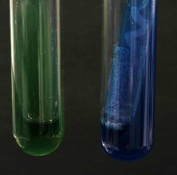
184 Intended Use Malonate Broth is used for differentiating Enterobacter from Escherichia based on malonate utilization. Principles of the Procedure Malonate Broth contains ammonium sulfate, which is the sole source of nitrogen in the medium; Sodium malonate is the sole source of carbon. Increased alkalinity resulting from malonate utilization causes the indicator, bromthymol blue, to change color from green to blue. Dr. S. S. Khoramrooz 189
185 Dr. S. S. Khoramrooz 190
186 Procedure 1. Inoculate tubes with a loopful of test organism. 2. Incubate at 35 2 C for hours. 3. Examine tubes for a change in the color of the medium from green to blue. Expected Results Malonate utilization is indicated by a change in the color of the medium from green to blue: Positive: Blue Negative: Green Dr. S. S. Khoramrooz 191

187 Dr. S. S. Khoramrooz 192
188 Intended Use SS Agar and Salmonella Shigella Agar are moderately selective and differential media for the isolation of pathogenic enteric bacilli, especially those belonging to the genus Salmonella. This formulation is not recommended for the primary isolation of Shigella. Dr. S. S. Khoramrooz 193
189 Principles of the Procedure SS Agar and Salmonella Shigella Agar are designated as moderately selective media based upon the degree of inhibition of gram-positive microorganisms that they inhibit due to their content of bile salts, brilliant green and citrates. Differentiation of enteric organisms is achieved by the incorporation of lactose in the medium. Organisms that ferment lactose produce acid which, in the presence of the neutral red indicator, results in the formation of red colonies. Lactose nonfermenters form colorless colonies. Dr. S. S. Khoramrooz 194
190 The latter group contains the majority of the intestinal pathogens, including Salmonella and Shigella. The sodium thiosulfate and ferric citrate enable the detection of hydrogen sulfide production as evidenced by colonies with black centers. Dr. S. S. Khoramrooz 195
191 Procedure A nonselective medium should also be streaked to increase the chance of recovery when the population of gram-negative organisms is low and to provide an indication of other organisms present in the specimen. Incubate plates, protected from light, at 35 ± 2 C for hours. If negative after 24 hours, reincubate an additional 24 hours. Dr. S. S. Khoramrooz 196

192 Dr. S. S. Khoramrooz 197

193 Expected Results Typical colonial morphology on Salmonella Shigella Agar is as follows: Dr. S. S. Khoramrooz 198
194 Dr. S. S. Khoramrooz 199
195 Limitation of the Procedure Due to the relatively high level of selectivity, some Shigella strains may not grow on SS Agar and Salmonella Shigella Agar and, therefore, these media are not recommended for the primary isolation of Shigella. Media recommended for the isolation of Shigella are Hektoen Enteric and XLD agars. Dr. S. S. Khoramrooz 200
196 Intended Use Selenite Broth (Selenite-F Broth) is used as an enrichment medium for the isolation of Salmonella from feces, urine, water, foods and other materials of sanitary importance. Principles of the Procedure The peptone provides essential nitrogenous and carbon compounds. The lactose in the medium serves to maintain a uniform ph. Dr. S. S. Khoramrooz 201
197 When selenite is reduced by the growth of bacteria, alkali is produced, and such increase in ph would lessen the toxicity of the selenite and result in overgrowth of extraneous bacteria. The acid produced by lactose fermentation serves to maintain a neutral or slightly decreased ph. The function of the phosphate is two-fold; it serves to maintain a stable ph and lessens the toxicity of the selenite, thus increasing the capacity of the medium. Sodium selenite inhibits many species of grampositive and gram-negative bacteria including enterococci and coliforms. Dr. S. S. Khoramrooz 202
198 Dr. S. S. Khoramrooz 203

199 Dr. S. S. Khoramrooz 204
Gram-negative rods. Enterobacteriaceae. Biochemical Reactions. Manal AL khulaifi
 Gram-negative rods Enterobacteriaceae Biochemical Reactions Bacteria Gram positive Gram negative Cocci Bacilli Cocci Rods Characters of Enterobacteriaceae All Enterobacteriaciae Gram-negative rods Reduce
Sections 11 & 12: Isolation and Identification of Enterobacteriaceae
 Sections 11 & 12: Isolation and Identification of Enterobacteriaceae The family Enterobacteriaceae includes many genera and species. The last edition of Bergey s Manual of Systematic Bacteriology (Vol.
IMViC: Indole, Methyl red, Voges-Proskauer, Citrate
 IMViC: Indole, Methyl red, Voges-Proskauer, Citrate + and H 2 S These 4 IMViC tests (actually 6 tests if you include motility and H 2 S) constitute, perhaps, the most critical tests used for identification
Microbiology Activity #6 Metabolism of Small Molecules.
 Microbiology Activity #6 Metabolism of Small Molecules. Analysis of Carbohydrate Metabolism Organisms that use CO 2 as a carbon source and fix the carbon into biomass are autotrophs, usually obtaining
6/28/2016. Growth Media and Metabolism. Complex Media. Defined Media. Made from complex and rich ingredients
 Growth Media and Metabolism Complex Media Made from complex and rich ingredients Ex. Soya protein extracts Milk protein extracts Blood products Tomato juice, etc. Exact chemical composition unknown Can
TSI AGAR INTENDED USE
 TSI AGAR INTENDED USE TSI (Triple Sugar Iron) Agar is used for the identification of enterobacteria by the rapid detection of the fermentation of lactose, glucose (with or without gas production) and of
Biochemical Testing Handout
 Biochemical Testing Handout As you guys know, the purpose of a medical microbiology laboratory is to mainly isolate and identify organisms to provide proper treatment. For this week we will focus on five
APPLICATION Detection and isolation of pathogenic intestinal bacteria including Shigella and Salmonella from surfaces, food, or liquid samples.
 HEK/SS Code 5543 COMING SOON! BioPaddles Colony Identification App Hektoen Enteric Agar (HEK) Salmonella Shigella Agar (SS) USE: Detection and isolation of pathogenic intestinal bacteria including Shigella
Exercise 15-B PHYSIOLOGICAL CHARACTERISTICS OF BACTERIA CONTINUED: AMINO ACID DECARBOXYLATION, CITRATE UTILIZATION, COAGULASE & CAMP TESTS
 Exercise 15-B PHYSIOLOGICAL CHARACTERISTICS OF BACTERIA CONTINUED: AMINO ACID DECARBOXYLATION, CITRATE UTILIZATION, COAGULASE & CAMP TESTS Decarboxylation of Amino Acids and Amine Production The decarboxylation
Pathogenic bacteria. Lab 6: Taxonomy: Kingdom: Bacteria Phylum: Proteobacteria Class: Gammaproteobacteria Order: Enterobacteriales
 Level 5 Pathogenic bacteria Lab 6: Family: Enterobacteriaceae Taxonomy: Kingdom: Bacteria Phylum: Proteobacteria Class: Gammaproteobacteria Order: Enterobacteriales Family: Enterobacteriaceae The prefix
Selective Growth Media for Differentiation and Detection of Escherichia Coli and Other Coliforms
 Page 1 of 5 Page 1 of 5 Return to Web Version Selective Growth Media for Differentiation and Detection of Escherichia Coli and Other Coliforms By: Jvo Siegrist, AnalytiX Volume 8 Article 4 E. coli and
ID of Most Common Bacterial Pathogens. CLS 417- Clinical Practice in Microbiology Miss Zeina Alkudmani
 ID of Most Common Bacterial Pathogens CLS 417- Clinical Practice in Microbiology Miss Zeina Alkudmani BACTERIA Gram Positive Gram Negative Cocci Bacilli Bacilli Cocci Coccobacilli - Staph - Strept - Clostridium
Biochemical tests. To identify bacteria, we must rely heavily on biochemical testing. The types of. for its identification.
 Biochemical tests To identify bacteria, we must rely heavily on biochemical testing. The types of بصمة اإلببام " thumbprint biochemical reactions each organism undergoes act as a " for its identification.
Multi-Biochemical Test System for Distinguishing
 APuPED MICROBIOLOGY, Sept. 1971, p. 8-1 Vol., No. Copyright 1971 American Society for Microbiology Printed in U.S.A. Multi-Biochemical Test System for Distinguishing Enteric and Other Gram-Negative Bacilli
Microbiological Methods V-A- 1 SALMONELLA SPECIES PRESUMPTIVE AND CONFIRMATION TESTS
 Microbiological Methods V-A- 1 PRESUMPTIVE AND CONFIRMATION TESTS PRINCIPLE SCOPE Enrichment and selective procedures are used to provide a reasonably sensitive, definitive and versatile means of qualitatively
EXERCISE. Proteins,Amino Acids, and Enzymes VII: Oxidase Test. Suggested Reading in Textbook. Pronunciation Guide. Materials per Student
 EXERCISE 30 Proteins,Amino Acids, SAFETY CONSIDERATIONS Be careful with the Bunsen burner flame. No mouth pipetting. The oxidase reagent is caustic. Avoid contact with eyes and skin. In case of contact,
Manal AL khulaifi. Enterobacteriaceae
 Enterobacteriaceae Characteristics E.coli Most significant species in the genus Important potential pathogen in humans Common isolate from colon flora Dry, pink (lactose positive) pink colony with area
Stool bench. Cultures: SARAH
 Stool bench The bacteria found in stool are representative of the bacteria that are present in the digestive system (gastrointestinal tract). Certain bacteria and fungi called normal flora inhabit everyone's
NOVASTREAK. Microbial Contamination Monitoring Device TYPICAL CULTURAL MORPHOLOGY Baird Parker Agar. S. aureus growth on Baird Parker Agar
 NOVASTREAK Microbial Contamination Monitoring Device TYPICAL CULTURAL MORPHOLOGY Baird Parker Agar S. aureus growth on Baird Parker Agar Baird Parker Agar is used for the selective isolation and enumeration
(1946), and Elek (1948) have described different methods. Stuart, van Stratum, and Rustigian (1945) found the method of Rustigian
, and Elek (1948) have described different methods. Stuart, van Stratum, and Rustigian (1945) found the method of Rustigian") A COMPARISON OF THE PHENYLPYRUVIC ACID REACTION AND THE UREASE TEST IN THE DIFFERENTIATION OF PROTEUS FROM OTHER ENTERIC ORGANISMS SVERRE DICK HENRIKSEN State Institute for Public Health, Bacteriological
A COMPARISON OF THE PHENYLPYRUVIC ACID REACTION AND THE UREASE TEST IN THE DIFFERENTIATION OF PROTEUS FROM OTHER ENTERIC ORGANISMS SVERRE DICK HENRIKSEN State Institute for Public Health, Bacteriological
Biochemical Differentiation of the Enterobacteriaceae
 APPLIED MICROBIOLOGY, Mar., 1966 Copyright 1966 American Society for Microbiology Vol. 14, No. 2 Printed in U.S.A. Biochemical Differentiation of the Enterobacteriaceae with the Aid of -Iron-Agar JANE
USE: Isolation and differentiation of Gram (-) enteric bacilli (MAC) / Coliform Testing / Recovery of Stressed Coliforms (EMB)
 enteric bacilli (MAC) / Coliform Testing / Recovery of Stressed Coliforms (EMB)") MAC/EMB Code 5544 MacConkey Agar (MAC) Eosin Methylene Blue Agar (EMB) USE: Isolation and differentiation of Gram (-) enteric bacilli (MAC) / Coliform Testing / Recovery of Stressed Coliforms (EMB) Side
MAC/EMB Code 5544 MacConkey Agar (MAC) Eosin Methylene Blue Agar (EMB) USE: Isolation and differentiation of Gram (-) enteric bacilli (MAC) / Coliform Testing / Recovery of Stressed Coliforms (EMB) Side
BACTERIAL EXAMINATION OF WATER
 BACTERIAL EXAMINATION OF WATER The bacteriological examination of water is performed routinely by water utilities and many governmental agencies to ensure a safe supply of water for drinking, bathing,
NOTE: Poor growth and a weak esculin reaction may be seen after 40 hours of incubation for some enterococci.
 LIS/EMB Code 5542 COMING SOON! BioPaddles Colony Identification App Listeria Agar (LIS) Eosin Methylene Blue Agar (EMB) USE: Enumeration and selective isolation of Listeria spp.(lis) Isolation and differentiation
KLIGLER IRON AGAR 1/5
 KLIGLER IRON AGAR INTENDED USE Kligler Iron Agar is used for the identification of enterobacteria by the rapid detection of lactose and glucose fermentation (with or without gas production), as well as
Lab #9. Introduction. Class samples:
 Lab #9 Introduction Food-borne illness is largely caused by the presence of bacteria in red meat. However, much of these harmful bacteria can be destroyed and prevented by sanitation and safe cooking practices.
Laboratorios CONDA, S.A. Distributed by Separations
 Culture Media as on Pharmacopoeia 7.3, Harmonized Method for Microbiological Examination of non sterile products -FORMULATIONS Buffered sodium chloride-peptone solution ph 7.0 Cat. Nº 1401 Potassium dihydrogen
HARMONISED PHARMACOPOEIA DEHYDRATED CULTURE MEDIA FOR SUPPORTING REGULATORY COMPLIANCE AVAILABLE NOW P O RTF O LIO.
 DEHYDRATED CULTURE MEDIA FOR ENHANCED P O RTF O LIO AVAILABLE NOW HARMONISED PHARMACOPOEIA SUPPORTING REGULATORY COMPLIANCE A Neogen Company THE GATEWAY TO MICROBIOLOGY INTRODUCTION Harmonised Pharmacopoeia;
S. aureus NCTC 6571, E. coli NCTC (antibiotic
 ISO Sensitivity Test Agar Code: KM1204 A semi-defined nutritionally rich sensitivity medium. It is composed of specially selected peptones with a small amount of glucose, solidified with a very pure agar
Motility-Indole-Lysine Medium for Presumptive
 JOURNAL OF CLINICAL MICROBIOLOGY, Sept. 1975, p. 247-252 Copyright (C 1975 American Society for Microbiology Vol. 2, No. 3 Printed in U.S.A. Motility-Indole-Lysine Medium for Presumptive Identification
AN NEXURE. B log Sodium chloride 5g Distilled water (DW) 1 Litre ph: ] g 100 ml pl-l: g Glucose
![AN NEXURE. B log Sodium chloride 5g Distilled water (DW) 1 Litre ph: ] g 100 ml pl-l: g Glucose](/thumbs/82/85681362.jpg "AN NEXURE. B log Sodium chloride 5g Distilled water (DW) 1 Litre ph: ] g 100 ml pl-l: g Glucose") AN NEXURE A. Composition of bacteriological media l. Alkaline Water (APW) B log Sodium chloride 5g Distilled water () 1 Litre ph: 9.110.] 2. Brilliant Green Bile Broth (BGLB ) Bile salt Brilliant green
AN NEXURE A. Composition of bacteriological media l. Alkaline Water (APW) B log Sodium chloride 5g Distilled water () 1 Litre ph: 9.110.] 2. Brilliant Green Bile Broth (BGLB ) Bile salt Brilliant green
Evaluation of the Enteric-Tek System for Identifying Enterobacteriaceae
 JOURNAL OF CLINICAL MICROBIOLOGY, Mar. 1982, p. 419-424 Vol. 15, No. 3 0095-1137/82/030419-06$02.00/0 Evaluation of the Enteric-Tek System for Identifying Enterobacteriaceae A. 0. ESAIAS,* D. L. RHODEN,
Rapid Microbiochemical Method for Presumptive Identification of Gastroenteritis-Associated Members of the Family Enterobacteriaceae
 JOURNAL OF CLINICAL MICROBIOLOGY, June 1985, p. 914-918 0095-1137/85/060914-05$02.00/0 Copyright 1985, American Society for Microbiology Vol. 21, No. 6 Rapid Microbiochemical Method for Presumptive Identification
A selective medium for the enumeration of coliforms in water and milk by the membrane filter method.
 ENDO BROTH A selective medium for the enumeration of coliforms in water and milk by the membrane filter method. Code: KM6612 Tryptone 5.00 Bacto Peptone 5.00 Soy Peptone 10.00 Yeast Extract 1.50 Lactose
Blue coloring. Enrichment medium for the simultaneous detection of total coliforms and Escherichia coli in water, foods and dairy products.
 s have proved to be a powerful tool in the identification of microorganisms due to their detection of specific enzymes produced by the target microorganism. The enzymes act as catalysts of the chromogenic,
National food safety standard. Food microbiological examination: Salmonella
 NATIONAL STANDARD OF THE PEOPLE S REPUBLIC OF CHINA GB 4789.4 2010 National food safety standard Food microbiological examination: Salmonella Issue date: 2010-03-26 Implementation date: 2010-06-01 Issued
Isolation of Shigellae
 APPuED MICROBIOLOGY, Jan. 1971, p. 32-37 Vol. 21, No. 1 Copyright 1971 American Society for Microbiology Printed in U.S.A. Isolation of Shigellae VIII. Comparison of Xylose Lysine Deoxycholate Agar, Hektoen
BACTERIAL EXAMINATION OF WATER
 BACTERIAL EXAMINATION OF WATER The bacteriological examination of water is performed routinely by water utilities and many governmental agencies to ensure a safe supply of water for drinking, bathing,
Identification of Unknown Indigenous Bacteria
 April 29, 2009 Identification of Unknown Indigenous Bacteria Introduction Many bacteria can be found in and on nearly all areas of the healthy human body. These bacteria are referred to as normal flora
1~~~~~~~~~~~~~~~~~~~~~~~~~~
 APPLIED AND ENVIRONMENTAL MICROBIOLOGY, Nov. 1985, p. 1213-1218 0099-2240/85/111213-06$02.00/0 Copyright C) 1985, American Society for Microbiology Vol. 50, No. 5 Characterization of Dysgonic, Heterotrophic
Gram-negative rods: Enterobacteriaceae Part II Common Organisms. Escherichia coli. Escherichia coli. Escherichia coli. CLS 418 Clinical Microbiology I
 Gram-negative rods: Enterobacteriaceae Part II Common Organisms Karen Honeycutt, M.Ed., MLS(ASCP) CM SM CM Session Enterobacteriaceae Antigens O somatic, part of cell wall (serogroup) Stimulates earliest
9.1 Introduction 9.2 Importance of Biochemical Tests 9.3 Biochemical Characteristics
 Food Microbiology and Safety Practical Manual PRACTICAL 9 Structure 9.1 Introduction 9.2 Importance of Biochemical Tests 9.3 Biochemical Characteristics BIOCHEMICAL TESTS BACTERIAL TESTING 9.3.1 Tests
MOTILE ENTEROCOCCI (STREPTOCOCCUS FAECIUM VAR. MOBILIS VAR. N.) ISOLATED FROM GRASS SILAGE
 ISOLATED FROM GRASS SILAGE") MOTILE ENTEROCOCCI (STREPTOCOCCUS FAECIUM VAR. MOBILIS VAR. N.) ISOLATED FROM GRASS SILAGE C. W. LANGSTON, JOYCE GUTIERREZ, AND CECELIA BOUMA Dairy Cattle Research Branch, Agricultural Research Center,
MOTILE ENTEROCOCCI (STREPTOCOCCUS FAECIUM VAR. MOBILIS VAR. N.) ISOLATED FROM GRASS SILAGE C. W. LANGSTON, JOYCE GUTIERREZ, AND CECELIA BOUMA Dairy Cattle Research Branch, Agricultural Research Center,
Received for publication 11 April 1975
 JOURNAL OF CLINICAL MICROBIOLOGY, Sept. 1975, p. 186-192 Copyright ) 1975 American Society for Microbiology Vol. 2, No. 3 Printed in U.S.A. Evaluation of the Enteric Analyzer for Identification of Enterobacteriaceae
WHO Global Foodborne Infections Network
 WHO Global Foodborne Infections Network (formerly WHO Global Salm-Surv) "A WHO network building capacity to detect, control and prevent foodborne and other enteric infections from farm to table Laboratory
Introduction to Microbiology BIOL 220, Summer Session 1, 1996 Exam # 2
 Name I. Multiple Choice (1 point each) Introduction to Microbiology BIOL 220, Summer Session 1, 1996 Exam # 2 D 1. Which transport process requires energy? A. Osmosis C. Diffusion B. Facilitated diffusion
Aim: To study the effect of ph on the action of salivary amylase. NCERT
 Exercise 28 Aim: To study the effect of ph on the action of salivary amylase. Principle: Optimal activity for most of the enzymes is generally observed between ph 5.0 and 9.0. However, a few enzymes, e.g.,
ا.م.د.هيفاء الحديثي. Enterobacteriaceae
 ا.م.د.هيفاء الحديثي Bacteriology Genus Salmonella Enterobacteriaceae - Pathogenic for human and animals - They are gram negative rods, motile with peritrichous flagella except Gallinarum-pullorum - Ferment
STUDIES ON THE ASAKUSA GROUP OF ENTEROBACTERIACEAE (EDWARDSIELLA TARDA)
") Japan. J. Med. Sci. Biol., 20, 205-212, 1967 STUDIES ON THE ASAKUSA GROUP OF ENTEROBACTERIACEAE (EDWARDSIELLA TARDA) RIICHI SAKAZAKI Department of Bacteriology I, National Institute of Health, Tokyo (Received:
Japan. J. Med. Sci. Biol., 20, 205-212, 1967 STUDIES ON THE ASAKUSA GROUP OF ENTEROBACTERIACEAE (EDWARDSIELLA TARDA) RIICHI SAKAZAKI Department of Bacteriology I, National Institute of Health, Tokyo (Received:
Scholars Research Library. Purification and characterization of neutral protease enzyme from Bacillus Subtilis
 Journal of Microbiology and Biotechnology Research Scholars Research Library J. Microbiol. Biotech. Res., 2012, 2 (4):612-618 (http://scholarsresearchlibrary.com/archive.html) Purification and characterization
I. Enterobacteriaceae (enteric = intestine) Enterics
 Enterics") I. Enterobacteriaceae (enteric = intestine) Enterics Enterics are ubiquitous in nature Except for few, most are present in the intestinal tract of animals and humans as commensal flora; therefore, they
I. Enterobacteriaceae (enteric = intestine) Enterics Enterics are ubiquitous in nature Except for few, most are present in the intestinal tract of animals and humans as commensal flora; therefore, they
A Single-Tube Screen for Salmonella and Shigella
 Microbiology and Infectious Disease / Tube Screen for Salmonella and Shigella A Single-Tube Screen for Salmonella and Shigella Gary W. Procop, MD, 1 Jacqueline D. Wallace, 1 Marion J. Tuohy, MT(ASCP),
SHIGELLA. Bacillary dysentery is caused by genus Shigella, named after Shiga who isolated them.
 24 SHIGELLA 24.1 INTRODUCTION Bacillary dysentery is caused by genus Shigella, named after Shiga who isolated them. OBJECTIVES After reading this lesson, you will be able to: describe the characteristics
320 MBIO Microbial Diagnosis. Aljawharah F. Alabbad Noorah A. Alkubaisi 2017
 320 MBIO Microbial Diagnosis Aljawharah F. Alabbad Noorah A. Alkubaisi 2017 Pathogens of the Urinary tract The urinary system is composed of organs that regulate the chemical composition and volume of
GB Translated English of Chinese Standard: GB NATIONAL STANDARD OF THE
 Translated English of Chinese Standard: GB4789.30-2016 www.chinesestandard.net Buy True-PDF Auto-delivery. Sales@ChineseStandard.net GB NATIONAL STANDARD OF THE PEOPLE S REPUBLIC OF CHINA GB 4789.30-2016
Materials and Methods
 Chapter-3 Materials and Methods 3.1 Isolation of Escherichia coli strain: 10 ml of sewage was collected and the residual solid matter was removed by filteration using Whatman No.1 filter paper. 0.1 ml
Comparison of Minitek and Conventional Methods for the
 JOURNAL OF CLINICAL MICROBIOLOGY, Oct. 1979, p. 409-414 Vol. 10, No. 4 0095-1 137/79/10-0409/06$02.00/0 Comparison of Minitek and Conventional Methods for the Biochemical Characterization of Oral Streptococci
Evaluation of Statens Serum Institut Enteric Medium for Detection of Enteric Pathogens
 JOURNAL OF CLINICAL MICROBIOLOGY, July 1999, p. 2312 2316 Vol. 37, No. 7 0095-1137/99/$04.00 0 Copyright 1999, American Society for Microbiology. All Rights Reserved. Evaluation of Statens Serum Institut
1430 West McCoy Lane Santa Maria, CA p:
 091217TR HardyCHROM BluEcoli 1 HardyCHROM Candida 2 HardyCHROM ECC 3 HardyCHROM ESBL 4 HardyCHROM Listeria 5 HardyCHROM MRSA 6 HardyCHROM O157 7 HardyCHROM Salmonella 8 HardyCHROM SS NoPRO 9 HardyCHROM
PROTEUS-PROVIDENCIA-MORGANELLA GENERA
 Gram-negative rods Proteus & Pseudomonas DR. HUDA ABO-ALEES 2014-2015 Objectives: Describe the morphology & physiology for Proteus & Pseudomonas. Determine the virulence factors of proteus and pseudomonas.
Phases of the bacterial growth:
 L3: Physiology of Bacteria: Bacterial growth Growth is the orderly increase in the sum of all the components of an organism. Cell multiplication is a consequence of growth, in unicellular organism, growth
Laboratory Protocol. November 2017 Version 3. Henrik Hasman, Yvonne Agersø and Lina M Cavaco (DTU Food)
") Laboratory Protocol Validation of selective MacConkey agar plates supplemented with 1 mg/l cefotaxime for monitoring of ESBL- and AmpCproducing E. coli in meat and caecal samples November 2017 Version
Laboratory Protocol Validation of selective MacConkey agar plates supplemented with 1 mg/l cefotaxime for monitoring of ESBL- and AmpCproducing E. coli in meat and caecal samples November 2017 Version
Detection of Hydrogen Sulfide Production by Bacteria using Paper Disc Methods12
 19581 PAPER DISCS FOR HYDROGEN SULFIDE TEST 193 enteric pathogens and coliform bacteria. J. Bacteriol., 67, 537-541. NETER, E. R. AND CLARK, D. 1944 The effectiveness of different culture media in the
This chapter provides tests for the estimation of the numble, a suitable, validated adaptation of a procedure set forth
 USP 35 Dietary Supplements / 2021 Microbial Enumeration Tests 955 Dietary Supplements General Chapters Information cessitates a modification of the procedure by (1) an increase INTRODUCTION 2021 MICROBIAL
Figure 2. Figure 1. Name: Bio AP Lab Organic Molecules
 Name: Bio AP Lab Organic Molecules BACKGROUND: A cell is a living chemistry laboratory in which most functions take the form of interactions between organic molecules. Most organic molecules found in living
GB Translated English of Chinese Standard: GB NATIONAL STANDARD OF
 Translated English of Chinese Standard: GB4789.40-2016 www.chinesestandard.net Sales@ChineseStandard.net GB NATIONAL STANDARD OF THE PEOPLE S REPUBLIC OF CHINA GB 4789.40-2016 National Food Safety Standard
Gram-negative rods Ferment glucose with acid production Reduce nitrates into nitrites Oxidase negative Facultative anaerobic
 Enterobacteriaceae Lecture -17 Dr.Baha,H. AL-Amiedi Ph. D.Microbiology Gram-negative rods Enterobacteriaceae Characters of Enterobacteriaceae EnterobacteriaciaeAll Gram-negative rods Ferment glucose with
Bacterial Metabolism & Growth Characteristics. Stijn van der Veen
 Bacterial Metabolism & Growth Characteristics Stijn van der Veen Differentiating bacterial species Morphology (shape) Composition (cell envelope and other structures) Metabolism & growth characteristics
Isolation and Biochemical Characterization of Lactobacillus species Isolated from Dahi
 International Journal of Current Microbiology and Applied Sciences ISSN: 2319-7706 Volume 5 Number 4 (2016) pp. 1042-1049 Journal homepage: http://www.ijcmas.com Original Research Article http://dx.doi.org/10.20546/ijcmas.2016.504.119
II- Streptococci. Practical 3. Objective: Required materials: Classification of Streptococci: Streptococci can be classified according to:
 Practical 3 II- Streptococci Objective: 1. Use of blood agar to differentiate between,, and hemolytic streptococci. 2. To know Gram reaction, shape and arrangement of streptococci. 3. To differentiate
Biological molecules = Biomolecules = Compounds of life
 Biological molecules = Biomolecules = Compounds of life Carbohydrates Proteins & Amino Acids Mono-saccharides Olego-saccharides Di-saccharides Poly-saccharides Lipids Oils & Fats Amino acids Proteins Enzymes
GI Micro Lab. B- After that the stool sample should be cultured on different types of media
 GI Micro Lab A- Stool sample is taken and added to Selenite broth to: - 1- Inhibit growth of normal flora bacteria 2- Enhance growth of pathogenic bacteria B- After that the stool sample should be cultured
staphylococci. They found that of 28 strains of staphylococci from foods STAPHYLOCOCCI AND RELATED VARIETIES
 A COMPARATIVE STUDY OF KNOWVN FOOD-POISONING STAPHYLOCOCCI AND RELATED VARIETIES JAMES B. EVANS AND C. F. NIVEN, JR. Division of Bacteriology, American Meat Institute Foundation, and the Department of
Infectious Disease Testing. UriSelect 4 Medium. Direct Identification Visibly Reliable
 Infectious Disease Testing Urielect 4 Medium Direct Identification Visibly Reliable Urielect 4 Non selective chromogenic medium for the isolation, differentiation and enumeration of urinary tract infections
CHAPTER V TAXONOMIC STUDIES OF THE SELECTED ISOLATE C 9
 CHAPTER V TAXONOMIC STUDIES OF THE SELECTED ISOLATE C 9 Selection of media for taxonomic studies: Culture media used for taxonomic studies on actinomycetes comprise: 1) Media used for characterization
Chapter # 3. Microbial Growth GROWTH
 Chapter # 3 Microbial Growth GROWTH It is defined as an increase in cellular constituents that may result in either Increase in cell number; or Increase in cell size However when it comes to microorganisms,
NON-LACTOSE FERMENTING BACTERIA FROM. While B. coli is generally accepted as a satisfactory index of
 NON-LACTOSE FERMENTING BACTERIA FROM POLLUTED WELLS AND SUB-SOIL' I. J. KLIGLER From the Laboratories of the Rockefeller Institute for Medical Research, New York Received for publication February 1, 1918
QUALITATIVE TESTS OF CARBOHYDRATE
 QUALITATIVE TESTS OF CARBOHYDRATE MACROMOLECULE CARBOHYDRATES Are the key source of energy used by living things. Also serve as extracellular structural elements as in cell wall of bacteria and plant.
CHAPTER IV ISOLATION AND IDENTIFICATION OF BACTERIA FROM SEPSIS SAMPLES
 62 CHAPTER IV ISOLATION AND IDENTIFICATION OF BACTERIA FROM SEPSIS SAMPLES 4.1 INTRODUCTION Infectious diseases remain major cause of mortality in both child and maternal populations. The mortality rate
Novobiocin-Brilliant Green-Glucose Agar: New Medium for
 APPLIED AND ENVIRONMENTAL MICROBIOLOGY, Sept. 1986, p. 539-545 0099-2240/86/090539-07$02.00/0 Copyright 1986, American Society for Microbiology Vol. 52, No. 3 Novobiocin-Brilliant Green-Glucose Agar: New
Orderly increase in all the chemical structures of the cell. Cell multiplication. Increase in the number of the cells
 GROWTH OF BACTERIA Growth Orderly increase in all the chemical structures of the cell Cell multiplication Increase in the number of the cells In natural habitat In or on another organism (infection) In
(LM pages 91 98) Time Estimate for Entire Lab: 2.5 to 3.0 hours. Special Requirements
 Time Estimate for Entire Lab: 2.5 to 3.0 hours. Special Requirements") Laboratory 7 Chemical Aspects of Digestion (LM pages 91 98) Time Estimate for Entire Lab: 2.5 to 3.0 hours Special Requirements Incubation. Students should start these sections at the beginning of the
Laboratory 7 Chemical Aspects of Digestion (LM pages 91 98) Time Estimate for Entire Lab: 2.5 to 3.0 hours Special Requirements Incubation. Students should start these sections at the beginning of the
QUALITATIVE ANALYSIS OF AMINO ACIDS AND PROTEINS
 QUALITATIVE ANALYSIS OF AMINO ACIDS AND PROTEINS Amino acids are molecules containing an amine group, a carboxylic acid group and a side chain that varies between different amino acids. Amino acids of
DR. HUDA ABO- ALEES GRAM-NEGATIVE BACILLI THE ENTERICS:
 DR. HUDA ABO- ALEES 214-2-15 GRAM-NEGATIVE BACILLI THE ENTERICS: Family Enterobacteriaceae: Genus Escherichia & Genus Klebsiella OBJECTIVES Describe the morphology & physiology for E.coli & Klebsiella
BCM 101 BIOCHEMISTRY Week 4 Practical Chemistry of proteins
 BCM 101 BIOCHEMISTRY Week 4 Practical Chemistry of proteins The word protein is derived from the Greek word proteios, which means of primary importance. In fact, proteins plays an important role in all
Available Online through
 ISSN: 0975-766X CODEN: IJPTFI Available Online through Review Article www.ijptonline.com BIOCHEMICAL TESTS FOR THE IDENTIFICATION OF BACTERIA Vinay Reddy Gopireddy* HOD, Dept of Microbiology, PMR Pg College,
Biochemical Characteristics and Identification of Enterobacteriaceae Isolated from Meats
 APPLIED AND ENVIRONMENTAL MICROBIOLOGY, Mar. 1981, P. 639-645 Vol. 41, No. 3 99-224/81/3639-7$2./ Biochemical Characteristics and Identification of Enterobacteriaceae Isolated from Meats MICHAEL E. STILES*
Figure 1. Bacterial growth curve.
 INTRODUCTION In order for suitable growth and division, a microorganism must be placed into a favorable environment. Bacterial growth refers to an increase in cell number rather than cell size. Bacteria
Detection of microbial enzyme : Amylase, lipase, gelatinase, catalase, urease, nitrate reductase, protease and coagulase
 Detection of microbial enzyme : Amylase, lipase, gelatinase, catalase, urease, nitrate reductase, protease and coagulase To detect amylase enzyme production Introduction: Amylase is hydrolytic enzyme produced
ENTEROBACTERIACEAE Escherichia coli Shigella Salmonella
 ENTEROBACTERIACEAE Enterobacteriaceae family contains a large number of genera that are biochemically and genetically related to one another. This group of organisms includes several that cause primary
Gram-Negative rods Introduction to
 Lec 5 Oral Microbiology Dr. Chatin Gram-Negative rods Introduction to Enterobacteriaceae Characteristics: جامعة تكريت كلية طب االسنان Small gram-negative rods (2-5 by 0.5 microns) Most motile with peritrichous
For example, monosaccharides such as glucose are polar and soluble in water, whereas lipids are nonpolar and insoluble in water.
 Biology 4A Laboratory Biologically Important Molecules Objectives Perform tests to detect the presence of carbohydrates, lipids, proteins, and nucleic acids Recognize the importance of a control in a biochemical
Simpson (1928), Julianelle (1937), Thompson and Khorazo. that the pathogenic strains, (Staphylococcus aureus and Staphylococcus
, Julianelle (1937), Thompson and Khorazo. that the pathogenic strains, (Staphylococcus aureus and Staphylococcus") THE RELATION OF AEROBIOSIS TO THE FERMENTATION OF MANNITOL BY STAPHYLOCOCCI EUGENIA VALENTINE COLWELL Laboratory of Industrial Hygiene Inc., New York City Received for publication August 5, 1938 While
THE RELATION OF AEROBIOSIS TO THE FERMENTATION OF MANNITOL BY STAPHYLOCOCCI EUGENIA VALENTINE COLWELL Laboratory of Industrial Hygiene Inc., New York City Received for publication August 5, 1938 While
Microbiological Quality of Non-sterile Products Culture Media for Compendial Methods
 Microbiological Quality of Non-sterile Products Culture Media for Compendial Methods The life science business of Merck operates as MilliporeSigma in the U.S. and Canada. Culture Media for Compendial Methods
GRAM-NEGATIVE BACILLI THE ENTERICS: Family Enterobacteriaceae: Genus Escherichia & Genus Klebsiella
 DR. HUDA ABO- ALEES 214-2-15 Obgectives: GRAM-NEGATIVE BACILLI THE ENTERICS: Family Enterobacteriaceae: Genus Escherichia & Genus Klebsiella Describe the morphology & physiology for E.coli & Klebsiella
UNDERSTANDING YOUR WATER PROFILE PRESENTED BY POULTRY PARTNERS AND AHPD
 UNDERSTANDING YOUR WATER PROFILE PRESENTED BY POULTRY PARTNERS AND AHPD WHY DOES IT MATTER? Water intake for commercial poultry breeds is 1.5-2x greater than feed intake Commercial birds drink more now
3.2 NUTRITIONAL REQUIREMENTS OF MICROBES: AN INTRODUCTION
 Food Microbiology and Safety Practical Manual PRACTICAL 3 Structure 3.1 Introduction CULTURE MEDIA 3.2 Nutritional Requirements of Microbes: An Introduction 3.3 Nutritional Types of Microorganisms 3.4
The composition can be adjusted / supplemented in order to achieve optimal performance.
 TECHNICAL DATA SHEET OXFORD AGAR DETECTION OF LISTERIA 1 INTENDED USE Oxford Agar is a selective medium used for the differentiation, the isolation and the enumeration of Listeria monocytogenes from milk