Biochemical tests. To identify bacteria, we must rely heavily on biochemical testing. The types of. for its identification.
|
|
|
- Alban Lawson
- 6 years ago
- Views:
Transcription
1 Biochemical tests To identify bacteria, we must rely heavily on biochemical testing. The types of بصمة اإلببام " thumbprint biochemical reactions each organism undergoes act as a " for its identification. Purposes of biochemical tests 1. Test for metabolism of carbohydrates and related products. Sugar fermentation test 2. Test for specific break down products Indole test Methyl Red and Voges Proskauer tests 3. Test to show ability to utilize a specific substance Citrate (Simon citrate medium) 4. Test for enzymes Catalase, oxidase, urease. 5. Test for metabolism of protein and amino acids 1- Fermentation of carbohydrates A wide variety of carbohydrates may be fermented by various bacteria in order to obtain energy and the types of carbohydrates which are fermented by a specific organism can serve as a diagnostic tool for the identification of that organism. Procedure: incubate tubes of media containing: 1. A single carbohydrate (such as lactose or glucose) 2. ph indicator (such as phenol red) 3. Durham tube (a small inverted tube to detect gas production). If the particular carbohydrate is fermented by the bacterium,acid end products will be produced which lowers the ph, causing the ph indicator to 1
, E coli ( produce acid & gas ) Alcaligenes faecalis (no acid or gas) 2- CATALASE TEST Bacterial cells produce hydrogen peroxide (H 2 O 2 ) during aerobic")

2 change color (phenol red turns yellow).if gas is produced along with the acid, it collects in the Durham tube as a gas bubble. Serratia marcescens ( produce acid), E coli ( produce acid & gas ) Alcaligenes faecalis (no acid or gas) 2- CATALASE TEST Bacterial cells produce hydrogen peroxide (H 2 O 2 ) during aerobic respiration. If hydrogen peroxide accumulates in the cell, it becomes toxic. For this reason, Most aerobic and facultatively anaerobic bacteria possess an enzyme called catalase, which breaks down hydrogen peroxide. However,some bacteria, such as species of Streptococcus and Enterococcus, lack this enzyme. These bacteria are easily distinguished from catalase-positive bacteria, such as species of Staphylococcus and Micrococcus. The catalase test is performed by adding 3% hydrogen peroxide (H 2 O 2 ) to an hour culture on an agar slant or glass slide. The culture is observed for the immediate appearance of bubbles. 2

3 Tips for Success 1- Do not use media containing blood, because red Blood cells contain catalase. 2- Use afresh bottle of hydrogen peroxide, because hydrogen peroxide is unstable. 3-To ensure its chemical reactivity, test the bottle's contents on a known catalase positive organism. Expected Results The catalase enzyme breaks down hydrogen peroxide in to water and oxygen The oxygen causes bubbles to form within seconds, indicating a positive test. The absence of bubbles is considered a negative test Staphylococcus epidermidis (+) Enterococcus faecalis, Streptococcus (-) 3
4 3- GELATIN Utilization Purpose and Procedure Summary Gelatin is a protein that is digested by bacterial extracellular enzymes called gelatinase. The end products of this reaction are amino acids that are transported in to the cell for utilization. Some bacteria, such as Pseudomonas aeruginosa (+) produce gelatinases, while others, such as Alcaligenes faecalis do not Nutrient gelatin tubes are used to determine whether an organism produces gelatinases, Nutrient gelatin contains beef extract and peptone to support growth, and enough gelatin (120g / liter ) to cause the medium to gel An isolate is inoculated in to a nutrient gelatin tube with a sterile transfer needle.the tube is incubated at 35C for hours. The medium is chilled thoroughly in a refrigerator before examination. Chilling is essential because gelatin is liquid at temperatures above 20c. After chilling, the medium is observed for gelling by carefully tilting the tube to the side. Tips for Success ا. tube 1- Compare results to an uninoculated control 2- Do not shake the tube when transferring it to the refrigerator ; gelatin digestion may have occurred only at the surface. 3- Incubate tubes for up to 7 days for slow gelatin utilize 4-Coagulase test The coagulase test is used to differentiate Staphylococcus aureus from coagulasenegative staphylococci. S.aureus produces two forms of coagulase (i.e., bound coagulase and free coagulase). 1- Bound coagulase, otherwise known 4
5 as "clumping factor", can be detected by carrying out a slide coagulase test, 2- free coagulase can be detected using a tube coagulase test. Slide test A slide coagulase test is run with a negative control to rule out auto agglutination. Two drops of saline are put on to the slide labeled with sample number, Test (T) and control (C). The two saline drops are emulsified with the test organism using a wire loop, straight wire, or wooden stick. A drop of plasma (rabbit plasma anticoagulated with EDTA is recommended) is placed on the inoculated saline drop corresponding to test, and mixed well, then the slide is rocked gently for about 10 seconds. If 'positive', macroscopic clumping would be observed in the plasma within 10 seconds, with no clumping in the saline drop. If 'negative', no clumping will be observed.. Tube test A fibrin clot formed in a test tube by the coagulase reaction The tube test uses rabbit plasma that has been inoculated with a staphylococcal colony (i.e., Grampositive cocci which are catalase positive). The tube is then incubated at 37C for 1.5 hours. If negative, then incubation is continued up to 18 hours. - If 'positive' (e.g., the suspect colony is S. aureus), the plasma will coagulate, resulting in a clot (sometimes the clot is so pronounced, the liquid will completely solidify). If 'negative', the plasma remains a liquid.. 5
6 List of coagulase-positive staphylococci: Staphylococcus aureus subsp. anaerobius, S. a. aureus, S. a. delphini, S. hyicus, S. intermedius, S. lutrae, and Staphylococcus schleiferi subsp. coagulans. List of coagulase-negative staphylococci of clinical significance: S. saprophyticus, S.cohnii subsp. cohnii 5-HYDROGEN SULFIDE (H2S) PRODUCTION Purpose and Procedure Summary Bacteria use the enzyme cysteine desulfurase to hydrolyze the amino acid cysteine, forming hydrogen sulfide as an end-product. Hydrogen sulfide(h2s) is also an end-product of the bacterial reduction of thiosulfate. Hydrogen sulfide is produced by Proteus vulgaris and Salmonella typhimurium, but not by Escherichia coli and Shigell aflexneri cysteine desulfurase cysteine NH 3 + pyruvic acid + H 2 S H 2 S+FeS FeS+H 2 S0 4 (blackening of medium) No cysteine desulfurase cysteine cysteine (no blackening of medium) SIM medium,also used to detect motility and indole production, is used to demonstrate hydrogen sulfide production. SIM medium contains peptone and beef extract to support growth. Peptone contains the amino acid cysteine. Sodium thiosulfate is also included in the medium. Ferrous sulfate is the source of iron used to detect hydrogen sulfide production from either cysteine or sodium thiosulfate Agar is added to make the medium semisolid Triple sugar iron agar and Kligler iron agar, are also used to demonstrate hydrogen sulfide production 6
7 An isolate is inoculated into a tube with a sterile transfer needle, The tube is incubated at 35C for hours before examination The medium is observed for blackening Tips for Success 1- Read hydrogen sulfide results first if determining indole production in the same. tubeاااا 2- Any blackening of the medium is considered a positive test. 6-INDOLE PRODUCTION Purpose and Procedure Summary Indole production is the "I" portion of the four IMViC tests used to characterize enteric bacteria The amino acid tryptophan can be broken down by the enzyme tryptophanase to form indole, pyruvic acid, and ammonia as endproducts. Tryptophanase differentiates the indole -positive enterics, such as Escherichia coli and Proteus vulgaris, from the indol-negative enterics, such as Serratia marcescens, Proteus mirabilis and Enterobacter aerogenes. SIM medium,also used to detect motility and hydrogen sulfide production, is used to demonstrate indole production. SIM medium contains peptone and beef extract to support growth. Peptone contains the amino acid tryptophan. Agar is added to make the medium semisolid An isolate is inoculated into a tube with a sterile transfer needle.the tube is incubated at 35C for hours. After incubation, five drops of Kovac's reagent are added to the agar surface Kovac's 7

8 amyl alcohol, hydrochloric acid, and para-dimethyl aminobenzaldehyde, which reacts with indole to form a red color (Ring) Tips for Success -Test the reactivity of the Kovac's reagent by using it on a known indole - positive organism - Watch for color development in the alcohol layer on the surface. 7- OXIDASE TEST Purpose and Procedure Summary The electron transport chain is a sequence of reactions that represent the final stage of bacterial cell respiration. The final reaction in this sequence is catalyzed by the enzyme cytochrome oxidase. In this final step, cytochrome oxidase oxidizes the electron transport molecule, cytochrome c, while reducing oxygen to form water. Bacteria that contain cytochrome oxidase, such as species of Pseudomonas, are 8

9 oxidase positive, while those that lack this enzyme, such as Escherichia coli and other enterics, are oxidase negative. The oxidase test requires the use of a reduced chemical reagent. This reagent does not interact directly with cytochrome oxidase, but instead interacts with the enzyme's product, oxidized cytochrome c.cytochrome c changes the reduced reagent to an oxidized form. The oxidase test can be performed either by adding oxidase reagent to bacterial growth on an agar plate or by transferring growth to a Dry Slide TM that already contains the oxidase reagent. After the growth is combined with oxidase reagent, color change is observed for up to 60seconds. cytochromeاااااااااااااااااااااااااااا oxidase cytochrome C (reduced) +O2... cytochrome C(oxidized) +H20 cytochrome C(oxidized) +oxidase reagent (reduced) cytochrome C(reduced) + oxidase reagent (oxidized) (colorless) (dark purple) 8-PHENYLALANINE DEAMINASE TEST Purpose and Procedure Summary The amino acid phenylalanine can be broken down by the enzyme phenylalanine deaminase to form phenylpyruvic acid and ammonia. Phenylalanine deaminase is produced by some enterics, such as Proteus vulgaris, 9
10 but not by others, such as Escherichia coli and Enterobacter aerogenes. Phenylalanine agar is used to detect the deamination of phenylalanine. This agar contains yeast extract to support growth, DL-phenylalanine, and agar. An isolate is inoculated onto a slant with a sterile transfer loop. The slant is incubated at 35C for hours. After incubation, four or five drops of 10% ferric chloride (FeCl 3 ) are added and rolled Over the surface of the slant. The slant is then observed for color change. phenylalanine deaminase - phenylalanine.. phenylpyruvic acid+nh3 phenylpyruvic acid+ added FeCl 3 =green color ( Proteus vulgaris ) no phenylalanine deaminase - phenylalanine... phenylalanine phenylalanine +added FeCl 3 =no green color (Escherichia coli) Tips for Success - Test the reactivity of the ferric chloride on a known positive. - Read this test within1-5 minutes,because the green color disappears. Expected Results Ferric chloride reacts with phenyl pyruvic acid to form a green color, A green color represents a positive test. No green color represents a negative test. 10

11 9-Citrate Utilization Test Citrate utilization is the "C" portion of the four IMViC tests, which are used to characterize enteric bacteria. Citrate is an organic molecule that can be utilized by bacteria that produce the enzyme citrase Simmons Citrate agar is a defined medium containing sodium citrate as the sole carbon source. The ph indicator bromothymol blue, will turn from green at neutral ph 6.9 to blue when a ph higher than 7.6 is reached ( alkaline ). If the citrate is utilized, the resulting growth will produce alkaline products changing the color of the medium from green to blue. (Blue color= positive reaction Klebsiella ) ; ( green color = negative reaction E.coli) Tips for Success Compare results to an uninoculated control to aid in the detection of color changes, Look for growth as well as color change, because only citrase - positive bacteria will grow on a medium that contains citrate as the only carbon source. 11

12 10- MOTILITY TEST Purpose and Procedure Summary Although the motility test is not a biochemical test, it is included here because it is often used to distinguish certain bacteria. The motility test determines the presence of flagella, external appendages used by bacteria for movement. Bacteria with flagella, such as Citrobacter freundi, are called motile,while bacteria without flagella, such as Staphylococcus epidermidis, are called non motile, Motility test medium, used to detect bacteria With flagella, contains beef extract and peptone to support growth, and 0.5% agar.the medium is semisolid because of the low concentration of agar, allowing movement of motile organisms through the medium. SIM medium is another semisolid medium used to determine bacterial motility. An isolate is inoculated in to a tube with a sterile transfer needle.the needle is inserted and withdrawn in a straight line in the center of the medium. The tube is incubated at 35C for hours before examining the growth along the line. 12

13 Tips for Success - Hold the tubes up to the light for better contrast When examining growth. - Compare results to an uninoculated tube held up to the light. Expected Results Bacteria with flagella spread away from the line of inoculation. When the tube is held up to the light, growth is seen macroscopically as turbidity extending through the semisolid medium. Growth away from the line of inoculation indicates that the organism is motile Bacteria without flagella do not spread away from the line of inoculation, so their growth does not extend into the medium.growth along the line of inoculation only indicates that the organism is non motile. 11- UREA UTILIZATION Purpose and Procedure Summary Some bacteria produce Urease, an enzyme capable Of breaking down urea. The breakdown of urea Within 24hours is a trait used to distinguish species of Proteus from other enteric bacteria. Urea broth contains yeast extract, urea, and the ph 13

14 indicator phenol red. Phenol red is yellow orange at the initial ph of 6.8 but changes to pinkish-red at a ph of 8.4. An isolate is inoculated into a tube with a sterile transfer loop. The tube is incubated at 35C for 24 hours before examination for color change. Tips for Success - Examine the color of broth with in 24hours; a pinkish-red color within 24hours distinguishes Species of Proteus; a pinkish-red color after24 hours indicates the slow urease activity of other enterics. Expected Results The enzyme urease breaks down urea in to alkaline end-products that raise the ph of the medium, causing the phenol red to turn a pinkish-red color There fore, a pinkish-red color represents a positive test for urea utilization. No color change represents a negative test. 14
15 12- Methyl red test & Voges-Proskauer test MR-VP test MR test: Principle to test the ability of the organism to produce acid end product from glucose fermentation, this is a qualitative test for acid production. VP test: To determine the ability of the organisms to produce neutral end product (acetoin) from glucose fermentation. procedure 1. Inoculate the tested organism in to 2 tubes of MR-VP broth 2. Incubate the tubes at 37 C for 24 hours 3. AFTER INCUBATION: Run the MR test in the tube 1, and the VP test in tube 2. For methyl red: Add 6-8 drops of methyl red reagent. For Voges-Proskauer: Add 12 drops of Barritt's A ( -naphthol), mix, 4 drops of Barritt's B (40% KOH), mix Let sit, for at least 1hour Results 1- MR results: Red: Positive MR (E. coli); Yellow: Negative MR (Klebsiella) 15
 13- DECARBOXYLASE TESTS Purpose and Procedure Summary Decarboxylase are bacterial enzymes that remove the carboxyl (-COOH) group from amino acids.")

16 2-Voges-Proskauer results Pink: Positive VP (Klebsiella), yellow: Negative VP (E. coli) 13- DECARBOXYLASE TESTS Purpose and Procedure Summary Decarboxylase are bacterial enzymes that remove the carboxyl (-COOH) group from amino acids. This process, called decarboxylation, is a step in the metabolism of amino acids. A specific decarboxylase enzyme exists for each amino acid, but only three are used to distinguish enteric bacteria: lysine decarboxylase, ornithine decarboxylase, and arginine decarboxylase, Lysine decarboxylase, for example, is produced by Enterobacter aerogenes, but not by Proteus vulgaris. Decarboxylase medium is supplemented with one of the three amino acids lysine, ornithine, or arginine to detect decarboxylation. 16
17 This medium also contains peptone and beef extract to support bacterial growth. The presence of a coenzyme called pyridoxal enhances decarboxylase activity. Glucose provides a fermentable carbohydrate. The ph indicator bromcresol purple, turns purple at a ph above 6.8 and yellow at a ph below 5.2. An isolate is inoculated into a tube with a sterile transfer loop. The tube is covered with 2-3 ml of sterile mineral oil to exclude oxygen. The lack of oxygen promotes the production of acids from glucose fermentation, thus creating the acidic environment necessary for the formation of decarboxylase. The tube is incubated at 35C for hours before examination. The medium is observed for color change. from yellow to purple A purple color represents a positive test. The medium remains acidic, and yellow, in tubes inoculated with bacteria that lack decarboxylase. A yellow color represents a negative test. are the same regardless of the amino acid tested. Tips for Success Compare results to an uninoculated control tube to determine whether changes have occurred in the medium. Any trace of purple is considered a positive test; color may vary from light purple to dark purple. Expected Results Acids from glucose fermentation lower the ph of the medium, causing the bromcresol purple to turn yellow. The acidic environment promotes the formation of decarboxylase in those bacteria that produce these enzymes. The resultant 17

18 decarboxylation results in alkaline end-products that raise the ph of the medium,causing the bromcresol purple to turn 14- STARCH HYDROLYSIS TEST Purpose and Procedure Summary Starch is a polysaccharide composed of repeating alpha-d-glucose subunits. Bacteria that produce the extracellular enzyme amylase, such as Bacillus cereus, break down starch into single subunits of alpha-d-glucose. These are transported in to the cell, where they are broken down in cell respiration. Starch agar is used to test for the breakdown of starch by amylase. This medium contains beef extract and peptone to support growth, soluble starch and agar. 18

19 An isolate is inoculated on to a plate with a sterile transfer loop. The plate is incubated at 35C for 48hours. After incubation, the plate is flooded with Gram's iodine, which reacts with starch to produce a purple -blue color. Tips for Success - Test the reactivity of the Gram's iodine by testing it on a known amylase-positive organism. Examine for color immediately after addition of Gram'siodine, because the color may fade. Expected Results Gram'siodine reacts with starch to produce a purple-blue color throughout the agar medium. A clear zone around bacterial growth indicates starch hydrolysis A purple-blue color to the edge of bacterial growth indicates no starch hydrolysis. 19
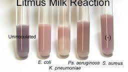
20 15-LITMUS MILK REACTIONS Purpose and Procedure Summary Bacteria can act on several different substrates in litmus milk, including lactose, casein, and litmus, causing a variety of reactions that are specific for each species of bacteria. Litmus milk contains skim milk,the source of lactose and casein, and litmus, the ph/oxidation reduction indicator. Litmus is purplish-blue at the initial ph of 6.8 in the uninoculated medium. Several other colors for litmus are possible after bacterial growth occurs, depending on the action of the isolate. An isolate is inoculated into a tube with a sterile transfer loop. The tube is incubated at 35 C for24-48 hours before it is examined for changes. 20
21 Tips for Success - Compare results to an uninoculated control tube to determine whether changes have occurred in the medium. An additional incubation of hours may Be necessary for some reactions to develop. The blue of the alkaline reaction is most evident at the top of the medium,while the white of litmus reduction is most evident at the bottom Expected Results The variety of reactions possible in litmus milk, are the following: lactose fermentation (A): Lactose fermentation releases lactic acid, which lowers the ph of the medium to 4.5, causing the litmus to turn pink. A pink color indicates lactose fermentation alkaline reaction (Alk): The action of bacteria on the nitrogen-containing components of skim milk causes ammonia to be released into the medium. As a result, the ph increases to 8.3-and the litmus turns blue.a blue color indicates an alkaline reaction clot formation (C): Either the precipitation of casein by lactic acid or the action of the enzymerenn in on casein may cause the formation of a clot, which appears as a white mass in the bottom of the tube reduction of litmus (R): If litmus is used as an electron acceptor during lactose fermentation, it is 21
22 22
23 16- Triple sugar-iron test Triple sugar iron (TSI) agar and Kligler iron agar (KIA) are used to determine both carbohydrate fermentation and hydrogen sulfide production in enteric bacteria. These media contain a variety of substrates that enteric bacteria can utilize, including several carbohydrates, proteins, and thiosulfate. Different enteric bacteria utilize these substrates differently, so they can be distinguished based on their pattern of utilization. Both media contain beef extract, yeast extract, and peptone to support bacterial growth. The ferment able carbohydrates in TSI are glucose, lactose, and sucrose. KIA contains only glucose and lactose. Sodium thiosulfate is the source of sulfur for H 2 S production in both media.both TSI and KIA contain ferrous sulfate, which reacts withh 2 Sto form a black precipitate called ferrous sulfide. The ph indicator in both media is phenol red. Phenol red is red at the initial ph of 7.4 but turns yellow at an acidic ph and dark red at an alkaline ph. Agar is present in these two media as a solidifying agent. An isolate is inoculated on to a TSI or KIA slant using a sterile transfer needle. First,the butt is stabbed. Then,the needle is with drawn and the slant is streaked. The slant is incubated at 35 C for18-24 hours before examination for color changes. Procedure:- 1. Sterilize the inoculating needle in the blue flame of the Bunsen burner till red hot and then allowed to cool. 23
24 2. From the rack, take the broth tube containing the hour culture remove, theاااااااااا cap and flame the neck of the tube. 3. Using aseptic technique, take the culture of the organism from the tube with the needle. 4. Again flame the neck of the tube and replace the tube in the test tube rack. 5. Take a sterile TSI slant tube from the rack, remove the cap and flame the tube. neckااااااااااا of the 6. Stab the needle containing the pure culture in to the medium, up to the butt surface ofاااااااااا the TSI tube, and then streak the needle back and forth along the slant. theاofاااااااااا 7. Again flame the neck of the TSI tube, cap it and place it in the test tube rack. 8. Incubate at 37 o c for 18 to 24 hours. Expected Results: 1. Alkaline slant (red) and acid butt (yellow) with or without gas production (breaks in the agar butt): Only glucose fermentation has occurred. The organisms preferentially degrade glucose first. Since this substrate is present in minimal concentration, the small amount of the acid produced on the slant surface is oxidized rapidly. The peptones in the medium are also used in the production of alkali. At the butt, the acid reaction is maintained because of the reduced oxygen tension and slower growth of the organisms. 2. Acid slant (yellow) and acid butt (yellow) with or without gas production: 24
25 Lactose or sucrose fermentation has occurred. Since these substances are present in higher concentrations, they serve as substrates for continued fermentative activities with maintenance of an acid reaction in both the slant and the butt. 3. Alkaline slant (red) and alkaline butt (red) or no change (orange-red) butt: No carbohydrate fermentation has occurred. Instead; peptones are catabolized under anaerobic and /or aerobic conditions resulting in alkaline ph due to production of ammonia. If only aerobic degradation of peptones occurs, the alkaline reaction is evidenced only on the slant surface. If there is aerobic and anaerobic utilization of peptone, the alkaline reaction is present on the slant and the butt. 4. Hydrogen sulfide (H2S) production: Some bacteria utilize thiosulfate anion as a terminal electron acceptor, reducing it to sulfide. If this occurs, the newly-formed hydrogen sulfide (H2S) reacts with ferrous sulfate in the medium to form ferrous sulfide, which is visible as a black precipitate. The blackening of the medium is almost always observed in the butt (bottom) of the medium. 5. Carbon dioxide (CO2) production: It is recognized simply as bubbles of gas between the agar and the wall of the tube or within the agar itself. The carbon dioxide production is sufficient to split the agar into two or more sections. To obtain accurate results, it is absolutely essential to observe the cultures within hours following incubation. This will ensure that the carbohydrate substrates have not been depleted and that degradation of peptones yielding alkaline end products has not taken place. 25

26 Result (slant/butt) Symbol Interpretation 1 Red/Yellow K/A Glucose fermentation only, peptone catabolized. 2 Yellow/Yellow A/A Glucose and lactose and/or sucrose fermentation. 3 Red/Red K/K No fermentation, Peptone catabolized. 4 Yellow/Yellow with bubbles A/A,G Glucose and lactose and/or sucrose fermentation, Gas produced. 5 Red/Yellow with bubbles K/A,G Glucose fermentation only, Gas produced. 6 7 Red/Yellow with bubbles and black precipitate Yellow/Yellow with bubbles and black precipitate K/A,G,H2S A/A,G,H2S Glucose fermentation only, Gas produced, H2S produced. Glucose and lactose and/or sucrose fermentation, Gas produced, H2S produced. 8 Red/Yellow with black precipitate K/A,H2S Glucose fermentation only, H2S produced. 9 Yellow/Yellow with black precipitate A/A,H2S Glucose and lactose and/or sucrose fermentation, H2S produced. 13-LITMUS MILK REACTIONS Purpose and Procedure Summary Bacteria can act on several different substrates in litmus milk, including lactose, casein, and litmus, causing a variety of reactions that are specific for each species 26
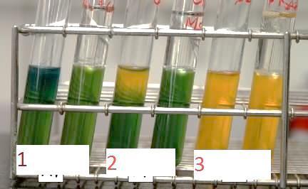
27 of bacteria. Litmus milk contains skim milk,the source of lactose and casein, and litmus, the ph/oxidation reduction indicator. Litmus is purplish-blue at the initial ph of 6.8 in the uninoculated medium. Several other colors for litmus are possible after bacterial growth occurs, depending on the action of the isolate. An isolate is inoculated into a tube with a sterile transfer loop. The tube is incubated at 35 C for24-48 hours before it is examined for changes. Tips for Success - Compare results to an uninoculated control tube to determine whether changes have occurred in the medium. An additional incubation of hours may Be necessary for some reactions to develop. The blue of the alkaline reaction is most evident at the top of the medium,while the white of litmus reduction is most evident at the bottom. 17- Oxidation - Fermentation Test (O-F test) Principle To determine the oxidative or fermentative metabolism of a carbohydrate or its non-utilization. Fermentation is an anaerobic process and bacterial fomenters of carbohydrates are usually facultative anaerobes. Oxidation is an aerobic process and bacterial oxidizers are usually strict aerobes. 27
28 Hugh and Leif son medium is a semi-solid medium in tubes containing glucose and a ph indicator bromothymol blue which turn to yellow in acids production and turn to blue in alkalinity. Carbohydrate conversion to acidic products can occur either aerobically by oxidation or an aerobically by fermentation. Bacteria that oxidize carbohydrates are called aerobes, while bacteria that ferment carbohydrates are called facultative anaerobes. The oxidation-fermentation (O-F) test is used to determine whether bacteria are aerobes or facultative anaerobes. The O-F test distinguishes Gram negative rods that are aerobes, such as Pseudomonas aeruginosa, from those that are facultative anaerobes, such as Escherichia coli. The O-F test also distinguishes Gram-positive cocci that are aerobes, such as species of Micrococcus,from those that are facultative anaerobes, such as species of Staphylococcus. Oxidation-fermentation (O-F) medium is used for this test. This medium contains a low concentration of peptone, but enough to support growth. A low concentration of peptone is essential to limit the formation of alkaline products that would neutralize the effect of acidic products. A carbohydrate, such as glucose, lactose, maltose, sucrose, mannitol,or xylose, is added at a high concentration. This high concentration promotes carbohydrate utilization, with the resulting formation of acidic products. The ph indicator, bromothymol blue,is green at the initial ph of 7.1 and yellow at a ph of 6.0. Agar is added at a low concentration to make the medium semisolid. Although phenol red broth can also be Used for the O-F test, it is not recommended because it has a high concentration of peptone, which May result in the formation of sufficient alkaline products to neutralize the effect of acidic products. 28
29 An isolate is inoculated into two tubes of O-F Medium with a sterile transfer needle. The medium in one tube is then covered with 2-3 ml of sterile mineral oil to create an anaerobic environment. The medium in the other tube is left open to the air to. provide an aerobic environment. The two tubes are incubated at 35C for 48 hours before examination for color change. Procedure Two tubes are inoculated by stabbing and one is immediately sealed with oil to produce anaerobic conditions 1. Organisms that cannot break down the carbohydrate aerobically or anaerobically eg Alcaligenes feacalis produce an alkaline reaction in the open tube and no change in the covered tube 2. Oxidizing organisms Pseudomonas spp produce an acid reaction in the open tube only 3. Fermenting organisms Enterobacteriaceae produce an acid reaction throughout the medium in both tubes 29
30 30
Sections 11 & 12: Isolation and Identification of Enterobacteriaceae
 Sections 11 & 12: Isolation and Identification of Enterobacteriaceae The family Enterobacteriaceae includes many genera and species. The last edition of Bergey s Manual of Systematic Bacteriology (Vol.
Biochemical Testing Handout
 Biochemical Testing Handout As you guys know, the purpose of a medical microbiology laboratory is to mainly isolate and identify organisms to provide proper treatment. For this week we will focus on five
Exercise 15-B PHYSIOLOGICAL CHARACTERISTICS OF BACTERIA CONTINUED: AMINO ACID DECARBOXYLATION, CITRATE UTILIZATION, COAGULASE & CAMP TESTS
 Exercise 15-B PHYSIOLOGICAL CHARACTERISTICS OF BACTERIA CONTINUED: AMINO ACID DECARBOXYLATION, CITRATE UTILIZATION, COAGULASE & CAMP TESTS Decarboxylation of Amino Acids and Amine Production The decarboxylation
Gram-negative rods. Enterobacteriaceae. Biochemical Reactions. Manal AL khulaifi
 Gram-negative rods Enterobacteriaceae Biochemical Reactions Bacteria Gram positive Gram negative Cocci Bacilli Cocci Rods Characters of Enterobacteriaceae All Enterobacteriaciae Gram-negative rods Reduce
Microbiology Activity #6 Metabolism of Small Molecules.
 Microbiology Activity #6 Metabolism of Small Molecules. Analysis of Carbohydrate Metabolism Organisms that use CO 2 as a carbon source and fix the carbon into biomass are autotrophs, usually obtaining
IMViC: Indole, Methyl red, Voges-Proskauer, Citrate
 IMViC: Indole, Methyl red, Voges-Proskauer, Citrate + and H 2 S These 4 IMViC tests (actually 6 tests if you include motility and H 2 S) constitute, perhaps, the most critical tests used for identification
6/28/2016. Growth Media and Metabolism. Complex Media. Defined Media. Made from complex and rich ingredients
 Growth Media and Metabolism Complex Media Made from complex and rich ingredients Ex. Soya protein extracts Milk protein extracts Blood products Tomato juice, etc. Exact chemical composition unknown Can
EXERCISE. Proteins,Amino Acids, and Enzymes VII: Oxidase Test. Suggested Reading in Textbook. Pronunciation Guide. Materials per Student
 EXERCISE 30 Proteins,Amino Acids, SAFETY CONSIDERATIONS Be careful with the Bunsen burner flame. No mouth pipetting. The oxidase reagent is caustic. Avoid contact with eyes and skin. In case of contact,
ID of Most Common Bacterial Pathogens. CLS 417- Clinical Practice in Microbiology Miss Zeina Alkudmani
 ID of Most Common Bacterial Pathogens CLS 417- Clinical Practice in Microbiology Miss Zeina Alkudmani BACTERIA Gram Positive Gram Negative Cocci Bacilli Bacilli Cocci Coccobacilli - Staph - Strept - Clostridium
TSI AGAR INTENDED USE
 TSI AGAR INTENDED USE TSI (Triple Sugar Iron) Agar is used for the identification of enterobacteria by the rapid detection of the fermentation of lactose, glucose (with or without gas production) and of
Pathogenic bacteria. Lab 6: Taxonomy: Kingdom: Bacteria Phylum: Proteobacteria Class: Gammaproteobacteria Order: Enterobacteriales
 Level 5 Pathogenic bacteria Lab 6: Family: Enterobacteriaceae Taxonomy: Kingdom: Bacteria Phylum: Proteobacteria Class: Gammaproteobacteria Order: Enterobacteriales Family: Enterobacteriaceae The prefix
9.1 Introduction 9.2 Importance of Biochemical Tests 9.3 Biochemical Characteristics
 Food Microbiology and Safety Practical Manual PRACTICAL 9 Structure 9.1 Introduction 9.2 Importance of Biochemical Tests 9.3 Biochemical Characteristics BIOCHEMICAL TESTS BACTERIAL TESTING 9.3.1 Tests
Microbiological Methods V-A- 1 SALMONELLA SPECIES PRESUMPTIVE AND CONFIRMATION TESTS
 Microbiological Methods V-A- 1 PRESUMPTIVE AND CONFIRMATION TESTS PRINCIPLE SCOPE Enrichment and selective procedures are used to provide a reasonably sensitive, definitive and versatile means of qualitatively
Lab #9. Introduction. Class samples:
 Lab #9 Introduction Food-borne illness is largely caused by the presence of bacteria in red meat. However, much of these harmful bacteria can be destroyed and prevented by sanitation and safe cooking practices.
Detection of microbial enzyme : Amylase, lipase, gelatinase, catalase, urease, nitrate reductase, protease and coagulase
 Detection of microbial enzyme : Amylase, lipase, gelatinase, catalase, urease, nitrate reductase, protease and coagulase To detect amylase enzyme production Introduction: Amylase is hydrolytic enzyme produced
APPLICATION Detection and isolation of pathogenic intestinal bacteria including Shigella and Salmonella from surfaces, food, or liquid samples.
 HEK/SS Code 5543 COMING SOON! BioPaddles Colony Identification App Hektoen Enteric Agar (HEK) Salmonella Shigella Agar (SS) USE: Detection and isolation of pathogenic intestinal bacteria including Shigella
Bacterial Metabolism & Growth Characteristics. Stijn van der Veen
 Bacterial Metabolism & Growth Characteristics Stijn van der Veen Differentiating bacterial species Morphology (shape) Composition (cell envelope and other structures) Metabolism & growth characteristics
Principles of biochemical tests commonly used in the identification of gram-negative bacteria
 Dr. Khoramrooz 1 In the name of God Department Of Microbiology Yasouj University of Medical Science Principles of biochemical tests commonly used in the identification of gram-negative bacteria By: Dr.
Identification of Unknown Indigenous Bacteria
 April 29, 2009 Identification of Unknown Indigenous Bacteria Introduction Many bacteria can be found in and on nearly all areas of the healthy human body. These bacteria are referred to as normal flora
(1946), and Elek (1948) have described different methods. Stuart, van Stratum, and Rustigian (1945) found the method of Rustigian
, and Elek (1948) have described different methods. Stuart, van Stratum, and Rustigian (1945) found the method of Rustigian") A COMPARISON OF THE PHENYLPYRUVIC ACID REACTION AND THE UREASE TEST IN THE DIFFERENTIATION OF PROTEUS FROM OTHER ENTERIC ORGANISMS SVERRE DICK HENRIKSEN State Institute for Public Health, Bacteriological
A COMPARISON OF THE PHENYLPYRUVIC ACID REACTION AND THE UREASE TEST IN THE DIFFERENTIATION OF PROTEUS FROM OTHER ENTERIC ORGANISMS SVERRE DICK HENRIKSEN State Institute for Public Health, Bacteriological
CHAPTER IV ISOLATION AND IDENTIFICATION OF BACTERIA FROM SEPSIS SAMPLES
 62 CHAPTER IV ISOLATION AND IDENTIFICATION OF BACTERIA FROM SEPSIS SAMPLES 4.1 INTRODUCTION Infectious diseases remain major cause of mortality in both child and maternal populations. The mortality rate
NOTE: Poor growth and a weak esculin reaction may be seen after 40 hours of incubation for some enterococci.
 LIS/EMB Code 5542 COMING SOON! BioPaddles Colony Identification App Listeria Agar (LIS) Eosin Methylene Blue Agar (EMB) USE: Enumeration and selective isolation of Listeria spp.(lis) Isolation and differentiation
GB Translated English of Chinese Standard: GB NATIONAL STANDARD OF THE
 Translated English of Chinese Standard: GB4789.30-2016 www.chinesestandard.net Buy True-PDF Auto-delivery. Sales@ChineseStandard.net GB NATIONAL STANDARD OF THE PEOPLE S REPUBLIC OF CHINA GB 4789.30-2016
USE: Isolation and differentiation of Gram (-) enteric bacilli (MAC) / Coliform Testing / Recovery of Stressed Coliforms (EMB)
 enteric bacilli (MAC) / Coliform Testing / Recovery of Stressed Coliforms (EMB)") MAC/EMB Code 5544 MacConkey Agar (MAC) Eosin Methylene Blue Agar (EMB) USE: Isolation and differentiation of Gram (-) enteric bacilli (MAC) / Coliform Testing / Recovery of Stressed Coliforms (EMB) Side
MAC/EMB Code 5544 MacConkey Agar (MAC) Eosin Methylene Blue Agar (EMB) USE: Isolation and differentiation of Gram (-) enteric bacilli (MAC) / Coliform Testing / Recovery of Stressed Coliforms (EMB) Side
6 The chemistry of living organisms
 Living organisms are composed of about 22 different chemical elements. These are combined to form a great variety of compounds. Six major elements make up almost 99% of the mass of the human body, as shown
Tests for Carbohydrates
 Goals bserve physical and chemical properties of some common carbohydrates. Use physical and chemical tests to distinguish between monosaccharides, disaccharides, and polysaccharides. Identify an unknown
Lab 6: Cellular Respiration
 Lab 6: Cellular Respiration Metabolism is the sum of all chemical reactions in a living organism. These reactions can be catabolic or anabolic. Anabolic reactions use up energy to actually build complex
NOVASTREAK. Microbial Contamination Monitoring Device TYPICAL CULTURAL MORPHOLOGY Baird Parker Agar. S. aureus growth on Baird Parker Agar
 NOVASTREAK Microbial Contamination Monitoring Device TYPICAL CULTURAL MORPHOLOGY Baird Parker Agar S. aureus growth on Baird Parker Agar Baird Parker Agar is used for the selective isolation and enumeration
AN NEXURE. B log Sodium chloride 5g Distilled water (DW) 1 Litre ph: ] g 100 ml pl-l: g Glucose
![AN NEXURE. B log Sodium chloride 5g Distilled water (DW) 1 Litre ph: ] g 100 ml pl-l: g Glucose](/thumbs/82/85681362.jpg "AN NEXURE. B log Sodium chloride 5g Distilled water (DW) 1 Litre ph: ] g 100 ml pl-l: g Glucose") AN NEXURE A. Composition of bacteriological media l. Alkaline Water (APW) B log Sodium chloride 5g Distilled water () 1 Litre ph: 9.110.] 2. Brilliant Green Bile Broth (BGLB ) Bile salt Brilliant green
AN NEXURE A. Composition of bacteriological media l. Alkaline Water (APW) B log Sodium chloride 5g Distilled water () 1 Litre ph: 9.110.] 2. Brilliant Green Bile Broth (BGLB ) Bile salt Brilliant green
HARMONISED PHARMACOPOEIA DEHYDRATED CULTURE MEDIA FOR SUPPORTING REGULATORY COMPLIANCE AVAILABLE NOW P O RTF O LIO.
 DEHYDRATED CULTURE MEDIA FOR ENHANCED P O RTF O LIO AVAILABLE NOW HARMONISED PHARMACOPOEIA SUPPORTING REGULATORY COMPLIANCE A Neogen Company THE GATEWAY TO MICROBIOLOGY INTRODUCTION Harmonised Pharmacopoeia;
S. aureus NCTC 6571, E. coli NCTC (antibiotic
 ISO Sensitivity Test Agar Code: KM1204 A semi-defined nutritionally rich sensitivity medium. It is composed of specially selected peptones with a small amount of glucose, solidified with a very pure agar
Multi-Biochemical Test System for Distinguishing
 APuPED MICROBIOLOGY, Sept. 1971, p. 8-1 Vol., No. Copyright 1971 American Society for Microbiology Printed in U.S.A. Multi-Biochemical Test System for Distinguishing Enteric and Other Gram-Negative Bacilli
LAB 4 Macromolecules
 LAB 4 Macromolecules Overview In addition to water and minerals, living things contain a variety of organic molecules. Most of the organic molecules in living organisms are of 4 basic types: carbohydrate,
Manal AL khulaifi. Enterobacteriaceae
 Enterobacteriaceae Characteristics E.coli Most significant species in the genus Important potential pathogen in humans Common isolate from colon flora Dry, pink (lactose positive) pink colony with area
Selective Growth Media for Differentiation and Detection of Escherichia Coli and Other Coliforms
 Page 1 of 5 Page 1 of 5 Return to Web Version Selective Growth Media for Differentiation and Detection of Escherichia Coli and Other Coliforms By: Jvo Siegrist, AnalytiX Volume 8 Article 4 E. coli and
CHAPTER V TAXONOMIC STUDIES OF THE SELECTED ISOLATE C 9
 CHAPTER V TAXONOMIC STUDIES OF THE SELECTED ISOLATE C 9 Selection of media for taxonomic studies: Culture media used for taxonomic studies on actinomycetes comprise: 1) Media used for characterization
Figure 2. Figure 1. Name: Bio AP Lab Organic Molecules
 Name: Bio AP Lab Organic Molecules BACKGROUND: A cell is a living chemistry laboratory in which most functions take the form of interactions between organic molecules. Most organic molecules found in living
KLIGLER IRON AGAR 1/5
 KLIGLER IRON AGAR INTENDED USE Kligler Iron Agar is used for the identification of enterobacteria by the rapid detection of lactose and glucose fermentation (with or without gas production), as well as
Phases of the bacterial growth:
 L3: Physiology of Bacteria: Bacterial growth Growth is the orderly increase in the sum of all the components of an organism. Cell multiplication is a consequence of growth, in unicellular organism, growth
INTERNATIONAL JOURNAL OF ENGINEERING SCIENCES & RESEARCH TECHNOLOGY
 [Ravish, 2(2): Feb., 2013] ISSN: 2277-9655 IJESRT INTERNATIONAL JOURNAL OF ENGINEERING SCIENCES & RESEARCH TECHNOLOGY Isolation And Characterization Of Proteolytic Bacteria And Its Protease Himani Ravish
Ch 07. Microbial Metabolism
 Ch 07 Microbial Metabolism SLOs Differentiate between metabolism, catabolism, and anabolism. Fully describe the structure and function of enzymes. Differentiate between constitutive and regulated enzymes.
LAB 5 - Enzymes BACKGROUND INFORMATION
 LAB 5 - Enzymes BACKGROUND INFORMATION Chemical Reactions The cells of organisms, from bacteria to plants to animals, carry out hundreds to thousands of chemical reactions that must be properly coordinated
Figure 1. Bacterial growth curve.
 INTRODUCTION In order for suitable growth and division, a microorganism must be placed into a favorable environment. Bacterial growth refers to an increase in cell number rather than cell size. Bacteria
Laboratorios CONDA, S.A. Distributed by Separations
 Culture Media as on Pharmacopoeia 7.3, Harmonized Method for Microbiological Examination of non sterile products -FORMULATIONS Buffered sodium chloride-peptone solution ph 7.0 Cat. Nº 1401 Potassium dihydrogen
(LM pages 91 98) Time Estimate for Entire Lab: 2.5 to 3.0 hours. Special Requirements
 Time Estimate for Entire Lab: 2.5 to 3.0 hours. Special Requirements") Laboratory 7 Chemical Aspects of Digestion (LM pages 91 98) Time Estimate for Entire Lab: 2.5 to 3.0 hours Special Requirements Incubation. Students should start these sections at the beginning of the
Laboratory 7 Chemical Aspects of Digestion (LM pages 91 98) Time Estimate for Entire Lab: 2.5 to 3.0 hours Special Requirements Incubation. Students should start these sections at the beginning of the
Microbiology lab 2017
 Assignments General Directives The first page must include the following information: The assignment number Course code :BIO3126 Your name or names Your group number The date Assignments may be done and
National food safety standard. Food microbiological examination: Salmonella
 NATIONAL STANDARD OF THE PEOPLE S REPUBLIC OF CHINA GB 4789.4 2010 National food safety standard Food microbiological examination: Salmonella Issue date: 2010-03-26 Implementation date: 2010-06-01 Issued
QUALITATIVE TESTS OF CARBOHYDRATE
 QUALITATIVE TESTS OF CARBOHYDRATE MACROMOLECULE CARBOHYDRATES Are the key source of energy used by living things. Also serve as extracellular structural elements as in cell wall of bacteria and plant.
320 MBIO Microbial Diagnosis. Aljawharah F. Alabbad Noorah A. Alkubaisi 2017
 320 MBIO Microbial Diagnosis Aljawharah F. Alabbad Noorah A. Alkubaisi 2017 Pathogens of the Urinary tract The urinary system is composed of organs that regulate the chemical composition and volume of
Medical Microbiology
 Lecture 5!!!!!!ƒš!!Œ!!! š!!œ!! Œ!!!! Dr. Ismail I. Daood Medical Microbiology!! Systematic Bacteriology Gram-Positive Cocci : GENUS : Staphylococcus : The general properties of Staphylococcus are Gram-
BACTERIAL EXAMINATION OF WATER
 BACTERIAL EXAMINATION OF WATER The bacteriological examination of water is performed routinely by water utilities and many governmental agencies to ensure a safe supply of water for drinking, bathing,
Staining Technology and Bright- Field Microscope Use
 Staining Technology and Bright- Field Microscope Use 2 Abstract We will introduce bright-field microscope use, practice Gram staining with foodborne pathogens, and practice endospore staining with Bacillus
Introduction to Microbiology BIOL 220, Summer Session 1, 1996 Exam # 2
 Name I. Multiple Choice (1 point each) Introduction to Microbiology BIOL 220, Summer Session 1, 1996 Exam # 2 D 1. Which transport process requires energy? A. Osmosis C. Diffusion B. Facilitated diffusion
A.F. GENITAL SYSTEM. ITEMS NECESSARY BUT NOT INCLUDED IN THE KIT A.F. GENITAL SYSTEM Reagent (ref ) Mycoplasma Transport Broth (ref.
 Mycoplasma Transport Broth (ref.") A.F. GENITAL SYSTEM ENGLISH System for detection, count and susceptibility test of pathogenic urogenital microorganisms DESCRIPTION A.F. GENITAL SYSTEM is a 24-well system containing desiccated biochemical
A.F. GENITAL SYSTEM ENGLISH System for detection, count and susceptibility test of pathogenic urogenital microorganisms DESCRIPTION A.F. GENITAL SYSTEM is a 24-well system containing desiccated biochemical
Biochemical Differentiation of the Enterobacteriaceae
 APPLIED MICROBIOLOGY, Mar., 1966 Copyright 1966 American Society for Microbiology Vol. 14, No. 2 Printed in U.S.A. Biochemical Differentiation of the Enterobacteriaceae with the Aid of -Iron-Agar JANE
Guided Inquiry Skills Lab. Additional Lab 1 Making Models of Macromolecules. Problem. Introduction. Skills Focus. Materials.
 Additional Lab 1 Making Models of Macromolecules Guided Inquiry Skills Lab Problem How do monomers join together to form polymers? Introduction A small number of elements make up most of the mass of your
Digestive Enzyme Lab
 Digestive Enzyme Lab Objectives 1. To describe the function of enzymes 2. To define: reactants, products, activation energy 3. To describe the enzymatic digestion of carbohydrates by salivary amylase 4.
Name: Period: Date: Testing for Biological Macromolecules Lab
 Testing for Biological Macromolecules Lab Introduction: All living organisms are composed of various types of organic molecules, such as carbohydrates, starches, proteins, lipids and nucleic acids. These
II- Streptococci. Practical 3. Objective: Required materials: Classification of Streptococci: Streptococci can be classified according to:
 Practical 3 II- Streptococci Objective: 1. Use of blood agar to differentiate between,, and hemolytic streptococci. 2. To know Gram reaction, shape and arrangement of streptococci. 3. To differentiate
MOTILE ENTEROCOCCI (STREPTOCOCCUS FAECIUM VAR. MOBILIS VAR. N.) ISOLATED FROM GRASS SILAGE
 ISOLATED FROM GRASS SILAGE") MOTILE ENTEROCOCCI (STREPTOCOCCUS FAECIUM VAR. MOBILIS VAR. N.) ISOLATED FROM GRASS SILAGE C. W. LANGSTON, JOYCE GUTIERREZ, AND CECELIA BOUMA Dairy Cattle Research Branch, Agricultural Research Center,
MOTILE ENTEROCOCCI (STREPTOCOCCUS FAECIUM VAR. MOBILIS VAR. N.) ISOLATED FROM GRASS SILAGE C. W. LANGSTON, JOYCE GUTIERREZ, AND CECELIA BOUMA Dairy Cattle Research Branch, Agricultural Research Center,
D. glycerol and fatty acids 4. Which is an example of an inorganic compound?
 Name: ate: 1. Glucose and maltose are classified as organic compounds because they are both 3. Which process is most directly responsible for the production of O 2 in these sugar solutions?. carbon-containing
For example, monosaccharides such as glucose are polar and soluble in water, whereas lipids are nonpolar and insoluble in water.
 Biology 4A Laboratory Biologically Important Molecules Objectives Perform tests to detect the presence of carbohydrates, lipids, proteins, and nucleic acids Recognize the importance of a control in a biochemical
1~~~~~~~~~~~~~~~~~~~~~~~~~~
 APPLIED AND ENVIRONMENTAL MICROBIOLOGY, Nov. 1985, p. 1213-1218 0099-2240/85/111213-06$02.00/0 Copyright C) 1985, American Society for Microbiology Vol. 50, No. 5 Characterization of Dysgonic, Heterotrophic
Chapter 5 MITOCHONDRIA AND RESPIRATION 5-1
 Chapter 5 MITOCHONDRIA AND RESPIRATION All organisms must transform energy. This energy is required to maintain a dynamic steady state, homeostasis, and to insure continued survival. As will be discussed
Scholars Research Library. Purification and characterization of neutral protease enzyme from Bacillus Subtilis
 Journal of Microbiology and Biotechnology Research Scholars Research Library J. Microbiol. Biotech. Res., 2012, 2 (4):612-618 (http://scholarsresearchlibrary.com/archive.html) Purification and characterization
BCH302 [Practical] 1
![BCH302 [Practical] 1](/thumbs/81/83984357.jpg "BCH302 [Practical] 1") BCH302 [Practical] 1 Carbohydrates are defined as the polyhydroxy aldehydes or polyhydroxy ketones. Most, but not all carbohydrate have a formula (CH 2 O)n (hence the name hydrate of carbon). Sugars ends
BCH302 [Practical] 1 Carbohydrates are defined as the polyhydroxy aldehydes or polyhydroxy ketones. Most, but not all carbohydrate have a formula (CH 2 O)n (hence the name hydrate of carbon). Sugars ends
Experiment 9 Amino Acids and Proteins
 Experiment 9 Amino Acids and Proteins Proteins are very important biological molecules, with many possible functions. Enzymes are proteins that catalyze biological reactions. There are transport proteins
QUALITATIVE ANALYSIS OF AMINO ACIDS AND PROTEINS
 QUALITATIVE ANALYSIS OF AMINO ACIDS AND PROTEINS Amino acids are molecules containing an amine group, a carboxylic acid group and a side chain that varies between different amino acids. Amino acids of
Aim: To study the effect of ph on the action of salivary amylase. NCERT
 Exercise 28 Aim: To study the effect of ph on the action of salivary amylase. Principle: Optimal activity for most of the enzymes is generally observed between ph 5.0 and 9.0. However, a few enzymes, e.g.,
Chapter 9: Cellular Respiration
 Chapter 9: Cellular Respiration To perform their many tasks, living cells require energy from outside sources. Energy stored in food utimately comes from the sun. Photosynthesis makes the raw materials
BACTERIAL EXAMINATION OF WATER
 BACTERIAL EXAMINATION OF WATER The bacteriological examination of water is performed routinely by water utilities and many governmental agencies to ensure a safe supply of water for drinking, bathing,
Detection of Hydrogen Sulfide Production by Bacteria using Paper Disc Methods12
 19581 PAPER DISCS FOR HYDROGEN SULFIDE TEST 193 enteric pathogens and coliform bacteria. J. Bacteriol., 67, 537-541. NETER, E. R. AND CLARK, D. 1944 The effectiveness of different culture media in the
Enzymes - Exercise 3 - Rockville
 Enzymes - Exercise 3 - Rockville Objectives -Understand the function of an enzyme. -Know what the substrate, enzyme, and the product of the reaction for this lab. -Understand how at various environments
Lab 2. The Chemistry of Life
 Lab 2 Learning Objectives Compare and contrast organic and inorganic molecules Relate hydrogen bonding to macromolecules found in living things Compare and contrast the four major organic macromolecules:
Characterization of Bacteria by Their Degradation of Amino Acids
 APPLIED MICROBIOLOGY, Oct. 1968, P. 1591-1595 Copyright 1968 American Society for Microbiology Vol. 16, No. 10 Printed in U.S.A. Characterization of Bacteria by Their Degradation of Amino Acids M. J. PICKETT
Orderly increase in all the chemical structures of the cell. Cell multiplication. Increase in the number of the cells
 GROWTH OF BACTERIA Growth Orderly increase in all the chemical structures of the cell Cell multiplication Increase in the number of the cells In natural habitat In or on another organism (infection) In
Labquality External Quality Assesment Programmes General Bacteriology 1 1/2010
 Labquality External Quality Assesment Programmes General Bacteriology 1 1/2010 Photos and text: Markku Koskela, M.D., Ph.D. Clinical microbiology specialist Oulu, Finland Sample 1/2010 Pus from an infected
BACTERIAL CONTAMINANTS ASSOCIATED WITH COMMERCIAL POULTRY FEEDS IN ENUGU NIGERIA
 Int. J. LifeSc. Bt & Pharm. Res. 2013 Onyeze Rosemary C et al., 2013 Research Paper ISSN 2250-3137 www.ijlbpr.com Vol. 2, No. 3, July 2013 2013 IJLBPR. All Rights Reserved BACTERIAL CONTAMINANTS ASSOCIATED
9. At about 0 C., most enzymes are (1.) inactive (2.) active (3.) destroyed (4.) replicated
 inactive (2.) active (3.) destroyed (4.) replicated") Study Guide 1. Which of the following enzymes would digest a fat? (1.) sucrase (2.) fatase (3.) protease (4.) lipase 2. At high temperatures, the rate of enzyme action decreases because the increased heat
Study Guide 1. Which of the following enzymes would digest a fat? (1.) sucrase (2.) fatase (3.) protease (4.) lipase 2. At high temperatures, the rate of enzyme action decreases because the increased heat
OCR (A) Biology A-level
 Biology A-level") OCR (A) Biology A-level Topic 2.2: Biological molecules Notes Water Water is a very important molecule which is a major component of cells, for instance: Water is a polar molecule due to uneven distribution
OCR (A) Biology A-level Topic 2.2: Biological molecules Notes Water Water is a very important molecule which is a major component of cells, for instance: Water is a polar molecule due to uneven distribution
staphylococci. They found that of 28 strains of staphylococci from foods STAPHYLOCOCCI AND RELATED VARIETIES
 A COMPARATIVE STUDY OF KNOWVN FOOD-POISONING STAPHYLOCOCCI AND RELATED VARIETIES JAMES B. EVANS AND C. F. NIVEN, JR. Division of Bacteriology, American Meat Institute Foundation, and the Department of
ISOLATION OF BACTERIAL ORGANISMS FROM BILE AND INTESTINAL CONTENT OF APPARENTLY HEALTHY SLAUGHTERED CHICKENS IN JOS AND ENVIRONS
 ISOLATION OF BACTERIAL ORGANISMS FROM BILE AND INTESTINAL CONTENT OF APPARENTLY HEALTHY SLAUGHTERED CHICKENS IN JOS AND ENVIRONS BOT C.J 1* ; WOMA T. Y 2 ; ABIAYI E 3 ; MANGUT P. E 1 ; and ISHAYA D 4 1
Biology 20 Laboratory Life s Macromolecules OBJECTIVE INTRODUCTION
 Biology 20 Laboratory Life s Macromolecules OBJECTIVE To observe and record reactions between three classes of macromolecules in the presence of simple chemical indictors. To be able to distinguish positive
Materials and Methods
 Chapter-3 Materials and Methods 3.1 Isolation of Escherichia coli strain: 10 ml of sewage was collected and the residual solid matter was removed by filteration using Whatman No.1 filter paper. 0.1 ml
Lecture 3. Microbial Physiology
 Micro-Biology For 3 rd Sem. Students of ISM-IUK, Bishkek Lecture 3 Microbial Physiology LECTURE OBJECTIVES 1. Bacterial Growth 2. Growth Requirements 3. Nutritional types of microorganisms 4. Enzymes,
GB Translated English of Chinese Standard: GB NATIONAL STANDARD OF
 Translated English of Chinese Standard: GB4789.40-2016 www.chinesestandard.net Sales@ChineseStandard.net GB NATIONAL STANDARD OF THE PEOPLE S REPUBLIC OF CHINA GB 4789.40-2016 National Food Safety Standard
Macromolecules Materials
 Macromolecules Materials Item per bench per class Test tubes 19 a bunch Benedict s reagent 1 bottle 6 Iodine bottle 1 bottle 6 Sudan IV bottle 1 bottle 6 Biuret s Bottle 1 bottle 6 250 ml beaker 1 6 heat
Investigation: Enzymes
 Investigation: Enzymes INTRODUCTION: What would happen to your cells if they made a poisonous chemical? You might think that they would die. In fact, your cells are always making poisonous chemicals. They
Biological molecules = Biomolecules = Compounds of life
 Biological molecules = Biomolecules = Compounds of life Carbohydrates Proteins & Amino Acids Mono-saccharides Olego-saccharides Di-saccharides Poly-saccharides Lipids Oils & Fats Amino acids Proteins Enzymes
Microbial Quality Analysis of Milk and Flavoured Milk Products from Local Vendors in Vellore
 Microbial Quality Analysis of Milk and Flavoured Milk Products from Local Vendors in Vellore Aditya Sood*, Ridhi Sood, Abhijit Kumar, Gaganjot Kaur, Candy Sidhu Assistant Professor, Chandigarh University,
Blue coloring. Enrichment medium for the simultaneous detection of total coliforms and Escherichia coli in water, foods and dairy products.
 s have proved to be a powerful tool in the identification of microorganisms due to their detection of specific enzymes produced by the target microorganism. The enzymes act as catalysts of the chromogenic,
STUDIES ON THE PROTEOLYTIC BACTERIA OF MILK
 STUDIES ON THE PROTEOLYTIC BACTERIA OF MILK III. ACTION OF PROTEOLYTIC BACTERIA OF MILK ON CASEIN AND GELATIN WILLIAM C. FRAZIER AND PHILIP RUPP From the Research Laboratories, Bureau of Dairy Industry,
Simpson (1928), Julianelle (1937), Thompson and Khorazo. that the pathogenic strains, (Staphylococcus aureus and Staphylococcus
, Julianelle (1937), Thompson and Khorazo. that the pathogenic strains, (Staphylococcus aureus and Staphylococcus") THE RELATION OF AEROBIOSIS TO THE FERMENTATION OF MANNITOL BY STAPHYLOCOCCI EUGENIA VALENTINE COLWELL Laboratory of Industrial Hygiene Inc., New York City Received for publication August 5, 1938 While
THE RELATION OF AEROBIOSIS TO THE FERMENTATION OF MANNITOL BY STAPHYLOCOCCI EUGENIA VALENTINE COLWELL Laboratory of Industrial Hygiene Inc., New York City Received for publication August 5, 1938 While
Isolation and Biochemical Characterization of Lactobacillus species Isolated from Dahi
 International Journal of Current Microbiology and Applied Sciences ISSN: 2319-7706 Volume 5 Number 4 (2016) pp. 1042-1049 Journal homepage: http://www.ijcmas.com Original Research Article http://dx.doi.org/10.20546/ijcmas.2016.504.119
Interpretation Guide. Enterobacteriaceae Count Plate
 Interpretation Guide The 3M Petrifilm Enterobacteriaceae Count Plate is a sample-ready-culture medium system that contains modified Violet Red Bile Glucose (VRBG) nutrients, a cold-watersoluble gelling
Cellular Respiration: Harvesting Chemical Energy CHAPTER 9
 Cellular Respiration: Harvesting Chemical Energy CHAPTER 9 9.1 Metabolic pathways that release energy are exergonic and considered catabolic pathways. Fermentation: partial degradation of sugars that occurs
ا.م.د.هيفاء الحديثي. Enterobacteriaceae
 ا.م.د.هيفاء الحديثي Bacteriology Genus Salmonella Enterobacteriaceae - Pathogenic for human and animals - They are gram negative rods, motile with peritrichous flagella except Gallinarum-pullorum - Ferment
Available Online through
 ISSN: 0975-766X CODEN: IJPTFI Available Online through Review Article www.ijptonline.com BIOCHEMICAL TESTS FOR THE IDENTIFICATION OF BACTERIA Vinay Reddy Gopireddy* HOD, Dept of Microbiology, PMR Pg College,
Background knowledge
 Background knowledge This is the required background knowledge: State three uses of energy in living things Give an example of an energy conversion in a living organism State that fats and oils contain