Ultrasound of Mid and Hindfoot Pathology
|
|
|
- Ronald Kevin Woods
- 5 years ago
- Views:
Transcription
1 Ultrasound of Mid and Hindfoot Pathology Levon N. Nazarian, M.D. Professor of Radiology Thomas Jefferson University Hospital
2 Disclosures None relevant to this presentation
3 Educational Objective Following the presentation, participant should be able to: Discuss the role of US in imaging the mid and hind foot Identify mid and hind foot pathologies seen on US
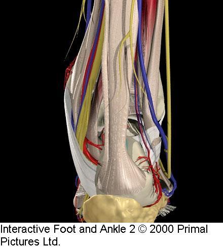
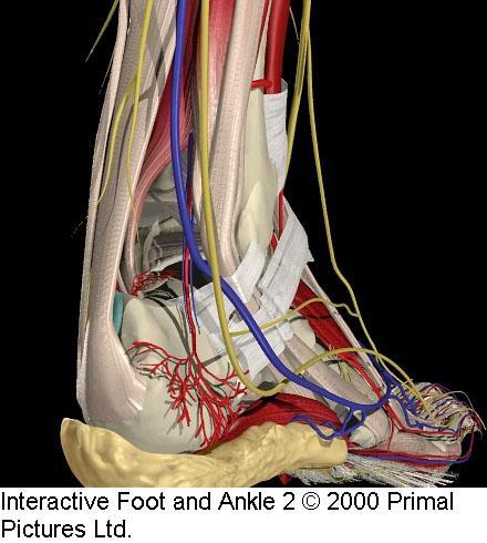
4 Posterior Compartment
5 Achilles Tendon Most commonly injured tendon No sheath: surrounded by two echogenic lines (paratenon) Normal AP thickness 5-6 mm

6 Longitudinal Achilles Tendon

7 Achilles Tendon Insertion


8 Transverse Achilles Tendon Direct contact Heaped-up scanning gel
9 Complete Achilles Rupture Weekend warriors Hematoma and fat may occupy torn area Image in plantar flexion to measure retraction


10 Complete Achilles Rupture Sagittal Transverse

11 Complete Achilles Tear: Extended Field of View


12 Complete Achilles Rupture Dorsiflexion Plantar flexion

13 Pitfall: Do not mistake plantaris tendon for intact Achilles fibers

14 Achilles Rupture With Refractive Shadowing

15 Achilles Rupture With Refractive Shadowing

16 Achilles Rupture With Refractive Shadowing

17 Achilles Rupture With Refractive Shadowing

18 Pitfall: Do Not Mistake Thickened Paratenon for Intact Achilles Fibers

19 High Grade Partial Achilles Tear

20 High Grade Partial Achilles Tear

21 High Grade Partial Achilles Tear

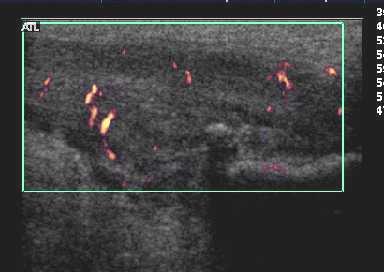
22 High Grade Partial Achilles Tear

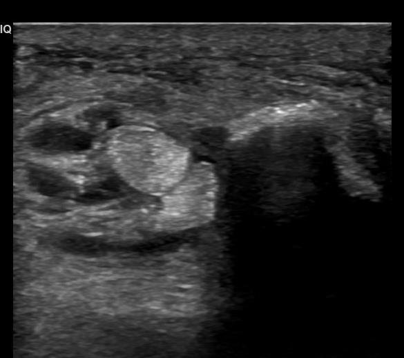
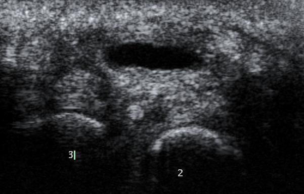
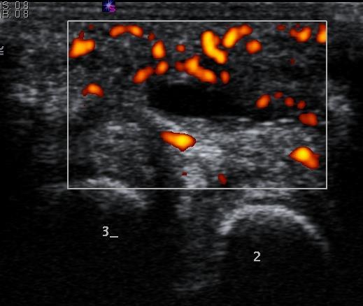
23 High Grade Partial Achilles Tear
24 Intrasubstance Achilles Tear
25 Chronic Tendinitis (Tendinosis) Thickened, heterogeneous tendon Nodular hypoechoic areas Partial tears Calcification May or may not see flow
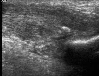
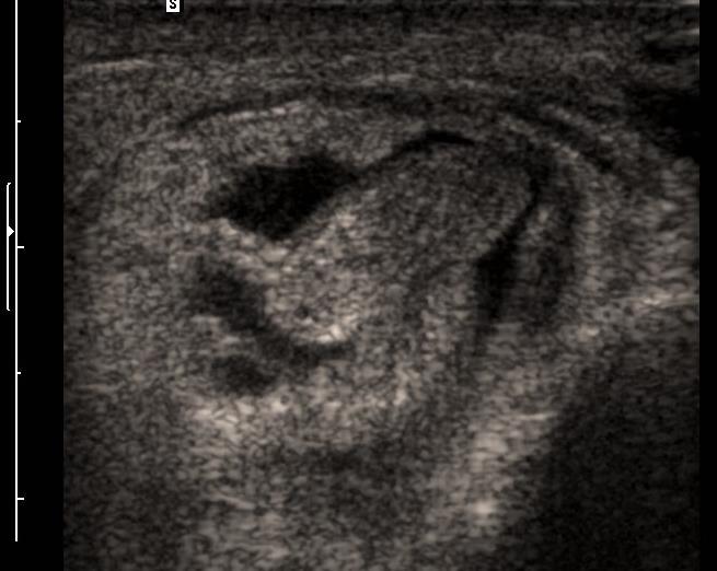
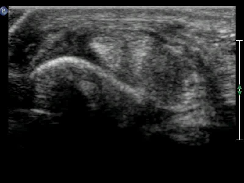
26 Insertional Achilles Tendinosis
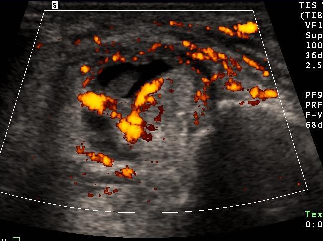
27 Midsubstance Achilles Tendinosis

28 Achilles Tendinosis Calc
29 Retrocalcaneal Bursitis


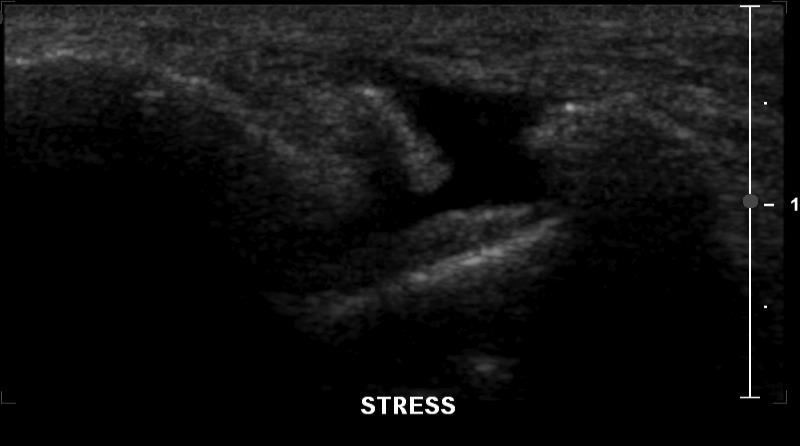
30 53 Year Old Man with Foot Pain


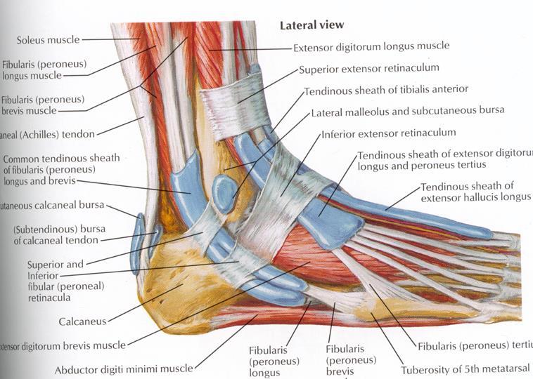
31 Aspiration Performed with 18 G Needle Result: Monosodium Urate Crystals Needle tip

32 Fluid in Superficial Achilles Bursa

33 Os Trigonum

34 Posterior Ankle Impingement With Os Trigonum

35 Posterior Ankle Impingement With Os Trigonum
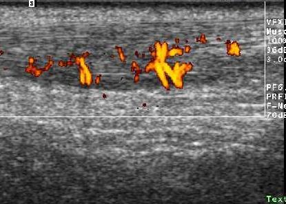
36
37

38 Peroneal Tendons: Axial PL PB

39 Peroneal Tendons: Axial

40 Peroneal Tendons: Axial

41 Peroneal Tendons: Axial

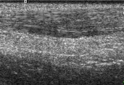
42 Peroneal Tendons: Longitudinal PL PB
43 Peroneal Tenosynovitis
44 Peroneal Tenosynovitis
45 Peroneal Tenosynovitis
46 Peroneal Tenosynovitis

47 Peroneus Brevis Tendinosis

48 Dx: Peroneus Brevis Split PB PL PB Tear

49 Peroneal Retinaculum: Normal

50 Peroneal Retinaculum: Abnormal

51 Normal Peroneal Tendon Motion
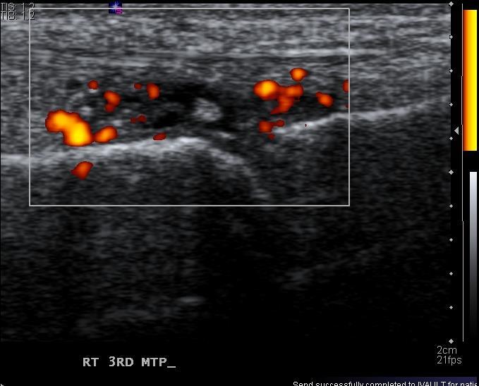
52 Peroneal Subluxation Injury to peroneal retinaculum Tendons exit groove anteriorly and laterally Elicited with dorsiflexion and eversion
53 Peroneal Subluxation
54 Peroneal Subluxation

55 Peroneus Brevis Tear, Subluxation
56 Intrasheath Subluxation

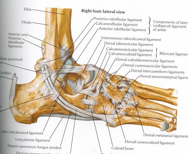
57 Peroneal Tendons: Intrasheath Subluxation Type A


58 Peroneal Tendons: Intrasheath Subluxation Type B

59 Distal Peroneus Brevis 5 th MT

60 Lateral Band Plantar Fasciitis

61 Lateral Band Plantar Fasciitis

62 Avulsion Fracture at Base of 5 th Metatarsal

63 Avulsion Fracture at Base of 5 th Metatarsal

64 Lateral Ankle Ligaments
65 Anterior Talofibular Ligament Most commonly injured ankle ligament Spectrum of injury from mild sprain to complete tear May see associated avulsion fracture

66 ATFL

67 Normal ATFL

68 ATFL Sprain
69 Normal Contralateral ATFL


70 Chronic ATFL Sprain Fibula Talus

71 ATFL Sprain

72 Anterolateral Ankle Impingement

73 Anterior Ankle Impingement

74 Anterior Inferior Tibiofibular Ligament

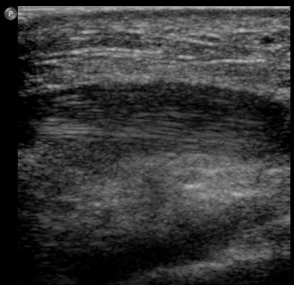
75 Calcaneofibular Ligament From: Sarrafian SK: Anatomy of the Foot and Ankle, 2 nd Ed., JB Lippincott

76 CFL Sprain

77 High Ankle Sprain
78 Plantar Fasciitis Painful condition, common in runners US diagnosis: Thickening (greater than 3-4mm) Heterogeneity Calcification Partial tears

79 Plantar Fascia: Anatomic Dissection

80 C Normal Plantar Fascia

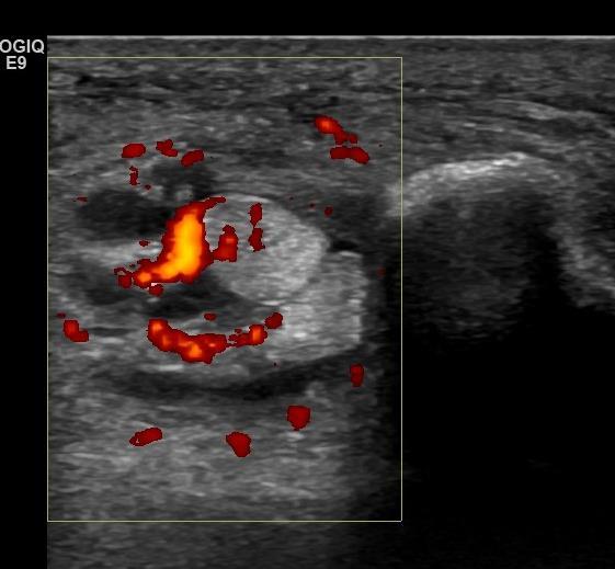
81 Plantar Fasciitis


82 Plantar Fibromatosis 44 year-old woman Longitudinal Transverse


83 Plantar Fibromatosis 44 year-old woman Contralateral side: same abnormality

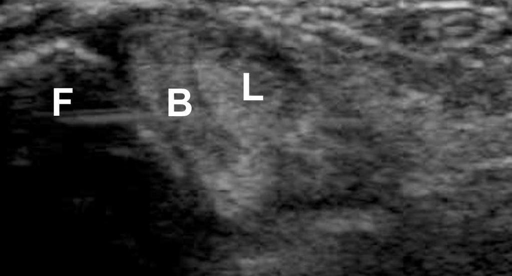
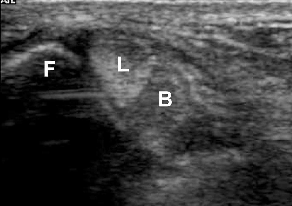
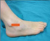
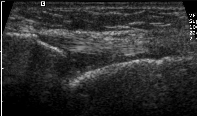
84 Ankle Joint Effusion

85 Degenerative Arthropathy Talonavicular Joint

86 Rheumatoid Arthritis Talonavicular Joint
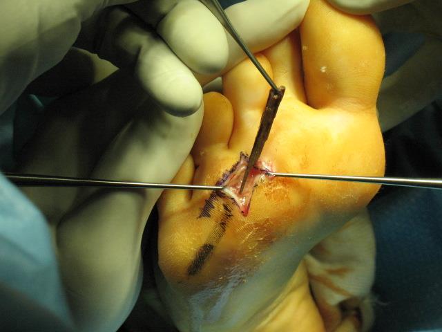
87 Rheumatoid Arthritis

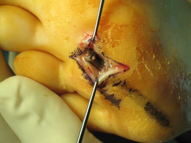
88 Gouty Arthritis

89 Gout: Periarticular Erosion
90 Adventitial Bursitis

91 Foot Ganglion
92 Foreign Bodies

93 Foreign Bodies

94 Surgical Hardware in Toe

95 Screw Impales FHL

96 Screw Impales FHL
97 Conclusions US is an effective tool for a wide range of foot and ankle abnormalities First line modality for: Tendons Plantar fasciitis Ligamentous instability/ impingement Ganglion cysts
Knee, Ankle, and Foot: Normal and Abnormal Features with MRI and Ultrasound Correlation. Disclosures. Outline. Joint Effusion. Suprapatellar recess
 Knee, Ankle, and Foot: Normal and Abnormal Features with MRI and Ultrasound Correlation Jon A. Jacobson, M.D. Professor of Radiology Director, Division of Musculoskeletal Radiology University of Michigan
17/10/2017. Foot and Ankle
 17/10/2017 Alicia M. Yochum RN, DC, DACBR, RMSK Foot and Ankle Plantar Fasciitis Hallux Valgus Deformity Achilles Tendinosis Posterior Tibialis Tendon tendinopathy Stress Fracture Ligamentous tearing Turf
Ankle Tendons in Athletes. Laura W. Bancroft, M.D.
 Ankle Tendons in Athletes Laura W. Bancroft, M.D. Outline Protocols Normal Anatomy Tendinopathy, partial and complete tears Posterior tibial, Flexor Hallucis Longus, Achilles, Peroneal and Anterior Tibial
Ankle Injuries. Resident Guidebook. Achilles tendon sprain/tear. Peroneal tendinopathy Peroneal subluxation. Extensor Hallucis Longus Tenosynovitis
 Ankle Injuries Achilles tendon sprain/tear Peroneal tendinopathy Peroneal subluxation Extensor Hallucis Longus Tenosynovitis Weber Fracture Stress fracture Calcaneal bursitis Calcaneal fracture Base of
Urgent Cases and Foreign Bodies
 Urgent Cases and Foreign Bodies Catherine J. Brandon, MD, MS University of Michigan Ann Arbor, MI, USA Introduction: Patients added on to the schedule from the emergency department or as urgent add-on
Shane A. Shapiro, M.D. Assistant Professor, Orthopedic Surgery Mayo Clinic 2012 MFMER slide MFMER slide-3
 Ultrasound Foot and Ankle Pathology Disclosures None relevant Shane A. Shapiro, M.D. Assistant Professor, Orthopedic Surgery Mayo Clinic Florida @ShaneShapiroMD 2012 MFMER slide-2 Foot and Ankle Fundamentals
Imaging of Ankle and Foot pain
 Imaging of Ankle and Foot pain Pramot Tanutit, M.D. Department of Radiology Faculty of Medicine, Prince of Songkla University 1 Outlines Plain film: anatomy Common causes of ankle and foot pain Exclude:
11/2/17. Lateral Collateral Complex Medial Collateral Complex Distal Tibiofibular Syndesmosis Spring Ligament
 Andrew J Grainger Leeds, UK Lateral Collateral Complex ial Collateral Complex Distal Tibiofibular Syndesmosis Spring Ligament Brief anatomy review Scan tips and tricks Pathological appearances andrewgrainger@nhs.net
Anatomy of Foot and Ankle
 Anatomy of Foot and Ankle Surface anatomy of the ankle & foot Surface anatomy of the ankle & foot Medial orientation point medial malleous sustentaculum tali tuberosity of navicular TA muscle TP muscle
Ultrasound Evaluation of Posteromedial Ankle Pathology. Andrew C Cordle, M.D., Ph.D. 9/21/2018
 Ultrasound Evaluation of Posteromedial Ankle Pathology Andrew C Cordle, M.D., Ph.D. 9/21/2018 Overview: Pathology of the Posteromedial Ankle Flexor Tendon Pathology Accessory Navicular Bone Pathology Tarsal
Musculoskeletal Ultrasound Technical Guidelines. VI. Ankle
 European Society of MusculoSkeletal Radiology Musculoskeletal Ultrasound Technical Guidelines VI. Ankle Ian Beggs, UK Stefano Bianchi, Switzerland Angel Bueno, Spain Michel Cohen, France Michel Court-Payen,
Why? Ultrasound of the Foot. Ultrasound of the Foot. General Rules. Plantar Fascia. Plantar Fasciitis 18/09/2018
 Ultrasound of the Foot Why? Ultrasound of the Foot Plantar fasciitis Plantar fascia fibromatosis Morton s neuroma Intermetatarsal bursitis Adventitial bursitis Plantar plate tears MTP joint synovitis Ganglia
Outline. Ankle/Foot Anatomy Ankle Sprains Ottawa Ankle Rules DDx: The Sprain That Wasn t
 Ankle Injuries Outline Ankle/Foot Anatomy Ankle Sprains Ottawa Ankle Rules DDx: The Sprain That Wasn t Anatomy: Ankle Mortise Bony Anatomy Lateral Ligament Complex Medial Ligament Complex Ankle Sprains
Index. Clin Sports Med 23 (2004) Note: Page numbers of article titles are in boldface type.
 Note: Page numbers of article titles are in boldface type.") Clin Sports Med 23 (2004) 169 173 Index Note: Page numbers of article titles are in boldface type. A Achilles enthesopathy, calcaneal spur with, 133 clinical presentation of, 135 136 definition of, 131
Clin Sports Med 23 (2004) 169 173 Index Note: Page numbers of article titles are in boldface type. A Achilles enthesopathy, calcaneal spur with, 133 clinical presentation of, 135 136 definition of, 131
Sports Injuries of the Ankle and Ankle Arthritis. Mr Amit Amin Consultant Foot and Ankle Surgeon Parkside Hospital
 Sports Injuries of the Ankle and Ankle Arthritis Mr Amit Amin Consultant Foot and Ankle Surgeon Parkside Hospital Impingement Painful mechanical limitation of full ankle movement secondary to osseous
Ultrasound examination of the ankle: most prevalent disease in our environment
 Ultrasound examination of the ankle: most prevalent disease in our environment Poster No.: C-0155 Congress: ECR 2013 Type: Educational Exhibit Authors: M. E. Moral Molero, R. J. Megales Navarro, L. Aguilar
The Lower Limb VII: The Ankle & Foot. Anatomy RHS 241 Lecture 7 Dr. Einas Al-Eisa
 The Lower Limb VII: The Ankle & Foot Anatomy RHS 241 Lecture 7 Dr. Einas Al-Eisa Ankle joint Synovial, hinge joint Allow movement of the foot in the sagittal plane only (1 degree of freedom): dorsiflexion:
Extraarticular Lateral Ankle Impingement
 Extraarticular Lateral Ankle Impingement Poster No.: C-1282 Congress: ECR 2016 Type: Educational Exhibit Authors: C. Cevikol; Keywords: Trauma, Diagnostic procedure, MR, CT, Musculoskeletal system, Musculoskeletal
Clin Podiatr Med Surg 19 (2002) Index
 Index") Clin Podiatr Med Surg 19 (2002) 335 344 Index Note: Page numbers of article titles are in bold face type. A Accessory soleus muscle, magnetic resonance imaging of, 300 Achilles tendon injury of, magnetic
Clin Podiatr Med Surg 19 (2002) 335 344 Index Note: Page numbers of article titles are in bold face type. A Accessory soleus muscle, magnetic resonance imaging of, 300 Achilles tendon injury of, magnetic
Copyright 2004, Yoshiyuki Shiratori. All right reserved.
 Ankle and Leg Evaluation 1. History Chief Complaint: A. What happened? B. Is it a sharp or dull pain? C. How long have you had the pain? D. Can you pinpoint the pain? E. Do you have any numbness or tingling?
Ankle Sprains and Their Imitators
 Ankle Sprains and Their Imitators Mark Halstead, MD Dr. Mark Halstead is the Associate Professor of the Departments of Orthopedics and Pediatrics at Washington University School of Medicine; Director of
Lateral ankle pain: what is the problem?
 Lateral ankle pain: what is the problem? Poster No.: C-1121 Congress: ECR 2013 Type: Educational Exhibit Authors: J. LEE, S. J. Lee, H. J. Choo, H. W. Jeong, Y.-M. Park, S. J. Choi; Busan/KR Keywords:
Clarification of Terms
 Clarification of Terms The plantar aspect of the foot refers to the role or its bottom The dorsal aspect refers to the top or its superior portion The ankle and foot perform three main functions: 1. shock
SURGICAL AND APPLIED ANATOMY
 Página 1 de 9 Copyright 2001 Lippincott Williams & Wilkins Bucholz, Robert W., Heckman, James D. Rockwood & Green's Fractures in Adults, 5th Edition SURGICAL AND APPLIED ANATOMY Part of "47 - ANKLE FRACTURES"
Review relevant anatomy of the foot and ankle. Learn the approach to examining the foot and ankle
 Objectives Review relevant anatomy of the foot and ankle Learn the approach to examining the foot and ankle Learn the basics of diagnosis and treatment of ankle sprains Overview of other common causes
Podiatry Ultrasound Report Templates
 Podiatry Ultrasound Report Templates 1 st Edition Compiled exclusively for the clients of Fisher Biomedical Inc. Podiatric Ultrasound Report Templates Welcome to our first edition of sample podiatric ultrasound
THE LOWER EXTREMITY EXAM FOR THE FAMILY PRACTITIONER
 THE LOWER EXTREMITY EXAM FOR THE FAMILY PRACTITIONER Melinda A. Scott, D.O. Orthopedic Associates of Dayton Board Certified in Primary Care Sports Medicine GOALS Identify landmarks necessary for exam of
OVERUSE AND SPORTS-RELATED INJURIES. Overuse and sports-related injuries of the ankle and hindfoot: MR imaging findings
 OVERUSE ND SPORTS-RELTED INJURIES CHPTER 10 Overuse and sports-related injuries of the ankle and hindfoot: MR imaging findings Elizabeth S. Sijbrandij 1, d P.G. van Gils 1, Eduard E. de Lange 2 From the
5 COMMON INJURIES IN THE FOOT & ANKLE
 5 COMMON INJURIES IN THE FOOT & ANKLE MICHAEL P. CLARE, MD FLORIDA ORTHOPAEDIC INSTITUTE TAMPA, FL USA MECHANISM OF INJURY HOW DID IT HAPPEN? HIGH ENERGY VS LOW ENERGY DIRECTION OF FORCES INVOLVED LIVING
ELENI ANDIPA General Hospital of Athens G. Gennimatas
 ELENI ANDIPA General Hospital of Athens G. Gennimatas Technological advances over the last years have caused a dramatic improvement in ultrasound quality and resolution An established imaging modality
Anterior Impingement
 Anterior Impingement Ziali Sivardeen BMedSci, (MRCS), AFRCS, FRCS (Tr & Orth) Consultant Trauma and Orthopaedic Surgeon (Shoulder, Knee and Sports Injuries) Aims Causes of Anterior Ankle Pain Ankle Impingement
V E R I TAS MGH 1811 MGH 1811 V E R I TAS. *Gerber JP. Persistent disability with ankle sprains. Foot Ankle Int 19: , 1998.
 MGH 1811 Management of Ankle Instability Richard J. de Asla, M.D. V E R I TAS MGH 1811 I have no potential conflicts with this presentation. V E R I TAS It s just a sprain Lateral Ankle Sprains Most common
Prevention and Treatment of Injuries. Anatomy. Anatomy. Tibia: the second longest bone in the body
 Prevention and Treatment of Injuries The Ankle and Lower Leg Westfield High School Houston, Texas Anatomy Tibia: the second longest bone in the body Serves as the principle weight-bearing bone of the leg.
Leg. Dr. Heba Kalbouneh Associate Professor of Anatomy and Histology
 Leg Dr. Heba Kalbouneh Associate Professor of Anatomy and Histology Skin of the Leg Cutaneous Nerves Medially: The saphenous nerve, a branch of the femoral nerve supplies the skin on the medial surface
Ankle impingement syndromes - pictorial review.
 Ankle impingement syndromes - pictorial review. Poster No.: P-0148 Congress: ESSR 2015 Type: Educational Poster Authors: R. D. T. Mesquita, J. Pinto, J. L. Rosas, A. Vieira ; Porto/PT, 1 2 2 3 1 1 3 Matosinhos/PT,
Ankle impingement syndromes - pictorial review.
 Ankle impingement syndromes - pictorial review. Poster No.: P-0148 Congress: ESSR 2015 Type: Educational Poster Authors: R. D. T. Mesquita, J. Pinto, J. L. Rosas, A. Vieira ; Porto/PT, 1 2 2 3 1 1 3 Matosinhos/PT,
Case 1 7 yo male Right elbow injury 3 months ago Medial elbow pain and tenderness over medial epicondyle Long arm cast given but off himself 1 month a
 Case presentations Case 1 7 yo male Right elbow injury 3 months ago Medial elbow pain and tenderness over medial epicondyle Long arm cast given but off himself 1 month after Progressive limited elbow flexion
Everything. You Should Know. About Your Ankles
 Everything You Should Know About Your Ankles How Your Ankle Works The ankle joint is a hinge type joint that participates in movement and is involved in lower limb stability. There are 2 types of motions
Imaging of posterior ankle pain : Main etiologies and differential diagnosis
 Imaging of posterior ankle pain : Main etiologies and differential diagnosis Poster No.: C-2399 Congress: ECR 2017 Type: Educational Exhibit Authors: W. Frikha, M. MECHRI, S. boukriba, H. RIAHI, M. CHELLI
Disclosures. Syndesmosis Injury. Syndesmosis Ligaments. Objectives. Mark M. Casillas, M.D.
 Disclosures Syndesmosis Injury No relevant disclosures Mark M. Casillas, M.D. 1 Objectives Syndesmosis Ligaments Understand the syndesmosis anatomy and function Classify syndesmosis injuries Describe treatment
Ankle and Foot Orthopaedic Tests Orthopedics and Neurology DX 612
 Ankle and Foot Orthopaedic Tests Orthopedics and Neurology DX 612 James J. Lehman, DC, MBA, DABCO University of Bridgeport College of Chiropractic Ankle & Foot Anatomy Stability of the ankle is dependent
Pragmatic ultrasound in the diagnosis of soft tissue rheumatic pain. Plamen Todorov
 Pragmatic ultrasound in the diagnosis of soft tissue rheumatic pain Plamen Todorov INTRODUCTION Soft tissue rheumatism: nonsystemic, focal pathological syndromes involving the periarticular structures.
Case Report Painful Os Peroneum Syndrome: Underdiagnosed Condition in the Lateral Midfoot Pain
 Case Reports in Radiology Volume 2016, Article ID 8739362, 4 pages http://dx.doi.org/10.1155/2016/8739362 Case Report Painful Os Peroneum Syndrome: Underdiagnosed Condition in the Lateral Midfoot Pain
Peripheral Nerve Ultrasound
 Peripheral Nerve Ultrasound Jon A. Jacobson, M.D. Professor of Radiology Director, Division of Musculoskeletal Radiology University of Michigan Normal Peripheral Nerve Ultrasound appearance: Hypoechoic
Ultrasound of the Knee
 Ultrasound of the Knee Jon A. Jacobson, M.D. Professor of Radiology Director, Division of Musculoskeletal Radiology University of Michigan Disclosures: Consultant: Bioclinica Book Royalties: Elsevier Advisory
بسم هللا الرحمن الرحيم
 بسم هللا الرحمن الرحيم Laboratory RHS 221 Manual Muscle Testing Theory 1 hour practical 2 hours Dr. Ali Aldali, MS, PT Department of Physical Therapy King Saud University Talocrural and Subtalar Joint
BIOMECHANICS OF ANKLE FRACTURES
 BIOMECHANICS OF ANKLE FRACTURES William R Reinus, MD MBA FACR Significance of Ankle Fractures Most common weight-bearing Fx 70% of all Fxs Incidence is increasing Bimodal distribution Men 15-24 Women over
Ultrasound Evaluation of Masses
 Ultrasound Evaluation of Masses Jon A. Jacobson, M.D. Professor of Radiology Director, Division of Musculoskeletal Radiology University of Michigan Disclosures: Consultant: Bioclinica Advisory Panel: GE,
US finding of the shoulder (with live demonstration) 인제의대상계백병원 안재기
 인제의대상계백병원 안재기") US finding of the shoulder (with live demonstration) 인제의대상계백병원 안재기 Shoulder US Biceps tendon & Rotator Cuff Long Head of Biceps Tendon Subscapularis tendon Supraspinatus tendon Infraspinatus tendon Teres
US finding of the shoulder (with live demonstration) 인제의대상계백병원 안재기 Shoulder US Biceps tendon & Rotator Cuff Long Head of Biceps Tendon Subscapularis tendon Supraspinatus tendon Infraspinatus tendon Teres
Main Menu. Ankle and Foot Joints click here. The Power is in Your Hands
 1 The Ankle and Foot Joints click here Main Menu Copyright HandsOn Therapy Schools 2009 K.8 http://www.handsonlineeducation.com/classes/k8/k8entry.htm[3/27/18, 1:40:03 PM] Ankle and Foot Joint 26 bones
OTM Lecture Gait and Somatic Dysfunction of the Lower Extremity
 OTM Lecture Gait and Somatic Dysfunction of the Lower Extremity Somatic Dysfunction Tenderness Asymmetry Range of Motion Tissue Texture Changes Any one of which must be present to diagnosis somatic dysfunction.
Longitudinal Split of the Peroneus Longus and Peroneus Brevis Tendons with Disruption of the Superior Peroneal Retinaculum
 Longitudinal Split of the Peroneus Longus and Peroneus Brevis Tendons with Disruption of the Superior Peroneal Retinaculum Gregory C. Diaz, MD, Marnix van Holsbeeck, MD, Jon A. Jacobson, MD Longitudinal
ANKLE JOINT ANATOMY 3. TALRSALS = (FOOT BONES) Fibula. Frances Daly MSc 1 CALCANEUS 2. TALUS 3. NAVICULAR 4. CUBOID 5.
 Fibula. Frances Daly MSc 1 CALCANEUS 2. TALUS 3. NAVICULAR 4. CUBOID 5.") ANKLE JOINT ANATOMY The ankle joint is a synovial joint of the hinge type. The joint is formed by the distal end of the tibia and medial malleolus, the fibula and lateral malleolus and talus bone. It is
ANKLE JOINT ANATOMY The ankle joint is a synovial joint of the hinge type. The joint is formed by the distal end of the tibia and medial malleolus, the fibula and lateral malleolus and talus bone. It is
MRI of the Ankle and Foot
 Acta Radiológica Portuguesa, Vol.XX, nº 79, pág. 55-63, Jul.-Set., 2008 MRI of the Ankle and Foot Mark Anderson University of Virginia Health Sciences Center, Charlottesville, Virginia discuss the basic
A Patient s Guide to Ankle Anatomy
 A Patient s Guide to Ankle Anatomy 1436 Exchange Street Middlebury, VT 05753 Phone: 802-388-3194 Fax: 802-388-4881 cvo@champlainvalleyortho.com DISCLAIMER: The information in this booklet is compiled from
THE JOURNAL OF NUCLEAR MEDICINE Vol. 56 No. 3 March 2015 Rauscher et al.
 Supplemental Figure 1 Correlation analysis of tracer between and subsequent as assessed by SUV max in focal lesions (A). x-axis displays quantitative values as obtained by, and y-axis displays corresponding
Rotator Cuff and Biceps Pathology
 Rotator Cuff and Biceps Pathology Jon A. Jacobson, M.D. Professor of Radiology Director, Division of Musculoskeletal Radiology University of Michigan Disclosures: Consultant: Bioclinica Advisory Board:
Foot and Ankle Complaints.
 Foot and Ankle Complaints www.fisiokinesiterapia.biz INTRODUCTION Anatomy and Function Foot Ankle Common complaints Common diagnoses FOOT AND ANKLE ANATOMY 26 bones and 2 sesamoids Forefoot Metatarsals
BLUE SKY SCHOOL OF PROFESSIONAL MASSAGE AND THERAPEUTIC BODYWORK Musculoskeletal Anatomy & Kinesiology KNEE & ANKLE MUSCLES
 BLUE SKY SCHOOL OF PROFESSIONAL MASSAGE AND THERAPEUTIC BODYWORK Musculoskeletal Anatomy & Kinesiology KNEE & ANKLE MUSCLES MSAK201-I Session 3 1) REVIEW a) THIGH, LEG, ANKLE & FOOT i) Tibia Medial Malleolus
Sports Injuries of the Foot and Ankle Dominic Nielsen. Parkside Hospital Ashtead Hospital St George s
 Sports Injuries of the Foot and Ankle Dominic Nielsen Parkside Hospital Ashtead Hospital St George s Themes Ankle instability Ankle impingement Stress fractures 5 th MT fractures Peroneal subluxation Ankle
3/6/2012 STATE OF THE ART: FOOT AND ANKLE GENERAL KNOWLEDGE 1. TRASP REHABILITATION CONTENTS. General knowledge Trasp Prevention
 STATE OF THE ART: FOOT AND ANKLE ILITATION Fabienne Van De Steene. CONTENTS General knowledge Trasp Prevention Rehab Ankle sprain CAI Achilles tendon Plantar fasciitis Take home message 2 1. TRASP Ankle
"The Role of Dynamic Ultrasound and MRI in the poorly resolving ankle sprain."
 "The Role of Dynamic Ultrasound and MRI in the poorly resolving ankle sprain." Poster No.: P-0007 Congress: ESSR 2013 Type: Scientific Exhibit Authors: J. M. Zietkiewicz, P. Mercouris, M. C. Marshall;
The Leg. Prof. Oluwadiya KS
 The Leg Prof. Oluwadiya KS www.oluwadiya.sitesled.com Compartments of the leg 4 Four Compartments: 1. Anterior compartment Deep fibular nerve Dorsiflexes the foot and toes 2. Lateral Compartment Superficial
Title. Issue Date Right.
 NOSITE: Nagasaki University's c Title uthor(s) Dynamic supination and hindfoot var tendons of both peroneus longus and Matsubayashi, Shohei; Tsujimoto, i Citation cta medica Nagasakiensia, 61(2), p Issue
A Patient s Guide to Ankle Anatomy
 A Patient s Guide to Ankle Anatomy 245 North College Lafayette, LA 70506 Phone: 337.232.5301 Fax: 337.237.6504 DISCLAIMER: The information in this booklet is compiled from a variety of sources. It may
Physical Examination of the Foot & Ankle
 Inspection Standing, feet straight forward facing toward examiner Swelling Deformity Flatfoot (pes planus and hindfoot valgus) High arch (pes cavus and hindfoot varus) Peek-a-boo heel Varus Too many toes
Surgery-Ortho. Fractures of the tibia and fibula. Management. Treatment of low energy fractures. Fifth stage. Lec-6 د.
 Fifth stage Lec-6 د. مثنى Surgery-Ortho 28/4/2016 Indirect force: (low energy) Fractures of the tibia and fibula Twisting: spiral fractures of both bones Angulatory: oblique fractures with butterfly segment.
PRIMARY CARE EXAMINATION OF KEY JOINTS. Thomas M. Howard, MD, FACSM FFPC Sports Medicine
 PRIMARY CARE EXAMINATION OF KEY JOINTS Thomas M. Howard, MD, FACSM FFPC Sports Medicine General exam principles: Expose entire joint and opposite limb for comparison Have a Differential Diagnosis Exam
Section Three: The Leg, Ankle, and Foot Lecture: Review of Clinical Anatomy, Patterns of Dysfunction and Injury, and
 Section Three: The Leg, Ankle, and Foot Lecture: Review of Clinical Anatomy, Patterns of Dysfunction and Injury, and Treatment Implications for the Leg, Ankle, and Foot Levels I and II Demonstration and
Original Report. Sonography of Ankle Tendon Impingement with Surgical Correlation
 Downloaded from www.ajronline.org by 162.158.89.91 on 08/23/18 from IP address 162.158.89.91. Copyright RRS. For personal use only; all rights reserved Monisha Shetty 1 David P. Fessell 1 John E. Femino
Index. Clin Podiatr Med Surg 22 (2005) Note: Page numbers of article titles are in bold face type.
 Note: Page numbers of article titles are in bold face type.") Clin Podiatr Med Surg 22 (2005) 137 141 Index Note: Page numbers of article titles are in bold face type. A Achilles tendinopathy, 19 43. See also Achilles classification of, 20 differential diagnosis
Clin Podiatr Med Surg 22 (2005) 137 141 Index Note: Page numbers of article titles are in bold face type. A Achilles tendinopathy, 19 43. See also Achilles classification of, 20 differential diagnosis
13/05/14. Lower Limb Injuries Below the Knee
 Lower Limb Injuries Below the Knee Dr Peter Friis Brisbane Orthopaedic & Sports Medicine Centre At least 1 previous injury or reduced lower limb funcjon score had a significant increased risk of sustaining
ii ANKLE INJURIES SPECIFIC TRAINING AFTER INJURY TO THE FOOT OR ANKLE
 40 Ankle injuries are among the most common injuries in sport. Ankle sprain (which is a mechanism rather than a diagnosis) is the most common injury in virtually all epidemiological studies. Being the
ANKLE SPRAIN: DIAGNOSIS AND THERAPY STARTS WITH KNOWLEDGE OF ANATOMY
 ANKLE SPRAIN: DIAGNOSIS AND THERAPY STARTS WITH KNOWLEDGE OF ANATOMY Written by Pau Golanó, Spain and Jordi Vega, Switzerland A thorough knowledge of anatomy is imperative for adequate assessment of joint
A Patient s Guide to Ankle Anatomy
 A Patient s Guide to Ankle Anatomy Pond View Professional Park 301 Professional View Drive Freehold, NJ 07728 Phone: 732-720-2555 DISCLAIMER: The information in this booklet is compiled from a variety
Anatomy and evaluation of the ankle.
 Anatomy and evaluation of the ankle www.fisiokinesiterapia.biz Ankle Anatomical Structures Tibia Fibular Talus Tibia This is the strongest largest bone of the lower leg. It bears weight and the bone creates
Anatomy MCQs Week 13
 Anatomy MCQs Week 13 1. Posterior to the medial malleolus of the ankle: The neurovascular bundle lies between Tibialis Posterior and Flexor Digitorum Longus The tendon of Tibialis Posterior inserts into
Arthroscopy Of the Ankle.
 Arthroscopy Of the Ankle www.fisiokinesiterapia.biz Ankle Arthroscopy Anatomy Patient setup Portal placement Procedures Complications Anatomy Portals Anterior Anteromedial Anterolateral Anterocentral Posterior
Sports Injuries of the Foot and Ankle. Mark McEleney, MD University of Iowa College of Medicine Refresher Course for the Family Physician 4/4/2018
 Sports Injuries of the Foot and Ankle Mark McEleney, MD University of Iowa College of Medicine Refresher Course for the Family Physician 4/4/2018 I. Objectives A. By the end of the lecture attendees will
Point of Care Ultrasound on the Field of Play K AT I E N ANOS, MD
 Point of Care Ultrasound on the Field of Play K AT I E N ANOS, MD H I GH P ERFORMANCE S PORTS MEDICINE P HYSI ATRIST, P R ACTICING S PORTS MEDI CINE No disclosures No disclosures Who am I? Objectives Over
Therapeutic Foot Care Certificate Program Part I: Online Home Study Program
 Therapeutic Foot Care Certificate Program Part I: Online Home Study Program 1 Anatomy And Terminology Of The Lower Extremity Joan E. Edelstein, MA, PT, FISPO Associate Professor of Clinical Physical Therapy
Talus Fractures: When and Why on Screws and Plates
 Talus Fractures: When and Why on Screws and Plates Frank A. Liporace, MD Associate Professor Director of Orthopaedic Research New York University / Hospital for Joint Diseases, NY, NY Director Orthopaedic
Introduction to Anatomy. Dr. Maher Hadidi. Laith Al-Hawajreh. Mar/25 th /2013
 Introduction to Anatomy Dr. Maher Hadidi Laith Al-Hawajreh 22 Mar/25 th /2013 Lower limb - The leg The skeleton of the leg is formed by two bones: 1) Medial: Tibia 2) Lateral: Fibula The two bones are
Bone Marrow Edema Patterns in the Ankle and Hindfoot: Distinguishing MRI Features
 Musculoskeletal Imaging Pictorial Essay Rios et al. MRI of the Ankle and Hindfoot Musculoskeletal Imaging Pictorial Essay Adriana Martins Rios 1 Zehava Sadka Rosenberg 2 Jenny Teresa Bencardino 2 Silvia
Sonography of Knee and Calf Pain: the differential considerations
 Sonography of Knee and Calf Pain: the differential considerations Dr. Lisa L. S.Wong Consultant Radiologist St Paul s Hospital Outline Ultrasound techniques Common pathologies in calf and posterior knee
Management of Chronic Lateral Ligament Instability
 Management of Chronic Lateral Ligament Instability Bony Anatomy Curved trochlear surface of talus produces a cone-shaped articulation whose apex is directed medially; thus the fan-shaped deltoid is all
The Egyptian Journal of Hospital Medicine (October 2017) Vol.69 (3), Page
 Vol.69 (3), Page") The Egyptian Journal of Hospital Medicine (October 2017) Vol.69 (3), Page 2016-2024 Role of MRI in Evaluation of Traumatic Ankle Injuries Mervat Mohamed Ibrahim Ali Elgohary*, Susan Adel Ali Abdul Rahim*,
The Egyptian Journal of Hospital Medicine (October 2017) Vol.69 (3), Page 2016-2024 Role of MRI in Evaluation of Traumatic Ankle Injuries Mervat Mohamed Ibrahim Ali Elgohary*, Susan Adel Ali Abdul Rahim*,
Ankle Injuries: Anatomical and Biomechanical Considerations Necessary for the Development of an Injury Prevention Program
 0196-6011 /80/0103-0171$02.00/0 THE JOURNAL OF ORTHOPAEDIC AND SPORTS PHYSICAL THERAPY Copyright O 1980 by The Orthopaedic and Sports Medicine Sections of the American Physical Therapy Association Ankle
Ankle and hindfoot Note medial malleolus, lateral malleolus, inferior tibiofibular joint, talocrural joint and subtalar joint form the 3 joint complex
 Session 4 Look at the ankle (talocrural joint) and the subtalar joint (hind foot) Anatomy of the joints Muscles and how the joints move (biomechanics) Structure of tendons and Achilles tendinitis Some
Posterior Ankle Impingement: Don t Get Pinched
 Posterior Ankle Impingement: Don t Get Pinched 11 th Annual Sports Medicine Continuing Education Conference Gregory P Witkowski, MD Orthopaedic Trauma and Foot/Ankle Surgery Disclosures I have nothing
Joints of the Lower Limb II
 Joints of the Lower Limb II Lecture Objectives Describe the components of the knee and ankle joint. List the ligaments associated with these joints and their attachments. List the muscles acting on these
musculoskeletal system anatomy muscles of foot sheet done by: dina sawadha & mohammad abukabeer
 musculoskeletal system anatomy muscles of foot sheet done by: dina sawadha & mohammad abukabeer Extensor retinaculum : A- superior extensor retinaculum (SER) : originates from the distal ends of the tibia
Ankle Injuries. Ankle Sprain. Range of Motion. The most likely diagnosis is lateral ligament sprain. Dorsiflexion Plantarflexion Inversion
 Ankle Injuries Dr Peter Brukner, OAM Sports Physician Associate Professor Centre for Sports Medicine Research & Education The University of Melbourne Adjunct Professor School of Human Movement Studies
Anatomy of Ankle & Foot. Chang-Hyung Lee, M.D., Ph.D. Physical Medicine & Rehabilitation Samsung Medical Center
 Anatomy of Ankle & Foot Chang-Hyung Lee, M.D., Ph.D. Physical Medicine & Rehabilitation Samsung Medical Center Ankle Introduction Most frequently injured major joint 3 main articulation: distal tibiofibular
Ligament lesions of the ankle. Marc C. Attinger
 Ligament lesions of the ankle Marc C. Attinger Anatomy Mechanism of injury Each lig with its function during ROM in dorsiflexion/er ATFL slack, CFL tight in plantarflexion/ir CFL slack, ATFL tight Acute
MR Imaging of normal ankle anatomy: What the radiologists need to know
 MR Imaging of normal ankle anatomy: What the radiologists need to know Poster No.: P-0110 Congress: ESSR 2013 Type: Scientific Exhibit Authors: M. M. Simonet Redondo, I. Santos Gomez, A. Marin Canete,
CHAPTER 80 BASIC CONSIDERATIONS
 Página 1 de 32 Copyright 2001 Lippincott Williams & Wilkins Loeser, John D. Bonica's Management of Pain, 3rd Edition CHAPTER 80 BASIC CONSIDERATIONS Part of "CHAPTER 80 - Pain in the Leg, Ankle, and Foot"
Introduction. The primary function of the ankle and foot is to absorb shock and impart thrust to the body during walking.
 The ankle 1 Introduction The primary function of the ankle and foot is to absorb shock and impart thrust to the body during walking. OSTEOLOGRY The term ankle refers primarily to the talocrural joint,
Craig S. Radnay, M.D. 1/27/2016. Access to the Talus for Treatment of Osteochondral Lesions. Epidemiology of OLT. Treatment of OLT
 Access to the Talus for Treatment of Osteochondral Lesions Craig S. Radnay, MD, MPH ISK Institute for Orthopaedics and Sports Medicine NYU/Hospital for Joint Diseases Tampa, FL January 23, 2016 Epidemiology