Sonography of Knee and Calf Pain: the differential considerations
|
|
|
- Barbra Pearson
- 5 years ago
- Views:
Transcription
1 Sonography of Knee and Calf Pain: the differential considerations Dr. Lisa L. S.Wong Consultant Radiologist St Paul s Hospital Outline Ultrasound techniques Common pathologies in calf and posterior knee pain Common pathologies in anterior, medial and lateral knee pain Advanced Specialty Program for Diagnostic Radiographers in Ultrasonography, Musculoskeletal Ultrasound Symposium, 6 th November 2010 Differential Considerations in Calf and Posterior Knee Pain Deep venous thrombosis Muscle injury e.g. strain or contusion Achilles tendinosis or tendon tear Ruptured Baker s cyst Differential Considerations in Calf and Posterior Knee Pain Deep venous thrombosis Muscle injury e.g. strain or contusion Achilles tendinosis or tendon tear Ruptured Baker s cyst Ultrasound Techniques and Approach Linear transducer High resolution >10 MHz Know anatomical landmarks to identify different musculoskeletal structures Extended field-of-view allows better display of muscle anatomy and pathology and enhances cross-specialty communication Ultrasound Techniques and Approach Clinical history very important Ultrasound palpation Comparison with contralateral side is important to pick up subtle changes Correlate with radiographs when necessary
2 Differential Considerations in Calf Pain Deep venous thrombosis Muscle injury Achilles tendon tear Ruptured Baker s cyst Direct MUSCLE INJURY Contusion Laceration Indirect DOMS (Delayed Onset Muscle Soreness) Strain MUSCLE CONTUSION Direct blunt trauma to muscle Most common lower extremities Early mobilization rapid recovery Can grade by amount of restricted motion MUSCLE CONTUSION Ultrasound features: Increased muscle girth Typically no significant fiber disruption Increase echogenicity depending on amount of haemorrhage MUSCLE LACERATION Ultrasound features: Abrupt muscle fibers disruption Sharp interphase at the muscle laceration Increase echogenicity depending on amount of haemorrhage Direct MUSCLE INJURY Contusion Laceration Indirect DOMS (Delayed Onset Muscle Soreness) Strain
3 DELAYED ONSET MUSCLE SORENESS (DOMS) Reversible structural damage within hours No history of trauma nor acute onset of pain Pain hours post exercise People unaccustomed to vigorous exercise Initiation or resumption of training Self limited subsides within 1 week usually No permanent damage to muscle function DELAYED ONSET MUSCLE SORENESS (DOMS) Ultrasound Usually normal May have perifascial fluid-like collections MUSCLE STRAINS Indirect injury excessive stretch during muscle contraction Muscles at risk crossing two joints eccentric (lengthening) contraction E.G. gastrocnemius, hamstrings, quadriceps Predispositions Lack stretching warm up Lack conditioning fatigue Steroid injections MUSCLE STRAINS Diagnosis usually straightforward Pain and tenderness Patient feels intense sudden pain - pop May feel gap Musculotendinous junction is the weak link May have extensive bleeding First degree MUSCLE STRAINS No significant loss of strength or ROM Typically less than 5% fiber disruption Second degree partial tear Partial loss strength Can be subdivided by extent Third degree complete tear 1 st DEGREE MUSCLE STRAIN ULTRASOUND Swollen muscle with either increased (blood products) or decreased echogenicity (oedema) Subtle changes picked up by comparing with contralateral DDx: muscle contusion (differs in mechanism of injury) infection or inflammation (myositis) iatrogenic (e.g. RT)


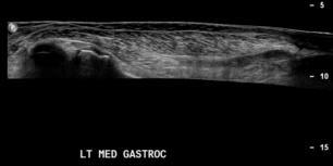
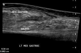
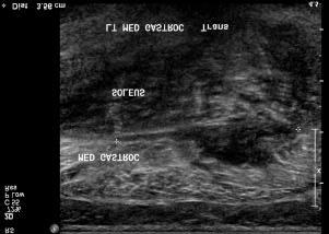
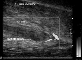
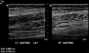
4 RT GASTROC LT GASTROC GASTROCNEMIUS STRAIN 2 nd DEGREE MUSCLE STRAIN Second degree partial tear involves < 1/3 of fibres - low grade involves 1/3 to 2/3 of fibres moderate grade involves more than 2/3 of fibres high grade T2W GASTROCNEMIUS STRAIN 2 nd DEGREE MUSCLE STRAIN SOLEUS STRAIN ULTRASOUND Swollen Architectural distortion at myotendinous junction Heterogeneous echogenicity due to blood products and oedema Muscle disruption Haematoma Perifascial fluid LS TS


5 2 nd DEGREE MUSCLE STRAIN 2 nd DEGREE MUSCLE STRAIN ULTRASOUND Haematoma follow-up necessary to exclude haemorrhage associated with an intramuscular tumour 3 rd DEGREE MUSCLE STRAIN 65 y.o. female with calf pain Third degree complete tear complete discontinuity of fibres retraction of fibres haematoma at site of rupture chronic muscle atrophy

6


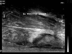
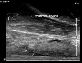
7 MUSCLE-APONEUROSIS TEAR Commonest site in the calf between the distal part of medial head of gastrocnemius and soleus muscle - Tennis leg Characteristic Pop Common in sports requiring jumps or speed MUSCLE-APONEUROSIS TEAR Ultrasound: Rounding of distal gastrocnemius fibres at the myoaponeuroticattachment with the soleus Fluid/haematoma along the aponeurosis Perifascial fluid Echogenic oedema in the muscles and subcutaneous fat NORMAL Achilles Tendon Conjoined tendon of medial & lateral gastrocnemius and soleus muscles Lacks tendon sheath, enclosed by paratenon Tear most common at myotendinous junction in lower calf Sudden pain at lower calf as if being kicked by someone May have pop sound Ultrasound Achilles Tendon Tear Ruptured tendon stump wavy, retracted Oedema increased echogenicity Fluid or haematoma at tear gap Report Site of tear from calcanealinsertion Complete vs partial (beware of intact plantaris tendon) Tear gap Dynamic study apposition of the tendon stumps Achilles Tendon Tear


8 Achilles Tendon Tear Achilles Tendon Tear Dynamic study Dynamic study


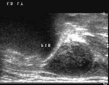
9 Dynamic study Achilles Tendon Tear Achilles Tendon Tear Plantaris Follows medial side of Achilles to insert either anteromedially on Achilles or on calcaneus Plantaris absent in 7-10% Intact plantaris tendon in the presence of full thickness Achilles tendon tear may be mistaken as partial Achilles tendon tear Baker s cyst Synovial cyst in posterior aspect of knee Formed by escape of effusion from the knee with subsequent trapping of fluid by one-way valvular mechanism in the gastrocnemius-semimembranosus bursa Causes: Large effusion e.g. internal derangement of the knee Arthritis e.g. rheumatoid arthritis Clinically: Asymptomatic if small Complained of mass if large Sudden severe pain if rupture Ultrasound Location: between the medial gastrocnemiusmuscle and semimembranosusmuscle/tendon


10 Baker s Cyst Baker s Cyst Positioning of the knee Patient lie flat and knee flexed Placed knee over a triangular soft pad Manoeuvres to deliver stress to certain ligaments e.g. valgus stress to detect MCL injury


 MRI better")


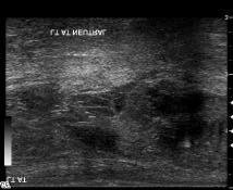
11 Differential Considerations in Knee Pain Problem-based approach Ultrasound good for superficial structures Know limitations of ultrasound MRI if suspect deep intra-articular knee pathology Differential diagnoses Anterior knee pain Muscle Injury Quadriceps muscles Mucoiddegeneration or tear Quadriceps tendon Patellar tendon Patellar retinaculi Distended suprapatellar bursa underlying internal joint derangement, synovitis Pre-patellar bursitis Patellar cartilage tear (ultrasound only see femoral trochlear cartilage ) MRI better Patellar fracture Anterior compartment muscles Quadriceps muscles Rectus femoris, vastus medialis, vastus lateralis and vastus intermedius Common tendon inserting to superior pole of the patella Garrett, Jr. W.E. Am. J. Sports Med 1996


12 Ruptured rectus femoris muscle Ruptured rectus femoris muscle with retracted tendon Patellar Tendon Jumper s Knee Overuse syndrome due to sudden or repetitive extension of the knee Athletes involved in kicking, jumping and running Spectrum of patellar tendinosis (mucoid degeneration or fibrinous necrosis) and tear +/- secondary inflammatory response at patellar insertion Ultrasound features Hypoechoic thickening of the PT Loss of normal fibrillary pattern Increased vascularity Jumper s Knee 22 y.o. anterior knee pain after sports session at school Avulsion of medial patellar retinaculum from patella


")
13 Effusion in suprapatellar bursa Differential diagnoses Medial Knee pain Muscle injury Vastus medialis muscle Mucoid degeneration or tear of Medial collateral ligament Medial patellar retinaculum Pes anserinus goose -feet Sartorius, Gracilisand Semitendinosus tendons Medial meniscus pathology (MRI should be performed) Medial compartment cartilage pathology (MRI)


14 MCL Tear Differential diagnoses Lateral knee pain Muscle Injury Vastus lateralis Mucoid degeneration or tear of: Iliotibial band Lateral collateral ligament Biceps femoris tendon Popliteus tendon Lateral patellar retinaculum Lateral meniscus pathology ( better with MRI) Lateral Landmarks and Structures Lateral Landmarks Lateral femoral epicondyle Fibular head Gerdy s tubercle of tibia Structures Iliotibial band G. tubercle Biceps femoris tendon inserts on fibular head Lateral collateral ligament lateral femoral epicondyle to fibular head Popliteus tendon inserts on femoral sulcus deep to LCL Lateral meniscus BF ITB FEMUR TIBIA Grant s Atlas of Anatomy


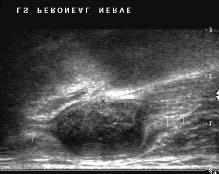
15 FIBULA FEMUR TIBIA PERONEAL NERVE PERONEAL NERVE TUMOUR PERONEAL NERVE TUMOUR


16 HETEROTOPIC OSSIFICATION Preferred term over myositis ossificans Risk of development related to injury severity Complication of contusion Most common second decade Symptoms may last for months pain, swelling, tenderness and palpable mass Most common quadriceps and brachialis muscles May spontaneously resolve HETEROTOPIC OSSIFICATION US findings Echogenic foci or interphase due to calcification Sometimes better than radiographs in subtle cases Correlation with radiographs or even CT Peripheral rind of mineralisation Cortex of adjacent bone intact and not in continuity with lesion Conclusion Ultrasound is excellent for differentiating causes of calf pain Ultrasound is good for superficial structures and pathology of the knee but limitations for assessing deep structures of the knee Problem-based approach is recommended THANKYOU!
Ultrasound of the Knee
 Ultrasound of the Knee Jon A. Jacobson, M.D. Professor of Radiology Director, Division of Musculoskeletal Radiology University of Michigan Disclosures: Consultant: Bioclinica Book Royalties: Elsevier Advisory
Ultrasound of the Knee Joint. Jun Sung Park,M.D. Bundang General Hospital Dept. of Rehabilitation Medicine
 Ultrasound of the Knee Joint Jun Sung Park,M.D. Bundang General Hospital Dept. of Rehabilitation Medicine Clinical History and P/E Chronic or Acute Symptoms Chronic Sx. : possible of systemic articular
Knee, Ankle, and Foot: Normal and Abnormal Features with MRI and Ultrasound Correlation. Disclosures. Outline. Joint Effusion. Suprapatellar recess
 Knee, Ankle, and Foot: Normal and Abnormal Features with MRI and Ultrasound Correlation Jon A. Jacobson, M.D. Professor of Radiology Director, Division of Musculoskeletal Radiology University of Michigan
RN(EC) ENC(C) GNC(C) MN ACNP *** MECHANISM OF INJURY.. MOST IMPORTANT *** - Useful in determining mechanism of injury / overuse
 ENC(C) GNC(C) MN ACNP *** MECHANISM OF INJURY.. MOST IMPORTANT *** - Useful in determining mechanism of injury / overuse") HISTORY *** MECHANISM OF INJURY.. MOST IMPORTANT *** Age of patient Sport / Occupation - Certain conditions are more prevalent in particular age groups (Osgood Schlaters in youth / Degenerative Joint Disease
HISTORY *** MECHANISM OF INJURY.. MOST IMPORTANT *** Age of patient Sport / Occupation - Certain conditions are more prevalent in particular age groups (Osgood Schlaters in youth / Degenerative Joint Disease
In the name of god. Knee. By: Tofigh Bahraminia Graduate Student of the Pathology Sports and corrective actions. Heat: Dr. Babakhani. Nov.
 In the name of god Knee By: Tofigh Bahraminia Graduate Student of the Pathology Sports and corrective actions Heat: Dr. Babakhani Nov. 2014 1 Anatomy-Bones Bones Femur Medial/lateral femoral condyles articulate
MR DIAGNOSTICS OF MUSCLE TRAUMA. Ivo Nikolov, M.D., Radiologist - Spectar Imaging Centre, Sofia
 MR DIAGNOSTICS OF MUSCLE TRAUMA Ivo Nikolov, M.D., Radiologist - Spectar Imaging Centre, Sofia Мyofibrils Сonnective tissue Fibers - Endomysium Fascicle - Permysium Мuscle - Еpimysium Question of the
Ultrasound and MRI Findings of Tennis Leg with Differential Diagnosis.
 Ultrasound and MRI Findings of Tennis Leg with Differential Diagnosis. Poster No.: R-0057 Congress: 2015 ASM Type: Educational Exhibit Authors: M. George, A. Thomas, R. Dutta, K. Gummalla; Singapore/SG
Please differentiate an internal derangement from an external knee injury.
 Knee Orthopaedic Tests Sports and Knee Injuries James J. Lehman, DC, MBA, DABCO University of Bridgeport College of Chiropractic Knee Injury Strain, Sprain, Internal Derangement Anatomy of the Knee Please
Imaging lower limb injuries of the myotendinous junction in elite athletes
 Imaging lower limb injuries of the myotendinous junction in elite athletes Poster No.: P-0121 Congress: ESSR 2013 Type: Authors: Keywords: DOI: Scientific Exhibit R. Chowdhury, G. Rajeswaran, J. Lee, J.
Recognizing common injuries to the lower extremity
 Recognizing common injuries to the lower extremity Bones Femur Patella Tibia Tibial Tuberosity Medial Malleolus Fibula Lateral Malleolus Bones Tarsals Talus Calcaneus Metatarsals Phalanges Joints - Knee
Urgent Cases and Foreign Bodies
 Urgent Cases and Foreign Bodies Catherine J. Brandon, MD, MS University of Michigan Ann Arbor, MI, USA Introduction: Patients added on to the schedule from the emergency department or as urgent add-on
CHAPTER 8: THE BIOMECHANICS OF THE HUMAN LOWER EXTREMITY
 CHAPTER 8: THE BIOMECHANICS OF THE HUMAN LOWER EXTREMITY _ 1. The hip joint is the articulation between the and the. A. femur, acetabulum B. femur, spine C. femur, tibia _ 2. Which of the following is
Compression Tension Shear
 Bertram Zarins, MD Physics 3 Forces Compression Tension Shear Current Clinical Issues in Primary Care Bone resists all 3 forces Muscles, tendons, & ligaments resist tension Epi- Meta- Dia- Apo- Physis
Joints of the Lower Limb II
 Joints of the Lower Limb II Lecture Objectives Describe the components of the knee and ankle joint. List the ligaments associated with these joints and their attachments. List the muscles acting on these
Imaging the Knee 17/10/2017. Friction syndrome Common in runners or cyclists Fluid between ITB and Lateral femoral condyle
 17/10/2017 Imaging the Knee Alicia M. Yochum RN, DC, DACBR, RMSK Iliotibial Band Syndrome Ligamentous Tears (ACL, PCL, MCL, LCL) Meniscal Tears Cartilage Degeneration Quadriceps/Patellar tendinosis Osteochondral
Knee Joint Assessment and General View
 Knee Joint Assessment and General View Done by; Mshari S. Alghadier BSc Physical Therapy RHPT 366 m.alghadier@sau.edu.sa http://faculty.sau.edu.sa/m.alghadier/ Functional anatomy The knee is the largest
Case study #11 Rt. knee
 The patient is a 55 year old female who presents with bilateral knee pain. Patient is a collegiate softball coach and has a very active lifestyle and career that is hampered by her chronic knee pain. She
Ligamentous and Meniscal Injuries: Diagnosis and Management
 Ligamentous and Meniscal Injuries: Diagnosis and Management Daniel K Williams, MD Franciscan Physician Network Orthopedic Specialists September 29, 2017 No Financial Disclosures INTRODUCTION Overview of
Prevention and Treatment of Injuries. Anatomy. Anatomy. Chapter 20 The Knee Westfield High School Houston, Texas
 Prevention and Treatment of Injuries Chapter 20 The Knee Westfield High School Houston, Texas Anatomy MCL, Medial Collateral Ligament LCL, Lateral Collateral Ligament PCL, Posterior Cruciate Ligament ACL,
Ultrasound Evaluation of Masses
 Ultrasound Evaluation of Masses Jon A. Jacobson, M.D. Professor of Radiology Director, Division of Musculoskeletal Radiology University of Michigan Disclosures: Consultant: Bioclinica Advisory Panel: GE,
ACL Athletic Career. ACL Rupture - Warning Features Intensive pain Immediate swelling Locking Feel a Pop Dead leg Cannot continue to play
 FIMS Ambassador Tour to Eastern Europe, 2004 Belgrade, Serbia Montenegro Acute Knee Injuries - Controversies and Challenges Professor KM Chan OBE, JP President of FIMS Belgrade ACL Athletic Career ACL
IMAGING OF LOWER LIMB MUSCLE INJURY Written by Justin Lee and Jeremiah Healy, UK
 SPORTS RADIOLOGY IMAGING OF LOWER LIMB MUSCLE INJURY Written by Justin Lee and Jeremiah Healy, UK Sports-related skeletal muscle injuries are a common cause of loss of playing time in both the amateur
MRI of Skeletal Muscle - Traumatic Injuries
 Acta Radiológica Portuguesa, Vol.XXII, nº 86, pág. 133-143, Abr.-Jun., 2010 MRI of Skeletal Muscle - Traumatic Injuries Zehava S. Rosenberg Department of Radiology, NYU Hospital for Joint Diseases MRI
Copyright 2012 by The McGraw-Hill Companies, Inc. All rights reserved. McGraw-Hill/Irwin
 CHAPTER 8: THE LOWER EXTREMITY: KNEE, ANKLE, AND FOOT KINESIOLOGY Scientific Basis of Human Motion, 12 th edition Hamilton, Weimar & Luttgens Presentation Created by TK Koesterer, Ph.D., ATC Humboldt State
Distal Femoral Resection
 Distal Femoral Resection Annie Arteau, Bruno Fuchs Introduction This text is a general description of a distal femoral resection. Focus is on anatomical structures and muscle resection. Each femoral resection
Knee Ultrasonography step by step
 Knee Ultrasonography step by step Poster No.: C-2809 Congress: ECR 2018 Type: Educational Exhibit Authors: J. A. Torres de Abreu Macedo, N. Pereira da Silva, A. I. Aguiar, F. Alves, F. Caseiro Alves; Coimbra/PT
Learning IRM. The Knee: lateral ligaments and anatomical quadrants.
 Learning IRM. The Knee: lateral ligaments and anatomical quadrants. Poster No.: C-1733 Congress: ECR 2014 Type: Educational Exhibit Authors: A. Amador Gil, M. D. C. Jurado Gómez, V. de Lara Bendahan ;
Prevention and Treatment of Injuries. The Femur. Quadriceps 12/11/2017
 Prevention and Treatment of Injuries The Thigh, Hip, Groin, and Pelvis Oak Ridge High School Conroe, Texas The Femur Is the longest and the second strongest bone in the body and is designed to permit maximum
Lecture 09. Popliteal Fossa. BY Dr Farooq Khan Aurakzai
 Lecture 09 Popliteal Fossa BY Dr Farooq Khan Aurakzai Dated: 14.02.2018 What is popliteus? Introduction Anything relating to, or near the part of the leg behind the knee. From New Latin popliteus the muscle
Sports Medicine 15. Unit I: Anatomy. The knee, Thigh, Hip and Groin. Part 4 Anatomies of the Lower Limbs
 Sports Medicine 15 Unit I: Anatomy Part 4 Anatomies of the Lower Limbs The knee, Thigh, Hip and Groin Anatomy of the lower limbs In Part 3 of this section we focused upon 11 of the 12 extrinsic muscles
The Lower Limb II. Anatomy RHS 241 Lecture 3 Dr. Einas Al-Eisa
 The Lower Limb II Anatomy RHS 241 Lecture 3 Dr. Einas Al-Eisa Tibia The larger & medial bone of the leg Functions: Attachment of muscles Transfer of weight from femur to skeleton of the foot Articulations
Standardised. knee. scanning of the. Basic pathology. Nemanja Damjanov. University of Belgrade Institute of Rheumatology
 Standardised scanning of the Nemanja Damjanov University of Belgrade Institute of Rheumatology knee Basic pathology Disclosure Lecturer: Pfizer, Abbvie, Roche, MSD, Boehringer-Ingelheim, Gedeon Richter,
Musculoskeletal Ultrasound of the Knee, Foot and ankle
 Musculoskeletal Ultrasound of the Knee, Foot and ankle ADVANCED TEAM PHYSICIAN COURSE SAN DIEGO, CALIFORNIA DECEMBER 11TH 2016 Jonathan S. Halperin MD Learning objec-ves: Understand the basics of knee,
The Hip (Iliofemoral) Joint. Presented by: Rob, Rachel, Alina and Lisa
 Joint. Presented by: Rob, Rachel, Alina and Lisa") The Hip (Iliofemoral) Joint Presented by: Rob, Rachel, Alina and Lisa Surface Anatomy: Posterior Surface Anatomy: Anterior Bones: Os Coxae Consists of 3 Portions: Ilium Ischium Pubis Bones: Pubis Portion
The Hip (Iliofemoral) Joint Presented by: Rob, Rachel, Alina and Lisa Surface Anatomy: Posterior Surface Anatomy: Anterior Bones: Os Coxae Consists of 3 Portions: Ilium Ischium Pubis Bones: Pubis Portion
Chapter 10. The Knee Joint. The Knee Joint. Bones. Bones. Bones. Bones. Knee joint. Manual of Structural Kinesiology R.T. Floyd, EdD, ATC, CSCS
 The Knee Joint Chapter 10 The Knee Joint Manual of Structural Kinesiology R.T. Floyd, EdD, ATC, CSCS 2007 McGraw-Hill Higher Education. All rights reserved. 10-1 Knee joint largest joint in body very complex
The Muscular System. Chapter 10 Part D. PowerPoint Lecture Slides prepared by Karen Dunbar Kareiva Ivy Tech Community College
 Chapter 10 Part D The Muscular System Annie Leibovitz/Contact Press Images PowerPoint Lecture Slides prepared by Karen Dunbar Kareiva Ivy Tech Community College Table 10.14: Muscles Crossing the Hip and
Human Anatomy Biology 351
 Human Anatomy Biology 351 Lower Limb Please place your name on the back of the last page of this exam. You must answer all questions on this exam. Because statistics demonstrate that, on average, between
The Knee Joint By Prof. Dr. Muhammad Imran Qureshi
 The Knee Joint By Prof. Dr. Muhammad Imran Qureshi Structurally, it is the Largest and the most complex joint in the body because of the functions that it performs: Allows mobility (flexion/extension)
emoryhealthcare.org/ortho
 COMMON SOCCER INJURIES Oluseun A. Olufade, MD Assistant Professor, Department of Orthopedics and PM&R 1/7/18 GOALS Discuss top soccer injuries and treatment strategies Simplify hip and groin injuries in
THE LOWER EXTREMITY EXAM FOR THE FAMILY PRACTITIONER
 THE LOWER EXTREMITY EXAM FOR THE FAMILY PRACTITIONER Melinda A. Scott, D.O. Orthopedic Associates of Dayton Board Certified in Primary Care Sports Medicine GOALS Identify landmarks necessary for exam of
Myology of the Knee. PTA 105 Kinesiology
 Myology of the Knee PTA 105 Kinesiology Objectives Describe the planes of motion and axes of rotation of the knee joint Visualize the origins and insertions of the muscles about the knee List the innervations
Point of Care Ultrasound on the Field of Play K AT I E N ANOS, MD
 Point of Care Ultrasound on the Field of Play K AT I E N ANOS, MD H I GH P ERFORMANCE S PORTS MEDICINE P HYSI ATRIST, P R ACTICING S PORTS MEDI CINE No disclosures No disclosures Who am I? Objectives Over
Contents of the Posterior Fascial Compartment of the Thigh
 Contents of the Posterior Fascial Compartment of the Thigh 1-Muscles: B i c e p s f e m o r i s S e m i t e n d i n o s u s S e m i m e m b r a n o s u s a small part of the adductor magnus (h a m s t
DIAGNOSIS AND EARLY MANAGEMENT OF KNEE INJURIES
 DIAGNOSIS AND EARLY MANAGEMENT OF KNEE INJURIES INTRODUCTION: The knee is a common site of athletic injury. The knee injuries can be classified either into traumatic or acute and chronic, with overuse
MRI grading of postero-lateral corner and anterior cruciate ligament injuries
 MRI grading of postero-lateral corner and anterior cruciate ligament injuries Poster No.: C-2533 Congress: ECR 2012 Type: Educational Exhibit Authors: J. Lopes Dias, J. A. Sousa Pereira, L. Fernandes,
The examination of the painful knee. Maja K Artandi, MD, FACP Clinical Associate Professor of Medicine Stanford University
 The examination of the painful knee Maja K Artandi, MD, FACP Clinical Associate Professor of Medicine Stanford University Objectives of the talk By the end of this talk you will know The important anatomy
Muscles of the Hip 1. Tensor Fasciae Latae O: iliac crest I: lateral femoral condyle Action: abducts the thigh Nerve: gluteal nerve
 Muscles of the Hip 1. Tensor Fasciae Latae O: iliac crest I: lateral femoral condyle Action: abducts the thigh Nerve: gluteal nerve 2. Gluteus Maximus O: ilium I: femur Action: abduct the thigh Nerve:
Imaging the Athlete s Knee. Peter Lowry, MD Musculoskeletal Radiology University of Colorado
 Imaging the Athlete s Knee Peter Lowry, MD Musculoskeletal Radiology University of Colorado None Disclosures Knee Imaging: Radiographs Can be performed weight-bearing or non-weight-bearing View options
and K n e e J o i n t Is the most complicated joint in the body!!!!
 K n e e J o i n t K n e e J o i n t Is the most complicated joint in the body!!!! 1-Consists of two condylar joints between: A-The medial and lateral condyles of the femur and The condyles of the tibia
The Knee. Prof. Oluwadiya Kehinde
 The Knee Prof. Oluwadiya Kehinde www.oluwadiya.sitesled.com The Knee: Introduction 3 bones: femur, tibia and patella 2 separate joints: tibiofemoral and patellofemoral. Function: i. Primarily a hinge joint,
1-Muscles: 2-Blood supply: Branches of the profunda femoris artery. 3-Nerve supply: Sciatic nerve
 1-Muscles: B i c e p s f e m o r i s S e m i t e n d i n o s u s S e m i m e m b r a n o s u s a small part of the adductor magnus (h a m s t r i n g p a r t o r i s c h i a l p a r t ) 2-Blood supply:
Case study #12 Left knee
 The patient is a 55 year old female who presents with bilateral knee pain. Patient is a collegiate softball coach and has a very active lifestyle and career that is hampered by her chronic knee pain. She
The Knee. Clarification of Terms. Osteology of the Knee 7/28/2013. The knee consists of: The tibiofemoral joint Patellofemoral joint
 The Knee Clarification of Terms The knee consists of: The tibiofemoral joint Patellofemoral joint Mansfield, p273 Osteology of the Knee Distal Femur Proximal tibia and fibula Patella 1 Osteology of the
Ultrasound of Mid and Hindfoot Pathology
 Ultrasound of Mid and Hindfoot Pathology Levon N. Nazarian, M.D. Professor of Radiology Thomas Jefferson University Hospital Disclosures None relevant to this presentation Educational Objective Following
A Patient s Guide to Pes Anserine Bursitis
 A Patient s Guide to Pes Anserine Bursitis 1436 Exchange Street Middlebury, VT 05753 Phone: 802-388-3194 Fax: 802-388-4881 cvo@champlainvalleyortho.com DISCLAIMER: The information in this booklet is compiled
Mohammad Ashraf. Abdulrahman Al-Hanbali. Ahmad Salman. 1 P a g e
 - 7 Mohammad Ashraf Abdulrahman Al-Hanbali Ahmad Salman 1 P a g e Structures under the cover of Gluteus Maximus: 1-Bones: Ileum, Femur (Head, greater trochanter and gluteal tuberosity), Ischium (ischial
POSTEROLATERAL CORNER RECONSTRUCTION WHEN AND HOW?
 OTHER KNEE SURGERIES POSTEROLATERAL CORNER RECONSTRUCTION WHEN AND HOW? Written by Jacques Ménétrey, Eric Dromzée and Philippe M. Tscholl, Switzerland Injury of the posterolateral corner (PLC) is relatively
11/16/2015. No disclosures or conflicts of interest relevant to the presentation
 Travis C. Burns, MD SAMMC, Ft Sam Houston, Tx Chief, Sports Medicine Advanced Concepts in Sports Medicine Nov 6 8, 2015 Las Vegascourse.com No disclosures or conflicts of interest relevant to the presentation
Hip and Knee Approaches
 Hip and Knee Approaches Professor VLADIMIR STAVREV, MD, PhD, DMSc GEORGI MINEV MD DEPARTMENT OF ORTHOPEDICS AND TRAUMATOLOGY, MEDICAL UNIVERSITY -PLOVDIV Anterior View Hip Joint Posterior View - Hip Joint
Knee Contusions and Stress Injuries. Laura W. Bancroft, M.D.
 Knee Contusions and Stress Injuries Laura W. Bancroft, M.D. Objectives Review 5 types of contusion patterns Pivot shift Dashboard Hyperextension Clip Lateral patellar dislocation Demonstrate various stress
Prevention and Treatment of Injuries. Anatomy. Anatomy. Tibia: the second longest bone in the body
 Prevention and Treatment of Injuries The Ankle and Lower Leg Westfield High School Houston, Texas Anatomy Tibia: the second longest bone in the body Serves as the principle weight-bearing bone of the leg.
MUSCLE CONTUSION (CORK)
") MUSCLE CONTUSION (CORK) Introduction Muscle contusions are essentially bruises of a muscle. There is a direct blunt crushing injury which disrupts the muscle to a variable degree, depending on the force
MUSCLE CONTUSION (CORK) Introduction Muscle contusions are essentially bruises of a muscle. There is a direct blunt crushing injury which disrupts the muscle to a variable degree, depending on the force
MY PATIENT HAS KNEE PAIN. David Levi, MD Chief, Division of Musculoskeletal l limaging Atlantic Medical Imaging
 MY PATIENT HAS KNEE PAIN David Levi, MD Chief, Division of Musculoskeletal l limaging Atlantic Medical Imaging Causes of knee pain Non traumatic Trauma Osteoarthritis Patellofemoral pain Menisci or ligaments
HIP THIGH KNEE Ultrasound Fundamental
 HIP THIGH KNEE Ultrasound Fundamental STAFFORDSHIRE MSK STUDY DAY BMUS Kirstie Godson Academic lecturer Leeds University Why Ultrasound Readily available. Shorter waiting times. Less expensive than other
Unusual Lateral Presentation of Popliteal Cyst
 Unusual Lateral Presentation of Popliteal Cyst Tarek Hemmali,* Abstract: The most common cyst occurs in the popliteal region is the popliteal cyst and over the past years it has been received much clinical
To describe he knee joint, ligaments, structure & To list the main features of other lower limb joints
 To describe he knee joint, ligaments, structure & neurovascular supply To demonstrate the ankle joint anatomy To list the main features of other lower limb joints To list main groups of lymph nodes in
Knee Injury Assessment
 Knee Injury Assessment Clinical Anatomy p. 186 Femur Medial condyle Lateral condyle Femoral trochlea Tibia Intercondylar notch Tibial tuberosity Tibial plateau Fibula Fibular head Patella Clinical Anatomy
Multi-ligamentous knee injuries - MRI injury patterns at a glance
 Multi-ligamentous knee injuries - MRI injury patterns at a glance Poster No.: P-0068 Congress: ESSR 2015 Type: Educational Poster Authors: A. Rastogi, D. Whelan, R. Martin, W. Mak, D. Pearce ; 1 1 1 2
Chapter 20 The knee and related structures
 Chapter 20 The knee and related structures Athletic Training Spring 2014 Jihong Park Bones & joints Femur, tibia, fibula, & patella Femur & tibia Weight bearing & muscle attachment Patella functions Anterior
Muscles of the lower extremities. Dr. Nabil khouri MD, MSc, Ph.D
 Muscles of the lower extremities Dr. Nabil khouri MD, MSc, Ph.D Posterior leg Popliteal fossa Boundaries Biceps femoris (superior-lateral) Semitendinosis and semimembranosis (superior-medial) Gastrocnemius
DISSECTION SCHEDULE. Session I - Hip (Front) & Thigh (Superficial)
 & Thigh (Superficial)") DISSECTION SCHEDULE Session I - Hip (Front) & Thigh (Superficial) Surface anatomy Inguinal region Gluteal region Thigh Leg Foot bones Hip bone Femur Superficial fascia Great saphenous vein Superficial
DISSECTION SCHEDULE Session I - Hip (Front) & Thigh (Superficial) Surface anatomy Inguinal region Gluteal region Thigh Leg Foot bones Hip bone Femur Superficial fascia Great saphenous vein Superficial
Muscles of Lesson Five. Muscular Nomenclature and Kinesiology - Two. Muscles of Lesson Five, cont. Chapter 16
 Chapter 16 Muscular Nomenclature and Kinesiology - Two Lessons 5-6 Muscles of Lesson Five Iliopsoas (psoas major, iliacus) Hip outward rotators (piriformis, gemellus superior, gemellus inferior, obturator
UNIT 7 JOINTS. Knee and Ankle Joints DR. ABDEL-MONEM A. HEGAZY
 UNIT 7 JOINTS Knee and Ankle Joints BY DR. ABDEL-MONEM A. HEGAZY (Degree in Bachelor of Medicine and Surgery with honor 1983, Dipl."Gynaecology and Obstetrics "1989, Master "Anatomy and Embryology "1994,
Differential Diagnosis
 Case 31yo M who sustained an injury to L knee while playing Basketball approximately 2 weeks ago. He describes pivoting and hyperextending his knee, which swelled over the next few days. He now presents
HUMAN BODY COURSE LOWER LIMB NERVES AND VESSELS
 HUMAN BODY COURSE LOWER LIMB NERVES AND VESSELS October 22, 2010 D. LOWER LIMB MUSCLES 2. Lower limb compartments ANTERIOR THIGH COMPARTMENT General lfunction: Hip flexion, knee extension, other motions
Overview Ligament Injuries. Anatomy. Epidemiology Very commonly injured joint. ACL Injury 20/06/2016. Meniscus Tears. Patellofemoral Problems
 Overview Ligament Injuries Meniscus Tears Pankaj Sharma MBBS, FRCS (Tr & Orth) Consultant Orthopaedic Surgeon Manchester Royal Infirmary Patellofemoral Problems Knee Examination Anatomy Epidemiology Very
Exercise Science Section 4: Joint Mechanics and Joint Injuries
 Exercise Science Section 4: Joint Mechanics and Joint Injuries An Introduction to Health and Physical Education Ted Temertzoglou Paul Challen ISBN 1-55077-132-9 Types of Joints Fibrous joint Cartilaginous
A calf strain often occurs when the calf muscles are working eccentrically ( working while under a stretch), such as coming down from a jump, and
, such as coming down from a jump, and") A calf strain often occurs when the calf muscles are working eccentrically ( working while under a stretch), such as coming down from a jump, and also during the time when you are about to push off to
A calf strain often occurs when the calf muscles are working eccentrically ( working while under a stretch), such as coming down from a jump, and also during the time when you are about to push off to
Unraveling the Mystery of Knee Pain #2: Client History & The 23 Injuries Common to the Knee
 Unraveling the Mystery of Knee Pain #2: Client History & The 23 Injuries Common to the Knee Instructor: Ben Benjamin, Ph.D. Instructor: Ben Benjamin, Ph.D. 1 Webinar Goals Understand the significance of
Unraveling the Mystery of Knee Pain #2: Client History & The 23 Injuries Common to the Knee
 Unraveling the Mystery of Knee Pain #2: Client History & The 23 Injuries Common to the Knee Instructor: Ben Benjamin, Ph.D. 1 Instructor: Ben Benjamin, Ph.D. Webinar Goals Understand the significance of
Due in Lab weeks because of Thanksgiving Prelab #10. Homework #8. Both sides! Both sides!
 Lab 8 MUSCLES Due in Lab 10 2 weeks because of Thanksgiving Prelab #10 Both sides! Homework #8 Both sides! Refer to Muscles 22-23 Naming of muscles Origin Site of muscle attachment that doesn t move during
Human Anatomy Biology 351
 Human Anatomy Biology 351 Lower Limb Please place your name on the back of the last page of this exam. You must answer all questions on this exam. Because statistics demonstrate that, on average, between
Muscle Testing of Knee Extensors. Yasser Moh. Aneis, PhD, MSc., PT. Lecturer of Physical Therapy Basic Sciences Department
 Muscle Testing of Knee Extensors Yasser Moh. Aneis, PhD, MSc., PT. Lecturer of Physical Therapy Basic Sciences Department Muscle Testing of Knee Extensors othe Primary muscle Quadriceps Femoris -Rectus
Practical 1 Worksheet
 Practical 1 Worksheet ANATOMICAL TERMS 1. Use the word bank to fill in the missing words. reference side stand body arms palms anatomical forward All anatomical terms have a(n) point which is called the
Personal use only. MRI of the extensor mechanism of the knee. 5 th Musculoskeletal MRI meeting. Falkowski, MD, MHBA
 MRI of the extensor mechanism of the knee 5 th Musculoskeletal MRI meeting Falkowski, MD, MHBA Outline extensor mechanism - anatomy - pathology - controversies anterior knee pain biomechanics 05.05.2018
W. Dilworth Cannon, M.D. Professor of Clinical Orthopaedic Surgery University of California San Francisco
 Knee Pain And Injuries In Adults W. Dilworth Cannon, M.D. Professor of Clinical Orthopaedic Surgery University of California San Francisco Pain Control Overview Narcotics rarely necessary after 1 st 1-2
Pragmatic ultrasound in the diagnosis of soft tissue rheumatic pain. Plamen Todorov
 Pragmatic ultrasound in the diagnosis of soft tissue rheumatic pain Plamen Todorov INTRODUCTION Soft tissue rheumatism: nonsystemic, focal pathological syndromes involving the periarticular structures.
PRIMARY CARE EXAMINATION OF KEY JOINTS. Thomas M. Howard, MD, FACSM FFPC Sports Medicine
 PRIMARY CARE EXAMINATION OF KEY JOINTS Thomas M. Howard, MD, FACSM FFPC Sports Medicine General exam principles: Expose entire joint and opposite limb for comparison Have a Differential Diagnosis Exam
Patellofemoral Pathology
 Patellofemoral Pathology Matthew Murray, MD UT Health Science Center/UT Medicine Sports Medicine and Arthroscopic Surgery I have disclosed that I am a consultant for Biomet Orthopaedics. Anterior Knee
The Knee. Tibio-Femoral
 The Knee Tibio-Femoral Osteology Distal Femur with Proximal Tibia Largest Joint Cavity in the Body A modified hinge joint with significant passive rotation Technically, one degree of freedom (Flexion/Extension)
Financial Disclosure. Medial Collateral Ligament
 Matthew Murray, M.D. UTHSCSA Sports Medicine Financial Disclosure Dr. Matthew Murray has no relevant financial relationships with commercial interests to disclose. Medial Collateral Ligament Most commonly
The thigh. Prof. Oluwadiya KS
 The thigh Prof. Oluwadiya KS www.oluwadiya.com The Thigh: Boundaries The thigh is the region of the lower limb that is approximately between the hip and knee joints Anteriorly, it is separated from the
PAIN DIAGRAM. Knee and Lower Leg. Osteoarthritis of the hip and thigh (see Hip and Thigh section) Fracture (intercondylar) tear (MCL)
 Fracture (intercondylar) tear (MCL)") PAIN DIAGRAM Knee and Lower Leg Patellofemoral instability Osteochondritis dissecans (see Pediatric Orthopaedics section) Osteoarthritis of the hip and thigh (see Hip and Thigh section) Iliotibial band
PAIN DIAGRAM Knee and Lower Leg Patellofemoral instability Osteochondritis dissecans (see Pediatric Orthopaedics section) Osteoarthritis of the hip and thigh (see Hip and Thigh section) Iliotibial band
Musculoskeletal Examination Benchmarks
 Musculoskeletal Examination Benchmarks _ The approach to examining the musculoskeletal system is the same no matter what joint or limb is being examined. The affected and contralateral region should both
Sports Medicine Unit 16 Elbow
 Sports Medicine Unit 16 Elbow I. Bones a. b. c. II. What movements does the elbow perform? a. Flexion b. c. Pronation d. III. Muscles in motion a. FLEXION (supinated) i Brachialis (pronated) ii (neutral)
American College of Physicians 2013 Ohio Chapter Scientific Meeting Columbus, OH October 11, 2013
 American College of Physicians 2013 Ohio Chapter Scientific Meeting Columbus, OH October 11, 2013 Paul J. Gubanich, MD, MPH Assistant Professor of Internal Medicine/Sports Medicine Team Physician, Ohio
11/15/2018. Temporalis Elevates & retracts mandible. Masseter = Prime mover of jaw closure. Levator scapulae Supraspinatus Clavicle.
 Due in Lab 10 Lab 8 MUSCLES 2 weeks because of Thanksgiving Prelab #10 Both sides! Homework #8 Both sides! Refer to Muscles 22-23 Examples of Origin & Insertion Naming of muscles Origin Site of muscle
ORTHOPAEDIC SUMMIT 2016
 Injuries to the Extensor Mechanism Quadriceps & Patellar tendon Pearls and Pitfalls M.Mike Malek, M.D. Washington Orthopaedic and Knee Clinic ORTHOPAEDIC SUMMIT 2016 Las Vegas, Nevada December 8, 2016
Muscles of the Thigh. 6.1 Identify, describe the attachments of and deduce the actions of the muscles of the thigh: Anterior group
 Muscles of the Thigh 6.1 Identify, describe the attachments of and deduce the actions of the muscles of the thigh: Anterior group Sartorius: This is a long strap like muscle with flattened tendons at each
Index. Note: Page numbers of article titles are in boldface type. Magn Reson Imaging Clin N Am 11 (2003)
") Magn Reson Imaging Clin N Am 11 (2003) 373 378 Index Note: Page numbers of article titles are in boldface type. A Achilles tendon, injuries of, in athletic activities, 296 Achilles tendonitis, 296 297
Magn Reson Imaging Clin N Am 11 (2003) 373 378 Index Note: Page numbers of article titles are in boldface type. A Achilles tendon, injuries of, in athletic activities, 296 Achilles tendonitis, 296 297