LOWER LIMB. As we know the bony part of the body is divided into Axial and Appendicular (upper and lower Limbs)
|
|
|
- Curtis Webster
- 6 years ago
- Views:
Transcription
1
2 LOWER LIMB As we know the bony part of the body is divided into Axial and Appendicular (upper and lower Limbs) Bones of the Lower limb: 1-Pelvic Girdle: composed of: 1. Right hip bone : is formed by 3 bones connected with each other at a cup-shaped cavity called acetabulum, the union site of these bones used to be cartilage, then they Ossify at age of 18 years. A- Ilium ( Superior ) B- Ischium (Post.): you sit on it, C- Pubis (Ant.) 2. left hip bone 3. Sacrum (Post.) 4. Coccyx -tail bone-(post.) These bones are united by 4 joints: A. Sacroiliac Joint : between Sacrum and Ilium, left and right B. Sacrococcygeus joint : between Sacrum and Coccyx C. Symphysis Pubis : between the right and left hip bone 2-Femur: it is a long bone (the longest bone in the body), it is convex ant. Concave post. Which gives the bone strength. It has; if it was vertical it might cause crash when jumping. A- Proximal End: a. Head b. Neck C. Greater Trochanter (lateral) d. Lesser Trochanter (medial) e. Intertrochanteric line (between Greater and Lesser Trochanters anterior) B- Distal End: a. Medial Condyle and Epicondyle b. Lateral Condyle and Epicondyle c. Patellar Surface of Femur, which is designed for Patella to slide over, and it is covered by Hyaline Cartilage. (Anterior view) d. Intercondylar Fossa, which is between the 2 condyles, and for the 2 Cruciate ligaments (posterior view) e. Medial and Lateral Supracondylar line C- Shaft: Anteriorly: it is convex and smooth. Posteriorly: it is concave and rough, it is rough because it has linea Aspra, which is a rough line for Muscles attachment.
3 D- Head covered with Hyaline Cartilage E- Neck The Axis of the Neck and the Axis of the Femur forms an Angle, in Males it is but in Females it is smaller than this. 3-Tibia: It contains Tibial Tuberosity, which is very important for Muscles attachment. 4-Fibula Layers of the Thigh: 1- Skin 2- Superficial Fascia: contains 2 veins: Great and Small saphenous veins, which is common to have varicose vein, especially in women. These 2 veins carry blood AGAINST gravity, unlike the Cephalic and Basilic Veins. A common Disorder in these 2 Veins is stagnation of blood, which causes a change the cylindrical shape of the vein to swollen vein (dilated vein). So, the wall of the Vein will be thinner, which may cause leaking of blood below skin. 3- Deep Fascia (Fascia Lata): it is thick and composed of dense connective tissue, mainly composed of Collagen fiber type 1, which is stronger than steel in compliance and stretching. The Deep Fascia sends 3 symptoms that divide the thigh into 3 Compartments. The Septums are: a. Lateral b. Posterior c. Medial They divide the thigh into 3 compartments The Compartments are: a- Anterior ( Extensor Comp.): its muscles are extensors for the knee joint and supply by femoral nerve. b- Posterior (Flexor Comp.): its muscles are flexor for the knee joint. c- Medial (Adductor Comp.): its muscles are adductors for the hip joint and supply by obturator nerve. The Deep Fascia is thickened lateral to form a ligament extending from the Ilium to the Tibia, called the ilitibial Tract, which confirm the extension of the knee (lock the knee). Therefore, Gymnastic Athletes when they land on the ground with an extended knee, it may cause injure ligaments of the knee. Each compartment has it is own muscles, nerve supply, arterial supply and common action. In fetus the lower limb rotated 90 Degrees medially just like the heart, that s why the anterior compartment contains extensor muscles, unlike the upper limb. In addition, some new born may suffer from congenital dislocation of hip (congenital hip dysplasia), because the lower limb did not rotate perfectly.
4 Nerves of the Lower Limb i. Lumber Plexus: originated from ventral rami of L1-L5 1. Femoral Nerve: originated from L2-L4 posterior divisions of Lumber Plexus, supplies the extensor muscles of the thigh. 2. Obturator Nerve: originated from L2-L4 anterior divisions of Lumber Plexus, supplies the adductor muscles of the thigh. 3. Sciatic Nerve: originated from ventral rami of L4, L5, S1, S2, S3, so it is originated from sacral and lumber plexus (post. Comp of thigh and leg) 4. Superior Gluteal Nerve: supplies Gluteus Minimus and Medias and Tensor Fascia Lata. 5. Inferior Gluteal Nerve : supplies ONLY Gluteus Maximus Anterior Compartment: All supplied by Femoral Nerve, Except Psoas muscle. Sartorius: which means tailor in Latin, this muscle originates from lateral and inserts in medial side of the thigh. Action: flex the Hip and the Knee Rectus Femoris: prime extensor. Action: flexion of the Hip, Extension of the Knee Vastus Lateralis Vastus Medialis Vastus Intermedius Action of the Vastus Muscles: Extension of the Knee The Vastus muscles and Rectus Femoris are called Quadriceps Femoris. Runners may be subjected of paralysis of the Quadriceps Femoris Muscles. Psoas (with silent P): Nerve Supply: Lumber Plexus Action: Prime Flexor of the Hip joint Iliacus Nerve supply: Femoral Nerve Action Prime Flexor of the Hip joint Note that: both the Psoas and iliacus are inserted on the lesser trochanter; so any fracture in it will cause losing the ability of flexing the thigh at the hip joint.
5 Rule: if a muscle crosses two joints; it performs two opposite actions except for Sartorius; because it changes its axis. In case if you have any note especially on the medial compartment & the leg; please contact Omar Sami on FB as he added them to the summary. Medial Compartment: All supplied by Obturator Nerve and all are Adductors. Adductor Longus Adductor Brevis Adductor Magnus Action of the Adductor muscles is adduction of the Hip joint Gracillis: Action: Adducts the Hip join, and flexes the Knee joint. Paralysis of this muscle occurs in Horse racers, which is called Charlie's horse. Pectineus: Action : Adduction of the Hip joint *Adductor Magnus: This muscle is located between the medial and Post. Compartment, it has 2 parts: a. Adductor part: Action: Adduction of Hip Joint. Nerve: Obturator Nerve. b. Hamstring part: Action: extension of Hip. Nerve: Sciatic Nerve. Adductor Tubercle (Opening): A defect between the insertion of the Adductor and Hamstring part of the Adductor Magnus, and it contains popliteal Artery and Vein. Adductor Magnus is pierced by 4 branches from the Deep Femoral Artery (Profunda Femoris, which is the largest branch of the femoral Artery), these branches perforate Adductor Magnus and supply it, to reach the Post. Compartment. ( as. In the slides Means Arteries) Arterial supply of the medial compartment: 1- Obturator artery. 2- Deep femoral artery; which is the largest branch of the femoral artery, also called profundus femorus artery. Both Gracilis and Pectineus muscles adducts the thigh at the hip joint; while gracillis also flexes the leg at the knee joint.
6 Adductor opening: Passes through it the femoral artery to be called popliteal artery. Passes above it the femoral vein to be called the popliteal vein. We have five extensors for the hip joint and they are: 1- Semitendinosus. 2- Semimembranosus. 3- Biceps femorus. 4- Hamstring part of the adductor magnus. 5- Gluteus maximus. Remember that: Obturator internous belongs to the gluteal region while the obturator externous belongs to the medial compartment. And both of them causes lateral rotation of the thigh at the hip joint. Gluteal region Gluteal Region is formed by the hip bones, which is composed of Ilium, Ischium, and Pubis. The external aspect of the ilium shows 3 lines: a- Anterior Gluteal Line b- Medial Gluteal Line c- Posterior Gluteal Line These lines give Origins for the Gluteal Muscles Bones of the Gluteal Region: 1- Ilium: it looks like the Oriental Fan, it has: i. body, ii. Iliac Crest: upper free border iii. Ant. Sup. Iliac spine: Ant. End iv. Post. Sup. Iliac Spine: Post End 2- Ischium: It has : i. Ischial Tuberosity: the part we sit on ii. Ischial Spine: separating greater and lesser Sciatic notch *Sciatic Nerve (Largest Nerve in the body) passes in the Greater Sciatic foramen.
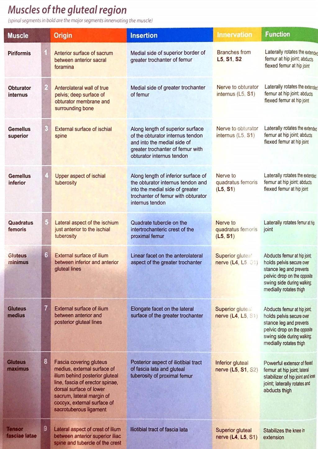
7 * These Notches are Inverted by the Sacrotuberus ligament, which change these notches into 2 Foramens: A. Greater Sciatic Foramen: the gateway of the Pelvis B. Lesser Sciatic Foramen: the gateway of the Perineum (private area) * notice that the gluteal region is divided into four quarters by two lines & The safest area to give an Intramuscular Injection is in the upper lateral quarter of the gluteal Region (Rule of thumb is applied here, refer to the picture); because it has the minimal amount of nerves and blood vessels. Layers of the Gluteal Region: 1. Thick Skin 2. Very thick Superficial Fascia 3. Deep Fascia 4. Muscles (most of the muscles are called Gluteus) Muscles of the Gluteal Area a) Gluteus Maximus: largest Muscle in the body(mass and weight) b) Gluteus Medias: c) Gluteus Minimus: d) Piriformis: e) Obturator Internus: f) Gemellus superior: Action: Extension, Lateral Rotation of Hip & confirm the extension of the knee joint. Nerve supply: Inferior Gluteal Nerve Action: Abduction of the hip (prevent tilting of the pelvis during walking). Nerve supply: Superior Gluteal Nerve Action: Abduction of the hip (prevent tilting of the pelvis) Nerve supply: Superior Gluteal Nerve Action: Lateral Rotation of the Hip joint Nerve supply: Sacral Plexus Action: Lateral Rotation of the Hip Nerve supply: Nerve to Obturator Internus Action: Lateral Rotation of the Hip
8 g) Gemellus inferior: h) Quadratus Femoris: i) Tensor Fascia Lata: Nerve supply: Nerve to Obturator Internus Action: Lateral Rotation of the Hip Nerve supply: Nerve to Quadratus Femoris Action: Lateral Rotation of the Hip Nerve supply: Nerve to Quadratus Femoris Action: Confirm Extension of the knee joint. Nerve supply: Superior Gluteal Nerve *The Piriformis is the KEY of Gluteal Region, Most structure that passes superior to it are called superior (Superior Gluteal VAN), and Most structure passing inferior to it are called inferior (Inferior Gluteal VAN). *The abdominal Aorta when it reaches L4, it divides into right common Iliac and left common iliac, each gives External iliac which continue as Femoral Artery, and Internal iliac that will supply Pelvis. The superior and inferior gluteal arteries are branches from the internal iliac artery. The Posterior Compartment of the Thigh Muscles *All originates from Ischial Tuber. *All supplied by Sciatic Nerve. *All Entend the Hip. *Most Flex the Knee. *All Supplied by Deep Branch of Femoral Artery. *SGS area: is the spot that (Sartorius and Gracillis and Semitendinosus) are inserted. 1. Semitendinosus. 2. Semimembranosus. 3. Biceps: a. Long Head. b. Short Head: only Flexes the Knee because it crosses only 1 Joint. Sciatic Nerve: o Origin: from ventral rami of L4, L5, S1, S2, S3, so it is originated from sacral and lumber plexus o Supplies: (post. Comp of thigh and leg). o Coarse: Leaves the Pelvis through Greater Sciatic Foramen(gateway of the Pelvis), then passes below Piriformis, then it gives 2 branches: 1) Tibial Nerve


9 2) Fibial Nerve (common peroneal nerve) Popliteal fossa: It s a diamond shaped space located behind the Knee Joint Bounded: SuperioMedially by: Semitendinosus and semimembranosus SuperoLaterally by: Biceps Femoris InferioMedially by: Medial head of Gastrocnemius InferioLaterally by: Lateral head of Gastrocnemius Contents: (from superficial to deep) Important 1. Small Saphenous Vein 2. Sciatic Nerve and branches 3. Popliteal Vein 4. Popliteal Artery (it is so deep so you cant measure pulse) When you sit on a chair and you feel Numbness in your feet, it is caused by compression of sciatic Nerve from the Popliteal Fossa. The Neurovascular bundle here is not arranged as supposed to be (VAN, Vein, Artery, Nerve), because here it is in a transition state, they were anterior then became posterior. The following table may help you in memorizing
10
11 The Leg I tried to make it easier by writing the information as points rather than paragraphs. Tibia is the only weight bearing bone in the leg. Its proximal end has 2 condyles while its distal end has medial malleolus. Fibula is only for muscle attachments. Tibial tuberosity is the site for the attachment of quadriceps muscle (patellar ligament). Anterior border & medial surface of the tibia are subcutaneous; which means you can feel them easily. The anterior border is called Shin ; this is the site that all of us suffered from its pain through childhood; when slipping one the stairs. Shaft of the Tibia is triangular in cross section 3 borders & 3 surfaces. Distal end of the tibia articulates with Talus bone in the foot. The fibula, however, articulates with the lateral condyle of the tibia. Remember that: the neck of the fibula is a very important clinical landmark; to be clarified later. Posterior surface of the tibia has an oblique line called soleal line. The ankle joint is U shaped & is formed of: 1- Lateral malleolus. 2- Medial malleolus. 3- Inferior articulating surface of tibia. Deep fascia of leg sends 2 intermuscular septa; one anterior & the other posterior so the leg is divided into three compartments. Notice that: peroneus (Latin) is the same as Fibula (Greek)
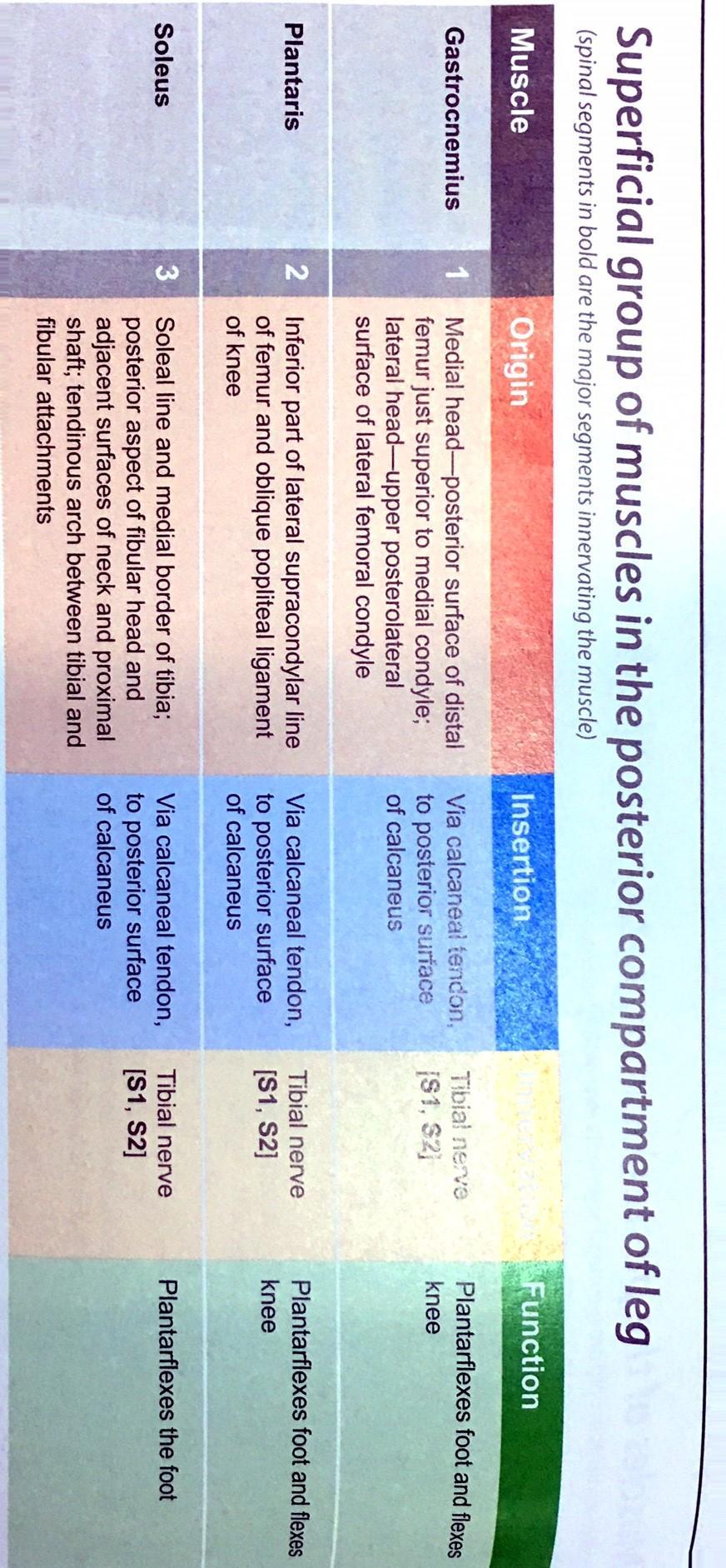
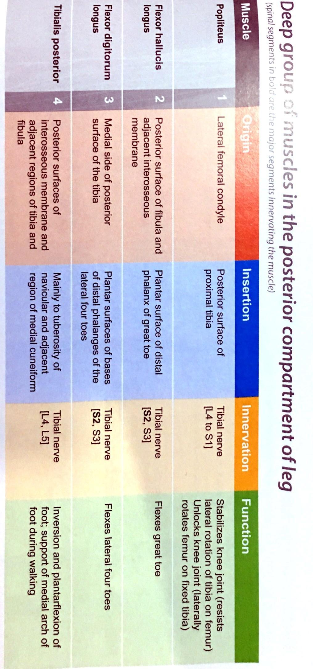
12 Soleus muscle gives you the initiation for running & walking. When the Femoral artery passes the popliteal fossa; it is now called popliteal artery. Notice that the popliteal artery bifurcates at the lower border of the popliteus muscle into: 1- Anterior Tibial artery (Ant. Compartment) 2- Posterior Tibial artery (Post. + Lateral compartment) It is worth noticing that the Sciatic nerve divides into two branches: a- Tibial nerve; which passes between the 2 heads of Gastrocnemius, to supply the posterior compartment. b- Common peroneal nerve (fibular nerve); which wined around the neck of the fibula. So, if the neck of Fibula is fractured, what might happen? - Well, as the fibular nerve supplies the anterior & lateral compartment through its 2 branches; loss of the function of extensors will occur. Remember that: - Deep Fibular Nerve supplies the Anterior Compartment. - Superficial Fibular Nerve supplies the Lateral Compartment. There is a tendon located posteriorly; you can easily feel it; called Calcaneal Tendon OR Ackhilles Tendon; which is mainly made by soleus muscle. The importance of this tendon is that it helps in Soleal Pump; which means to pump venous blood against gravity. Good to know:
13 - That Planatris muscle has a small belly and a long tendon; yet it is important in tendon transplant. - Popliteus muscle, which extends from lateral to medial, causes Unlocking of the knee; through Lateral Rotation of the femur. - Popliteus muscle also initiates flexion of the extended knee. The following tables summarize the action & nerve supply of the leg muscles. Notice that the doctor has mentioned the functions simply, yet the book is more detailed in some of the functions.
14
15
16
 & Extension of the ankle joint =")
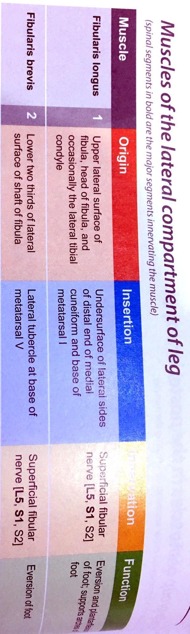
17 Remember that any Tibialis causes Inversion (sole in) & any Fibularis causes eversion (sole out) & Extension of the ankle joint = Dorsi-Flexion
18 & Flexion of the ankle joint = Plantar-Flexion My students We Are Done. Many thanks to Dr. Ahmad Gharaibeh GOOD LUCK TO YOU ALL Dr. Maher Taha Hadidi
Muscles of the lower extremities. Dr. Nabil khouri MD, MSc, Ph.D
 Muscles of the lower extremities Dr. Nabil khouri MD, MSc, Ph.D Posterior leg Popliteal fossa Boundaries Biceps femoris (superior-lateral) Semitendinosis and semimembranosis (superior-medial) Gastrocnemius
Lower limb summary. Anterior compartment of the thigh. Done By: Laith Qashou. Doctor_2016
 Lower limb summary Done By: Laith Qashou Doctor_2016 Anterior compartment of the thigh Sartorius Anterior superior iliac spine Upper medial surface of shaft of tibia 1. Flexes, abducts, laterally rotates
The Hip (Iliofemoral) Joint. Presented by: Rob, Rachel, Alina and Lisa
 Joint. Presented by: Rob, Rachel, Alina and Lisa") The Hip (Iliofemoral) Joint Presented by: Rob, Rachel, Alina and Lisa Surface Anatomy: Posterior Surface Anatomy: Anterior Bones: Os Coxae Consists of 3 Portions: Ilium Ischium Pubis Bones: Pubis Portion
The Hip (Iliofemoral) Joint Presented by: Rob, Rachel, Alina and Lisa Surface Anatomy: Posterior Surface Anatomy: Anterior Bones: Os Coxae Consists of 3 Portions: Ilium Ischium Pubis Bones: Pubis Portion
The thigh. Prof. Oluwadiya KS
 The thigh Prof. Oluwadiya KS www.oluwadiya.com The Thigh: Boundaries The thigh is the region of the lower limb that is approximately between the hip and knee joints Anteriorly, it is separated from the
lesser trochanter of femur lesser trochanter of femur iliotibial tract (connective tissue) medial surface of proximal tibia
 medial surface of proximal tibia") LOWER LIMB MUSCLES OF THE APPENDICULAR SKELETON The muscles that act on the lower limb fall into three groups: those that move the thigh, those that move the lower leg, and those that move the ankle, foot,
LOWER LIMB MUSCLES OF THE APPENDICULAR SKELETON The muscles that act on the lower limb fall into three groups: those that move the thigh, those that move the lower leg, and those that move the ankle, foot,
Muscles of Lesson Five. Muscular Nomenclature and Kinesiology - Two. Muscles of Lesson Five, cont. Chapter 16
 Chapter 16 Muscular Nomenclature and Kinesiology - Two Lessons 5-6 Muscles of Lesson Five Iliopsoas (psoas major, iliacus) Hip outward rotators (piriformis, gemellus superior, gemellus inferior, obturator
Muscles of Gluteal Region
 1 The Gluteal Region In the gluteal region the skin is tough with many layers underneath. Directly under it is the superficial fascia followed by the deep fascia then the muscles and the bones of the thigh.
Muscles of the Hip 1. Tensor Fasciae Latae O: iliac crest I: lateral femoral condyle Action: abducts the thigh Nerve: gluteal nerve
 Muscles of the Hip 1. Tensor Fasciae Latae O: iliac crest I: lateral femoral condyle Action: abducts the thigh Nerve: gluteal nerve 2. Gluteus Maximus O: ilium I: femur Action: abduct the thigh Nerve:
Human Anatomy Biology 351
 Human Anatomy Biology 351 Lower Limb Please place your name on the back of the last page of this exam. You must answer all questions on this exam. Because statistics demonstrate that, on average, between
Mohammad Ashraf. Abdulrahman Al-Hanbali. Ahmad Salman. 1 P a g e
 - 7 Mohammad Ashraf Abdulrahman Al-Hanbali Ahmad Salman 1 P a g e Structures under the cover of Gluteus Maximus: 1-Bones: Ileum, Femur (Head, greater trochanter and gluteal tuberosity), Ischium (ischial
Gluteal region DR. GITANJALI KHORWAL
 Gluteal region DR. GITANJALI KHORWAL Gluteal region The transitional area between the trunk and the lower extremity. The gluteal region includes the rounded, posterior buttocks and the laterally placed
The Muscular System. Chapter 10 Part D. PowerPoint Lecture Slides prepared by Karen Dunbar Kareiva Ivy Tech Community College
 Chapter 10 Part D The Muscular System Annie Leibovitz/Contact Press Images PowerPoint Lecture Slides prepared by Karen Dunbar Kareiva Ivy Tech Community College Table 10.14: Muscles Crossing the Hip and
Human Anatomy Biology 351
 Human Anatomy Biology 351 Lower Limb Please place your name on the back of the last page of this exam. You must answer all questions on this exam. Because statistics demonstrate that, on average, between
ANATYOMY OF The thigh
 ANATYOMY OF The thigh 1- Lateral cutaneous nerve of the thigh Ι) Skin of the thigh Anterior view 2- Femoral branch of the genitofemoral nerve 5- Intermediate cutaneous nerve of the thigh 1, 2 and 3 are
Anatomage Table Instructors Guide- Lower Limb
 The Lower Limb Anatomage Table Instructors Guide- Lower Limb Table of Contents Lower Limb 1- The Skeletal System...3 1: Hip Bone...3 2: Hip Joint and Femur...4 3: Patella and Knee Joint...7 4: Tibia, Fibula,
Main Menu. Joint and Pelvic Girdle click here. The Power is in Your Hands
 1 Hip Joint and Pelvic Girdle click here Main Menu K.6 http://www.handsonlineeducation.com/classes//k6entry.htm[3/23/18, 2:01:12 PM] Hip Joint (acetabular femoral) Relatively stable due to : Bony architecture
Muscles of the Thigh. 6.1 Identify, describe the attachments of and deduce the actions of the muscles of the thigh: Anterior group
 Muscles of the Thigh 6.1 Identify, describe the attachments of and deduce the actions of the muscles of the thigh: Anterior group Sartorius: This is a long strap like muscle with flattened tendons at each
Contents of the Posterior Fascial Compartment of the Thigh
 Contents of the Posterior Fascial Compartment of the Thigh 1-Muscles: B i c e p s f e m o r i s S e m i t e n d i n o s u s S e m i m e m b r a n o s u s a small part of the adductor magnus (h a m s t
Anatomy & Physiology. Muscles of the Lower Limbs.
 Anatomy & Physiology Muscles of the Lower Limbs http://www.ishapeup.com/musclecharts.html Muscles of the Lower Limbs Among the strongest muscles in the body. Because pelvic girdle is composed of heavy,
Lectures of Human Anatomy
 Lectures of Human Anatomy Lower Limb Gluteal Region and Hip Joint By DR. ABDEL-MONEM AWAD HEGAZY M.B. with honor 1983, Dipl."Gynecology and Obstetrics "1989, Master "Anatomy and Embryology" 1994, M.D.
Identify the muscles associated with the medial compartment of the thigh. Identify the attachment points of the medial thigh muscles.
 L 8 A B O R A T O R Y Thigh MEDIAL THIGH Identify the muscles associated with the medial compartment of the thigh. Identify the attachment points of the medial thigh muscles. Identify the actions of these
Topic 7: Hip and pelvis. Parts of the hip. Parts of the femur
 Topic 7: Hip and pelvis Parts of the hip Parts of the femur Classifying the hip joint Ball and socket Synovial Multiaxial Movements of the hip: Abduction/adduction Flexion/extension Medial/lateral rotation
The University Of Jordan Faculty Of Medicine THE LOWER LIMB. Dr.Ahmed Salman Assistant Prof. of Anatomy. The University Of Jordan
 The University Of Jordan Faculty Of Medicine THE LOWER LIMB Dr.Ahmed Salman Assistant Prof. of Anatomy. The University Of Jordan Gluteal Region Cutaneous nerve supply of (Gluteal region) 1. Lateral cutaneous
ANATOMY TEAM GLUTEAL REGION & BACK OF THIGH
 ANATOMY TEAM GLUTEAL REGION & BACK OF THIGH OBJECTIVES By the end of this lecture, the student should be able to identify and discuss: Contents of gluteal region: Groups of Glutei muscles and small muscles
Hip joint and pelvic girdle. Lower Extremity. Pelvic Girdle 6/5/2017
 Hip joint and pelvic girdle Lower Extremity The relationship between the pelvic girdle and hip is similar to that between the shoulder girdle and shoulder joint. The lower limbs are attached to the axial
musculoskeletal system anatomy nerves of the lower limb 2 done by: Dina sawadha & mohammad abukabeer
 musculoskeletal system anatomy nerves of the lower limb 2 done by: Dina sawadha & mohammad abukabeer #Sacral plexus : emerges from the ventral rami of the spinal segments L4 - S4 and provides motor and
lower limb Anterior Compartment: lecture 3 The deep fascia ( fascia lata) divides the thigh into 3 compartments:
 divides the thigh into 3 compartments:") lower limb lecture 3 The deep fascia ( fascia lata) divides the thigh into 3 compartments: 1. Anterior Extensor compartment 2. Medial Adductor compartment 3. Posterior Flexor compartment Anterior Compartment:
lower limb lecture 3 The deep fascia ( fascia lata) divides the thigh into 3 compartments: 1. Anterior Extensor compartment 2. Medial Adductor compartment 3. Posterior Flexor compartment Anterior Compartment:
1-Muscles: 2-Blood supply: Branches of the profunda femoris artery. 3-Nerve supply: Sciatic nerve
 1-Muscles: B i c e p s f e m o r i s S e m i t e n d i n o s u s S e m i m e m b r a n o s u s a small part of the adductor magnus (h a m s t r i n g p a r t o r i s c h i a l p a r t ) 2-Blood supply:
ANATYOMY OF The thigh
 ANATYOMY OF The thigh 1- Lateral cutaneous nerve of the thigh Ι) Skin of the thigh Anterior view 2- Femoral branch of the genitofemoral nerve 5- Intermediate cutaneous nerve of the thigh 1, 2 and 3 are
Lower Limb Nerves. Clinical Anatomy
 Lower Limb Nerves Clinical Anatomy Lumbar Plexus Ventral rami L1 L4 Supplies: Abdominal wall External genitalia Anteromedial thigh Major nerves.. Lumbar Plexus Nerves relation to psoas m. : Obturator n.
The Lower Limb II. Anatomy RHS 241 Lecture 3 Dr. Einas Al-Eisa
 The Lower Limb II Anatomy RHS 241 Lecture 3 Dr. Einas Al-Eisa Tibia The larger & medial bone of the leg Functions: Attachment of muscles Transfer of weight from femur to skeleton of the foot Articulations
MUSCLES OF THE LOWER LIMBS
 MUSCLES OF THE LOWER LIMBS Naming, location and general function Dr. Nabil khouri ROLES THAT SHOULD NOT BE FORGOTTEN Most anterior compartment muscles of the hip and thigh Flexor of the femur at the hip
rotation of the hip Flexion of the knee Iliac fossa of iliac Lesser trochanter Femoral nerve Flexion of the thigh at the hip shaft of tibia
 Anatomy of the lower limb Anterior & medial compartments of the thigh Dr. Hayder The fascia lata encloses the entire thigh like a sleeve/stocking. Three intramuscular fascial septa (lateral, medial, and
Posterior compartment of the thigh. Dr. Heba Kalbouneh Associate Professor of Anatomy and Histology
 Posterior compartment of the thigh Dr. Heba Kalbouneh Associate Professor of Anatomy and Histology Posterior compartment of the thigh 1-Muscles: Biceps femoris Semitendinosus Semimembranosus Adductor magnus
The psoas minor is medial to the psoas major. The iliacus is a fan-shaped muscle that when contracted helps bring the swinging leg forward in walking
 1 p.177 2 3 The psoas minor is medial to the psoas major. The iliacus is a fan-shaped muscle that when contracted helps bring the swinging leg forward in walking and running. The iliopsoas and adductor
Muscles of the Gluteal Region
 Muscles of the Gluteal Region 1 Some of the most powerful in the body Extend the thigh during forceful extension Stabilize the iliotibial band and thoracolumbar fascia Related to shoulders and arms because
CHAPTER 8: THE BIOMECHANICS OF THE HUMAN LOWER EXTREMITY
 CHAPTER 8: THE BIOMECHANICS OF THE HUMAN LOWER EXTREMITY _ 1. The hip joint is the articulation between the and the. A. femur, acetabulum B. femur, spine C. femur, tibia _ 2. Which of the following is
Anterior and Medial compartments of the thigh. Dr. Heba Kalbouneh Associate Professor of Anatomy and Histology
 Anterior and Medial compartments of the thigh Dr. Heba Kalbouneh Associate Professor of Anatomy and Histology Terms Related to Movements Movement Flexion Extension Abduction Adduction Medial (internal)
Lumbar Plexus. Ventral rami L1 L4 Supplies: Major nerves.. Abdominal wall External genitalia Anteromedial thigh
 Lower Limb Nerves Lectures Objectives Describe the structure and relationships of the plexuses of the lower limb. Describe the course, relationships and structures supplied for the major nerves of the
DISSECTION SCHEDULE. Session I - Hip (Front) & Thigh (Superficial)
 & Thigh (Superficial)") DISSECTION SCHEDULE Session I - Hip (Front) & Thigh (Superficial) Surface anatomy Inguinal region Gluteal region Thigh Leg Foot bones Hip bone Femur Superficial fascia Great saphenous vein Superficial
DISSECTION SCHEDULE Session I - Hip (Front) & Thigh (Superficial) Surface anatomy Inguinal region Gluteal region Thigh Leg Foot bones Hip bone Femur Superficial fascia Great saphenous vein Superficial
Copyright 2003 Pearson Education, Inc. publishing as Benjamin Cummings. Dr. Nabil Khouri MD, MSc, Ph.D
 Dr. Nabil Khouri MD, MSc, Ph.D Pelvic Girdle (Hip) Organization of the Lower Limb It is divided into: The Gluteal region The thigh The knee The leg The ankle The foot The thigh and the leg have compartments
Human Anatomy Biology 255
 Human Anatomy Biology 255 Exam #4 Please place your name and I.D. number on the back of the last page of this exam. You must answer all questions on this exam. Because statistics demonstrate that, on average,
Myology of the Knee. PTA 105 Kinesiology
 Myology of the Knee PTA 105 Kinesiology Objectives Describe the planes of motion and axes of rotation of the knee joint Visualize the origins and insertions of the muscles about the knee List the innervations
Lecture 08 THIGH MUSCLES ANTERIOR COMPARTMENT. Dr Farooq Khan Aurakzai. Dated:
 Lecture 08 THIGH MUSCLES ANTERIOR COMPARTMENT BY Dr Farooq Khan Aurakzai Dated: 11.02.2017 INTRODUCTION to the thigh Muscles. The musculature of the thigh can be split into three sections by intermuscular
In-Depth Foundations: Anatomy Terms to Know
 Be familiar with / able to identify and define all the following parts. The Spine Cranium Vertebrae Cervical, Thoracic, Lumbar Sacrum Coccyx Bones of Upper Body Cranium Mastoid process; Occipital condyle,
Bones of Lower Limb. Dr. Heba Kalbouneh Associate Professor of Anatomy and Histology
 Bones of Lower Limb Dr. Heba Kalbouneh Associate Professor of Anatomy and Histology Bones of the lower limb Hip Bone Made up of 3 bones: 1) Ilium (flat), superior in position 2) Ischium (L), postero-inferior
Lower Limb Dr. Robin Paudel
 Lower Limb n What is a limb? n Skeleton n Joints n Pelvis or limb girdle n Hip/Hip Muscles n Lumber and sacral plexus getting spinal nerves out onto limb n Muscles anterior and posterior compartments n
First practical session. Bones of the gluteal region
 First practical session 2017 Bones of the gluteal region The Hip bone The hip bone is made of: 1 The ilium: superior in position 2 The ischium:postero-inferior in position 3 The pubis: antero-inferior
ANATYOMY OF The thigh
 ANATYOMY OF The thigh 1- Lateral cutaneous nerve of the thigh Ι) Skin of the thigh Anterior view 2- Femoral branch of the genitofemoral nerve 1, 2 and 3 are From the lumber plexus 5- Intermediate cutaneous
Practical 1 Worksheet
 Practical 1 Worksheet ANATOMICAL TERMS 1. Use the word bank to fill in the missing words. reference side stand body arms palms anatomical forward All anatomical terms have a(n) point which is called the
Lesson 24. A & P Hip
 Lesson 24 A & P Hip 1 Aims of the Session This session will allow candidates to have an understanding of the bony prominences and soft tissues of the hip 2 Learning Outcomes By the end of the lesson the
Figure 1 - Hip and Pelvis
 Hip Figure 1 - Hip and Pelvis The terms hip and pelvis are frequently used interchangeably, but strictly speaking, the pelvis is a girdle of bones and the hip is a joint. The pelvis consists of The sacrum
this makes sense, however this is lower order thinking and does not solve the lower leg
 Functional Knee Valgus in a Barbell Squat 1 One of the most common lower leg dysfunction we see in athletes, particularly general population is functional knee valgus, or better referred to as the knees
It is formed by fusion of 3 bones: I. Ilium (superior bone). II. Pubis (antero-inferior bone). III. Ischium (postero-inferior bone).
. II. Pubis (antero-inferior bone). III. Ischium (postero-inferior bone).") It is formed by fusion of 3 bones: I. Ilium (superior bone). II. Pubis (antero-inferior bone). III. Ischium (postero-inferior bone). Pubis Acetabulum Ana (242 ) The three constituent of bones of the hip
It is formed by fusion of 3 bones: I. Ilium (superior bone). II. Pubis (antero-inferior bone). III. Ischium (postero-inferior bone). Pubis Acetabulum Ana (242 ) The three constituent of bones of the hip
Bones of the Lower Limb Bone Structure Description Notes. border of the superior ramus. inferolaterally from the pubic symphysis
 Bones of the Lower Limb Bone Structure Description Notes pubis an angulated bone the forms the anterior part of the pelvis one of three bones that form the os coxae: ilium, ischium, pubis; its forms 1/5
HUMAN BODY COURSE LOWER LIMB NERVES AND VESSELS
 HUMAN BODY COURSE LOWER LIMB NERVES AND VESSELS October 22, 2010 D. LOWER LIMB MUSCLES 2. Lower limb compartments ANTERIOR THIGH COMPARTMENT General lfunction: Hip flexion, knee extension, other motions
Baraa Ayed حسام أبو عوض. Ahmad Salman. 1 P a g e
 4 Baraa Ayed حسام أبو عوض Ahmad Salman 1 P a g e Today we are going to cover these concepts: Iliotibial tract Anterior compartment of the thigh and the hip Medial compartment of the thigh Femoral triangle
The Hip Joint. Shenequia Howard David Rivera
 The Hip Joint Shenequia Howard David Rivera Topics Of Discussion Movement Bony Anatomy Ligamentous Anatomy Muscular Anatomy Origin/Insertion/Action/Innervation Common Injuries MOVEMENT Flexion Extension
Where should you palpate the pulse of different arteries in the lower limb?
 Where should you palpate the pulse of different arteries in the lower limb? The femoral artery In the femoral triangle, its pulse is easily felt just inferior to the inguinal ligament midway between the
Acland's DVD Atlas of Human Anatomy. Transcript for Volume Robert D Acland
 Acland's DVD Atlas of Human Anatomy Transcript for Volume 2 2007 Robert D Acland This free downloadable pdf file is to be used for individual study only. It is not to be reproduced in any form without
MUSCULOSKELETAL LOWER LIMB
 MUSCULOSKELETAL LOWER LIMB Spinal Cord Lumbar and Sacral Regions Spinal cord Dorsal root ganglion Conus medullaris Cauda equina Dorsal root ganglion of the fifth lumbar nerve End of subarachnoid space
The Knee. Clarification of Terms. Osteology of the Knee 7/28/2013. The knee consists of: The tibiofemoral joint Patellofemoral joint
 The Knee Clarification of Terms The knee consists of: The tibiofemoral joint Patellofemoral joint Mansfield, p273 Osteology of the Knee Distal Femur Proximal tibia and fibula Patella 1 Osteology of the
Muscles to know. Lab 21. Muscles of the Pelvis and Lower Limbs. Muscles that Position the Lower Limbs. Generally. Muscles that Move the Thigh
 Muscles to know Lab 21 Muscles of the Pelvis, Leg and Foot psoas major iliacus gluteus maximus gluteus medius sartorius quadriceps femoris (4) gracilus adductor longus biceps femoris semitendinosis semimembranosus
Scapula Spine Lateral edge of clavicle. Medial border Scapula. Medial border of Scapula, between superior angle and root of spine. Scapula.
 Muscle attachments and actions answer sheet Muscle Origins insertions Movements Joints crossed Trapezius Base of skull Spinous process of C7 Thoracic Spine Lateral edge of clavicle Elevation Retraction
The Lower Limb. Anatomy RHS 241 Lecture 2 Dr. Einas Al-Eisa
 The Lower Limb Anatomy RHS 241 Lecture 2 Dr. Einas Al-Eisa The bony pelvis Protective osseofibrous ring for the pelvic viscera Transfer of forces to: acetabulum & head of femur (when standing) ischial
5 Testing the Muscles of the Lower Extremity
 C H A P T E R 5 Testing the Muscles of the Lower Extremity Hip Flexion Hip Flexion, Abduction, and External Rotation with Knee Flexion Hip Extension Hip Abduction Hip Abduction from Flexed Position Hip
Organization of the Lower Limb
 Organization of the Lower Limb Limb Development Lower limb develops in an aterolateral position at the level of the L2 to S3 trunk segments Great toe positioned cephalic direction with the soles of the
ANATOMY. Lecturer : Maher Hadidi Done by: ,,Subject : lecture#: ~ Date:
 ANATOMY,,Subject : Lecturer : Maher Hadidi Done by: ~~ lecture#: ~ Date: GftJteal Reaion ' ' v Highest point of iliac cres Safe site for (/M) injection Horizantal line Sciatic nerve Ischial tubrosity
Leg. Dr. Heba Kalbouneh Associate Professor of Anatomy and Histology
 Leg Dr. Heba Kalbouneh Associate Professor of Anatomy and Histology Skin of the Leg Cutaneous Nerves Medially: The saphenous nerve, a branch of the femoral nerve supplies the skin on the medial surface
Sports Medicine 15. Unit I: Anatomy. The knee, Thigh, Hip and Groin. Part 4 Anatomies of the Lower Limbs
 Sports Medicine 15 Unit I: Anatomy Part 4 Anatomies of the Lower Limbs The knee, Thigh, Hip and Groin Anatomy of the lower limbs In Part 3 of this section we focused upon 11 of the 12 extrinsic muscles
Bony Anatomy. Femur. Femoral Head Femoral Neck Greater Trochanter Lesser Trochanter Intertrochanteric Crest Intertrochanteric Line Gluteal Tuberosity
 Hip Anatomy Bony Anatomy Femur Femoral Head Femoral Neck Greater Trochanter Lesser Trochanter Intertrochanteric Crest Intertrochanteric Line Gluteal Tuberosity Bony Anatomy Pelvic Girdle Acetabulum 3 bones
The Leg. Prof. Oluwadiya KS
 The Leg Prof. Oluwadiya KS www.oluwadiya.sitesled.com Compartments of the leg 4 Four Compartments: 1. Anterior compartment Deep fibular nerve Dorsiflexes the foot and toes 2. Lateral Compartment Superficial
5.1 Identify, describe the attachments of and deduce the actions of the muscles of the thigh:
 5.1 Identify, describe the attachments of and deduce the actions of the muscles of the thigh: Anterior group Proximal attachment Distal attachment Sartorius ASIS» Upper part of shaft tibia (middle surface)»
BONES JOINTS MUSCLES OF THE LOWER LIMB
 BONES JOINTS MUSCLES OF THE LOWER LIMB LOWER LIMB: BONES LOWER LIMB GLUTEAL REGION consists of 6 major segments: FEMORAL REGION (THIGH) KNEE REGION LEG REGION TALOCRURAL REGION (ANKLE) FOOT REGION LOWER
Anatomy of the lower limb
 Anatomy of the lower limb 1. Bones of the lower limb Pelvis Hip bone/coxal bone Acetabulum o Acetabular margin o Acetabular fossa o Acetabular notch o Lunate surface Ischiopubic ramus Obturator foramen
LAB Notes#1. Ahmad Ar'ar. Eslam
 LAB Notes#1 Ahmad Ar'ar Eslam 1 P a g e Anatomy lab Notes Lower limb bones :- Pelvic girdle: It's the connection between the axial skeleton and the lower limb; it's made up of one bone called the HIP BONE
Organization of the Lower Limb
 Organization of the Lower Limb Most illustrations from: Thieme Atlas of Anatomy: Musculoskeletal System. M Schuenke, et al, 2006. Anatomy: A Regional Atlas of the Human Body. Carmine Clemente, 4th edition.
Gluteal Region and Back of Thigh
 Gluteal Region and Back of Thigh Musculoskeletal block- Anatomy-lecture 14 Editing file Objectives Know contents of gluteal region: 1. Groups of Glutei muscles and small muscles (Lateral Rotators). 2.
Muscle Testing of Knee Extensors. Yasser Moh. Aneis, PhD, MSc., PT. Lecturer of Physical Therapy Basic Sciences Department
 Muscle Testing of Knee Extensors Yasser Moh. Aneis, PhD, MSc., PT. Lecturer of Physical Therapy Basic Sciences Department Muscle Testing of Knee Extensors othe Primary muscle Quadriceps Femoris -Rectus
Figure 7: Bones of the lower limb
 BONES OF THE APPENDICULAR SKELETON The appendicular skeleton is composed of the 126 bones of the appendages and the pectoral and pelvic girdles, which attach the limbs to the axial skeleton. Although the
Anatomy images for MSS practical exam- 2019
 Anatomy images for MSS practical exam- 2019 Ilium Ischium Pubis Acetabulaum Iliac crest Iliac tubercle ASIS (muscle and ligament attached) AIIS (muscle attached) PSIS PIIS Ischial spine Ischial tuberosity
musculoskeletal system anatomy nerves of the lower limb 1 done by: dina sawadha & mohammad abukabeer
 musculoskeletal system anatomy nerves of the lower limb 1 done by: dina sawadha & mohammad abukabeer What is the importance of plexuses? plexuses provides us the advantage of a phenomenon called convergence
TABLE OF MUSCLES OF LOWER EXTREMITY 2018zillmusom ANTERIOR THIGH
 TABLE OF MUSCLES OF LOWER EXTREMITY 2018zillmusom ANTERIOR THIGH MUSCLE ORIGIN INSERTION ACTION NERVE Iliopsoas Ilium, vertebra Femur Flex hip joint Femoral nerve (T12-L5) Pectineus Pubis Femur Flex hip
The Lower Limb. Sevda LAFCI FAHRİOĞLU, MD.PhD.
 The Lower Limb Sevda LAFCI FAHRİOĞLU, MD.PhD. The Lower Limb The bones of the lower limb form the inferior part of the appendicular skeleton the organ of locomotion for bearing the weight of body stronger
Organization of the Lower Limb Audrone Biknevicius, Ph.D. Dept. Biomedical Sciences, OU HCOM at Dublin Clinical Anatomy Immersion 2014
 Organization of the Lower Limb Audrone Biknevicius, Ph.D. Dept. Biomedical Sciences, OU HCOM at Dublin Clinical Anatomy Immersion 2014 www.thestudio1.co.za LIMB FUNCTION choco-locate.com blog.coolibar.com
The os coxae or hip bone consists of three flat bones, ilium, ischium and pubis, which fuse together to form the acetabulum.
 The os coxae The os coxae or hip bone consists of three flat bones, ilium, ischium and pubis, which fuse together to form the acetabulum. The ilium extends from the acetabulum upwards forming the lateral
Due in Lab weeks because of Thanksgiving Prelab #10. Homework #8. Both sides! Both sides!
 Lab 8 MUSCLES Due in Lab 10 2 weeks because of Thanksgiving Prelab #10 Both sides! Homework #8 Both sides! Refer to Muscles 22-23 Naming of muscles Origin Site of muscle attachment that doesn t move during
To describe he knee joint, ligaments, structure & To list the main features of other lower limb joints
 To describe he knee joint, ligaments, structure & neurovascular supply To demonstrate the ankle joint anatomy To list the main features of other lower limb joints To list main groups of lymph nodes in
Peripheral Nervous System: Lower Body
 Peripheral Nervous System: Lower Body MSTN121 - Neurophysiology Session 11 Department of Myotherapy Lumbar Plexus Iliohypogastric nerve (T12-L1) Motor: Transverse abdominis and internal obliques Sensory:
Anatomy&Embryology Final Exam
 Anatomy&Embryology Final Exam Done by : Maha AbuAjamieh 1. Fertilisation is more likely to occur: A) 12-24 hours after ovulation. B) 24-40 hours after ovulation. C) 40-72 hours after ovulation. 2. wrong
Regional Anaesthesia
 Regional Anaesthesia Lower limb anatomy and blocks Hip and Knee Joint Hip Joint: Nerve supply Lumbar plexus Femoral nerve through the nerve to the Rectus Femoris Ant division of the Obturator nerve The
Introduction to Anatomy. Dr. Maher Hadidi. Laith Al-Hawajreh. Mar/25 th /2013
 Introduction to Anatomy Dr. Maher Hadidi Laith Al-Hawajreh 22 Mar/25 th /2013 Lower limb - The leg The skeleton of the leg is formed by two bones: 1) Medial: Tibia 2) Lateral: Fibula The two bones are
Anatomy MCQs Week 13
 Anatomy MCQs Week 13 1. Posterior to the medial malleolus of the ankle: The neurovascular bundle lies between Tibialis Posterior and Flexor Digitorum Longus The tendon of Tibialis Posterior inserts into
The Hay is in the Barn
 Anatomy 1 Practical 1 Review Made by Forrest Allen (nerd) Edited by TJ Williamson (not nerd) The Hay is in the Barn 2019 Thunderbringers Too much to handle https://www.youtube.com/watch?v=glii-kaza d8
Muscle Anatomy Review Chart
 Muscle Anatomy Review Chart BACK Superficial (5) Trapezius Transverse cervical a. Latissimus dorsi Thoracodorsal a. Rhomboideus major Dorsal scapular a. Rhomboideus minor Levator scapulae Intermediate
11/15/2018. Temporalis Elevates & retracts mandible. Masseter = Prime mover of jaw closure. Levator scapulae Supraspinatus Clavicle.
 Due in Lab 10 Lab 8 MUSCLES 2 weeks because of Thanksgiving Prelab #10 Both sides! Homework #8 Both sides! Refer to Muscles 22-23 Examples of Origin & Insertion Naming of muscles Origin Site of muscle
Applied anatomy of the hip and buttock
 CHAPTER CONTENTS The hip joint e9 Capsule and ligaments e9 s e0 Flexor muscles................... e0 Extensor muscles.................. e Abductor muscles.................. e Adductor muscles..................
Lab Activity 9. Appendicular Skeleton Martini Chapter 8. Portland Community College BI 231
 Lab Activity 9 Appendicular Skeleton Martini Chapter 8 Portland Community College BI 231 Appendicular Skeleton Upper & Lower extremities Shoulder Girdle Pelvic Girdle 2 Humerus 3 Humerus: Proximal End
VENOUS DRAINAGE OF THE LOWER LIMB
 Anatomy of the lower limb Superficial veins & nerve injuries Dr. Hayder VENOUS DRAINAGE OF THE LOWER LIMB The venous drainage of the lower limb is of huge clinical & surgical importance. Since the venous
Bio 113 Anatomy and Physiology The Muscles. Muscles of the Head and Neck. Masseter. Orbicularis occuli. Orbicularis oris. Sternocleidomastoid
 Bio 113 Anatomy and Physiology The Muscles Muscles of the Head and Neck Masseter Orbicularis occuli Orbicularis oris Sternocleidomastoid Temporalis BIO 113 Fall 2011 Muscles Page 1 of 5 Muscles of the