Thigh, Hip, & Low Back Evaluation.
|
|
|
- Anastasia Stanley
- 6 years ago
- Views:
Transcription
1 Thigh, Hip, & Low Back Evaluation
2 Thigh Injuries Quad contusions - Myositis Ossificans Trochanteric bursitis - snapping hip Ischial bursitis - bench-warmer s bursitis Strains Fractures - femur, stress, apophysitis IT band friction syndrome may predispose an athlete - leg length difference
3 Hip Injuries Fractures pelvis, ischial tuberosity (hamstring attachment) Iliac crest contusion - hip pointer Sprains hip Hip dislocation Posterior dislocation lower leg position adduction & internal rotation Piriformis syndrome Proximal insertion: sacrum Distal insertion: medial aspect of greater trochanter *can compress sciatic nerve between ischium & greater trochanter if tight
4 Lowback Injuries Contusions, lacerations, subluxations, sprains, strains Disc & Neurologic Pathology Fractures - stress, apophysitis Spondylosis- arthritis or osteoarthritis of the vertebrae; results in pressures being placed on the vertebral nerve roots Spondylolysis- degeneration of a vertebral structure secondary to repetitive stress most commonly affecting pars interarticularis but with no displacement of the vertebral body (Scotty dog) Spondylolisthesis - anterior slippage of vertebrae superior to pathological site; causes increased back pain upon extension
 joints Capsule around the joint, bursa")

5 Anatomy Lumbar vertebrae (transverse processes, spinous process) Sacroiliac (SI) joints Capsule around the joint, bursa (trochanteric, ischial) Muscles Erector Spinae, Adductors, Abductors, Hamstrings, Quadriceps, External rotators of hip Intervertebral discs Sacrum


6 Anatomy Pelvic bone - divided into 3 areas Ilium - upper two fifths Ischium - posterior & lower two fifths Pubis - anterior & lower one fifth Femur
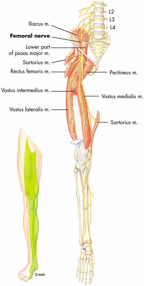
7 Anatomy Lumbar plexus (T12-L5, some say L3-S1) Femoral Nerve - L2, L3, & L4 n. roots converge (anterior branches form Obturator n.) Sacral plexus (L4-S5) Sciatic n. 1) Tibial n., 2) Common Peroneal n., 3) Slip of Tibial n. innervates hamstring Femoral Triangle femoral n., femoral a. & v. Sartorius lateral edge Adductor longus medial edge Inguinal ligament superior edge


8
")
 Keeps us")


9 Anatomy Ligaments Iliofemoral ligament (Y ligament of Bigelow) Reinforces anterior joint capsule (limits hyperextension) Keeps us upright Pubofemoral ligament Limits abduction & hyperextension Inguinal ligament Runs from ASIS to pubic symphysis Superior border of femoral triangle
10 Evaluation - History MOI direct blow vs. overuse Pain Pain scale, Type, Location, Time, Consistency (constant/intermittent) Radiating, numbness/tingling, burning, aching, throbbing Onset Acute vs. Chronic Training Techniques Changes in intensity, frequency, & duration of training, surface (terrain, hills), shoes Prior history Legg-Calve-Perthes disease avascular necrosis of prox. femoral epiphysis
11 Evaluation - Observation Deformities Musculature Bony Nelaton s line - ASIS to ischial tuberosity Hip angulations Angle of Inclination angular relationship between femur & tibia Frontal plane Coxa valga, coxa vara, patellar position Normally 125º ( slightly ) Angle of Torsion relationship between femoral head & shaft Transverse plane Normally 15º Anteversion: internal femoral rotation, toed-in gait (pigeontoed), squinting patellae Retroversion: external femoral rotation, toed-out gait (duckfooted), frog-eyed patellae
12 Evaluation - Observation Leg Length Discrepancy True Leg Length ASIS to medial malleolus BONY Apparent Leg Length Umbilicus to medial malleolus SOFT TISSUE Greater than ¼ difference is considered a discrepancy Level of iliac crest when standing Gait Level of iliac crest ROM Limp
 Lateral curve - scoliosis Pelvic")
13 Evaluation - Observation Spinal column curvature Lumbar spine Lordotic curve (sway back) Thoracic spine Kyphotic curve Cervical spine Lordotic curve (hunch back) Lateral curve - scoliosis Pelvic Tilt
14 Evaluation - Palpation Use discretion when palpating in this region! Provide privacy when palpating area! Bony Step-off deformity of lumbar spine (spondylolisthesis) T7 vertebrae inferior angle of scapula L3 posterior from umbilicus L4 level of iliac crest L5 bilateral dimples (may vary) S2 PSIS level Femoral Triangle
15 Evaluation Palpation Muscles Hip joint & pelvic girdle muscles Anterior - primarily hip flexion Iliopsoas Pectineus Rectus femoris Sartorius Medial primarily hip adduction Adductor brevis Adductor longus Adductor magnus Gracilis Posterior - primarily hip extension Gluteus maximus Biceps femoris Semitendinosus Semimembranosus External rotators Lateral - primarily hip abduction Gluteus medius Gluteus minimus External rotators Tensor fasciae latae
16 Evaluation ROM active, passive, resistive (knee flex/ext) Hip flexion neutral to Hip extension neutral to Hip abduction neutral to Hip adduction neutral to 30 Internal rotation neutral to 45 External rotation neutral to 50 Trunk rotation, lateral bending, flexion, extension Active standing position Beevor s Sign - Partial Sit-up (T5-T12 n. innervation) Anterior, Posterior, Left & Right Lateral pelvic rotation True Leg Length Discrepancy Test Apparent Leg Length Discrepancy Test
17 Evaluation Stress Tests Musculature tests Trendelenburg s Test gluteus medius Thomas Test hip flexor tightness Rectus femoris vs. Iliopsoas Ober Test IT band Noble s Compression Test IT band Ely s Test Rectus femoris (PROM)
18 Evaluation Stress Tests Ligamentous testing no specific tests Neurologic testing Beevor s Sign thoracic n. inhibition Piriformis Test Piriformis Syndrome impingement of sciatic n. from spasm of piriformis Resisted hip abd. while seated can duplicate pain caused by this syndrome Straight Leg Raise (SLR) test sciatic n. irritation or disc (Passive) Well SLR test disc (opposite side) Increased Intrathecal Pressure Valsalva test (maneuver) herniated disc Milgram test disc (active double SLR) Kernig s test or Kernig/Brudzinski test disc (active SLR w/ knee extended) SLR Slump test sciatic or other neurologic Quadrant test nerve vs. facet
19 Evaluation Stress Tests Neurological testing Femoral Nerve Stretch Test - disc Single Leg Stance Test lumbar spine or SI area Lower Quarter (Extremity) Neurological Screen (p. 352) Sensory testing L1- S2 Motor testing L1-S2 L1 & 2 hip flexion L3 knee extension L4 dorsiflexion L5 great toe extension S1 plantarflexion Reflex testing L4 (patellar tendon), S1 (achilles) Hip Scouring test articular cartilage of femur or acetabulum, labral tear
20 Evaluation Stress Tests Sacroiliac joint problems SI Compression SI Distraction FABER(E) test Gaenslen s test Long Sit test rotated ilium March test Other Spring Test - hyper/hypomobility Hoover test malingering
21 On-field Evaluation On-field Neurologic tests History: MOI, location of pain, peripheral symptoms (pain, weakness, numbness) Inspection: position, posture, willingness to move Neurologic: sensory & motor tests Palpation: bony & musculature
The Hip (Iliofemoral) Joint. Presented by: Rob, Rachel, Alina and Lisa
 Joint. Presented by: Rob, Rachel, Alina and Lisa") The Hip (Iliofemoral) Joint Presented by: Rob, Rachel, Alina and Lisa Surface Anatomy: Posterior Surface Anatomy: Anterior Bones: Os Coxae Consists of 3 Portions: Ilium Ischium Pubis Bones: Pubis Portion
The Hip (Iliofemoral) Joint Presented by: Rob, Rachel, Alina and Lisa Surface Anatomy: Posterior Surface Anatomy: Anterior Bones: Os Coxae Consists of 3 Portions: Ilium Ischium Pubis Bones: Pubis Portion
The Hip Joint. Shenequia Howard David Rivera
 The Hip Joint Shenequia Howard David Rivera Topics Of Discussion Movement Bony Anatomy Ligamentous Anatomy Muscular Anatomy Origin/Insertion/Action/Innervation Common Injuries MOVEMENT Flexion Extension
RN(EC) ENC(C) GNC(C) MN ACNP *** MECHANISM OF INJURY.. MOST IMPORTANT ***
 ENC(C) GNC(C) MN ACNP *** MECHANISM OF INJURY.. MOST IMPORTANT ***") HISTORY *** MECHANISM OF INJURY.. MOST IMPORTANT *** Age of patient - Certain conditions are more prevalent in particular age groups (Hip pain in children may refer to the knee from Legg-Calve-Perthes
HISTORY *** MECHANISM OF INJURY.. MOST IMPORTANT *** Age of patient - Certain conditions are more prevalent in particular age groups (Hip pain in children may refer to the knee from Legg-Calve-Perthes
Main Menu. Joint and Pelvic Girdle click here. The Power is in Your Hands
 1 Hip Joint and Pelvic Girdle click here Main Menu K.6 http://www.handsonlineeducation.com/classes//k6entry.htm[3/23/18, 2:01:12 PM] Hip Joint (acetabular femoral) Relatively stable due to : Bony architecture
Muscles of the lower extremities. Dr. Nabil khouri MD, MSc, Ph.D
 Muscles of the lower extremities Dr. Nabil khouri MD, MSc, Ph.D Posterior leg Popliteal fossa Boundaries Biceps femoris (superior-lateral) Semitendinosis and semimembranosis (superior-medial) Gastrocnemius
The Lower Limb. Anatomy RHS 241 Lecture 2 Dr. Einas Al-Eisa
 The Lower Limb Anatomy RHS 241 Lecture 2 Dr. Einas Al-Eisa The bony pelvis Protective osseofibrous ring for the pelvic viscera Transfer of forces to: acetabulum & head of femur (when standing) ischial
Lesson 24. A & P Hip
 Lesson 24 A & P Hip 1 Aims of the Session This session will allow candidates to have an understanding of the bony prominences and soft tissues of the hip 2 Learning Outcomes By the end of the lesson the
Bony Anatomy. Femur. Femoral Head Femoral Neck Greater Trochanter Lesser Trochanter Intertrochanteric Crest Intertrochanteric Line Gluteal Tuberosity
 Hip Anatomy Bony Anatomy Femur Femoral Head Femoral Neck Greater Trochanter Lesser Trochanter Intertrochanteric Crest Intertrochanteric Line Gluteal Tuberosity Bony Anatomy Pelvic Girdle Acetabulum 3 bones
Hip joint and pelvic girdle. Lower Extremity. Pelvic Girdle 6/5/2017
 Hip joint and pelvic girdle Lower Extremity The relationship between the pelvic girdle and hip is similar to that between the shoulder girdle and shoulder joint. The lower limbs are attached to the axial
Figure 1 - Hip and Pelvis
 Hip Figure 1 - Hip and Pelvis The terms hip and pelvis are frequently used interchangeably, but strictly speaking, the pelvis is a girdle of bones and the hip is a joint. The pelvis consists of The sacrum
Muscles of the Hip 1. Tensor Fasciae Latae O: iliac crest I: lateral femoral condyle Action: abducts the thigh Nerve: gluteal nerve
 Muscles of the Hip 1. Tensor Fasciae Latae O: iliac crest I: lateral femoral condyle Action: abducts the thigh Nerve: gluteal nerve 2. Gluteus Maximus O: ilium I: femur Action: abduct the thigh Nerve:
THE HIP. Cooler than cool, the pinnacle of what is "it". Beyond all trends and conventional coolness.
 THE HIP Cooler than cool, the pinnacle of what is "it". Beyond all trends and conventional coolness. Objectives Hip anatomy Causes of hip pain Hip exam Anatomy Bones Ilium Anterior Superior Iliac Spine
Hip Anatomy. Bony. The Athletic Hip: Anatomy and Common Injuries. Kyle Wilkens MSPAS, PA-C, ATC/L October 8, 2013
 The Athletic Hip: Anatomy and Common Injuries Kyle Wilkens MSPAS, PA-C, ATC/L October 8, 2013 Hip Anatomy Bony Femur Ischium Ilium Pubis Sacrum Coccyx Soft Tissue Joints Muscles -Pubic Symphisis Labrum
Joints of the lower limb
 Joints of the lower limb 1-Type: Hip joint Synovial ball-and-socket joint 2-Articular surfaces: a- head of femur b- lunate surface of acetabulum Which is deepened by the fibrocartilaginous labrum acetabulare
The Lower Limb II. Anatomy RHS 241 Lecture 3 Dr. Einas Al-Eisa
 The Lower Limb II Anatomy RHS 241 Lecture 3 Dr. Einas Al-Eisa Tibia The larger & medial bone of the leg Functions: Attachment of muscles Transfer of weight from femur to skeleton of the foot Articulations
Lectures of Human Anatomy
 Lectures of Human Anatomy Lower Limb Gluteal Region and Hip Joint By DR. ABDEL-MONEM AWAD HEGAZY M.B. with honor 1983, Dipl."Gynecology and Obstetrics "1989, Master "Anatomy and Embryology" 1994, M.D.
Overview. Overview. Introduction. Introduction Anatomy History Examination Common Disorders. Introduction Anatomy History Examination Common Disorders
 Common Hip Disorders in Figure Skaters 14 th Annual Meeting of Sports Medicine and Science in Figure Skating January 25, 2009 8:15-8:45am Robert J. Dimeff, MD Medical Director of Sports Medicine Overview
The psoas minor is medial to the psoas major. The iliacus is a fan-shaped muscle that when contracted helps bring the swinging leg forward in walking
 1 p.177 2 3 The psoas minor is medial to the psoas major. The iliacus is a fan-shaped muscle that when contracted helps bring the swinging leg forward in walking and running. The iliopsoas and adductor
The thigh. Prof. Oluwadiya KS
 The thigh Prof. Oluwadiya KS www.oluwadiya.com The Thigh: Boundaries The thigh is the region of the lower limb that is approximately between the hip and knee joints Anteriorly, it is separated from the
Scapula Spine Lateral edge of clavicle. Medial border Scapula. Medial border of Scapula, between superior angle and root of spine. Scapula.
 Muscle attachments and actions answer sheet Muscle Origins insertions Movements Joints crossed Trapezius Base of skull Spinous process of C7 Thoracic Spine Lateral edge of clavicle Elevation Retraction
Lower limb summary. Anterior compartment of the thigh. Done By: Laith Qashou. Doctor_2016
 Lower limb summary Done By: Laith Qashou Doctor_2016 Anterior compartment of the thigh Sartorius Anterior superior iliac spine Upper medial surface of shaft of tibia 1. Flexes, abducts, laterally rotates
Gluteal region DR. GITANJALI KHORWAL
 Gluteal region DR. GITANJALI KHORWAL Gluteal region The transitional area between the trunk and the lower extremity. The gluteal region includes the rounded, posterior buttocks and the laterally placed
The University Of Jordan Faculty Of Medicine THE LOWER LIMB. Dr.Ahmed Salman Assistant Prof. of Anatomy. The University Of Jordan
 The University Of Jordan Faculty Of Medicine THE LOWER LIMB Dr.Ahmed Salman Assistant Prof. of Anatomy. The University Of Jordan Gluteal Region Cutaneous nerve supply of (Gluteal region) 1. Lateral cutaneous
Muscles of Lesson Five. Muscular Nomenclature and Kinesiology - Two. Muscles of Lesson Five, cont. Chapter 16
 Chapter 16 Muscular Nomenclature and Kinesiology - Two Lessons 5-6 Muscles of Lesson Five Iliopsoas (psoas major, iliacus) Hip outward rotators (piriformis, gemellus superior, gemellus inferior, obturator
In-Depth Foundations: Anatomy Terms to Know
 Be familiar with / able to identify and define all the following parts. The Spine Cranium Vertebrae Cervical, Thoracic, Lumbar Sacrum Coccyx Bones of Upper Body Cranium Mastoid process; Occipital condyle,
Human Anatomy Biology 351
 Human Anatomy Biology 351 Lower Limb Please place your name on the back of the last page of this exam. You must answer all questions on this exam. Because statistics demonstrate that, on average, between
rotation of the hip Flexion of the knee Iliac fossa of iliac Lesser trochanter Femoral nerve Flexion of the thigh at the hip shaft of tibia
 Anatomy of the lower limb Anterior & medial compartments of the thigh Dr. Hayder The fascia lata encloses the entire thigh like a sleeve/stocking. Three intramuscular fascial septa (lateral, medial, and
ANATYOMY OF The thigh
 ANATYOMY OF The thigh 1- Lateral cutaneous nerve of the thigh Ι) Skin of the thigh Anterior view 2- Femoral branch of the genitofemoral nerve 5- Intermediate cutaneous nerve of the thigh 1, 2 and 3 are
CHAPTER 8: THE BIOMECHANICS OF THE HUMAN LOWER EXTREMITY
 CHAPTER 8: THE BIOMECHANICS OF THE HUMAN LOWER EXTREMITY _ 1. The hip joint is the articulation between the and the. A. femur, acetabulum B. femur, spine C. femur, tibia _ 2. Which of the following is
lesser trochanter of femur lesser trochanter of femur iliotibial tract (connective tissue) medial surface of proximal tibia
 medial surface of proximal tibia") LOWER LIMB MUSCLES OF THE APPENDICULAR SKELETON The muscles that act on the lower limb fall into three groups: those that move the thigh, those that move the lower leg, and those that move the ankle, foot,
LOWER LIMB MUSCLES OF THE APPENDICULAR SKELETON The muscles that act on the lower limb fall into three groups: those that move the thigh, those that move the lower leg, and those that move the ankle, foot,
Evaluation of the Hip
 Evaluation of the Hip Adam Lewno, DO PCSM Fellow, University of Michigan Primary Care Sports Update 2017 Disclosures Financial: None Images: I would like to acknowledge the work of the original owners
Identify the muscles associated with the medial compartment of the thigh. Identify the attachment points of the medial thigh muscles.
 L 8 A B O R A T O R Y Thigh MEDIAL THIGH Identify the muscles associated with the medial compartment of the thigh. Identify the attachment points of the medial thigh muscles. Identify the actions of these
FUNCTIONAL ANATOMY AND EXAM OF THE HIP, GROIN AND THIGH
 FUNCTIONAL ANATOMY AND EXAM OF THE HIP, GROIN AND THIGH Peter G Gerbino, MD, FACSM Orthopedic Surgeon Monterey Joint Replacement and Sports Medicine Monterey, CA TPC, San Diego, 2017 The lecturer has no
The Muscular System. Chapter 10 Part D. PowerPoint Lecture Slides prepared by Karen Dunbar Kareiva Ivy Tech Community College
 Chapter 10 Part D The Muscular System Annie Leibovitz/Contact Press Images PowerPoint Lecture Slides prepared by Karen Dunbar Kareiva Ivy Tech Community College Table 10.14: Muscles Crossing the Hip and
Anterior and Medial compartments of the thigh. Dr. Heba Kalbouneh Associate Professor of Anatomy and Histology
 Anterior and Medial compartments of the thigh Dr. Heba Kalbouneh Associate Professor of Anatomy and Histology Terms Related to Movements Movement Flexion Extension Abduction Adduction Medial (internal)
Hip Joint DX 612 Orthopedics and Neurology
 Hip Joint DX 612 Orthopedics and Neurology James J. Lehman, DC, MBA, DABCO University of Bridgeport College of Chiropractic Hip Anatomy Palpation Point tenderness Edema Symmetry Hip ROM Hip Contracture
Hip Anatomy. Hip Joint DX 612 Orthopedics and Neurology. Hip ROM. Palpation
 Hip Joint DX 612 Orthopedics and Neurology Hip Anatomy James J. Lehman, DC, MBA, DABCO University of Bridgeport College of Chiropractic Palpation Hip ROM Point tenderness Edema Symmetry Hip Contracture
Human Anatomy Biology 255
 Human Anatomy Biology 255 Exam #4 Please place your name and I.D. number on the back of the last page of this exam. You must answer all questions on this exam. Because statistics demonstrate that, on average,
Anatomy & Physiology. Muscles of the Lower Limbs.
 Anatomy & Physiology Muscles of the Lower Limbs http://www.ishapeup.com/musclecharts.html Muscles of the Lower Limbs Among the strongest muscles in the body. Because pelvic girdle is composed of heavy,
Myology of the Knee. PTA 105 Kinesiology
 Myology of the Knee PTA 105 Kinesiology Objectives Describe the planes of motion and axes of rotation of the knee joint Visualize the origins and insertions of the muscles about the knee List the innervations
Lower Limb Dr. Robin Paudel
 Lower Limb n What is a limb? n Skeleton n Joints n Pelvis or limb girdle n Hip/Hip Muscles n Lumber and sacral plexus getting spinal nerves out onto limb n Muscles anterior and posterior compartments n
Baraa Ayed حسام أبو عوض. Ahmad Salman. 1 P a g e
 4 Baraa Ayed حسام أبو عوض Ahmad Salman 1 P a g e Today we are going to cover these concepts: Iliotibial tract Anterior compartment of the thigh and the hip Medial compartment of the thigh Femoral triangle
ANATYOMY OF The thigh
 ANATYOMY OF The thigh 1- Lateral cutaneous nerve of the thigh Ι) Skin of the thigh Anterior view 2- Femoral branch of the genitofemoral nerve 1, 2 and 3 are From the lumber plexus 5- Intermediate cutaneous
Human Anatomy Biology 351
 Human Anatomy Biology 351 Lower Limb Please place your name on the back of the last page of this exam. You must answer all questions on this exam. Because statistics demonstrate that, on average, between
Applied anatomy of the hip and buttock
 CHAPTER CONTENTS The hip joint e9 Capsule and ligaments e9 s e0 Flexor muscles................... e0 Extensor muscles.................. e Abductor muscles.................. e Adductor muscles..................
First practical session. Bones of the gluteal region
 First practical session 2017 Bones of the gluteal region The Hip bone The hip bone is made of: 1 The ilium: superior in position 2 The ischium:postero-inferior in position 3 The pubis: antero-inferior
ANATYOMY OF The thigh
 ANATYOMY OF The thigh 1- Lateral cutaneous nerve of the thigh Ι) Skin of the thigh Anterior view 2- Femoral branch of the genitofemoral nerve 5- Intermediate cutaneous nerve of the thigh 1, 2 and 3 are
Muscles of the Thigh. 6.1 Identify, describe the attachments of and deduce the actions of the muscles of the thigh: Anterior group
 Muscles of the Thigh 6.1 Identify, describe the attachments of and deduce the actions of the muscles of the thigh: Anterior group Sartorius: This is a long strap like muscle with flattened tendons at each
Balanced Body Movement Principles
 Balanced Body Movement Principles How the Body Works and How to Train it. Module 3: Lower Body Strength and Power Developing Strength, Endurance and Power The lower body is our primary source of strength,
Where should you palpate the pulse of different arteries in the lower limb?
 Where should you palpate the pulse of different arteries in the lower limb? The femoral artery In the femoral triangle, its pulse is easily felt just inferior to the inguinal ligament midway between the
Mohammad Ashraf. Abdulrahman Al-Hanbali. Ahmad Salman. 1 P a g e
 - 7 Mohammad Ashraf Abdulrahman Al-Hanbali Ahmad Salman 1 P a g e Structures under the cover of Gluteus Maximus: 1-Bones: Ileum, Femur (Head, greater trochanter and gluteal tuberosity), Ischium (ischial
ANATOMY TEAM GLUTEAL REGION & BACK OF THIGH
 ANATOMY TEAM GLUTEAL REGION & BACK OF THIGH OBJECTIVES By the end of this lecture, the student should be able to identify and discuss: Contents of gluteal region: Groups of Glutei muscles and small muscles
THE LOWER EXTREMITY EXAM FOR THE FAMILY PRACTITIONER
 THE LOWER EXTREMITY EXAM FOR THE FAMILY PRACTITIONER Melinda A. Scott, D.O. Orthopedic Associates of Dayton Board Certified in Primary Care Sports Medicine GOALS Identify landmarks necessary for exam of
Acland's DVD Atlas of Human Anatomy. Transcript for Volume Robert D Acland
 Acland's DVD Atlas of Human Anatomy Transcript for Volume 2 2007 Robert D Acland This free downloadable pdf file is to be used for individual study only. It is not to be reproduced in any form without
Lower Limb Nerves. Clinical Anatomy
 Lower Limb Nerves Clinical Anatomy Lumbar Plexus Ventral rami L1 L4 Supplies: Abdominal wall External genitalia Anteromedial thigh Major nerves.. Lumbar Plexus Nerves relation to psoas m. : Obturator n.
lower limb Anterior Compartment: lecture 3 The deep fascia ( fascia lata) divides the thigh into 3 compartments:
 divides the thigh into 3 compartments:") lower limb lecture 3 The deep fascia ( fascia lata) divides the thigh into 3 compartments: 1. Anterior Extensor compartment 2. Medial Adductor compartment 3. Posterior Flexor compartment Anterior Compartment:
lower limb lecture 3 The deep fascia ( fascia lata) divides the thigh into 3 compartments: 1. Anterior Extensor compartment 2. Medial Adductor compartment 3. Posterior Flexor compartment Anterior Compartment:
MUSCULOSKELETAL LOWER LIMB
 MUSCULOSKELETAL LOWER LIMB Spinal Cord Lumbar and Sacral Regions Spinal cord Dorsal root ganglion Conus medullaris Cauda equina Dorsal root ganglion of the fifth lumbar nerve End of subarachnoid space
Diagnostic Approach to Hip Pain. Zoë J. Foster, MD October 3, 2018
 Diagnostic Approach to Hip Pain Zoë J. Foster, MD October 3, 2018 Disclosures I have nothing to disclose. Objectives Review examination of the hip, including special tests Discuss differential diagnosis
Organization of the Lower Limb Audrone Biknevicius, Ph.D. Dept. Biomedical Sciences, OU HCOM at Dublin Clinical Anatomy Immersion 2014
 Organization of the Lower Limb Audrone Biknevicius, Ph.D. Dept. Biomedical Sciences, OU HCOM at Dublin Clinical Anatomy Immersion 2014 www.thestudio1.co.za LIMB FUNCTION choco-locate.com blog.coolibar.com
Lumbar Plexus. Ventral rami L1 L4 Supplies: Major nerves.. Abdominal wall External genitalia Anteromedial thigh
 Lower Limb Nerves Lectures Objectives Describe the structure and relationships of the plexuses of the lower limb. Describe the course, relationships and structures supplied for the major nerves of the
Posterior compartment of the thigh. Dr. Heba Kalbouneh Associate Professor of Anatomy and Histology
 Posterior compartment of the thigh Dr. Heba Kalbouneh Associate Professor of Anatomy and Histology Posterior compartment of the thigh 1-Muscles: Biceps femoris Semitendinosus Semimembranosus Adductor magnus
DISSECTION SCHEDULE. Session I - Hip (Front) & Thigh (Superficial)
 & Thigh (Superficial)") DISSECTION SCHEDULE Session I - Hip (Front) & Thigh (Superficial) Surface anatomy Inguinal region Gluteal region Thigh Leg Foot bones Hip bone Femur Superficial fascia Great saphenous vein Superficial
DISSECTION SCHEDULE Session I - Hip (Front) & Thigh (Superficial) Surface anatomy Inguinal region Gluteal region Thigh Leg Foot bones Hip bone Femur Superficial fascia Great saphenous vein Superficial
CHAPTER 19. The Hip and Pelvis KEY TERMS OBJECTIVES
 CHAPTER 19 The Hip and Pelvis KEY TERMS adductor muscles coccyx greater sciatic notch hamstring muscles hip flexors iliac crest iliac crest contusion iliac fossa ilium ischium obturator foramina pubis
5 Testing the Muscles of the Lower Extremity
 C H A P T E R 5 Testing the Muscles of the Lower Extremity Hip Flexion Hip Flexion, Abduction, and External Rotation with Knee Flexion Hip Extension Hip Abduction Hip Abduction from Flexed Position Hip
Muscle Testing of Knee Extensors. Yasser Moh. Aneis, PhD, MSc., PT. Lecturer of Physical Therapy Basic Sciences Department
 Muscle Testing of Knee Extensors Yasser Moh. Aneis, PhD, MSc., PT. Lecturer of Physical Therapy Basic Sciences Department Muscle Testing of Knee Extensors othe Primary muscle Quadriceps Femoris -Rectus
Muscles of Gluteal Region
 1 The Gluteal Region In the gluteal region the skin is tough with many layers underneath. Directly under it is the superficial fascia followed by the deep fascia then the muscles and the bones of the thigh.
Anatomage Table Instructors Guide- Lower Limb
 The Lower Limb Anatomage Table Instructors Guide- Lower Limb Table of Contents Lower Limb 1- The Skeletal System...3 1: Hip Bone...3 2: Hip Joint and Femur...4 3: Patella and Knee Joint...7 4: Tibia, Fibula,
Muscles of the Gluteal Region
 Muscles of the Gluteal Region 1 Some of the most powerful in the body Extend the thigh during forceful extension Stabilize the iliotibial band and thoracolumbar fascia Related to shoulders and arms because
Information within the handout. Brief Introduction Anatomy & Biomechanics Assessment & Diagnosis Treatment through Muscle Energy
 Manual Medicine Diagnosis and Treatment for Somatic Dysfunction of the Pelvis Through Muscle Energy Greenman s Priciples of Manual Medicine (5 th Ed.)- Lisa DeStefano,DO Speaker disclosure I declare I
Sports Medicine 15. Unit I: Anatomy. The knee, Thigh, Hip and Groin. Part 4 Anatomies of the Lower Limbs
 Sports Medicine 15 Unit I: Anatomy Part 4 Anatomies of the Lower Limbs The knee, Thigh, Hip and Groin Anatomy of the lower limbs In Part 3 of this section we focused upon 11 of the 12 extrinsic muscles
MUSCLES OF THE LOWER LIMBS
 MUSCLES OF THE LOWER LIMBS Naming, location and general function Dr. Nabil khouri ROLES THAT SHOULD NOT BE FORGOTTEN Most anterior compartment muscles of the hip and thigh Flexor of the femur at the hip
musculoskeletal system anatomy nerves of the lower limb 2 done by: Dina sawadha & mohammad abukabeer
 musculoskeletal system anatomy nerves of the lower limb 2 done by: Dina sawadha & mohammad abukabeer #Sacral plexus : emerges from the ventral rami of the spinal segments L4 - S4 and provides motor and
Topic 7: Hip and pelvis. Parts of the hip. Parts of the femur
 Topic 7: Hip and pelvis Parts of the hip Parts of the femur Classifying the hip joint Ball and socket Synovial Multiaxial Movements of the hip: Abduction/adduction Flexion/extension Medial/lateral rotation
CLINICS IN SPORTS MEDICINE
 Clin Sports Med 25 (2006) 365 369 CLINICS IN SPORTS MEDICINE A Acetabular labrum, tears of, hip arthroscopy in, 264 Acetabular rim, trimming of, and labral repair, new method for, 293 297 Acetabulum, femoral
GET HIP! CAPA 2015 Annual Conference WHAT IS HIP? HIP JOINT. Bradford H. Stiles, M.D., FAAFP
 GET HIP! Bradford H. Stiles, M.D., FAAFP WHAT IS HIP? HIP JOINT Synovial ball-and-socket joint Articulation between femoral head and acetabulum Acetabulum formed by the confluence of pelvis bones (ilium,
Bones of the Lower Limb Bone Structure Description Notes. border of the superior ramus. inferolaterally from the pubic symphysis
 Bones of the Lower Limb Bone Structure Description Notes pubis an angulated bone the forms the anterior part of the pelvis one of three bones that form the os coxae: ilium, ischium, pubis; its forms 1/5
Hip Injuries & Arthroscopy in Athletes
 Hip Injuries & Arthroscopy in Athletes John P Salvo, MD Sports Medicine Rothman Institute Philadelphia, PA EATA Annual Meeting January, 2011 Hip Injuries & Arthroscopy in Anatomy History Physical Exam
LOWER LIMB. As we know the bony part of the body is divided into Axial and Appendicular (upper and lower Limbs)
") LOWER LIMB As we know the bony part of the body is divided into Axial and Appendicular (upper and lower Limbs) Bones of the Lower limb: 1-Pelvic Girdle: composed of: 1. Right hip bone : is formed by 3
LOWER LIMB As we know the bony part of the body is divided into Axial and Appendicular (upper and lower Limbs) Bones of the Lower limb: 1-Pelvic Girdle: composed of: 1. Right hip bone : is formed by 3
From Childhood to Adulthood OMT for LOWER EXTREMITY Hip, Knee, Ankle, Foot. Objectives
 From Childhood to Adulthood OMT for LOWER EXTREMITY Hip, Knee, Ankle, Foot Jan Hendryx, DO, FAAO Peek n Peak CME March 1, 2019 Objectives 1. Demonstrate knowledge of the anatomy of the lower extremity-
LAB Notes#1. Ahmad Ar'ar. Eslam
 LAB Notes#1 Ahmad Ar'ar Eslam 1 P a g e Anatomy lab Notes Lower limb bones :- Pelvic girdle: It's the connection between the axial skeleton and the lower limb; it's made up of one bone called the HIP BONE
Biomechanics of the Upper Extremity Shoulder and Hip
 Biomechanics of the Upper Extremity Shoulder and Hip www.fisiokinesiterapia.biz The Shoulder Common Injuries Shoulder Joint - Bones Anatomical Structures Bursa - Fibrous, fluid-filled sac that reduces
Lecture 08 THIGH MUSCLES ANTERIOR COMPARTMENT. Dr Farooq Khan Aurakzai. Dated:
 Lecture 08 THIGH MUSCLES ANTERIOR COMPARTMENT BY Dr Farooq Khan Aurakzai Dated: 11.02.2017 INTRODUCTION to the thigh Muscles. The musculature of the thigh can be split into three sections by intermuscular
Organization of the Lower Limb
 Organization of the Lower Limb Most illustrations from: Thieme Atlas of Anatomy: Musculoskeletal System. M Schuenke, et al, 2006. Anatomy: A Regional Atlas of the Human Body. Carmine Clemente, 4th edition.
DR. (PROF.) ANIL ARORA MS
 ANIL ARORA MS") Hip Examination DR. (PROF.) ANIL ARORA MS (Ortho) DNB (Ortho) Dip SIROT (USA) FAPOA (Korea), FIGOF (Germany), FJOA (Japan) Commonwealth Fellow Joint Replacement (Royal National Orthopaedic Hospital, London,
Hip Examination DR. (PROF.) ANIL ARORA MS (Ortho) DNB (Ortho) Dip SIROT (USA) FAPOA (Korea), FIGOF (Germany), FJOA (Japan) Commonwealth Fellow Joint Replacement (Royal National Orthopaedic Hospital, London,
Muscles to know. Lab 21. Muscles of the Pelvis and Lower Limbs. Muscles that Position the Lower Limbs. Generally. Muscles that Move the Thigh
 Muscles to know Lab 21 Muscles of the Pelvis, Leg and Foot psoas major iliacus gluteus maximus gluteus medius sartorius quadriceps femoris (4) gracilus adductor longus biceps femoris semitendinosis semimembranosus
Organization of the Lower Limb
 Organization of the Lower Limb Limb Development Lower limb develops in an aterolateral position at the level of the L2 to S3 trunk segments Great toe positioned cephalic direction with the soles of the
this makes sense, however this is lower order thinking and does not solve the lower leg
 Functional Knee Valgus in a Barbell Squat 1 One of the most common lower leg dysfunction we see in athletes, particularly general population is functional knee valgus, or better referred to as the knees
BONES JOINTS MUSCLES OF THE LOWER LIMB
 BONES JOINTS MUSCLES OF THE LOWER LIMB LOWER LIMB: BONES LOWER LIMB GLUTEAL REGION consists of 6 major segments: FEMORAL REGION (THIGH) KNEE REGION LEG REGION TALOCRURAL REGION (ANKLE) FOOT REGION LOWER
The hip: Built for endurance and mobility
 The hip: Built for endurance and mobility The hip joint Some anatomical landmarks Innominate Ilium, pubis, ischium Sacrum Iliac crests Asis Psis Pubic tubercle Acetabulum Femur Head of femur Neck of femur
MR Imaging in Athlete s Hip/Pelvis
 MR Imaging in Athlete s Hip/Pelvis Tara Lawrimore, MD FRCPC Department of Radiology Musculoskeletal Division Massachusetts General Hospital Harvard Medical School No disclosures MR and Hip Pain in the
The Knee. Clarification of Terms. Osteology of the Knee 7/28/2013. The knee consists of: The tibiofemoral joint Patellofemoral joint
 The Knee Clarification of Terms The knee consists of: The tibiofemoral joint Patellofemoral joint Mansfield, p273 Osteology of the Knee Distal Femur Proximal tibia and fibula Patella 1 Osteology of the
Copyright 2003 Pearson Education, Inc. publishing as Benjamin Cummings. Dr. Nabil Khouri MD, MSc, Ph.D
 Dr. Nabil Khouri MD, MSc, Ph.D Pelvic Girdle (Hip) Organization of the Lower Limb It is divided into: The Gluteal region The thigh The knee The leg The ankle The foot The thigh and the leg have compartments
12. Acromioclavicular (AC) Joint Compression Test (Shear)
 Joint Compression Test (Shear)") List of Some Common Orthopedic tests: Shoulder 1. Empty Can (Supraspinatus) Test 2. Yergason Test 3. Speed's Test 4. Ludington's Sign 5. Drop Arm Test 6. Apley's Scratch Test 7. Cross-Over Impingement
List of Some Common Orthopedic tests: Shoulder 1. Empty Can (Supraspinatus) Test 2. Yergason Test 3. Speed's Test 4. Ludington's Sign 5. Drop Arm Test 6. Apley's Scratch Test 7. Cross-Over Impingement
Lowe w r e r B ody Resistance Training
 Lower Body Resistance Training Tibialis Anterior Extensor Hallucis Longus Extensor Digitorum Longus Proneus Tertius AROM 25-40 degrees Extensor active-insufficiency Flexion & Eversion (Pronation) Flexion
SKELETAL MUSCLE ANATOMY
 SKELETAL MUSCLE ANATOMY OUTLINE I. Anatomical Terms of Motion II. Head, Face & Neck Muscles III. Anterior Torso Muscles IV. Posterior Torso Muscles V. Arm & Shoulder Muscles VI. Leg & Hip Muscles 2 ANATOMICAL
REVIEW OF LOWER EXTREMITY
 REVIEW OF LOWER EXTREMITY I. OVERVIEW - UPPER AND LOWER EXTREMITY ROTATION, DERMATOME MAP, REFLEXES II. REGIONS - HIP, KNEE, ANKLE, FOOT DEVELOPMENT OF EXTREMITIES: ROTATION CLAPPING BABY'S HANDS AND FEET
BLUE SKY SCHOOL OF PROFESSIONAL MASSAGE AND THERAPEUTIC BODYWORK Musculoskeletal Anatomy & Kinesiology POSTURE & GAIT ASSESSMENT
 BLUE SKY SCHOOL OF PROFESSIONAL MASSAGE AND THERAPEUTIC BODYWORK Musculoskeletal Anatomy & Kinesiology POSTURE & GAIT ASSESSMENT MSAK201-I Session 4 LEARNING OBJECTIVES: By the end of session 4, the student
As for the forelimb, treatment of condition of the hindlimb may be treated by both localised therapy, applying the laser
 MLS Master Class - Veterinary Imaging Presented by CelticSMR Ltd Free Phone (UK): 0800 279 9050 International: +44 (0) 1646 603150 AUTHOR DETAILS Carl Gorman BVSc MRCVS PUBLISHER DETAILS Mike Howe B Vet
OVERVIEW OF THE SIX FLEXIBILITY HIGHWAYS
 A P P E N D I X O N E OVERVIEW OF THE SIX FLEXIBILITY HIGHWAYS THE ANTERIOR FLEXIBILITY HIGHWAY STRETCHING THE ANTERIOR FLEXIBILITY HIGHWAY LEVEL 1 LEVEL 2 PHOTO A1.1A AND B STRETCHING THE ANTERIOR FLEXIBILITY
EXAMINATION OF HIP. A. Inspection Examination
 EXAMINATION OF HIP History: What is your trouble? Pain, stiffness, limp Please tell me more about your problem?.listen Listen for at least one minute: Let patient do the talking Do not ask leading question