2017 Resident Advanced Trauma Techniques Course COMPLICATIONS / CHALLENGES MALUNIONS/DEFORMITY
|
|
|
- Evelyn Howard
- 5 years ago
- Views:
Transcription


 position")


1 2017 Resident Advanced Trauma Techniques Course COMPLICATIONS / CHALLENGES MALUNIONS/DEFORMITY What is a Malunion? Definition: a fracture that has healed in a nonanatomic (i.e. deformed) position Must know normal parameters for limb alignment to determine if exists Thorough clinical & radiographic evaluation is paramount


2 What is a malunion? Limits of vary by bone and plane Somewhat arbitrary Merchant and Dietz, JBJS 1989 No long term effects on knee or ankle function at 29 years with >10 degrees after tibia fx Proximal vs distal RADIOGRAPHIC ASSESSMENT

3 Malunion: Radiographic Evaluation Objective radiographic analysis answers the following questions: 1) Is there a? 2) Where is the? 3) What are the characteristics of the? Recommended X-rays : Bilateral full length standing AP & lateral views Steps to Identify & characterize 1. Obtain X- rays 2. Analyze limb alignment 3. Evaluate joint orientation 4. Find apex (aka CORA) 5. Identify type 6. Determine plane of 7. Quantify magnitude of AP view taken with patella aimed forward, lateral view is 90 to AP Knees are fully extended May Use blocks to equalize limb lengths if necessary Feet should be plantigrade if possible Center x-ray beam at knee level for initial films, use magnification marker if possible Deformities near hip or ankle are often best assessed with repeat films with XR beam centered at that level
 line from center of femoral head to center of ankle 2.")
 How To determine Sagittal plane Alignment - Steps to Identify & characterize 1. Obtain X-rays 2.")
4 How to Determine Frontal Plane Alignment Steps to Identify & characterize 1. Obtain X-rays 2. Analyze limb alignment 3. Evaluate joint orientation 4. Find apex (aka CORA) 5. Identify type 6. Determine plane of 7. Quantify magnitude of 1. Draw mechanical axis of lower extremity (LE) line from center of femoral head to center of ankle 2. Measure the mechanical axis deviation (MAD) * distance from the knee joint center to the mechanical axis * center of knee joint is ½ way between tibial spines If MAD is abnormal, a is present Varus malalignment = medial MAD > 15mm Valgus malalignment = lateral MAD >3mm (varus malalignment) How To determine Sagittal plane Alignment - Steps to Identify & characterize 1. Obtain X-rays 2. Analyze limb alignment 3. Evaluate joint orientation 4. Find apex (aka CORA) 5. Identify type 6. Determine plane of 7. Quantify magnitude of 1. Draw mechanical axis of lower extremity (LE) line from center of femoral head to center of ankle Should pass anterior to hinge point of knee 2. Measure anterior cortical lines of femur & tibia Lines are collinear in normal limb If mechanical axis of anterior cortical lines abnormal, then flexion or hyperextension present B. Anterior cortical lines are abnormal & demonstrate a flexion

 5. Identify type 6. Quantify magnitude of Frontal Plane JOL 1.")
5 Determine Joint Line Orientation Steps to Identify & characterize 1. Obtain X-rays 2. Analyze limb alignment 3. Evaluate joint orientation frontal plane 4. Find apex (aka CORA) 5. Identify type 6. Determine plane of 7. Quantify magnitude of Draw Either Mechanical or Anatomic Axes Mechanical axis of femur and tibia Anatomic axis of the femur and tibia Joint Orientation Lines (JOL) Steps to Identify & characterize 1. Obtain X-rays 2. Analyze limb alignment 3. Evaluate joint orientation frontal plane 4. Find apex (aka CORA) 5. Identify type 6. Determine plane of 7. Quantify magnitude of Frontal Plane JOL 1. Proximal femur JOL: line connecting center of femoral head to tip of the greater trochanter 2. Distal femur JOL: line connecting most convex points of medial & lateral femoral condyles 3. Proximal tibia JOL: line connecting most concave points of medial & lateral tibial plateau 4. Distal tibia: line connecting medial and lateral margins of tibial plafond
 1.")
 4. Lateral distal tibial angle (LDTA) Joint Orientation Angles (Anatomic Axis) Joint Orientation Angles Steps to Identify & characterize 1. Obtain X-rays 2.")
6 Joint Orientation Angles Steps to Identify & characterize 1. Obtain X-rays 2. Analyze limb alignment 3. Evaluate joint orientation frontal plane 4. Find apex (aka CORA) 5. Identify type 6. Determine plane of 7. Quantify magnitude of Joint Orientation Angles (Mechanical Axis) 1. Proximal femoral angle Lateral proximal femoral (mlpfa) if mechanical axis used Medial proximal femoral angle if anatomic axis used (ampfa) 2. Lateral distal femoral angle (LDFA) 3. Medial proximal tibial angle (MPTA) 4. Lateral distal tibial angle (LDTA) Joint Orientation Angles (Anatomic Axis) Joint Orientation Angles Steps to Identify & characterize 1. Obtain X-rays 2. Analyze limb alignment 3. Evaluate joint orientation sagittal plane 4. Find apex (aka CORA) 5. Identify type 6. Determine plane of 7. Quantify magnitude of 3 Frontal Plane Joint Orientation Angles 1. Posterior distal femoral angle (PDFA) 2. Posterior proximal tibial angle (PPTA) 3. Anterior distal tibial angle (ADTA)
 5.")


7 Identify Apex Of The Deformity CORA (center of rotation and angulation) Steps to Identify & characterize 1. Obtain X-rays 2. Analyze limb alignment 3. Evaluate joint orientation sagittal plane 4. Find apex (aka CORA) 5. Identify type 6. Determine plane of 7. Quantify magnitude of Location of a within a deformed bone Located at the intersection of the proximal & distal axis lines Mechanical or anatomic axis can be used Anatomic axis best used in diaphyseal malunions Juxta-articular malunion: Use intersection of femoral mechanical axis and intersection of tibial mechanical axis (same as anatomic axis) Simple deformities have 1 apex, complex deformities can have multiple apices
 5. Identify type 6. Determine plane of 7.")
8 Identify Apex Of The Deformity CORA (center of rotation and angulation) Steps to Identify & characterize 1. Obtain X-rays 2. Analyze limb alignment 3. Evaluate joint orientation sagittal plane 4. Find apex (aka CORA) 5. Identify type 6. Determine plane of 7. Quantify magnitude of In this case, the intersection of the mechanical and anatomic axis are at the SAME location THUS If you rotate around the CORA you will correct the angular and restore the correct anatomic and mechanical axis In this case, the intersection of the mechanical and anatomic axis are at the SAME location CORA But they are NOT located at the apex of the..thus you have a translational as well HOWEVER>>>> Apex of


9 HOWEVER If you rotate around the CORA you will correct the angular and restore the correct anatomic and mechanical axis Without having to disturb the original malunion.. FRONTAL PLANE ALIGNMENT ANGULATION ASSESSMENT Asses degree of translation ( if any) If the intersection of the anatomic axis does NOT correspond to the actual location of the A compensatory translation is present Translation may represent unrecognized mal rotation

10 FRONTAL PLANE ALIGNMENT ANGULATION ASSESSMENT Any time you see a translational.on plain x-ray.. There has to be some degree of MAL ROTATIONAL component Determined with clinical exam or CT Types of Deformities ANGULATION MALROTATION CT EVALUATION CLINICAL DETERMINATION LEG LENGTH DESCREPANCY CT / SCANOGRAM TRANSLATION RELATIVE ALIGNMENT MEDULLARY CANALS OF


11 Relationship Of Translation To Mal-rotation With any translational there almost ALWAYS is a COMPENSATORY mal rotation Especially in the tibia!!...perform a thorough clinical exam to include evaluation of rotation Deformity Evaluation Rotation Exam (rotational profile) CT
12 Remember All Deformities Are Oblique Plane Deformities We think in terms of A/P and lateral deformities In reality all deformities are out of plane with respect to the x-ray views. Value of oblique radiographs. Best visualized by determining plane of true and magnitude of the using the graphic method. Oblique Plane Deformity Lateral View Define plane of


13 Oblique Plane Deformity A/P View Define plane of A/P ANGULATION


14 LATERAL ANGULATION A/P view 33 apex lateral 33

15 Lateral view 20 apex anterior 20 GRAPHIC METHOD L tibia 1. Plot the A/P and lateral degrees of angulation. 2. Draw resultant line and measure.gives true magnitude of in degrees 3. Measure angle in degrees.gives orientation of oblique plane 33 apex lat 35 oblique orientation 20 apex ant 40 Magnitude of true

16 GRAPHIC METHOD L tibia You will see the with its true magnitude measuring 40 If you angle the x-ray beam 90 to the true plane of the..you will see that the x-ray gives you the plane of maximal i.e will measure the 40 angle apex lat 35 oblique orientation 20 apex ant 40 Magnitude of true 40

17 GRAPHIC METHOD L tibia If you angle the x-ray beam 35 off the horizontal.you will see that the x-ray demonstrates the true plane of the. i.e you will see a straight bone From the horizontal is the true plane of the..an x-ray taken in line will demonstrate a straight tibia except for any translation present 33 apex lat 35 oblique orientation 20 apex ant Magnitude of true 40 X-ray beam is parallel to the true plane of the. Thus, you will see that the x-ray gives you a straight tibia except for the residual translation.. Any osteotomy will need to be carried out in this plane in order to correct all deformities

18 You can use this technique clinically to demonstrate the plane of maximal and observe the true plane of the 45 varus 15 apex posterior
19 15 apex posterior 18 external rotation, plane of maximal GRAPHIC METHOD oblique orientation 48 magnitude true 45 apex lateral INT ROTATE 18 will reveal leg with maximal


20 INT ROTATE 18 will reveal leg with maximal INT ROTATE 20 will reveal leg with no, you are looking parallel to the true plane of the Malunion: Treatment by type Length Descrepancy Acute or gradual correction possible Acute correction Bone ends acutely distracted or compressed to desired length Osteotomy stabilized with intramedullary nail or plate Bone graft utilized in acute distraction Gradual correction Distraction thru corticotomy or nonunion Bone formed by distraction osteogenesis Ex fix can correct all deformites simulatneously

21 Malunion: Treatment by type Angulation Acute or gradual correction possible Acute correction Dome osteotomies well suited for juxtaarticular deformities Wedge osteotomies work well in diaphyseal region Osteotomy typically stabilized with intramedullary nail or plate Gradual correction Single Apex, Correction: Closing wedge Single site to heal Biomechanically sound Soft tissue friendly Corrects angulation May shorten No graft required


22 The picture can't be displayed. Correction: Closing wedge Correction: Closing wedge
23 Complex Deformity, Acute Correction Determine apex and plane of Graphic method Mathematical methods Osteotomy apex with or without slide to achieve length and translation Plate or nail for fixation COMPLEX DEFORMITY Note that the CORA does NOT correspond to the apex location of the thus there is a translational as well AND MAL ROTATION..
24 The picture can't be displayed. The picture can't be displayed. LOTS OF MATH!!! SANGEORZAN, B et.al GREEN, S et.al
 Note that the CORA does NOT correspond to the apex location of the thus")

25 CORA DETERMINIATION INTERSECTION OF MECHANICAL AXIS OF FEMUR AND TIBIA GIVES LOCATION OF DEFORMITY (CORA) Note that the CORA does NOT correspond to the apex location of the thus there is a translational as well RELATIONSHIP OF ANGULATION TO TRANSLATION Intersection of anatomic axis does NOT correspond to the apex of the actual A compensatory translation is present.

26 Intersection of mechanical axis of femur and tibia Intersection of anatomic axis of femur (apex of the ) Note..Not at same level.thus a translational also exists!!! Along with angular correction a translational correction must also occur Surgical correction Plane of Degree of translation Perform oblique cut Slide for angular correction Distract for length De-Rotate


27 Cora IS not at apex of THUS a translational and malrotational is present Oblique multiplanar osteotomy slide to correct all deformities.


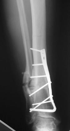
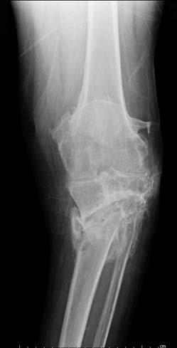
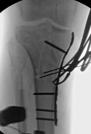
28 Common places to get into trouble Distal femur Proximal tibia Medial condylar component. Proximal femur Lack of fibular length restoration Pilon fractures Reduction at length with temp spanning ex fix

29 The picture can't be displayed. The picture can't be displayed.


30 The picture can't be displayed.
31
32
Multiapical Deformities p. 97 Osteotomy Concepts and Frontal Plane Realignment p. 99 Angulation Correction Axis (ACA) p. 99 Bisector Lines p.
 p. 99 Bisector Lines p.") Normal Lower Limb Alignment and Joint Orientation p. 1 Mechanical and Anatomic Bone Axes p. 1 Joint Center Points p. 5 Joint Orientation Lines p. 5 Ankle p. 5 Knee p. 5 Hip p. 8 Joint Orientation Angles
Normal Lower Limb Alignment and Joint Orientation p. 1 Mechanical and Anatomic Bone Axes p. 1 Joint Center Points p. 5 Joint Orientation Lines p. 5 Ankle p. 5 Knee p. 5 Hip p. 8 Joint Orientation Angles
PRINCIPLES OF MALUNIONS
 26 PRINCIPLES OF MLUNIONS Mark R. rinker and Daniel P. O Connor EVLUTION CLINICL RDIOGRPHIC EVLUTION OF THE VRIOUS DEFORMITY TYPES TRETMENT OSTEOTOMIES TRETMENT Y DEFORMITY TYPE TRETMENT Y DEFORMITY LOCTION
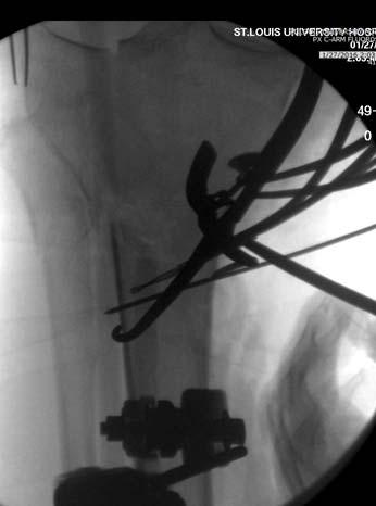
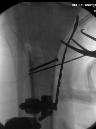
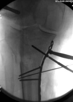
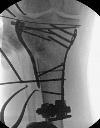
SCIENTIFIC POSTER #28 Tibia OTA 2016
 SCIENTIFIC POSTER #28 Tibia OTA 2016 Effectiveness of Complex Combined Nonunion/Malunion Correction, Utilizing a Hexapod External Fixator John Arvesen, MD 1 ; J. Tracy Watson, MD 2 ; Heidi Israel, PhD,
OPERATIVE TECHNIQUE. Limb Reconstruction System. Part B: Correction of Deformities. By Dr. S. Nayagam
 OPERATIVE TECHNIQUE 11 Limb Reconstruction System Part B: Correction of Deformities By Dr. S. Nayagam CONTENTS LIMB RECONSTRUCTION SYSTEM Part B: Correction of Deformities QUICK REFERENCE GUIDE... Page
Operative Technique. by PROF. NAYAGAM. LIMB RECONSTRUCTION SYSTEM Part B: Correction of Deformities
 Operative Technique by PROF. NAYAGAM LIMB RECONSTRUCTION SYSTEM Part B: Correction of Deformities 11 Quick Reference Guide CONTENTS LIMB RECONSTRUCTION SYSTEM Part A: General Principles Page N o I Introduction
Case Report. Antegrade Femur Lengthening with the PRECICE Limb Lengthening Technology
 Case Report Antegrade Femur Lengthening with the PRECICE Limb Lengthening Technology S. Robert Rozbruch, MD Hospital for Special Surgery New York, NY, USA ABSTRACT This is a case illustrating a 4.5 cm
Tibial deformity correction by Ilizarov method
 International Journal of Research in Orthopaedics http://www.ijoro.org Case Report DOI: http://dx.doi.org/10.18203/issn.2455-4510.intjresorthop20180422 Tibial deformity correction by Ilizarov method Robert
4/28/2010. Fractures. Normal Bone and Normal Ossification Bone Terms. Epiphysis Epiphyseal Plate (physis) Metaphysis
 Metaphysis") Fractures Normal Bone and Normal Ossification Bone Terms Epiphysis Epiphyseal Plate (physis) Metaphysis Diaphysis 1 Fracture Classifications A. Longitudinal B. Transverse C. Oblique D. Spiral E. Incomplete
Fractures Normal Bone and Normal Ossification Bone Terms Epiphysis Epiphyseal Plate (physis) Metaphysis Diaphysis 1 Fracture Classifications A. Longitudinal B. Transverse C. Oblique D. Spiral E. Incomplete
Correction of Traumatic Ankle Valgus and Procurvatum using the Taylor Spatial Frame: A Case Report
 The Foot and Ankle Online Journal Official publication of the International Foot & Ankle Foundation Correction of Traumatic Ankle Valgus and Procurvatum using the Taylor Spatial Frame: A Case Report by
Combined Tibial and Fibular Measurement for the Classification of Supramalleolar Deformity D O U G B E A M A N, M D P A X T O N G E H L I N G, B M
 Combined Tibial and Fibular Measurement for the Classification of Supramalleolar Deformity D O U G B E A M A N, M D P A X T O N G E H L I N G, B M Combined Tibial and Fibular Measurement for the Classification
S. Robert Rozbruch, MD. Chief, Limb Lengthening & Complex Reconstruction Service Professor of Clinical Orthopedic Surgery
 S. Robert Rozbruch, MD Chief, Limb Lengthening & Complex Reconstruction Service Professor of Clinical Orthopedic Surgery Small Bone Innovations: consultant and royalties Smith and Nephew: consultant External
Pocket Guide. Version 4.1: Fracture reduction and deformity correction software
 Pocket Guide www.spatialframe.com Version 4.1: Fracture reduction and deformity correction software Shoulder bolt Master tab Strut 5 Strut 1 Strut 6 Strut 4 Strut 2 Strut 3 ID band Figure 1 Frame assembly
Imaging assessment of Unicomp candidates!
 7th Advanced Course on Knee Surgery - 2018: Imaging assessment of Unicomp candidates! Presenter: Anders Troelsen, MD, ph.d., dr.med., Professor Distribution of the basic primary OA patterns Medial FT:
Practical Reduction Techniques: Diaphyseal Reduction. Philip Wolinsky University of California at Davis Medical Center
 OTA Specialty Day 2016 Practical Reduction Techniques: Diaphyseal Reduction Philip Wolinsky University of California at Davis Medical Center 8:55 am 9:55 am Tips and Tricks: Practical Reduction Techniques
FIBULAR & SYNDESMOSIS MALUNIONS
 FIBULAR & SYNDESMOSIS MALUNIONS MICHAEL P. CLARE, MD FLORIDA ORTHOPAEDIC INSTITUTE TAMPA, FL USA MORTISE INHERENTLY UNSTABLE LATERAL MALLEOLUS ACTS AS BUTTRESS / POST RESIST LATERAL TRANSLATION OF TALUS
The pilon tibiale fracture
 The pilon tibiale fracture Thomas Beck Spitalzentrum Oberwallis OTC Trauma course september 2017 xxx I have no financial relationships with commercial entities that produce healthcare related products.
Biomet Carbon Rail Deformity System. Surgical Technique
 Biomet Carbon Rail Deformity System Surgical Technique Contents Introduction... Page 1 System Components... Page 2 Indications, Contraindications, and Deformity Planning... Page 4 Deformity Planning-Identifying...
Malrotation can occur following closed intramedullary
 ORIGINAL ARTICLE The Effects of Femoral Shaft Malrotation on Lower Extremity Anatomy Joseph J. Gugenheim, MD,* Robert A. Probe, MD, and Mark R. Brinker, MD* Objective: To determine how axial rotation around
PRE & POST OPERATIVE RADIOLOGICAL ASSESSMENT IN TOTAL KNEE REPLACEMENT. Dr. Divya Rani K 2 nd Year Resident Dept. of Radiology
 PRE & POST OPERATIVE RADIOLOGICAL ASSESSMENT IN TOTAL KNEE REPLACEMENT Dr. Divya Rani K 2 nd Year Resident Dept. of Radiology PRE OPERATIVE ASSESSMENT RADIOGRAPHS Radiographs are used for assessment and
CORRECTIVE OSTEOTOMY BRINGING THE PLAN TO THE BONE (TRIGONOMETERY, GUIDE WIRES, SLA MODELING AND ART)
") CORRECTIVE OSTEOTOMY BRINGING THE PLAN TO THE BONE (TRIGONOMETERY, GUIDE WIRES, SLA MODELING AND ART) Randy J. Boudrieau, DVM, DACVS, DECVS Cummings School of Veterinary Medicine at Tufts University, North
CORRECTIVE OSTEOTOMY BRINGING THE PLAN TO THE BONE (TRIGONOMETERY, GUIDE WIRES, SLA MODELING AND ART) Randy J. Boudrieau, DVM, DACVS, DECVS Cummings School of Veterinary Medicine at Tufts University, North
Lower Extremity Fracture Management. Fractures of the Hip. Lower Extremity Fractures. Vascular Anatomy. Lower Extremity Fractures in Children
 Lower Extremity Fracture Management Brian Brighton, MD, MPH Levine Children s s Hospital Carolinas Medical Center Charlotte, NC Oscar Miller Day October 16, 2009 Lower Extremity Fractures in Children Anatomic
RADIOGRAPHY OF THE KNEE, PATELLA, and FEMUR
 RADIOGRAPHY OF THE KNEE, PATELLA, and FEMUR KNEE AP Projection Patient Position: Part Position: Leg in Center Femoral condyles Central Ray: - Asthenic patient - if ASIS to tabletop is < 19 cm Sthenic patient
RADIOGRAPHY OF THE ANKLE and LOWER LEG
 RADIOGRAPHY OF THE ANKLE and LOWER LEG Patient Position: ANKLE AP Projection Part Position: True Slight to place foot s long axis Center to Central Ray: to IR Midway Note: Ankle joint is to tips of malleoli
Extra-articular deformities in TKA
 Extra-articular deformities in TKA NOV Najaarscongres Donderdag 12 oktober 2017 Veldhoven G.G. van Hellemondt, MD Sint Maartenskliniek Nijmegen The Netherlands Disclosures Consultancy ZimmerBiomet Smith&Nephew
Calculation and Correction of Secondary Translation Deformities and Secondary Length Deformities
 Calculation and Correction of Secondary Translation Deformities and Secondary Length Deformities BRUNO GLADBACH, DMED*; ETIENNE HEIJENS, DMED*; JOACHIM PFEIL, PROFDMED*; DROR PALEY, MD abstract External
Massive Varus- Overview. Massive Varus- Classification. Massive Varus- Definition 07/02/14. Correction of Massive Varus Deformity in TKR
 07/02/14 Massive Varus- Overview Correction of Massive Varus Deformity in TKR Myles Coolican Val d Isere 2014 Massive Varus- Classification Classification Intra articular Massive Varus- Classification Classification
Axial alignment of the lower extremity in Chinese adults. Journal Of Bone And Joint Surgery - Series A, 2000, v. 82 n. 11, p.
 Title Axial alignment of the lower extremity in Chinese adults Author(s) Tang, WM; Zhu, YH; Chiu, KY Citation Journal Of Bone And Joint Surgery - Series A, 2000, v. 82 n. 11, p. 1603-1608 Issued Date 2000
EVOS MINI with IM Nailing
 Case Series Dr. John A. Scolaro EVOS MINI with IM Nailing A series of studies Introduction Intramedullary nailing has become the standard for many long bone fractures. Fracture reduction prior to nail
Masterclass. Tips and tricks for a successful outcome. E. Verhaven, M. Thaeter. September 15th, 2012, Brussels
 Masterclass Tips and tricks for a successful outcome September 15th, 2012, Brussels E. Verhaven, M. Thaeter Belgium St. Nikolaus-Hospital Orthopaedics & Traumatology Ultimate Goal of TKR Normal alignment
Arthrex Open Wedge Osteotomy Technique Designed in conjunction with:
 Arthrex Open Wedge Osteotomy Technique Designed in conjunction with: Dr. Giancarlo Puddu, M.D. Dr. Peter Fowler, M.D. Dr. Ned Amendola, M.D. To treat pain and instability associated with lower extremity
OPERATIVE TECHNIQUE. advanced Limb Reconstruction System. Limb Reconstruction System
 OPERATIVE TECHNIQUE advanced Limb Reconstruction System Limb Reconstruction System 1 INTRODUCTION 2 FEATURES AND BENEFITS Components for Standard Application Components for Gradual Correction Components
Surgery-Ortho. Fractures of the tibia and fibula. Management. Treatment of low energy fractures. Fifth stage. Lec-6 د.
 Fifth stage Lec-6 د. مثنى Surgery-Ortho 28/4/2016 Indirect force: (low energy) Fractures of the tibia and fibula Twisting: spiral fractures of both bones Angulatory: oblique fractures with butterfly segment.
Imaging of Cervical Spine Trauma Tudor H Hughes, M.D.
 Imaging of Cervical Spine Trauma Tudor H Hughes, M.D. General Considerations Most spinal fractures are due to a single episode of major trauma. Fatigue fractures of the spine are unusual except in the
PediLoc Extension Osteotomy Plate (PLEO)
") PediLoc Extension Osteotomy Plate (PLEO) Left PLEO Plates Sizes: 6, 8 and 10 hole plates Right PLEO Plates Sizes: 6, 8 and 10 hole plates PediLoc Extension Osteotomy Plate The technique description herein
PediLoc Extension Osteotomy Plate (PLEO) Left PLEO Plates Sizes: 6, 8 and 10 hole plates Right PLEO Plates Sizes: 6, 8 and 10 hole plates PediLoc Extension Osteotomy Plate The technique description herein
Lower Extremity Alignment: Genu Varum / Valgum
 Lower Extremity Alignment: Genu Varum / Valgum Arthur B Meyers, MD Nemours Children s Hospital & Health System Associate Professor of Radiology, University of Central Florida Clinical Associate Professor
Femoral Fractures in Adolescents: A Comparison of Four Methods of Fixation
 Femoral Fractures in Adolescents: A Comparison of Four Methods of Fixation By Leonhard E. Ramseier, MD, Joseph A. Janicki, MD, Shannon Weir, BSc, and Unni G. Narayanan, MBBS, MSc, FRCSC Investigation performed
Intramedullary Rodding of Distal Tibial Shaft Fractures with Intra Articular Extension
 Intramedullary Rodding of Distal Tibial Shaft Fractures with Intra Articular Extension My Name is Claude Sagi CSOT Tampa, FL 2018 Disclosures: None, I am just a simple man. This talk is about treating
Complex angular and torsional deformities (distal femoral malunions)
") Online Supplementary Material to: Complex angular and torsional deformities (distal femoral malunions) Preoperative planning using stereolithography and surgical correction with locking plate fixation
Online Supplementary Material to: Complex angular and torsional deformities (distal femoral malunions) Preoperative planning using stereolithography and surgical correction with locking plate fixation
TOTAL KNEE ARTHROPLASTY (TKA)
") TOTAL KNEE ARTHROPLASTY (TKA) 1 Anatomy, Biomechanics, and Design 2 Femur Medial and lateral condyles Convex, asymmetric Medial larger than lateral 3 Tibia Tibial plateau Medial tibial condyle: concave
TOTAL KNEE ARTHROPLASTY (TKA) 1 Anatomy, Biomechanics, and Design 2 Femur Medial and lateral condyles Convex, asymmetric Medial larger than lateral 3 Tibia Tibial plateau Medial tibial condyle: concave
ANATOMIC. Navigated Surgical Technique 4 in 1 TO.G.GB.016/1.0
 ANATOMIC Navigated Surgical Technique 4 in 1 TO.G.GB.016/1.0 SCREEN LAYOUT Take screenshot Surgical step Dynamic navigation zone Information area and buttons 2 SCREEN LAYOUT Indicates action when yellow
Is Distraction Histiogenesis a Reliable Method to Reconstruct Segmental Bone and Acquired Leg Length Discrepancy in Tibia Fractures and Non Unions?
 Is Distraction Histiogenesis a Reliable Method to Reconstruct Segmental Bone and Acquired Leg Length Discrepancy in Tibia Fractures and Non Unions? James J Hutson Jr MD Professor Orthopedic Trauma Ryder
9/24/2015. When Can I Use a SHS? When CAN T I Use a SHS? Sliding Hip Screw. Time proven. Technically simple. Cheap. Quick
 When Can I Use a SHS? Frank A. Liporace, MD Associate Professor Director of Orthopaedic Trauma Research Director of Orthopaedic Trauma Jersey City Medical Center New York University / Hospital for Joint
OPERATIVE TECHNIQUE. Knee Hinge (LRS Advanced System)
") OPERTIVE TECHNIQUE Knee Hinge (LRS dvanced System) 1 1 2 4 6 INTRODUCTION INDICTIONS FETURES ND BENEFITS EQUIPMENT REQUIRED KNEE HINGE SSEMBLY 8 17 TRUM KNEE DISLOCTION Orthofix wishes to thank the following
OPERTIVE TECHNIQUE Knee Hinge (LRS dvanced System) 1 1 2 4 6 INTRODUCTION INDICTIONS FETURES ND BENEFITS EQUIPMENT REQUIRED KNEE HINGE SSEMBLY 8 17 TRUM KNEE DISLOCTION Orthofix wishes to thank the following
Impact of Intramedullary Reaming Depth on Establishing Femoral Canal Axis in the Sagittal Plane
 Impact of Intramedullary Reaming Depth on Establishing Femoral Canal Axis in the Sagittal Plane Yifei Dai, Ph.D., Dwight Todd. Zimmer Inc, Warsaw, IN, USA. Disclosures: Y. Dai: 3A; zimmer inc. 4; zimmer
Two Plane Deformity Correction byilizarov Ring Fixatorlessons
 IOSR Journal of Dental and Medical Sciences (IOSR-JDMS) e-issn: 2279-0853, p-issn: 2279-0861.Volume 15, Issue 6 Ver. XII (June 2016), PP 46-51 www.iosrjournals.org Two Plane Deformity Correction byilizarov
X-Ray Rounds: (Plain) Radiographic Evaluation of the Ankle.
 Radiographic Evaluation of the Ankle.") X-Ray Rounds: (Plain) Radiographic Evaluation of the Ankle www.fisiokinesiterapia.biz Anatomy Complex hinge joint Articulations among: Fibula Tibia Talus Tibial plafond Distal tibial articular surface
X-Ray Rounds: (Plain) Radiographic Evaluation of the Ankle www.fisiokinesiterapia.biz Anatomy Complex hinge joint Articulations among: Fibula Tibia Talus Tibial plafond Distal tibial articular surface
CONTRIBUTING SURGEON. Barry Waldman, MD Director, Center for Joint Preservation and Replacement Sinai Hospital of Baltimore Baltimore, MD
 CONTRIBUTING SURGEON Barry Waldman, MD Director, Center for Joint Preservation and Replacement Sinai Hospital of Baltimore Baltimore, MD System Overview The EPIK Uni is designed to ease the use of the
QUICK REFERENCE GUIDE. Arthrodiatasis. Articulated Joint Distraction
 4 Arthrodiatasis Articulated Joint Distraction ARTHRODIATASIS OF THE HIP To prepare the assembly, remove the female component and replace it with the ProCallus articulated body for the hip. Remove cam
Closing Wedge Retrotubercular Tibial Osteotomy and TKA for Posttraumatic Osteoarthritis With Angular Deformity
 ORTHOPEDICS May 2009;32(5):360. Closing Wedge Retrotubercular Tibial Osteotomy and TKA for Posttraumatic Osteoarthritis With Angular Deformity by John P. Meehan, MD; Mohammad A. Khadder, MD; Amir A. Jamali,
Overview. Acceptance criteria for all protocols
 X-Ray protocol Overview The Smith & Nephew VISIONAIRE X-Ray protocol is essentially an AP leg length image. The images are preferred to be done erect, but can be done supine if necessary due to the type
BIOMECHANICAL EXAMINATION OF THE PEDIATRIC LOWER EXTREMITY
 BIOMECHANICAL EXAMINATION OF THE PEDIATRIC LOWER EXTREMITY B.Resseque, D.P.M. ARCH HEIGHT OFF WEIGHTBEARING Evaluate arch height by placing a ruler from the heel to the first metatarsal head Compare arch
5/31/2018. Ipsilateral Femoral Neck And Shaft Fractures. Ipsilateral Neck-Shaft Fractures Introduction. Ipsilateral Neck-Shaft Fractures Introduction
 Ipsilateral Femoral Neck And Shaft Fractures Exchange Nailing For Non- Union Donald Wiss MD Cedars-Sinai Medical Center Los Angeles, California Introduction Uncommon Injury Invariably High Energy Trauma
Basic Principles of Fractures & Easily Missed Fractures. Mr Irfan Merchant Trauma & Orthopaedic Registrar Bedford Hospital, East of England
 Basic Principles of Fractures & Easily Missed Fractures Mr Irfan Merchant Trauma & Orthopaedic Registrar Bedford Hospital, East of England Objectives Types Fracture Patterns Fracture Healing Assessing
Osteotomies for Cartilage Protections. Jeffrey Halbrecht,, MD San Francisco, Ca
 Osteotomies for Cartilage Protections Jeffrey Halbrecht,, MD San Francisco, Ca ACI/Osteotomy Osteotomy: Optimal Patient Selection Mechanical axis falls within involved compartment Mild joint space narrowing
Plaster-Wedging Technique:
 Plaster-Wedging Technique: An Appropriate, Safe, Quick, & Economical Method To Stretch Soft Tissue Contractures Of The Knee H.M. Steenbeek General Information. In the field of physical rehabilitation of
Failed Subtrochanteric Fracture How I Decide What to Do?
 Failed Subtrochanteric Fracture How I Decide What to Do? Gerald E. Wozasek Thomas M. Tiefenboeck 5 October 2016, Washington Medical University of Vienna, Department of Trauma Surgery ordination @wozasek.at
Lengthening & Deformity correction with. Fixator Assisted Nailing
 Lengthening & Deformity correction with Fixator Assisted Nailing External Fixation Used as *Intra-Op Alignment tool * for lengthening with the main intention of reducing External fixation time! Advantages
The space shuttle docking at the international space station
 Docking Tips The space shuttle docking at the international space station Bone gap after component fracture tibia A standard distal to proximal bone transport Apparently simple pictures of the bone transport
OPERATIVE TECHNIQUE. The Ring Fixator System. The Sheffield Ring Fixator - Limb Reconstruction. and Complex Trauma. By Prof. M.
 OPERATIVE TECHNIQUE 12 The Ring Fixator System Part C: The Sheffield Ring Fixator - Limb Reconstruction and Complex Trauma By Prof. M. Saleh CONTENTS Page N INTRODUCTION...................................................................................................................................
Definition of Anatomy. Anatomy is the science of the structure of the body and the relation of its parts.
 Definition of Anatomy Anatomy is the science of the structure of the body and the relation of its parts. Basic Anatomical Terms Anatomical terms for describing positions: Anatomical position: Supine position:
Overview. Acceptance criteria for all protocols
 X-Ray protocol Overview The Smith & Nephew VISIONAIRE X-Ray protocol is essentially an AP leg length image. The images are preferred to be done erect, but can be done supine if necessary due to the type
Large Distractor Femur
 Fracture Reduction and Provisional Stabilization Large Distractor Femur Surgical Technique Table of Contents Introduction Standard Femoral Distraction 2 Large Distractor System 4 Surgical Technique Prepare
Orthopedic Bone Nail System - Distal Femoral Nail Surgical Technique Manual
 Orthopedic Bone Nail System - Distal Femoral Nail Surgical Technique Manual Note: The surgical procedures should be performed under the guidance of qualified skilled orthopedic surgeons, and this surgical
General Concepts. Growth Around the Knee. Topics. Evaluation
 General Concepts Knee Injuries in Skeletally Immature Athletes Zachary Stinson, M.D. Increased rate and ability of healing Higher strength of ligaments compared to growth plates Continued growth Children
Minimally Invasive Plating of Fractures:
 Minimally Invasive Plating of Fractures: Advantages, Techniques and Trade-offs Matthew Garner, MD Created January 2016 OUTLINE Principles of fracture management The importance of vascular supply Equipment
Foot and ankle radiographic parameters: validity and reproducibility of biplane imaging system versus conventional radiography
 Foot and ankle radiographic parameters: validity and reproducibility of biplane imaging system versus conventional radiography Chamnanni Rungpai, MD; Jessica E. Goetz, PhD; Marut Arunakul, MD; Yubo, Gao,
Instructional Course Lecture 2011
 Instructional Course Lecture 2011 Yoon Hae Kwak Dept. of Orthopaedic Surgery Hallym University Sacred Heart Hospital Hallym University Medical Center Rotational and Angular variations of the lower extremities
TRK REVISION KNEE Surgical Technique
 1 TRK REVISION KNEE Surgical Technique 1. 2. 3. 4. 5. 6. 7. 8. 9. 10. INTERCONDYLAR RESECTION...... page FEMORAL STEM...... page NON CEMENTED FEMORAL STEM...... page TRIAL FEMORAL COMPONENTS...... page
Tibial Shaft Fractures
 Tibial Shaft Fractures Mr Krishna Vemulapalli Consultant Orthopaedics Surgeon Queens & King George Hospitals Queens Hospital 14/03/2018 Google Maps Map data 2018 Google 10 km Orthopaedics Department Covers
Ankle Valgus in Cerebral Palsy
 Ankle Valgus in Cerebral Palsy Freeman Miller Contents Introduction... 2 Natural History... 2 Treatment... 3 Diagnostic Evaluations... 3 Indications for Intervention... 3 Outcome of Treatment... 5 Complications
radiologymasterclass.co.uk
 http://radiologymasterclass.co.uk Hip X-ray anatomy - Normal AP (anterior-posterior) Shenton's line is formed by the medial edge of the femoral neck and the inferior edge of the superior pubic ramus Loss
Zimmer MIS Periarticular Distal Femoral Locking Plate
 For Clinical Evaluations Zimmer MIS Periarticular Distal Femoral Locking Plate Surgical Technique The Science of the Landscape Zimmer MIS Periarticular Distal Femoral Locking Plate Surgical Technique
ANATOMIC SURGICAL TECHNIQUE. 5 in 1. Conventional instrumentation 07/11/2013
 ANATOMIC SURGICAL TECHNIQUE 5 in 1 Conventional instrumentation PRO.GB.933/1.0 Octobre 2013 2 Tibial step 3 Intramedullary technique - Based on the preoperative plan, drill the medullary canal with the
LCP Anterior Ankle Arthrodesis Plates. Part of the Synthes Locking Compression Plate (LCP) System.
 System.") LCP Anterior Ankle Arthrodesis Plates. Part of the Synthes Locking Compression Plate (LCP) System. Technique Guide Instruments and implants approved by the AO Foundation Table of Contents Introduction
LCP Anterior Ankle Arthrodesis Plates. Part of the Synthes Locking Compression Plate (LCP) System. Technique Guide Instruments and implants approved by the AO Foundation Table of Contents Introduction
VARIABILITY OF THE POSTERIOR CONDYLAR ANGLE
 VARIABILITY OF THE POSTERIOR CONDYLAR ANGLE Łukasz Cieliński, Damian Kusz, Michał Wójcik Department of Orthopedics Medical University of Silesia in Katowice Introduction Correct positioning of implants
Fibula-related complications during bilateral tibial lengthening
 Acta Orthopaedica 2012; 83 (3): 271 275 271 Fibula-related complications during bilateral tibial lengthening 60 patients followed for mean 5 years Seung-Ju Kim, Mandar Vikas Agashe, Sang-Heon Song, and
Pre-operative evaluation
 Pre-operative evaluation Andrea Meyer-Lindenberg Clinic of Small Animal Surgery and eproduction Ludwig-Maximilians-University Munich Importance of pre-operative planning Evaluate patient before selecting
GAMMA LOCKING NAIL INSTRUMENTS OPERATIVE TECHNIQUE
 GAMMA LOCKING NAIL INSTRUMENTS OPERATIVE TECHNIQUE FEATURES AND BENEFITS The One Shot Device is a new component of the Gamma Locking Nail instrumentation system determining the correct position of the
Principles Starting Point Trajectory L/A/R Stable Construct. DISCLOSURES Hassan R. Mir, MD, MBA, FACS 5/16/2017
 DISCLOSURES Hassan R. Mir, MD, MBA, FACS Medical/Orthopaedic Publications Editorial/Governing Board OTA Newsletter Editor OsteoSynthesis, The JOT Online Discussion Forum Editor JOT Associate Editor JAAOS
Fractures of the tibia shaft treated with locked intramedullary nail Retrospective clinical and radiographic assesment
 ARS Medica Tomitana - 2013; 4(75): 197-201 DOI: 10.2478/arsm-2013-0035 Șerban Al., Botnaru V., Turcu R., Obadă B., Anderlik St. Fractures of the tibia shaft treated with locked intramedullary nail Retrospective
BIOTECH FUTURE KNEE Minimal Invasive (and classic) Surgical Technique
 Surgical Technique") BIOTECH FUTURE KNEE Minimal Invasive (and classic) Surgical Technique Page No. Product description 2. Pre-operational planning 3. 1st step Femoral resection 4. P/S Femoral component 7. 2nd step Proximal
BIOTECH FUTURE KNEE Minimal Invasive (and classic) Surgical Technique Page No. Product description 2. Pre-operational planning 3. 1st step Femoral resection 4. P/S Femoral component 7. 2nd step Proximal
Medical Devices DYNAMIC MULTIAXIAL FIXATOR
 Medical Devices DDDAF DYNAMIC MULTIAXIAL FIXATOR DAF DYNAMIC MULTIAXIAL FIXATOR It is a single plan external fixation tool. The ball-joint structure connects the clamp to the fixator body as an articular
Periarticular knee osteotomy
 Periarticular knee osteotomy Turnberg Building Orthopaedics 0161 206 4803 All Rights Reserved 2018. Document for issue as handout. Knee joint The knee consists of two joints which allow flexion (bending)
Biomet Multi-Axial Correction (MAC) System Correction Atlas
 System Correction Atlas") Biomet Multi-Axial Correction (MAC) System Correction Atlas Demonstrated with the XS MAC on a Left Tibia (CORA Centric Approach) Introduction... Page 1 Indications/ Contraindictions... Page 1 MAC Module
Biomet Multi-Axial Correction (MAC) System Correction Atlas Demonstrated with the XS MAC on a Left Tibia (CORA Centric Approach) Introduction... Page 1 Indications/ Contraindictions... Page 1 MAC Module
CLINICAL PRESENTATION AND RADIOLOGY QUIZ QUESTION
 Donald L. Renfrew, MD Radiology Associates of the Fox Valley, 333 N. Commercial Street, Suite 100, Neenah, WI 54956 11/24/2012 Radiology Quiz of the Week # 100 Page 1 CLINICAL PRESENTATION AND RADIOLOGY
GREENS SURGICALS. Redefining Excellence INSTRUMENT SYSTEM PREPARED BY: DR. VINAY KUMAR
 GREENS SURGICALS Redefining Excellence TIBIA AND FEMUR INSTRUMENT SYSTEM PREPARED BY: DR. VINAY KUMAR OPERATIVE TECHNIQUES INDEX SR.NO CONTENTS 1 LIST OF INSTRUMENT FOR TIBIA AND FEMUR. 2 RADIO GRAPH OF
Aesculap Targon TX. Intramedullary Nail for Tibial Fractures...going to X-tremes. Aesculap Orthopaedics
 Aesculap Targon TX Intramedullary Nail for Tibial Fractures...going to X-tremes Aesculap Orthopaedics Surgical Technique Preoperative Planning KH483 X-ray template Targon TX (KH483200 : 375 mm-420 mm KH48320
Where Is the Natural Internal-External Rotation Axis of the Tibia?
 Where Is the Natural Internal-External Rotation Axis of the Tibia? Daniel Boguszewski 1, Paul Yang 2, Nirav Joshi 2, Keith Markolf 1, Frank Petrigliano 1, David McAllister 1. 1 University of California
Alignment Rod. For intraoperatively confirming correction of the mechanical leg axis.
 Alignment Rod. For intraoperatively confirming correction of the mechanical leg axis. Easy to use Accuracy of surgery Reduces X-ray exposure Table of Contents Introduction Alignment Rod 2 Handling Technique
Modern Rx of Polio. with Ilizarov & new techniques
 Modern Rx of Polio with Ilizarov & new techniques Poliomyelitis Best Teacher of Orthopaedics Teaches Thorough clinical examination Muscle charting Gait Analysis Analysis of Joint Instability Precision
Post test for O&P 2 Hrs CE. The Exam
 Post test for O&P 2 Hrs CE The Exam This examination is taken in "open book" format. That means you are free to answer the questions after research or discussion with your fellow workers. We feel this
THE KNEE SOCIETY VIRTUAL FELLOWSHIP
 THE KNEE SOCIETY VIRTUAL FELLOWSHIP CHAPTER 2: RADIOGRAPHIC EVALUATION OF THE KNEE Radiographic Evaluation of the Knee Presented by: R. Michael Meneghini, MD COPYRIGHT 2016 THE KNEE SOCIETY Disclosures
Biomechanics of the Knee. Valerie Nuñez SpR Frimley Park Hospital
 Biomechanics of the Knee Valerie Nuñez SpR Frimley Park Hospital Knee Biomechanics Kinematics Range of Motion Joint Motion Kinetics Knee Stabilisers Joint Forces Axes The Mechanical Stresses to which
Preoperative Planning. The primary objectives of preoperative planning are to:
 Preoperative Planning The primary objectives of preoperative planning are to: - Determine preoperative leg length discrepancy. - Assess acetabular component size and placement. - Determine femoral component
Small-wire circular fixators and hybrid external fixation
 ORIGINAL ARTICLE Correction of Tibial Malunion and Nonunion With Six-Axis Analysis Deformity Correction Using the Taylor Spatial Frame David S. Feldman, MD, Steven S. Shin, MD, Sanjeev Madan, MD, and Kenneth
Diaphyseal Humerus Fractures. OTA Course Dallas, TX 1/20/17 Ellen Fitzpatrick MD
 Diaphyseal Humerus Fractures OTA Course Dallas, TX 1/20/17 Ellen Fitzpatrick MD OBJECTIVES TREATMENT OPTIONS SURGICAL INDICATIONS CONTROVERSIES IN MANAGEMENT Humerus Fractures Treatment Goals: Functional
ILIZAROV TECHNIQUE IN CORRECTING LIMBS DEFORMITIES: PRELIMINARY RESULTS
 Bahrain Medical Bulletin, Volume 17, Number 2, June 1995 Original ILIZAROV TECHNIQUE IN CORRECTING LIMBS DEFORMITIES: PRELIMINARY RESULTS Saleh W. Al-Harby, FRCS(Glasg)* This is a prospective study of
Kinematic vs. mechanical alignment: What is the difference?
 Kinematic vs. mechanical alignment: What is the difference? In this 4 Questions interview, Stephen M. Howell, MD, explains the potential benefits of 3D alignment during total knee replacement. Introduction
Routine Guide EXAMINATION PROJECTION CASSETTE SIZE NOTES PRINT ORIENTATION. 14x17 CW* 14x17LW 14x17LW. 14x17LW 14x17LW 14x17LW
 EXAMINATION PROJECTION CASSETTE SIZE NOTES PRINT ORIENTATION A-C Joints without weights with weights 14x17 CW* One 14x17 divided; both shoulders on one exposure. *If part does not fit, do 10x12s CW. Both
LEG LENGTH INEQUALITY: Sports Medicine Perspective
 LEG LENGTH INEQUALITY: Sports Medicine Perspective Debra A. Zillmer, M.D. M&M Orthopaedics, Ltd 18 Year Old Experienced Cross Country Runner: Sept Sr Year Pain in left lower leg with running Pain now prevents