Lower Extremity Alignment: Genu Varum / Valgum
|
|
|
- Donna Lindsey
- 6 years ago
- Views:
Transcription

1 Lower Extremity Alignment: Genu Varum / Valgum Arthur B Meyers, MD Nemours Children s Hospital & Health System Associate Professor of Radiology, University of Central Florida Clinical Associate Professor of Pediatric Radiology, Florida State University
2 Disclosures Author/editor for Amirsys/Elsevier, receiving royalties
3 Outline Definitions Normal knee alignment during development Genu Varum Genu Valgum
4 Outline Definitions Normal knee alignment during development Genu Varum Genu Valgum
5 Varus Valgus
6 Varus Distal to angulation Valgus Distal to angulation
7 Varus Distal to angulation Apex of angulation Valgus Distal to angulation Apex of angulation


8 Varus Distal to angulation Apex of angulation Valgus Distal to angulation Apex of angulation Midline Right Left


9 Varus Distal to angulation Apex of angulation Valgus Distal to angulation Apex of angulation Midline Right Left
10 Varus Distal to angulation deviates to midline Apex of angulation Valgus Distal to angulation Apex of angulation Midline Right Left
11 Varus Distal to angulation deviates to midline Apex of angulation points from midline Valgus Distal to angulation Apex of angulation Midline Right Left
12 Varus Distal to angulation deviates to midline Apex of angulation points from midline Valgus Distal to angulation Apex of angulation Midline Right Left


13 Varus Distal to angulation deviates to midline Apex of angulation points from midline Valgus Distal to angulation Apex of angulation Midline Right Left
14 Varus Distal to angulation deviates to midline Apex of angulation points from midline Valgus Distal to angulation deviates from midline Apex of angulation Midline Right Left
15 Varus Distal to angulation deviates to midline Apex of angulation points from midline Valgus Distal to angulation deviates from midline Apex of angulation points to midline Midline Right Left
16 Normal Knee Alignment
17 Tibiofemoral angle
18 Tibiofemoral angle On standing radiograph
19 Tibiofemoral angle On standing radiograph Angle between lines parallel to: Mid femoral diaphysis Mid tibial diaphysis
20 Tibiofemoral angle Salenius P, et al. Development of the tibiofemoral angle in children. J Bone Joint Surg Am Mar;57(2):
21 Tibiofemoral angle 0-1 yr varus Salenius P, et al. Development of the tibiofemoral angle in children. J Bone Joint Surg Am Mar;57(2):
22 Tibiofemoral angle 0-1 yr varus 1-2 yrs 0-10 varus Salenius P, et al. Development of the tibiofemoral angle in children. J Bone Joint Surg Am Mar;57(2):
23 Tibiofemoral angle 0-1 yr varus 1-2 yrs 0-10 varus 2-3 yrs 0-10 valgus Salenius P, et al. Development of the tibiofemoral angle in children. J Bone Joint Surg Am Mar;57(2):
24 Tibiofemoral angle 0-1 yr varus 1-2 yrs 0-10 varus 2-3 yrs 0-10 valgus 3-4 yrs 8-12 valgus Salenius P, et al. Development of the tibiofemoral angle in children. J Bone Joint Surg Am Mar;57(2):
25 Tibiofemoral angle 0-1 yr varus 1-2 yrs 0-10 varus 2-3 yrs 0-10 valgus 3-4 yrs 8-12 valgus 4-13 yrs ~6 valgus Salenius P, et al. Development of the tibiofemoral angle in children. J Bone Joint Surg Am Mar;57(2):
26 Developmental changes 1-year-old 3-year-old 13-year-old
27 Developmental changes 1-year-old 3-year-old 13-year-old Varus
28 Developmental changes 1-year-old 3-year-old 13-year-old Varus Valgus

29 Developmental changes 1-year-old 3-year-old 13-year-old Varus Valgus Mild Valgus
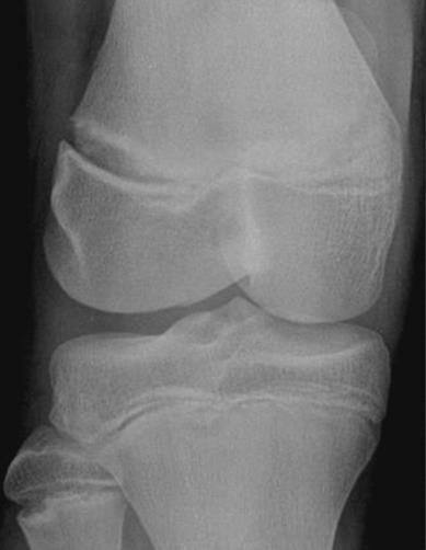
30 Genu varum
31 Genu varum / Bowing Angular deformity at the knee Apex of the deformity points away from the midline. Leg (below knee) deviates toward the midline. Common referral to orthopedic clinics Up-to-date, 2017
32 Genu varum / Bowing Angular deformity at the knee Apex of the deformity points away from the midline. Leg (below knee) deviates toward the midline. Common referral to orthopedic clinics Up-to-date, 2017
33 Genu varum / Bowing Differential diagnosis: Exaggerated physiologic bowing Blount disease Abnormal bones Metabolic bone disease Skeletal dysplasias Physeal/metaphyseal injury Up-to-date, 2017

34 Genu varum / Bowing Differential diagnosis: Exaggerated physiologic bowing Blount disease Abnormal bones Metabolic bone disease Skeletal dysplasias Physeal/metaphyseal injury Up-to-date, 2017
35 Exaggerated Physiologic Bowing
36 Exaggerated Physiologic Bowing Exaggeration of the normal age-related bowing Birth 2yrs Bilateral & symmetric Normal stature <2 sd of mean height for age & sex
37 Exaggerated Physiologic Bowing Radiographs 23-month-old
38 Exaggerated Physiologic Bowing Radiographs Varus angulation 20 varus 1-2 years nl = 0-10 varus 23-month-old
39 Exaggerated Physiologic Bowing Radiographs Varus angulation Medial tibial metaphysis Mild enlargement / depression Mild beaking No fragmentation 23-month-old
40 Exaggerated Physiologic Bowing Radiographs Varus angulation Medial tibial metaphysis Mild enlargement / depression Mild beaking No fragmentation
41 Exaggerated Physiologic Bowing Radiographs Varus angulation Medial tibial metaphysis Mild enlargement / depression Mild beaking No fragmentation Mild thickening of the medial tibal cortex
42 Exaggerated Physiologic Bowing Radiographs Varus angulation Medial tibial metaphysis Mild enlargement / depression Mild beaking No fragmentation Mild thickening of the medial tibal cortex Normal metaphysealdiaphyseal angle
43 Metaphyseal Diaphyseal Angle MDA the angle between:
44 Metaphyseal Diaphyseal Angle MDA the angle between: Line drawn along the proximal tibial metaphysis
45 Metaphyseal Diaphyseal Angle MDA the angle between: Line drawn along the proximal tibial metaphysis Line perpendicular to the long axis of the tibia 10
46 Metaphyseal Diaphyseal Angle MDA the angle between: Line drawn along the proximal tibial metaphysis Line perpendicular to the long axis of the tibia MDA values Physiologic bowing MDA typically < 11 Blount disease MDA typically > 11 Borderline

47 Exaggerated Physiologic Bowing Initial 7 months later
48 Blount Disease
49 Blount Disease (Tibia Vara) Developmental disorder with disrupted endochondral ossification of the medial proximal tibial physis Abnormal development of the proximal, medial tibial epiphysis/metaphysis
50 Blount Disease (Tibia Vara) Developmental disorder with disrupted endochondral ossification of the medial proximal tibial physis Abnormal development of the proximal, medial tibial epiphysis/metaphysis Angular deformites: Genu varum Procurvatum Internal rotation of the tibia
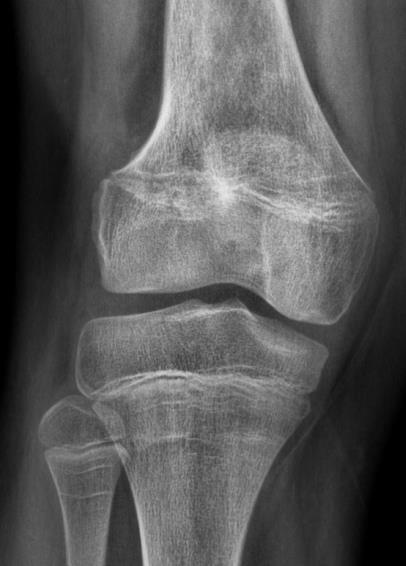
51 Blount Disease (Tibia Vara) Developmental disorder with disrupted endochondral ossification of the medial proximal tibial physis Abnormal development of the proximal, medial tibial epiphysis/metaphysis Angular deformites: Genu varum Procurvatum Internal rotation of the tibia Limb shortening Leg length discrepancy if asymmetric or unilateral
52 Blount Disease Risk factors: Early ambulation Obesity African or Scandinavian descent
53 Blount Disease Risk factors: Early ambulation Obesity African or Scandinavian descent Etiology: unknown (likely multifactorial) Risk factors of early ambulation & obesity suggest biomechanical component
54 Blount Disease Risk factors: Early ambulation Obesity African or Scandinavian descent Etiology: unknown (likely multifactorial) Risk factors of early ambulation & obesity suggest biomechanical component Two forms: Infantile or early onset < 4 years Late onset >4 yrs Juvenile 4-10 yrs Adolescent >10 yrs
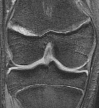
: 1758-1776.")
55 Langenskiöld Classification of Early Onset Blount Disease Sabharwal S. Blount Disease. J Bone Joint Surg Am, 2009 Jul 01; 91 (7):
56 Radiographic findings
57 Radiographic findings Standing AP radiograph
58 Radiographic findings Standing AP radiograph Genu Varum 15-month-old

59 Radiographic findings Standing AP radiograph Genu Varum Tibiofemoral angles ~ years nl = 0-10 varus 15-month-old
60 Radiographic findings Genu Varum (Standing AP radiograph) Increased metaphysealdiaphyseal angle (MDA) Physiologic bowing MDA typically < 11 Blount disease MDA typically > 11 Borderline
 Physiologic bowing MDA typically < 11 Blount disease MDA typically > 11 Borderline 8-11")
61 Radiographic findings Genu Varum (Standing AP radiograph) Increased metaphysealdiaphyseal angle (MDA) Physiologic bowing MDA typically < 11 Blount disease MDA typically > 11 Borderline
62 Radiographic findings Genu Varum (Standing AP radiograph) Increased metaphyseal-diaphyseal angle (MDA) 1 year later
63 Radiographic findings Genu Varum (Standing AP radiograph) Increased metaphyseal-diaphyseal angle (MDA) Widened medial tibial physis Medial tibial metaphysis Depression Beaked Irregular / fragmented
64 Radiographic findings Genu Varum (Standing AP radiograph) Increased metaphyseal-diaphyseal angle (MDA) Widened medial tibial physis Medial tibial metaphysis Depression Beaked Irregular / fragmented
65 Radiographic findings Genu Varum (Standing AP radiograph) Increased metaphyseal-diaphyseal angle (MDA) Widened medial tibial physis Medial tibial metaphysis Depression Beaked Irregular / fragmented
 Widened medial tibial physis Medial tibial metaphysis Depression Beaked Irregular /")
66 Radiographic findings Genu Varum (Standing AP radiograph) Increased metaphyseal-diaphyseal angle (MDA) Widened medial tibial physis Medial tibial metaphysis Depression Beaked Irregular / fragmented
 Widened medial tibial physis Medial tibial metaphysis Depression Beaked Irregular /")
67 Radiographic findings Genu Varum (Standing AP radiograph) Increased metaphyseal-diaphyseal angle (MDA) Widened medial tibial physis Medial tibial metaphysis Depression Beaked Irregular / fragmented
68 Radiographic findings Genu Varum (Standing AP radiograph) Increased metaphyseal-diaphyseal angle (MDA) Widened medial tibial physis Medial tibial metaphysis Depression Beaked Irregular / fragmented

69 Radiographic findings Genu Varum (Standing AP radiograph) Increased metaphyseal-diaphyseal angle (MDA) Widened medial tibial physis Medial tibial metaphysis Depression Beaked Irregular / fragmented Medial tibial epiphysis Abnormal/delayed ossification

70 Radiographic findings Angular deformities: Genu varum Lateral subluxation of the tibia Procurvatum

71 Radiographic findings Angular deformities: Genu varum Lateral subluxation of the tibia Procurvatum

72 Radiographic findings Angular deformities: Genu varum Lateral subluxation of the tibia Procurvatum Lateral radiograph 10 year old
73 Radiographic findings Angular deformities: Genu varum Lateral subluxation of the tibia Procurvatum Lateral radiograph

74 Radiographic findings Angular deformities: Genu varum Lateral subluxation of the tibia Procurvatum Lateral radiograph
75 MRI Findings
76 MRI Findings Medial Proximal Tibia
77 MRI Findings Medial Proximal Tibia Physis / metaphysis Physeal widening
78 MRI Findings Medial Proximal Tibia Physis / metaphysis Physeal widening Downsloping / irregularity

79 MRI Findings Medial Proximal Tibia Physis / metaphysis Physeal widening Downsloping / irregularity
80 MRI Findings Medial Proximal Tibia Physis / metaphysis Physeal widening Downsloping / irregularity Physeal Bridge
81 MRI Findings Medial Proximal Tibia Physis / metaphysis Physeal widening Downsloping / irregularity Physeal Bridge
82 MRI Findings Medial Proximal Tibia Physis / metaphysis Physeal widening Downsloping / irregularity Physeal Bridge Epiphyseal cartilage Far medial thick Central medial - thin Increased joint space

83 MRI Findings Medial Proximal Tibia Physis / metaphysis Physeal widening Downsloping / irregularity Physeal Bridge Epiphyseal cartilage Far medial thick Central medial - thin Increased joint space
84 MRI Findings Medial Proximal Tibia Physis / metaphysis Physeal widening Downsloping / irregularity Physeal Bridge Epiphyseal cartilage Far medial thick Central medial - thin Increased joint space Ho-Fung V, et al. Pediatr Radiol (2013)

85 MRI Findings Medial Proximal Tibia Physis / metaphysis Physeal widening Downsloping / irregularity Physeal Bridge Epiphyseal cartilage Far medial thick Central medial - thin Increased joint space Ho-Fung V, et al. Pediatr Radiol (2013)
86 MRI Findings Medial Proximal Tibia Physis / metaphysis Physeal widening Downsloping / irregularity Physeal Bridge Epiphyseal cartilage Far medial thick Central medial - thin Increased joint space Medial Meniscus Thickened +/- abnormal signal
87 MRI Findings Medial Proximal Tibia Physis / metaphysis Physeal widening Downsloping / irregularity Physeal Bridge Epiphyseal cartilage Far medial thick Central medial - thin Increased joint space Medial Meniscus Thickened +/- abnormal signal
88 MRI Findings Medial Proximal Tibia Physis / metaphysis Physeal widening Downsloping / irregularity Physeal Bridge Epiphyseal cartilage Far medial thick Central medial - thin Increased joint space Medial Meniscus Thickened +/- abnormal signal


89 MRI Findings Medial Proximal Tibia Physis / metaphysis Physeal widening Downsloping / irregularity Physeal Bridge Epiphyseal cartilage Far medial thick Central medial - thin Increased joint space Medial Meniscus Thickened +/- abnormal signal Angular deformities Medial and posterior downsloping
90 MRI Findings Other findings:
91 MRI Findings Other findings: Lateral proximal tibia Physis - widened/irregular Metaphysis

92 MRI Findings Other findings: Lateral proximal tibia Physis - widened/irregular Metaphysis

93 MRI Findings Other findings: Lateral proximal tibia Physis Metaphysis - irregular
94 MRI Findings Other findings: Lateral proximal tibia Physis Metaphysis Femur Epiphysis Metaphysis Physis


95 MRI Findings Other findings: Lateral proximal tibia Physis Metaphysis Femur Epiphysis Metaphysis Physis Ho-Fung V, et al. Pediatr Radiol (2013)

96 MRI Findings Other findings: Lateral proximal tibia Physis Metaphysis Femur Epiphysis Metaphysis Physis Ho-Fung V, et al. Pediatr Radiol (2013)

97 MRI Findings Other findings: Lateral proximal tibia Physis Metaphysis Femur Epiphysis Metaphysis Physis Soft tissues Perichondral menbrane Thickened ACL - laxity Ho-Fung V, et al. Pediatr Radiol (2013)
98 Treatment of Blount Disease
99 Treatment Conservative Observation Orthosis
100 Treatment Conservative Observation Orthosis Surgery Lateral hemiepiphyseodesis Resection of a physeal bar Realignment osteotomy

101 Treatment Conservative Observation Orthosis Surgery Lateral hemiepiphyseodesis Resection of a physeal bar Realignment osteotomy

102 Treatment Conservative Observation Orthosis Surgery Lateral hemiepiphyseodesis Resection of a physeal bar Realignment osteotomy

103 Treatment Conservative Observation Orthosis Surgery Lateral hemiepiphyseodesis Resection of a physeal bar Realignment osteotomy
104 Genu varum / Bowing Differential diagnosis: Exaggerated physiologic Blount disease Abnormal bones Metabolic bone disease Skeletal dysplasias Physeal/metaphyseal injury
105 Genu varum / Bowing Differential diagnosis: Exaggerated physiologic Blount disease Abnormal bones Metabolic bone disease Skeletal dysplasias Physeal/metaphyseal injury

106 Genu varum / Bowing Differential diagnosis: Exaggerated physiologic Blount disease Abnormal bones Metabolic bone disease Rickets Skeletal dysplasias Physeal/metaphyseal injury 3-year-old girl with rickets


107 Genu varum / Bowing Differential diagnosis: Exaggerated physiologic Blount disease Abnormal bones Metabolic bone disease Rickets Skeletal dysplasias Physeal/metaphyseal injury 3-year-old girl with rickets

108 Genu varum / Bowing Differential diagnosis: Exaggerated physiologic Blount disease Abnormal bones Metabolic bone disease Skeletal dysplasias Achondroplasia Physeal/metaphyseal injury 4-year-old boy with achondroplasia

109 Genu varum / Bowing Differential diagnosis: Exaggerated physiologic Blount disease Abnormal bones Metabolic bone disease Skeletal dysplasias Achondroplasia Physeal/metaphyseal injury 5-year-old boy with achondroplasia
110 Genu varum / Bowing Differential diagnosis: Exaggerated physiologic Blount disease Abnormal bones Metabolic bone disease Skeletal dysplasias Physeal/metaphyseal injury
111 Physeal/metaphyseal injury Chronic repetitive (overuse) injuries Physeal injury -> Bridge
112 Physeal/metaphyseal injury Chronic repetitive (overuse) injuries Physeal injury -> Bridge Can cause genu varum or valgum Depending on location of injury
113 Physeal/metaphyseal injury Chronic repetitive (overuse) injuries Physeal injury -> Bridge
114 Physeal/metaphyseal injury Chronic repetitive (overuse) injuries > Disruption of metaphyseal blood supply > Disruption of endochondral ossification 14-year-old competitive soccer player
115 Physeal/metaphyseal injury Chronic repetitive (overuse) injuries > Disruption of metaphyseal blood supply > Disruption of endochondral ossification 14-year-old competitive soccer player
116


117 2-month follow up after rest

118 Physeal/metaphyseal injury 11 years Laor T. et. al. Physeal Widening in the Knee Due to Stress Injury in Child Athletes. AJR 2006
119 Physeal/metaphyseal injury 11 years Laor T. et. al. Physeal Widening in the Knee Due to Stress Injury in Child Athletes. AJR 2006

120 Physeal/metaphyseal injury 11 years 2 year follow up Laor T. et. al. Physeal Widening in the Knee Due to Stress Injury in Child Athletes. AJR 2006


121 Physeal/metaphyseal injury 11 years 17 years Laor T. et. al. Physeal Widening in the Knee Due to Stress Injury in Child Athletes. AJR 2006
122 Genu valgum
123 Genu Valgum / Knock Knees Angular deformity at the knee Apex of the deformity points toward the midline. Leg (below knee) deviates away from the midline. Common referral to orthopedic clinics Up-to-date, 2017
124 Genu Valgum / Knock Knees Angular deformity at the knee Apex of the deformity points toward the midline. Leg (below knee) deviates away from the midline. Common referral to orthopedic clinics Up-to-date, 2017
125 Genu Valgum Differential diagnosis: Exaggerated physiologic Physeal/metaphyseal injuries Abnormal bones Tumors Up-to-date, 2017

126 Genu Valgum Differential diagnosis: Exaggerated physiologic Physeal/metaphyseal injuries Abnormal bones Tumors Up-to-date, 2017
127 Exaggerated Physiologic Valgum Exaggeration of the normal age-related valgus Age: 3-5 yrs Bilateral & symmetric Normal stature Absent or mild symptoms 3-years
128 Exaggerated Physiologic Valgum Exaggeration of the normal age-related valgus Age: 3-5 yrs Bilateral & symmetric Normal stature Absent or mild symptoms Valgus 3-4 yrs nl = 8-12


129 Exaggerated Physiologic Valgum Exaggeration of the normal age-related valgus Age: 3-5 yrs Bilateral & symmetric Normal stature Absent or mild symptoms 3-years 9-years
130 Genu Valgum Differential diagnosis: Exaggerated physiologic Physeal/metaphyseal injuries Chronic repetitive (overuse) injuries Physeal injury -> Bridge Cozen Phenomenon Abnormal bones Tumors
131 Physeal Injury -> Bridge Causes Trauma most common cause Other causes: Infection Radiation Medications Tumors
132 Risk Factors of Bridge Formation Severity of injury Growth potential Younger / less skeletal maturity Anatomic site Contour of the physis & growth rate
133 Risk Factors of Bridge Formation Anatomic site:
134 Risk Factors of Bridge Formation Anatomic site: Distal radius Distal femur
135 Risk Factors of Bridge Formation Anatomic site: Distal radius Distal femur
136 Risk Factors of Bridge Formation Anatomic site: Distal radius smooth, uniplanar Distal femur
137 Risk Factors of Bridge Formation Anatomic site: Distal radius smooth, uniplanar Distal femur - undulating, multiplanar physis
138 Risk Factors of Bridge Formation Anatomic site: Distal radius smooth, uniplanar Distal femur - undulating, multiplanar physis


139 Risk Factors of Bridge Formation Anatomic site: Distal radius smooth, uniplanar Distal femur - undulating, multiplanar physis Physeal fx: Radius >>> Femur % -> Bridges : Femur >>> Radius

140 10 year old girl
141 9 month follow up 10-year-old girl with history of a distal femur fracture
142 Imaging Physeal Bridges Radiographs Directly visualized Indirect evidence Narrowing of the physis Growth recovery lines 10-year-old girl with history of a distal femur fracture
143 Imaging Physeal Bridges Radiographs Directly visualized Indirect evidence Narrowing of the physis Growth recovery lines 10-year-old girl with history of a distal femur fracture
144 Imaging Physeal Bridges Radiographs Directly visualized Indirect evidence Narrowing of the physis Growth recovery lines 10-year-old girl with history of a distal femur fracture
145 Imaging Physeal Bridges Radiographs Directly visualized Indirect evidence Narrowing of the physis Growth recovery lines Normal - parallel physis 10-year-old girl with history of a distal femur fracture
146 Imaging Physeal Bridges Radiographs Directly visualized Indirect evidence Narrowing of the physis Growth recovery lines Normal - parallel physis 10-year-old girl with history of a distal femur fracture
147 Imaging Physeal Bridges Radiographs Directly visualized Indirect evidence Narrowing of the physis Growth recovery lines Normal - parallel physis Abnormal Oblique, converge at physeal bridge 10-year-old girl with history of a distal femur fracture
148 Imaging Physeal Bridges Radiographs Directly visualized Indirect evidence Narrowing of the physis Growth recovery lines Normal - parallel physis Abnormal Oblique, converge at physeal bridge 10-year-old girl with history of a distal femur fracture

149 Imaging Physeal Bridges Radiographs Directly visualized Indirect evidence Narrowing of the physis Growth recovery lines Normal - parallel physis Abnormal Oblique, converge at physeal bridge 10-year-old girl with history of a distal femur fracture
150 Treatment of Physeal Bridges Resection
151 Treatment of Physeal Bridges Resection considered if: Existing or developing deformity

152 Treatment of Physeal Bridges Resection considered if: Existing or developing deformity > 2 years or 2 cm of remaining growth
153 Treatment of Physeal Bridges Resection considered if: Existing or developing deformity > 2 years or 2 cm of remaining growth Bridge occupies < 50% of the physeal area GRE
154 Treatment of Physeal Bridges Resection considered if: Existing or developing deformity > 2 years or 2 cm of remaining growth Bridge occupies < 50% of the physeal area GRE

155 Treatment of Physeal Bridges Resection considered if: Existing or developing deformity > 2 years or 2 cm of remaining growth Bridge occupies < 50% of the physeal area GRE
156 Treatment of Physeal Bridges Resection considered if: Existing or developing deformity > 2 years or 2 cm of remaining growth Bridge occupies < 50% of the physeal area Total Area of Physis

157 Treatment of Physeal Bridges Resection considered if: Existing or developing deformity > 2 years or 2 cm of remaining growth Bridge occupies < 50% of the physeal area Area of Bridge _ Total Area of Physis
158 Treatment of Physeal Bridges Resection considered if: Existing or developing deformity > 2 years or 2 cm of remaining growth Bridge occupies < 50% of the physeal area Area of Bridge _ Total Area of Physis = % of Physis Occupied by Bridge

159 Physeal Bridge Treatment options: Bridge resection Osteotomy to correct angular deformities

160 Physeal Bridge Treatment options: Bridge resection Osteotomy to correct angular deformities Contralateral epiphysiodesis
161
162
163
164
165
166
167
168 Genu Valgum Differential diagnosis: Exaggerated physiologic Physeal/metaphyseal injuries Chronic repetitive (overuse) injuries Physeal injury -> Bridge Cozen Phenomenon Abnormal bones Tumors
169 Genu Valgum Differential diagnosis: Exaggerated physiologic Physeal/metaphyseal injuries Chronic repetitive (overuse) injuries Physeal injury -> Bridge Cozen Phenomenon Valgus deformity s/p proximal tibial fx Despite good alignment at fx site Burton A, et al. Cozen's phenomenon revisited. J Pediatr Orthop B Nov;25(6):

170 Genu Valgum Differential diagnosis: Exaggerated physiologic Physeal/metaphyseal injuries Chronic repetitive (overuse) injuries Physeal injury -> Bridge Cozen Phenomenon Valgus deformity s/p proximal tibial fx Despite good alignment at fx site Burton A, et al. Cozen's phenomenon revisited. J Pediatr Orthop B Nov;25(6):
171 Genu Valgum Differential diagnosis: Exaggerated physiologic Physeal/metaphyseal injuries Chronic repetitive (overuse) injuries Physeal injury -> Bridge Cozen Phenomenon Valgus deformity s/p proximal tibial fx Despite good alignment at fx site Burton A, et al. Cozen's phenomenon revisited. J Pediatr Orthop B Nov;25(6):

172 Genu Valgum Differential diagnosis: Exaggerated physiologic Physeal/metaphyseal injuries Chronic repetitive (overuse) injuries Physeal injury -> Bridge Cozen Phenomenon Valgus deformity s/p proximal tibial fx Despite good alignment at fx site Burton A, et al. Cozen's phenomenon revisited. J Pediatr Orthop B Nov;25(6):

173 Genu Valgum Differential diagnosis: Exaggerated Physiologic Physeal/metaphyseal injuries Chronic repetitive (overuse) injuries Physeal injury -> Bridge Cozen Phenomenon Most accepted theory Fx -> vascularity -> medial metaphyseal overgrowth Burton A, et al. Cozen's phenomenon revisited. J Pediatr Orthop B Nov;25(6):
174 Genu Valgum Differential diagnosis: Exaggerated Physiologic Physeal/metaphyseal injuries Abnormal bones Tumors
175 Genu Valgum Differential diagnosis: Exaggerated Physiologic Physeal/metaphyseal injuries Abnormal bones Metabolic bone disease Rickets 5-year-old boy with rickets

176 Genu Valgum Differential diagnosis: Exaggerated Physiologic Physeal/metaphyseal injuries Abnormal bones Metabolic bone disease Rickets 5-year-old boy with rickets

177 Genu Valgum Differential diagnosis: Exaggerated Physiologic Physeal/metaphyseal injuries Abnormal bones Metabolic bone disease Rickets 5-year-old boy with rickets
178 Genu Valgum Differential diagnosis: Exaggerated Physiologic Physeal/metaphyseal injuries Abnormal bones Metabolic bone disease Lysosomal storage disease Mucopolysaccharidosis IV (Morquio syndrome) Mucopolysaccharidosis IV
")
179 Genu Valgum Differential diagnosis: Exaggerated Physiologic Physeal/metaphyseal injuries Abnormal bones Metabolic bone disease Lysosomal storage disease Mucopolysaccharidosis IV (Morquio syndrome) Mucopolysaccharidosis IV
")
180 Genu Valgum Differential diagnosis: Exaggerated Physiologic Physeal/metaphyseal injuries Abnormal bones Metabolic bone disease Lysosomal storage disease Mucopolysaccharidosis IV (Morquio syndrome) Mucopolysaccharidosis IV
181 Genu Valgum Differential diagnosis: Exaggerated Physiologic Physeal/metaphyseal injuries Abnormal bones Metabolic bone disease Lysosomal storage disease Skeletal dysplasias Chondroectodermal dysplasia (Ellisvan Crevald) Ellis-van Crevald

 Ellis-van")
182 Genu Valgum Differential diagnosis: Exaggerated Physiologic Physeal/metaphyseal injuries Abnormal bones Metabolic bone disease Lysosomal storage disease Skeletal dysplasias Chondroectodermal dysplasia (Ellisvan Crevald) Ellis-van Crevald
 Ellis-van Crevald")
183 Genu Valgum Differential diagnosis: Exaggerated Physiologic Physeal/metaphyseal injuries Abnormal bones Metabolic bone disease Lysosomal storage disease Skeletal dysplasias Chondroectodermal dysplasia (Ellisvan Crevald) Ellis-van Crevald

184 Genu Valgum Differential diagnosis: Exaggerated Physiologic Physeal/metaphyseal injuries Abnormal bones Tumors Osteochondromas / Mutliple hereditary exostosis

185 Genu Valgum Differential diagnosis: Exaggerated Physiologic Physeal/metaphyseal injuries Abnormal bones Tumors Osteochondromas / Mutliple hereditary exostosis
186 Summary

187 Summary Normal developmental changes at the knee 1-year-old 3-year-old Varus Valgus


188 Summary Genu Varum Exaggerated Physiologic Blount Abnormal Bones Injuries Genu Valgum Exaggerated Physiologic Physeal/metaphyseal injuries Abnormal bones Tumors Physiologic Morquio Blount Osteochondromas
189 References Salenius P, et al. Development of the tibiofemoral angle in children. J Bone Joint Surg Am Mar;57(2): Sabharwal S. Blount disease. J Bone Joint Surg Am Jul;91(7): Ho-Fung V, et al. MRI evaluation of the knee in children with infantile Blount disease: tibial and extra-tibial findings. Pediatr Radiol Oct;43(10): Burton A, et al. Cozen's phenomenon revisited. J Pediatr Orthop B Nov;25(6): Laor T, et al. Physeal widening in the knee due to stress injury in child athletes. AJR Am J Roentgenol May;186(5):
General Concepts. Growth Around the Knee. Topics. Evaluation
 General Concepts Knee Injuries in Skeletally Immature Athletes Zachary Stinson, M.D. Increased rate and ability of healing Higher strength of ligaments compared to growth plates Continued growth Children
BOW LEGS (GENU VARUM)
") BOW LEGS (GENU VARUM) By Dr John Ebnezar INTRODUCTION Have you noticed how your knees look like? If you observe carefully you will see that both your knees are not parallel but deviated slightly outwards
BOW LEGS (GENU VARUM) By Dr John Ebnezar INTRODUCTION Have you noticed how your knees look like? If you observe carefully you will see that both your knees are not parallel but deviated slightly outwards
Physeal Fractures and Growth Arrest
 Physeal Fractures and Growth Arrest Raymond W. Liu, M.D. Victor M. Goldberg Master Clinician-Scientist in Orthopaedics Rainbow Babies and Children s Hospital Case Western Reserve University Outline General
Other Congenital & Developmental Knee & Leg Disease. Jong Sup Shim,M.D. Department of Orthopedic Surgery Samsung Medical Center
 Other Congenital & Developmental Knee & Leg Disease Jong Sup Shim,M.D. Department of Orthopedic Surgery Samsung Medical Center Sungkyunkwan University School of Medicine Rotational Deformity Intoeing Outtoeing
Valgus rotational -dome shaped -tibial osteotomy in treatment of tibia vara
 The Egyptian Journal of Hospital Medicine Vol., September I.S.S.N: Valgus rotational -dome shaped -tibial osteotomy in treatment of tibia vara Hossam Kandil MD., Department of Pediatric Orthopedic Surgery
Bow legs in young children can be a
 ONLINE EXCLUSIVE ORIGINAL RESEARCH Management of bow legs in children: A primary care protocol This protocol, which is designed to coincide with wellchild visits, distinguishes between normal physiologic
Orthotic Correction of Blount's Disease
 Orthotic Correction of Blount's Disease by Terry J. Supan, C.P.O. John M. Mazur, M.D. INTRODUCTION Infantile tibia vara is the result of abnormal growth in the proximal tibial epiphyseal late of the tibial
Development of genu varum in achondroplasia
 Development of genu varum in achondroplasia RELATION TO FIBULAR OVERGROWTH S. T. Lee, H. R. Song, R. Mahajan, V. Makwana, S. W. Suh, S. H. Lee From Korea University, Guro Hospital, Seoul, Korea Genu varum
Preliminary Outcome Using a New Free Motion Offloading KAFO for Postoperative Management of Hemiepiphysiodesis in Adolescent Tibia Vara
 Preliminary Outcome Using a New Free Motion Offloading KAFO for Postoperative Management of Hemiepiphysiodesis in Adolescent Tibia Vara Joseph W. Whiteside CO/LO 1399 E. Western Reserve Road, Poland, OH
Multiapical Deformities p. 97 Osteotomy Concepts and Frontal Plane Realignment p. 99 Angulation Correction Axis (ACA) p. 99 Bisector Lines p.
 p. 99 Bisector Lines p.") Normal Lower Limb Alignment and Joint Orientation p. 1 Mechanical and Anatomic Bone Axes p. 1 Joint Center Points p. 5 Joint Orientation Lines p. 5 Ankle p. 5 Knee p. 5 Hip p. 8 Joint Orientation Angles
Normal Lower Limb Alignment and Joint Orientation p. 1 Mechanical and Anatomic Bone Axes p. 1 Joint Center Points p. 5 Joint Orientation Lines p. 5 Ankle p. 5 Knee p. 5 Hip p. 8 Joint Orientation Angles
Other Congenital & Developmental Knee & Leg Disease. Jong Sup Shim,M.D. Department of Orthopedic Surgery Samsung Medical Center
 Other Congenital & Developmental Knee & Leg Disease Jong Sup Shim,M.D. Department of Orthopedic Surgery Samsung Medical Center Sungkyunkwan University School of Medicine Torsional Deformity (Rotational
4/28/2010. Fractures. Normal Bone and Normal Ossification Bone Terms. Epiphysis Epiphyseal Plate (physis) Metaphysis
 Metaphysis") Fractures Normal Bone and Normal Ossification Bone Terms Epiphysis Epiphyseal Plate (physis) Metaphysis Diaphysis 1 Fracture Classifications A. Longitudinal B. Transverse C. Oblique D. Spiral E. Incomplete
Fractures Normal Bone and Normal Ossification Bone Terms Epiphysis Epiphyseal Plate (physis) Metaphysis Diaphysis 1 Fracture Classifications A. Longitudinal B. Transverse C. Oblique D. Spiral E. Incomplete
COMMON MUSCULOSKELETAL PROBLEMS GROWTH AND DEVELOPMENT PATHOLOGIC VS. NORMAL
 COMMON MUSCULOSKELETAL PROBLEMS GROWTH AND DEVELOPMENT PATHOLOGIC VS. NORMAL Clifford L. Craig, M.D. M2 Musculoskeletal Fall 2008 I. ANGULAR AND TORSIONAL DEFORMITIES OF THE LOWER LIMBS Examination Relaxed,
2017 Resident Advanced Trauma Techniques Course COMPLICATIONS / CHALLENGES MALUNIONS/DEFORMITY
 2017 Resident Advanced Trauma Techniques Course COMPLICATIONS / CHALLENGES MALUNIONS/DEFORMITY What is a Malunion? Definition: a fracture that has healed in a nonanatomic (i.e. deformed) position Must
Physiological Genu Varum
 Acta Orthopaedica Scandinavica ISSN: 0001-6470 (Print) (Online) Journal homepage: http://www.tandfonline.com/loi/iort19 Physiological Genu Varum Lars Ingvar Hansson & Mohammed Zayer To cite this article:
Hemiepiphyseal stapling for treatment of genu valgum: A case report
 Hemiepiphyseal stapling for treatment of genu valgum: A case report Nina Agrawal, BA, Danielle Cameron, BA, Lawrence Wells, MD Abstract A 12-year-old girl underwent a bilateral distal femoral and proximal
, MD. physiologic. tibia varum. in utero (in. Disease in. variation. positioning. back and legs. instead of. Blount's. Infant with bowing in both legs
 North Jersey Orthopaedic Institute Rutgers, The Statee University of New Jersey 140 Bergen Street, D1610 Newark, NJ 07101 973-972-2150 South Orange Ambulatory Center Hackensack Medical Plaza Overlook Medical
ANTERIOR MEDIAL AND POSTERIOR MEDIAL DEFORMITY OF THE TIBIA
 ANTERIOR MEDIAL AND POSTERIOR MEDIAL DEFORMITY OF THE TIBIA 5 TH ANNUAL SLAOTI MEETING SAO PAOLO, BRAZIL OCTOBER 12-14, 2017 Richard M Schwend MD Professor Orthopaedics and Pediatrics Director of Research
Normal Values of Tibio-Femoral Angle in Nigerian Adolescents
 ISPUB.COM The Internet Journal of Orthopedic Surgery Volume 17 Number 1 Normal Values of Tibio-Femoral Angle in Nigerian Adolescents T BA, A AO, O AB Citation T BA, A AO, O AB.. The Internet Journal of
PEM GUIDE CHILDHOOD FRACTURES
 PEM GUIDE CHILDHOOD FRACTURES INTRODUCTION Skeletal injuries account for 10-15% of all injuries in children; 20% of those are fractures, 3 out of 4 fractures affect the physis or growth plate. Always consider
Early experience with medial femoral tension band plating in idiopathic genu valgum
 J Child Orthop (2011) 5:11 17 DOI 10.1007/s11832-010-0310-6 ORIGINAL CLINICAL ARTICLE Early experience with medial femoral tension band plating in idiopathic genu valgum Humberto Guzman Burt Yaszay Vanessa
Pediatric Fractures. Objectives. Epiphyseal Complex. Anatomy and Physiology. Ligaments. Bony matrix
 1 Pediatric Fractures Nicholas White, MD Assistant Professor of Pediatrics Eastern Virginia Medical School Attending, Pediatric Emergency Department Children s Hospital of The King s Daughters Objectives
Femoral deformity in tibia vara
 Washington University School of Medicine Digital Commons@Becker Open Access Publications 2-1-2006 Femoral deformity in tibia vara J. Eric Gordon St. Louis Shriners Hospital for Children David J. King Washington
Childhood Fractures. Incomplete fractures more common. Ligaments stronger than bone. Tendons stronger than bone. Fractures may be pathologic
 Childhood Fractures Incomplete fractures more common Plastic bowing Torus / Buckle Greenstick Ligaments stronger than bone Fracture patterns different Physeal injury, not dislocation Tendons stronger than
Lower Extremity Malalignment: When to Refer and When to Reassure?
 Lower Extremity Malalignment: When to Refer and When to Reassure? Mary Aschenbrener, PA-C Minnesota Academy of Physician Assistants 03/18/16 Cary H. Mielke, MD Chief of Staff Orthopaedic Burn Spinal cord
The Efficacy of Percutaneous Lateral Hemiepiphysiodesis on Angular Correction in Idiopathic Adolescent Genu Varum
 Original Article Clinics in Orthopedic Surgery 2016;8:92-98 http://dx.doi.org/10.4055/cios.2016.8.1.92 The Efficacy of Percutaneous Lateral Hemiepiphysiodesis on Angular Correction in Idiopathic Adolescent
Modified dome shaped proximal tibial osteotomy for treatment of infantile tibia vara
 International Journal of Research in Orthopaedics Geith MAA. Int J Res Orthop. 2016 Dec;2(4):394-399 http://www.ijoro.org Original Research Article DOI: http://dx.doi.org/10.18203/issn.2455-4510.intjresorthop20164175
Lower Extremity Fracture Management. Fractures of the Hip. Lower Extremity Fractures. Vascular Anatomy. Lower Extremity Fractures in Children
 Lower Extremity Fracture Management Brian Brighton, MD, MPH Levine Children s s Hospital Carolinas Medical Center Charlotte, NC Oscar Miller Day October 16, 2009 Lower Extremity Fractures in Children Anatomic
Brothers with genu recurvatum
 Short Communication Brothers with genu recurvatum Naoto Saito a, *, Keiji Tensyo b, Hiroshi Horiuchi b, Kaoru Aoki b, Seneki Kobayashi b, Hiroyuki Kato b, and Tomoki Kosho c adepartment of Applied Physical
Change in Effective Leg Length after Angular Deformity Correction by Hemiepiphyseal Stapling
 Original Article Clinics in Orthopedic Surgery 2010;2:85-89 doi:10.4055/cios.2010.2.2.85 Change in Effective Leg Length after Angular Deformity Correction by Hemiepiphyseal Stapling Ho-Joong Jung, MD,
Bilateral hip pain with right proximal femoral lesion
 Bilateral hip pain with right proximal femoral lesion Legg-Calve-Perthes Idiopathic osteonecrosis of the femoral head epiphysis during childhood First described by Arthur Thorton Legg in 1909 and published
PEDIATRIC OVERUSE INJURIES. Nick Monson, DO Assistant Professor University of Utah Orthopedic Center U of U Sports Medicine Symposium
 PEDIATRIC OVERUSE INJURIES Nick Monson, DO Assistant Professor University of Utah Orthopedic Center U of U Sports Medicine Symposium MINI-ME Little adults Different injury patterns Ligaments > bones Changing
The effect of proximal tibial fractures on the limb axis in children
 Acta Orthop. Belg., 2007, 73, 345-353 ORIGINAL STUDY The effect of proximal tibial fractures on the limb axis in children Sawas NENOPOULOS, Aristides VRETTAKOS, Nikolaos CHAFTIKIS, Theodoros BESLIKAS,
Will She Still Make the WNBA? Sports Injuries & Fractures
 Will She Still Make the WNBA? Sports Injuries & Fractures Aharon Z. Gladstein MD Pediatric Orthopaedic Surgery Pediatric Sports Medicine Sports Injuries Chronic (overuse) Acute Who can be treated in PCP
Intoeing: When to Worry? Sukhdeep K. Dulai SPORC 2018
 Intoeing: When to Worry? Sukhdeep K. Dulai SPORC 2018 What is it? Intoeing: When to worry? Why isn t it always cause for worry? What are the benign causes of intoeing? What are the pathologic causes of
Does langenskiold staging have a good prognostic value in late onset tibia vara?
 Khanfour Journal of Orthopaedic Surgery and Research 2012, 7:23 RESEARCH ARTICLE Open Access Does langenskiold staging have a good prognostic value in late onset tibia vara? Ashraf Ahmad Khanfour * Abstract
The Surgical Management of Rickets & Osteogenesis Imperfecta
 The Surgical Management of Rickets & Osteogenesis Imperfecta Dr Greg Firth Chris Hani Baragwanath Academic Hospital Department of Orthopaedics University of the Witwatersrand Rickets Inadequate mineralization
No Disclosures. Topics. Pediatric ACL Tears
 Knee Injuries in Skeletally Immature Athletes No Disclosures Zachary Stinson, M.D. 2 Topics ACL Tears and Tibial Eminence Fractures Meniscus Injuries Discoid Meniscus Osteochondritis Dessicans Patellar
Gentle Guided Growth to Correct Knock Knees and Bowed Legs in Children
 PATIENT INFORMATION Gentle Guided Growth to Correct Knock Knees and Bowed Legs in Children The Guided Growth System eight-plate quad-plate INTRODUCTION Children need gentle guidance and correction in many
Imaging assessment of Unicomp candidates!
 7th Advanced Course on Knee Surgery - 2018: Imaging assessment of Unicomp candidates! Presenter: Anders Troelsen, MD, ph.d., dr.med., Professor Distribution of the basic primary OA patterns Medial FT:
Large head Pliable rib cage, exposed liver and spleen Unprotected bowels Distended bladder avove pelvis. Open physeal plate and thick periosteum
 PHYSEAL INJURY 2008 년도소아정형외과학연수강좌인제대학교일산백병원주석규 2008년 11월 9일 Children and Adult Large head Pliable rib cage, exposed liver and spleen Unprotected bowels Distended bladder avove pelvis Open physeal plate
Trends in Pediatric Orthopedics. Kathleen Moen MD Swedish Pediatric Specialty Care NPPS 193 rd Meeting, Lake Chelan September 25, 2016
 Trends in Pediatric Orthopedics Kathleen Moen MD Swedish Pediatric Specialty Care NPPS 193 rd Meeting, Lake Chelan September 25, 2016 I have no Conflicts of Interest to disclose. Disclaimer: Final slides
BIOMECHANICAL EXAMINATION OF THE PEDIATRIC LOWER EXTREMITY
 BIOMECHANICAL EXAMINATION OF THE PEDIATRIC LOWER EXTREMITY B.Resseque, D.P.M. ARCH HEIGHT OFF WEIGHTBEARING Evaluate arch height by placing a ruler from the heel to the first metatarsal head Compare arch
There are various causes of coronal angular deformity
 ORIGINAL ARTICLE for Genu Valgum Deformities in Patients With Multiple Hereditary Exostoses: A Comparative Study of Patients With Deformities of Idiopathic Cause Seungcheol Kang, MD, Jae Young Kim, MD,
LIMB LENGTH DISCREPANCIES
 LIMB LENGTH DISCREPANCIES Jill C Flanagan, MD OBJECTIVES Evaluate the patient with a possible limb length difference (LLD) Understand general treatment principles when managing limb length differences
Options in the Young ACL Deficient Knee
 BOSTON SHOULDER AND SPORTS SYMPOSIUM 2013 Thomas M. DeBerardino, MD Disclosure Information Disclosure Information: The following relationships exist: Research Support from: 1. Musculoskeletal Transplant
Guided growth with tension band plate or definitive epiphysiodesis for treatment of limb length discrepancy?
 Borbas et al. Journal of Orthopaedic Surgery and Research (2019) 14:99 https://doi.org/10.1186/s13018-019-1139-4 RESEARCH ARTICLE Guided growth with tension band plate or definitive epiphysiodesis for
Tibial deformity correction by Ilizarov method
 International Journal of Research in Orthopaedics http://www.ijoro.org Case Report DOI: http://dx.doi.org/10.18203/issn.2455-4510.intjresorthop20180422 Tibial deformity correction by Ilizarov method Robert
Fractures of the Ankle Region in the Skeletally Immature Patient. The Salter Classification is Worthless!!
 Fractures of the Ankle Region in the Skeletally Immature Patient. The Salter Classification is Worthless!! Kaye E Wilkins D.V.M,M.D. President's Council/Dielmann Chair in Pediatric Orthopedics Professor
Growth disturbance after osteomyelitis of femoral condyles in infants
 Acta Orthopaedica Scandinavica ISSN: 0001-6470 (Print) (Online) Journal homepage: http://www.tandfonline.com/loi/iort19 Growth disturbance after osteomyelitis of femoral condyles in infants Anders Langenskiöld
BIOMECHANICAL EXAMINATION OF THE PEDIATRIC LOWER EXTREMITY 2017
 BIOMECHANICAL EXAMINATION OF THE PEDIATRIC LOWER EXTREMITY 2017 B. RESSEQUE, D.P.M., D.A.B.P.O. Professor, N.Y. College of Podiatric Medicine ARCH HEIGHT OFF WEIGHTBEARING Evaluate arch height by placing
Ankle Valgus in Cerebral Palsy
 Ankle Valgus in Cerebral Palsy Freeman Miller Contents Introduction... 2 Natural History... 2 Treatment... 3 Diagnostic Evaluations... 3 Indications for Intervention... 3 Outcome of Treatment... 5 Complications
Instructional Course Lecture 2011
 Instructional Course Lecture 2011 Yoon Hae Kwak Dept. of Orthopaedic Surgery Hallym University Sacred Heart Hospital Hallym University Medical Center Rotational and Angular variations of the lower extremities
Friday Teaching. Bones
 Friday Teaching Bones Regarding slipped femoral capital epiphysis It represents Salter Harris type V injury 20% are bilateral There is slight widening of the joint space Slip is typically posteromedial
1. Discuss some common pediatric problems seen in the clinic. Diagnosis Clinical examination (at birth and subsequent well-baby examinations)
") 1 Pediatric Orthopaedics for Primary Care Providers 2 Disclosure Statement No conflicts related to this presentation 3 4 Goals 1. Discuss some common pediatric problems seen in the clinic 2. Examination
1 Pediatric Orthopaedics for Primary Care Providers 2 Disclosure Statement No conflicts related to this presentation 3 4 Goals 1. Discuss some common pediatric problems seen in the clinic 2. Examination
Correction of Tibia Vara With Six-Axis Deformity Analysis and the Taylor Spatial Frame
 Journal of Pediatric Orthopaedics 23:387 391 2003 Lippincott Williams & Wilkins, Inc., Philadelphia Correction of Tibia Vara With Six-Axis Deformity Analysis and the Taylor Spatial Frame David S. Feldman,
Fibula-related complications during bilateral tibial lengthening
 Acta Orthopaedica 2012; 83 (3): 271 275 271 Fibula-related complications during bilateral tibial lengthening 60 patients followed for mean 5 years Seung-Ju Kim, Mandar Vikas Agashe, Sang-Heon Song, and
Distal tibial physeal arrest after meningococcal septicaemia
 CHILDREN S ORTHOPAEDICS Distal tibial physeal arrest after meningococcal septicaemia MANAGEMENT AND OUTCOME IN SEVEN ANKLES F. P. Monsell, J. R. Barnes, R. Kirubanandan, A. M. B. McBride From Bristol Royal
Citation Acta medica Nagasakiensia. 1997, 42
 NAOSITE: Nagasaki University's Ac Title Author(s) Dysplasia Epiphysealis Hemimelica o Uetani, Masataka; Hashmi, Rashid; H Hayashi, Tomayoshi Citation Acta medica Nagasakiensia. 1997, 42 Issue Date 1997-12-20
Correction of Traumatic Ankle Valgus and Procurvatum using the Taylor Spatial Frame: A Case Report
 The Foot and Ankle Online Journal Official publication of the International Foot & Ankle Foundation Correction of Traumatic Ankle Valgus and Procurvatum using the Taylor Spatial Frame: A Case Report by
Common Orthopaedic Injuries in Children
 Common Orthopaedic Injuries in Children Rakesh P. Mashru, M.D. Division of Orthopaedic Trauma Cooper University Hospital Cooper Medical School of Rowan University December 1, 2017 1 Learning Objectives
Nuts and Bolts. A Radiologist s Guide to Orthopedic Hardware Utilized in the Lower Extremities of Children
 #2649100 Nuts and Bolts A Radiologist s Guide to Orthopedic Hardware Utilized in the Lower Extremities of Children University of Wisconsin School of Medicine and Public Health Department of Radiology 1
Patterns of Premature Physeal Arrest: MR Imaging of 111 Children
 Kirsten Ecklund 1 Diego Jaramillo 2 Received May 18, 2001; accepted after revision September 25, 2001. Supported in part by grant AR 42396-05 from the National Institutes of Health. 1 Department of Radiology,
July 2011 Case of the Month. By Matt Grady, MD
 July 2011 Case of the Month By Matt Grady, MD CC: Knee Pain - Osteochondritis Dissecans or not? A Case Comparison HPI: The first patient is a 12 year old female swimmer with right knee pain. The pain started
ROTATIONAL & ANGULAR VARIATIONS IN CHILDREN:
 ROTATIONAL & ANGULAR VARIATIONS IN CHILDREN: IN-TOEING, OUT-TOEING, BOWED LEGS, AND KNOCK-KNEES Leigh Ann Lather MD FAAP 29 September, 2018 MSK Bootcamp I have no relevant financial relationships with
S. Robert Rozbruch, MD. Chief, Limb Lengthening & Complex Reconstruction Service Professor of Clinical Orthopedic Surgery
 S. Robert Rozbruch, MD Chief, Limb Lengthening & Complex Reconstruction Service Professor of Clinical Orthopedic Surgery Small Bone Innovations: consultant and royalties Smith and Nephew: consultant External
Valgus osteotomy by external fixation for treatment for developmental coxa vara
 Strat Traum Limb Recon (2013) 8:161 167 DOI 10.1007/s11751-013-0178-3 ORIGINAL ARTICLE Valgus osteotomy by external fixation for treatment for developmental coxa vara Hany Hefny Elhussein Mohamed Elmoatasem
OPERATIVE TECHNIQUE. Limb Reconstruction System. Part B: Correction of Deformities. By Dr. S. Nayagam
 OPERATIVE TECHNIQUE 11 Limb Reconstruction System Part B: Correction of Deformities By Dr. S. Nayagam CONTENTS LIMB RECONSTRUCTION SYSTEM Part B: Correction of Deformities QUICK REFERENCE GUIDE... Page
PRE & POST OPERATIVE RADIOLOGICAL ASSESSMENT IN TOTAL KNEE REPLACEMENT. Dr. Divya Rani K 2 nd Year Resident Dept. of Radiology
 PRE & POST OPERATIVE RADIOLOGICAL ASSESSMENT IN TOTAL KNEE REPLACEMENT Dr. Divya Rani K 2 nd Year Resident Dept. of Radiology PRE OPERATIVE ASSESSMENT RADIOGRAPHS Radiographs are used for assessment and
Operative Technique. by PROF. NAYAGAM. LIMB RECONSTRUCTION SYSTEM Part B: Correction of Deformities
 Operative Technique by PROF. NAYAGAM LIMB RECONSTRUCTION SYSTEM Part B: Correction of Deformities 11 Quick Reference Guide CONTENTS LIMB RECONSTRUCTION SYSTEM Part A: General Principles Page N o I Introduction
Normal lower limb variants in children
 Link to this article online for CPD/CME credits The Royal London & Barts and The London Children s Hospitals, Barts Health NHS Trust, London E1 1BB, UK Correspondence to: A Yeo andreayeo@doctors.org.uk
Pediatric Tibia Fractures Key Points. Christopher Iobst, MD
 Pediatric Tibia Fractures Key Points Christopher Iobst, MD Goals Bone to heal Return to full weight bearing Acceptable alignment rule of 10s 10 degrees of varus 8 degrees of valgus 12 degrees of procurvatum
Neonatal Orthopedic Conditions
 Neonatal Orthopedic Conditions Kyla Ortved, DVM, PhD, DACVS, DACVSMR kortved@vet.upenn.edu Learning Objectives Differentiate between the main equine pediatric orthopedic conditions Understand principles
Distal Femoral Osteotomy to Treat Patellar Instability with Valgus Lower Extremity Alignment in Adolescents
 Distal Femoral Osteotomy to Treat Patellar Instability with Valgus Lower Extremity Alignment in Adolescents Sheena R. Black, MD, Henry B. Ellis, MD, Philip L. Wilson, MD, David A. Podeszwa, MD LLRS Annual
Patellofemoral Instability Jacqueline Munch, MD April 23, 2016
 Patellofemoral Instability Jacqueline Munch, MD April 23, 2016 With many thanks to Beth Shubin Stein, MD What is the Problem??? THIS IS THE PROBLEM Patella Stability Factors contributing to stability Articular
Bow legs and knock knees: is it physiological or pathological?
 International Journal of Contemporary Pediatrics Ganavi R. Int J Contemp Pediatr. 2016 May;3(2):687691 http://www.ijpediatrics.com pissn 23493283 eissn 23493291 Clinical Perspective DOI: http://dx.doi.org/10.18203/23493291.ijcp20161068
BAD RESULTS OF CONSERVATIVE TREATMENT OF ACL TEARS IN CHILDREN. Guy BELLIER PARIS France
 BAD RESULTS OF CONSERVATIVE TREATMENT OF ACL TEARS IN CHILDREN Guy BELLIER PARIS France TREATMENT OF ACL TEARS IN CHILDREN CONTROVERSIAL DIAGNOSIS clinical exam X-rays (stress) M.R.I. arthroscopy ACL TEARS
Correction of Angular Deformities by Physeal Distraction
 Correction of Angular Deformities by Physeal Distraction Jose Cañadell, M.D., and Julio De Pablos, M.D. From the Department of Orthopaedic Surgery and Traumatology, Faculty of Medicine, University of Navarra,
Femoroacetabular impingement in adolescents and young adults an update
 U N I V E R S I T E T E T I B E R G E N Femoroacetabular impingement in adolescents and young adults an update Lene Bjerke Laborie, MD, PhD Paediatric Radiology Department, Haukeland University Hospital
Physeal injuries of the ankle joint constitute 11% of all
 ORIGINAL ARTICLE Outcome of Physeal and Epiphyseal Injuries of the Distal Tibia With Intra-Articular Involvement Savvas P. Nenopoulos, MD, Vasilios A. Papavasiliou, MD, and Athanasios V. Papavasiliou,
The use of the Taylor spatial frame in adolescent Blount s disease: is fibular osteotomy necessary?
 J Child Orthop (2008) 2:199 204 DOI 10.1007/s11832-008-0099-8 ORIGINAL CLINICAL ARTICLE The use of the Taylor spatial frame in adolescent Blount s disease: is fibular osteotomy necessary? Mark Eidelman
Presented By Dr Vincent VG An MD BSc (Adv) MPhil Dr Murilo Leie MD Mr Joshua Twiggs BEng Dr Brett A Fritsch MBBS FRACS (Orth) FAOrthA.
 MPhil Dr Murilo Leie MD Mr Joshua Twiggs BEng Dr Brett A Fritsch MBBS FRACS (Orth) FAOrthA.") A comparison of kinematic and mechanical alignment with regards to bony resection, soft tissue release, and deformity correction in total knee replacement Presented By Dr Vincent VG An MD BSc (Adv) MPhil
A comparison of kinematic and mechanical alignment with regards to bony resection, soft tissue release, and deformity correction in total knee replacement Presented By Dr Vincent VG An MD BSc (Adv) MPhil
CORRECTIVE OSTEOTOMY BRINGING THE PLAN TO THE BONE (TRIGONOMETERY, GUIDE WIRES, SLA MODELING AND ART)
") CORRECTIVE OSTEOTOMY BRINGING THE PLAN TO THE BONE (TRIGONOMETERY, GUIDE WIRES, SLA MODELING AND ART) Randy J. Boudrieau, DVM, DACVS, DECVS Cummings School of Veterinary Medicine at Tufts University, North
CORRECTIVE OSTEOTOMY BRINGING THE PLAN TO THE BONE (TRIGONOMETERY, GUIDE WIRES, SLA MODELING AND ART) Randy J. Boudrieau, DVM, DACVS, DECVS Cummings School of Veterinary Medicine at Tufts University, North
Closing Wedge Retrotubercular Tibial Osteotomy and TKA for Posttraumatic Osteoarthritis With Angular Deformity
 ORTHOPEDICS May 2009;32(5):360. Closing Wedge Retrotubercular Tibial Osteotomy and TKA for Posttraumatic Osteoarthritis With Angular Deformity by John P. Meehan, MD; Mohammad A. Khadder, MD; Amir A. Jamali,
Treatment Alternatives for Pediatric Femoral Fractures
 Treatment Alternatives for Pediatric Femoral Fractures Gregory A. Schmale, MD Seattle Children's Hospital, USA, gregory.schmale@seattlechildrens.org version 2 I have no conflicts of interest to report
Knee dislocation. Rare in children Physeal seperation > dislocation or ligament injury If dislocation occurs: Vascular compromise Compartment syndrome
 인제대학교일산백병원 주석규 Knee dislocation Rare in children Physeal seperation > dislocation or ligament injury If dislocation occurs: Vascular compromise Compartment syndrome Patellar dislocation Common in adolescent
LOCKING TEP LOCKING TITANIUM ELASTIC PIN INTRAMEDULLARY NAIL
 LOCKING TEP LOCKING TITANIUM ELASTIC PIN INTRAMEDULLARY NAIL ... Index -3 3-8 8 9 9 0 7 Introduction Features Indicatiıons Surgical Technique Femoral Surgical Technique Tibial Surgical Technique Ulna Radius
SC FE. Slipped Capital Femoral Epiphysis SPR
 SC FE Slipped Capital Femoral Epiphysis SPR Disclosures Lecture specific-nothing to disclose. Dr. Taragin is a member of the medical advisory board of Carestream Health. SAM Goal SCFE = SCIFI PRESENTATION
5/14/2013. Acute vs Chronic Mechanism of Injury:
 Third Annual Young Athlete Conference: The Lower Extremity February 22, 2013 Audrey Lewis, DPT Acute vs Chronic Mechanism of Injury: I. Direct: blow to the patella II. Indirect: planted foot with a valgus
Study of Ender s Nailing in Paediatric Tibial Shaft Fractures
 Study of Ender s Nailing in Paediatric Tibial Shaft Fractures Dr. Himanshu G. Ladani 1* 1 Ex. Assistant Professor of Orthopaedics, M.P.Shah Medical College, Jamnagar, Gujarat. ABSTRACT Background: Closed
Coronal angular deformity of the knee is a not uncommon
 ORIGINAL ARTICLE Hemiepiphysiodesis for Idiopathic Genu Valgum: Percutaneous Transphyseal Screw Versus Tension-band Plate Hoon Park, MD,* Minjung Park, MD,w Sung Min Kim, MD,* Hyun Woo Kim, MD,z and Dong
Epiphysiodesis of the greater trochanter in Legg-Calvé-Perthes disease : The importance of timing
 Acta Orthop. Belg., 2006, 72, 309-313 ORIGINAL STUDY Epiphysiodesis of the greater trochanter in Legg-Calvé-Perthes disease : The importance of timing Alexander VAN TONGEL, Guy FABRY From the University
Disclosures. Fracture vs. Break: Is There a Difference? Jennifer Weiner, MS, RN, CPNP AC/PC March 21, Fracture vs. Break. Learning Objectives
 39 th National Conference on Pediatric Health Care Fracture vs. Break: Is There a Difference? Jennifer Weiner, MS, RN, CPNP AC/PC March 21, 2018 March 19-22, 2018 CHICAGO Disclosures I have no conflicts
Case Report. Antegrade Femur Lengthening with the PRECICE Limb Lengthening Technology
 Case Report Antegrade Femur Lengthening with the PRECICE Limb Lengthening Technology S. Robert Rozbruch, MD Hospital for Special Surgery New York, NY, USA ABSTRACT This is a case illustrating a 4.5 cm
Non-inflammatory joint pain
 Non-inflammatory joint pain Lawrence Owino Okong o, Mmed (UoN); Mphil. (UCT). Lecturer, Department of Paediatrics and Child Health, University of Nairobi. Paediatrician/ Rheumatologist. INTRODUCTION Musculoskeletal
Current Thinking of the Osteochondroses. Diego Jaramillo, M.D., M.P.H. Department of Radiology Stanford Children s Hospital
 Current Thinking of the Osteochondroses Diego Jaramillo, M.D., M.P.H. Department of Radiology Stanford Children s Hospital What is an osteochondrosis? Abnormal endochondral ossification and epiphyseal
Growth modulation with a medial malleolar screw for ankle valgus deformity
 Acta Orthopaedica 015; 86 (5): 611 615 611 Growth modulation with a medial malleolar screw for ankle valgus deformity 79 children with 15 affected ankles followed until correction or physeal closure Martin
Mohammad Ayati,M.D Department of Orthopaedics, Yazd University of Medical Science.
 IN THE NAME OF GOD Mohammad Ayati,M.D Department of Orthopaedics, Yazd University of Medical Science. Devastating injury resulting from : high-energy usually from MVC or fall from height commonly a dashboard
Upper Extremity Fractures
 Upper Extremity Fractures Ranie Whatley, RN,FNP-C David W. Gray, MD Skeletal Trauma 10 to 15 % of all Childhood Injuries Physeal (Growth Plate) Injuries are ~ 15% of all Skeletal Injuries Orthopaedic Assessment
Practical Reduction Techniques: Diaphyseal Reduction. Philip Wolinsky University of California at Davis Medical Center
 OTA Specialty Day 2016 Practical Reduction Techniques: Diaphyseal Reduction Philip Wolinsky University of California at Davis Medical Center 8:55 am 9:55 am Tips and Tricks: Practical Reduction Techniques