ANEURYSMAL BONE CYST OF MANDIBLE - A CASE REPORT
|
|
|
- Martina Bridges
- 5 years ago
- Views:
Transcription
1 Case Report Received : Review completed : Accepted : ANEURYSMAL BONE CYST OF MANDIBLE - A CASE REPORT V. V. N Pavan Kumar,* Madhumati Singh,** Sandeep Kashyap,*** Madan Govindaswamy *Consultant, Department of Oral & Maxillofacial Surgery, Guntur, Andhra Pradesh, India **Professor & Head, Department of Oral & Maxillofacial Surgery, Rajarajeswari Dental College & Hospital, Bangalore, Karnataka, India ***Post Graduate Student, Department of Oral & Maxillofacial Surgery, Rajarajeswari Dental College & Hospital, Bangalore, Karnataka, India Consultant, Department of Oral & Maxillofacial Surgery, Bangalore, Karnataka, India ABSTRACT Aneurysmal bone cysts are rare benign lesions of bone tissue, infrequent in craniofacial skeleton with regard to other structures like long bones or the spine. They are composed of sinusoidal and vascular spaces blood-filled and surrounded by fibrous tissue septa. We present a case of a 42- year-old female with a big swelling in the left mandible associated to pain and rapid growth. On the X-ray study, an expansive multilocular and high vascularized bony lesion within the mandibular parasymphysis region was observed. It produced expansion and destruction of buccal cortex. An incisional biopsy was performed showing a fibrous tissue with blood-filled spaces lesion suggestive of an aneurysmal bone cyst. Surgical enucleation was done with curettage. Aneurysmal bone cysts are non-neoplastic but locally aggressive tumours with occasional rapid growth that may be differentiated from other multilocular process like ameloblastoma, ossifying fibroma, epithelial cyst, giant cell granuloma and sarcomas. Treatment of choice consists on conservative surgical excision of the mass with curettage or enucleation. When resection creates a big defect, primary surgical reconstruction is recommended. KEYWORDS: (ABC), Mandibular Tumours, Benign Bone Lesion INTRODUCTION Aneurysmal bone cyst is a benign cystic lesion of bone composed of blood filled spaces separated by connective tissue septa containing fibroblasts, osteoclast type giant cells and reactive woven bone 50% in long bones, 20% in the vertebral column. Mandible is a usual site of involvement in the craniofacial skeleton. Accounts for 1.5% of the non odontogenic, non epithelial cysts of the mandible, aneurysmal bone cysts consist of nonneoplastic benign bony lesions with multilocular appearance. They are considered as pseudocyst because of the lack of an epithelial lining. [1] They are principally located in long bone metaphysis like the femur and the tibia (more than 50% of aneurysmal bone cysts) and spine (12-30%). [2] The presence of these tumours in facial bones is infrequent, with a 2-12% of all the aneurysmal bone cysts of the body. [2-4] In case of craniofacial location, the lower jaw is more frequently affected than the maxilla with a proportion from 2:1 to 11:9. [4-6] The body and the mandibular ramus are the main location with rare case reports in the coronoid process and the mandibular condyle. [5-7] Age of presentation of aneurysmal bone cysts is the first two decades of life, being infrequent in patients up to 20 years. [1,2,8] There is a slight sex preponderance in females. [2,9] The clinical signs and symptoms of these lesions are nonspecific and may lead to further difficulty in diagnosis. [4] Occasionally, its presentation can be as a rapid expansive growing mass locally destructive that may be miss diagnosed as a highaggressive or a malignant neoplasm. [1-3,5] This rapid growth may be as a result of the erosion of one of the cortical plates of an asymptomatic slow IJOCR Jul - Sep 2013; Volume 1 Issue 1 56
2 growth lesion that becomes then symptomatic. [1,3] However, other authors consider a traumatic pathogenesis and local vascular alterations within a latent lesion as an explanation of this rapid progression in some aneurysmal bone cysts. [2,10] Although they are non-neoplastic lesions with possible local aggressiveness, a differential diagnosis with other multicystic processes like ameloblastoma, ossifying fibroma, epithelial cyst, giant cell granuloma and sarcomas should be established. [3,5,8] CASE REPORT A 42 year old female was referred to our department with a left mandibular swelling two months of evolution, associated to pain and rapid growth within the left mandibular parasymphysis. A panoramic radiography showed presence of a well-defined mixed radiolucent-radio-opaque lesion with cortical border between 33 and 34 measuring approx. 2.5x3 cm extending mesiodistally from the distal aspect of 33 to the mesial aspect of 34. Mesial displacement of 33 and lateral displacement of 34 also could be appreciated in the left mandibular parasymphysis region (Fig. 1). Absence of symptoms like fever or suppuration excluded an infectious process. Panoramic radiography shows a lytic and expansive lesion in the left mandibular parasymphysis showing a honeycomb and soap-bubble like appearance with undefined moth-eaten margins. Occlusal radiograph shows buccal cortical plate expansion with scanty trabeculae within it, the lesion measures 2x3cm extending from the distal aspect of 32 to the distal aspect of 35 (Fig. 1). Because of the aggressive pattern of the lesion and the suspicious of a high grade neoplasm, an incisional biopsy under local anesthesia was performed through an intraoral approach. In this biopsy a fibrotic lesion with blood-filled spaces was observed. The histopathological examination showed numerous small and large vascular spaces lined by endothelial cells. Abundant pools of RBCs were seen. Hemosiderin pigment was seen at places along with giant cells, which was suggestive of aneurismal bone cyst (Fig. 3). Surgical treatment with conservative resection of the lesion and enucleation with curettage was performed under general anaesthesia through intraoral approach. The jaw cortex was ballooned out and thinned by the mass with an egg-shell like appearance but without loss of continuity. The lesion was formed of solid spaces and blood-filled cavities (Fig. 2). There was no significative blood loss during surgery. The histopathological examination of the surgical sample revealed a cystic lesion with many dilated blood-filled spaces. The diagnosis was of an aneurismal bone cyst. No other associated lesions were confirmed in the histological diagnosis. The patient had a good evolution, showing a correct functional and aesthetic appearance four months after surgery, with a good healing of oral tissues in the postoperative photographs without signs of local recurrence (Fig. 4). DISCUSSION Aneurysmal bone cysts were first described in the literature by Jaffé et al. [11] The term aneurysmatic refers to the blow-out effect or expansion of the affected bone that appears in this type of lesions. This fact provides a radiolucent expansive imaging, frequently multilocular, in the X-ray studies. [3,12] Nowadays, ethiopathogenesis of these pseudocysts is still controversial. There are some theories that try to explain its origin and classify these cysts in primary or secondary lesions. Most of these cysts are considered as congenital primary lesions that may co-exist with other osseous pathologies. [6] Other authors suggest a vascular origin, in which local hemodynamic disturbances, like arterio-venous shunts or malformations, would lead to increased venous pressure and subsequent bony resorption and destruction of the vascular bed that would form these lesions. [5,11] Other different theory considers that aneurysmal bone cysts are secondary lesions related to degeneration of a preexisting bone lesion such as the central giant cell granuloma, fibrous dysplasia or ossifying and cementifying fibromas. However, this origin has not been demonstrated in histopathological examinations. [3] This occurs in the histological analysis of the present case, in which no clear microscopic signs of other pre-existing lesions were found. So we have to assume the primary origin of the lesion, in despite of the small radiolucency near the mandibular parasymphysis described in the panoramic radiography. Histologically, these cysts are described as osteolytic lesions with blood-filled cavities and sinusoidal spaces, separated by fibrous connective tissue septa with osteoid trabeculae. Variable IJOCR Jul - Sep 2013; Volume 1 Issue 1 57


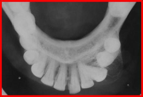
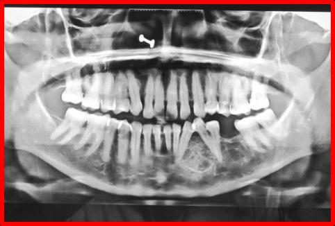


3 Fig. 1: Pre-Operative Photographs and X-Rays Fig. 2: Intra-Operative Photographs IJOCR Jul - Sep 2013; Volume 1 Issue 1 58


![[5] The most typical clinical presentation of these lesions is a well-defined swelling of soft tissues due to expansion of the adjacent bone, causing noticeable facial asymmetry.](/docs-images/94/121102434/images/4-2.jpg "[3,14,15] They usually present a slow progressive growth until cortical plates are eroded at any point and then show a rapid growth. Malocclusion can be a consequence of facial deformity.")
![[10] Pain is an infrequent symptom except of rapid growth cases as the present case.](/docs-images/94/121102434/images/4-4.jpg "[4] Other less common clinical presentations could be root resorption of teeth, disesthesias, proptosis, diplopia and progressive nasal obstruction in maxillary lesions.")
![[4,8] Solid aneurysmal bone cyst is usually asymptomatic whereas vascular form usually presents an invasive rapid growing evolution with extension to overlying tissues.](/docs-images/94/121102434/images/4-5.jpg "[5,13,14] MRI is mandatory in complex cases such as the reported case, in order to improve plain radiographic examination and CT-scan as it is more accurate in soft-tissue contrast.")
4 Fig. 3: Histopathological Sections Fig. 4: Post-Operative Healing After 1 Week and Four Months amount of hemosiderin and giant cells can be found. [1,2,13] This description is characteristic of the classic or vascular form, which is the most frequent. Solid form is the other histological type that represents only a 5% of all the cases. This form is a noncystic variant with solid graywhite tissue, hemorrhagic foci and abundant fibroblastic and fibrohistiocytic elements with osteoclast-like giant cells. Osteoblastic differentiation areas with osteoid and calcifying fibromyxoid tissue complete the picture. [2,5] A third variant mixed form demonstrates elements of both vascular and solid types. [5] The most typical clinical presentation of these lesions is a well-defined swelling of soft tissues due to expansion of the adjacent bone, causing noticeable facial asymmetry. [3,14,15] They usually present a slow progressive growth until cortical plates are eroded at any point and then show a rapid growth. Malocclusion can be a consequence of facial deformity. [10] Pain is an infrequent symptom except of rapid growth cases as the present case. [4] Other less common clinical presentations could be root resorption of teeth, disesthesias, proptosis, diplopia and progressive nasal obstruction in maxillary lesions. [4,8] Solid aneurysmal bone cyst is usually asymptomatic whereas vascular form usually presents an invasive rapid growing evolution with extension to overlying tissues. [5,13,14] MRI is mandatory in complex cases such as the reported case, in order to improve plain radiographic examination and CT-scan as it is more accurate in soft-tissue contrast. [12] Panoramic radiography frequently shows the presence of a cystic radiolucent imaging, usually multilocular, with a cystic meshwork divided by coarse septa. [12] Bony cortex can also be expanded. The multilocular effect gives this cyst the characteristical but no pathognomonical honeycomb and soapbubble - like appearance seen in other lesions such as giant cell granuloma, myxoma, IJOCR Jul - Sep 2013; Volume 1 Issue 1 59
5 desmoblastic fibroma, haemangioma, keratocyst, ameloblastoma and other tumours. [3,8,12] Occasionally, destruction of bony cortex may be identified, displaying a periosteal reaction imaging or ray-sun effect that is characteristic of osteosarcomas, with which differencial diagnosis should be done. [5,12] CT-scan accurately identifies tissue septa. MRI findings of fluid-fluid levels inside the lesion are high specific of aneurysmal bone cysts. [3,12] Angiography is only occasionally used in the diagnosis of these pseudocysts, however it may be necessary if an haemangioma or a high grade neoplams is suspected when MRI shows hyper vascularization. [4] Fine needle aspiration and incisional biopsy may be also performed when high grade tumours are supected. [3] Nowadays, treatment of choice is conservative surgical resection of the lesion. This must be limited to careful enucleation or curettage of the mass as it is a benign process. Segmentary resection must be done only in case of multiple recurrencies or extension to overlying tissues. [1,3,4,6] Recurrence rates range from 20% to 30% according to different series and seems to occur most frequently within the first year after surgery. [3,4,6] This is usually attributed to incomplete removal of the lesion especially in soft tissue invasive cases. [3,5,10,13] Several authors recommend immediate reconstruction of the defect with autogenous grafts in cases of aesthetic deformity and in cases with high risk of fractures and loss of mandibular continuity. [1,3,6,15] CONCLUSION As the radiological features of ABC are varied, resembling many lesions, histopathological analysis is a must for the diagnosis. BIBLIOGRAPHY 1. Kiattavorncharoen S, Joos U, Brinkschmidt C, Werkmeister R. Aneurysmal bone cyst of the mandible: a case report. Int J Oral Maxillofac Surg. 2003;32: Perrotti V, Rubini C, Fioroni M, Piattelli A. Solid aneurysmal bone cyst of the mandible. Int J Pediatr Otorhinolaryngol. 2004;68: Rapidis AD, Vallianatou D, Apostolidis C, Lagogiannis G. Large lytic lesion of the ascending ramus, the condyle, and the infratemporal region. J Oral Maxillofac Surg. 2004;62: Bataineh AB. Aneurysmal bone cysts of the maxilla: a clinicopathologic review. J Oral Maxillofac Surg. 1997;55: Motamedi MH. Destructive aneurysmal bone cyst of the mandibular condyle: report of a case and review of the literature. J Oral Maxillofac Surg. 2002;60: Martins WD, Fávaro DM. Aneurysmal bone cyst of the coronoid process of the mandible: a case report. J Contemp Dent Pract. 2005;6: Matsuura S, Tahara T, Ro T, Masumi T, Kasuya H, Yokota T. Aneurysmal bone cyst of the coronoid process of the mandible. Dentomaxillofac Radiol. 1999;28: López-Arcas JM, Cebrián L, González J, Burgueño M. Aneurysmal bone cyst of the mandible: case presentation and review of the literature. Med Oral Patol Oral Cir Bucal. 2007;12: Mankin HJ, Hornicek FJ, Ortiz-Cruz E, Villafuerte J, Gebhardt MC. Aneurysmal bone cyst: a review of 150 patients. J Clin Oncol. 2005;23: Struthers PJ, Shear M. Aneurysmal bone cyst of the jaws. (I). Clinicopathological features. Int J Oral Surg. 1984;13: Jaffé HL, Lichtenstein L. Solitary unicameral bone cyst. With emphasis on roentger picture, the pathological appearance and the pathogenesis. Arch Surg. 1942;44: Asaumi J, Konouchi H, Hisatomi M, Matsuzaki H, Shigehara H, Honda Y, et al. MR features of aneurysmal bone cyst of the mandible and characteristics distinguishing it from other lesions. Eur J Radiol. 2003;45: Motamedi MH, Yazdi E. Aneurysmal bone cyst of the jaws: analysis of 11 cases. J Oral Maxillofac Surg. 1994;52: Hernandez GA, Castro A, Castro G, Amador E. Aneurysmal bone cyst versus hemangioma of the mandible. Report of a long-term follow-up of a self-limiting case. Oral Surg Oral Med Oral Pathol. 1993;76: IJOCR Jul - Sep 2013; Volume 1 Issue 1 60
6 Gadre KS, Zubairy RA. Aneurysmal bone cyst of the mandibular condyle: report of a case. J Oral Maxillofac Surg. 2000;58: Source of Support: Nil Conflict of Interest: Nil IJOCR Jul - Sep 2013; Volume 1 Issue 1 61
Aneurysmal Bone Cyst of the Coronoid Process of the Mandible: A Case Report
 Aneurysmal Bone Cyst of the Coronoid Process of the Mandible: A Case Report Abstract A rare case of aneurysmal bone cyst (ABC) located in the ramus of the mandible with involvement of the coronoid process
Aneurysmal Bone Cyst of Mandible: A Case Report and Review of Literature
 CASE REPORT Aneurysmal Bone Cyst of Mandible: A Case Report and Review of Literature ABSTRACT An aneurysmal bone cyst is a rare benign expanding osteolytic lesion of bone characterized by replacement with
TRAUMATIC BONE CYST OF IDIOPATHIC ORIGIN? A REPORT OF TWO CASES
 Traumatic Bone Cyst Kumar S. et al 183 CASE REPORT TRAUMATIC BONE CYST OF IDIOPATHIC ORIGIN? A REPORT OF TWO CASES Kumar Satish 1, S. Padmashree 1, Jayalekshmy Rema 1 ABSTRACT BACKGROUND: Traumatic bone
CENTRAL GIANT CELL GRANULOMA PRESENTING AS UNILOCULAR RADIOLUCENCY IN POSTERIOR MANDIBLE A CASE REPORT
 IJCRR Section: Healthcare Sci. Journal Impact Factor 4.016 Case Report CENTRAL GIANT CELL GRANULOMA PRESENTING AS UNILOCULAR RADIOLUCENCY IN POSTERIOR MANDIBLE A CASE REPORT S. Aruleena Shaminey 1, G.
MARK D. MURPHEY MD, FACR. Physician-in-Chief, AIRP. Chief, Musculoskeletal Imaging
 ALPHABET SOUP AND CYSTIC LESIONS OF THE BONE MARK D. MURPHEY MD, FACR Physician-in-Chief, AIRP Chief, Musculoskeletal Imaging ALPHABET SOUP AND CYSTIC LESIONS OF THE BONE Giant cell tumor (GCT) Unicameral
Differential Diagnosis of Radiolucent Lesions of the Jaws
 Differential Diagnosis of Radiolucent Lesions of the Jaws Multilocular Multilocular Radiolucencies Odontogenic Keratocyst Botryoid Odontogenic Cyst Glandular odontogenic Cyst Invasive Ameloblastoma Central
JCD CASE REPORT ABSTRACT INTRODUCTION CASE REPORT
 JCD CASE REPORT Giant Aneurysmal 10.5005/jp-journals-10031-1161 Bone Cyst of the Mandible 1 Rajshri U Gurav, 2 Jigna Pathak, 3 Shilpa Patel, 4 Niharika Swain ABSTRACT An aneurysmal bone cyst (ABC) is a
Aneurysmal bone cyst of the mandible managed by conservative surgical therapy with preoperative embolization
 Imaging Science in Dentistry 2012; 42 : 35-9 http://dx.doi.org/10.5624/isd.2012.42.1.35 neurysmal bone cyst of the mandible managed by conservative surgical therapy with preoperative embolization Department
Aggressive Juvenile Ossifying Fibroma of the Anterior Mandible
 Case Report Aggressive Juvenile Ossifying Fibroma of the Anterior Mandible Dr.Ravikumar.R 1, Dr.Raghavendra.K 2, Dr.Santhosh Kumar 3 1 Senior Lecturer, 2 Reader Department of Oral Surgery, Sri Siddhartha
Peripheral Odontogenic Fibroma: A rare case report
 Annals of Dental Research (2013) Vol 3 (1): 10-14 HATAM Publishers: All Rights Reserved Annals of Dental Research www.hgpub.com www.adres.yolasite.com Case Report Peripheral Odontogenic Fibroma: A rare
AMELOBLASTIC FIBROMA: A RARE CASE REPORT
 Case Report International Journal of Dental and Health Sciences Volume 04, Issue 03 AMELOBLASTIC FIBROMA: A RARE CASE REPORT Namratha Patil 1 1.Sr lecturer, dept of oral medicine and radiology, KAHES VK
J of Evolution of Med and Dent Sci/ eissn , pissn / Vol. 3/ Issue 18/May 05, 2014 Page 4859
 CYSTIC DEGENERATION IN FOLLICULAR AMELOBLASTOMA: A CASE REPORT Neeraj Kumar 1, Niharika Rathore 2, Hemant Shakya 3, Anshuman Jamdade 4, Puneet Chitlangia 5 HOW TO CITE THIS ARTICLE: Neeraj Kumar, Niharika
Disclosures. Giant Cell Rich Tumors of Bone. Outline. The osteoclast. Giant cell rich tumors 5/21/11
 Disclosures Giant Cell Rich Tumors of Bone Andrew Horvai, MD, PhD Associate Clinical Professor, Pathology This lecture discusses "off label" uses of a number of pharmaceutical agents. The speaker is describing
Keratocystic Odontogenic Tumor : What radiologist needs to know?
 Keratocystic Odontogenic Tumor : What radiologist needs to know? Poster No.: C-0444 Congress: ECR 2014 Type: Authors: Keywords: DOI: Educational Exhibit K. El Karzazi, J. M. Villanueva Rincón, R. Corrales,
MULTILOCULAR AMELOBLASTOMA OF MANDIBLE-A CASE REPORT
 International Journal of Advancements in Research & Technology, Volume 2, Issue2, February-2013 1 MULTILOCULAR AMELOBLASTOMA OF MANDIBLE-A CASE REPORT DR SANTOSH KUMAR SUBUDHI*, DR SUMIT DASH**, DR.K..PREMANANDA***,
AMERICAN JOURNAL OF BIOLOGICAL AND PHARMACEUTICAL RESEARCH
 AMERICAN JOURNAL OF BIOLOGICAL AND PHARMACEUTICAL RESEARCH e-issn - 2348-2184 Print ISSN - 2348-2176 Journal homepage: www.mcmed.us/journal/ajbpr SOLID AND MULTICYSTIC FOLLICULAR AMELOBLASTOMA - A CASE
SURGICAL'MANAGEMENT'OF' PLEXIFORM'AMELOBLASTOMA:'A' CASE'REPORT'
 HARSH,Ashutosh* PUROHIT,Pragya* PUROHIT,NC** ADLAKHA,Twisha*** SURGICALMANAGEMENTOF PLEXIFORMAMELOBLASTOMA:A CASEREPORT ABSTRACT Ameloblastoma is a benign but locally invasive neoplasm of odontogenicepithelium.patientsusuallypresentlateafterthetumor
Maxilla and mandible benign lesions: Radiologic Findings and Differential Diagnosis in CT
 Maxilla and mandible benign lesions: Radiologic Findings and Differential Diagnosis in CT Poster No.: C-0964 Congress: ECR 2012 Type: Scientific Exhibit Authors: N. Lopez 1, E. Marcos Naranjo 2, M. D.
CENTRAL GIANT CELL GRANULOMA IN A 10 YEAR OLD CHILD A CASE REPORT
 International Journal of Innovation Sciences and Research Vol.4, No, 4, pp. 139-143, April- 2015 CASE STUDY Available online at http://www.ijisr.com CENTRAL GIANT CELL GRANULOMA IN A 10 YEAR OLD CHILD
Large Dentigerous Cyst
 Volume 16.2.1 Feb 2016 This Lecture Series qualifies for 0.5 Informal CPD Learning Hours Large Dentigerous Cyst By Dr Hassem Geha A 55 year-old male presented with a painless swelling in the right mandible.
The Radiology Assistant : Bone tumor - ill defined osteolytic tumors and tumor-like lesions
 Bone tumor - ill defined osteolytic tumors and tumor-like lesions Henk Jan van der Woude and Robin Smithuis Radiology department of the Onze Lieve Vrouwe Gasthuis, Amsterdam and the Rijnland hospital,
Glandular Odontogenic Cyst Coexisting with a Dentigerous Cyst: Case Report
 SmyrnaMed Case 2018;2(1): 1-5 ISSN (Online): 2564-6869 www.smyrnamed.com Glandular Odontogenic Cyst Coexisting with a Dentigerous Cyst: Case Report Assist.Prof.Dr. Serap Keskin Tunç 1, Dt. Erkan Feslihan
Case Report Basal Cell Ameloblastoma of Mandible: A Rare Case Report with Review
 Case Reports in Dentistry Volume 2013, Article ID 187820, 4 pages http://dx.doi.org/10.1155/2013/187820 Case Report Basal Cell Ameloblastoma of Mandible: A Rare Case Report with Review Hemant Shakya, 1
PACIFIC JOURNAL OF MEDICAL SCIENCES ISSN:
 PACIFIC JOURNAL OF MEDICAL SCIENCES {Formerly: Medical Sciences Bulletin} ISSN: 2072 1625 Pac. J. Med. Sci. (PJMS) www.pacjmedsci.com. Email: pacjmedsci@gmail.com. ADENOMATOID ODONTOGENIC TUMOR WITH RARE
Case Presentation: Figure 1. Palatal lesion. Figure 2. Panoramic xray
 Case Presentation: A 10-year-old girl presented with a hard palatal mass that had been progressively increasing in size for the last 4 months, as reported by her dentist. Two months previously, her dentist
An unusual site of Adenomatoid Odontogenic Tumor: A rare case report
 J. Int Oral Health 2010 Case Report All right reserved An unusual site of Adenomatoid Odontogenic Tumor: A rare case report Sapna Panjwani*, Anjana Bagewadi**, Vaishali Keluskar*** *Post Graduate Student
Case report: Central Giant Cell Granuloma of Mandible A Case Report
 Case report: Central Giant Cell Granuloma of Mandible A Case Report 1Dr Praveen Kumar, 2 Dr Balreddy P., 3 Dr Sridhar Reddy B., 4 Dr Sanjeev Naik, 5Dr. Sujitha Reddy Patil 1Post Graduate, Department of
Reconstruction of large mandibular defects
 Immediate Reconstruction of a Large Mandibular Defect of Locally Invasive Benign Lesions (A New Method) Gholamreza Shirani, OMFS, DDS, MS,* Mahnaz Arshad, DDS, 1 Farnoush Mohammadi, OMFS, DDS, MS* Tehran,
A Case Report of Odontogenic Keratocyst in Anterior Mandibule Position
 A Case Report of Odontogenic Keratocyst in Anterior Mandibule Position Malihe Moeini 1, Seyed Ehsan Anvar 2, Rasool Barzegari Bafghi 3* 1.Resident of Oral and Maxillofacial Radiology, Faculty of Dentistry,
Primary bone tumors > metastases from other sites Primary bone tumors widely range -from benign to malignant. Classified according to the normal cell
 Primary bone tumors > metastases from other sites Primary bone tumors widely range -from benign to malignant. Classified according to the normal cell counterpart and line of differentiation. Among the
The Radiology Assistant : Bone tumor - well-defined osteolytic tumors and tumor-like lesions
 Bone tumor - well-defined osteolytic tumors and tumor-like lesions Henk Jan van der Woude and Robin Smithuis Radiology department of the Onze Lieve Vrouwe Gasthuis, Amsterdam and the Rijnland hospital,
Annals and Essences of Dentistry
 aedj.2014.6.3.2.4 Central Calcifying Cystic Odontogenic Tumor Of Mandible A Case Report 1 Rajasekhar Gali 2 Madan Mohan Reddy 3 Vandana Raghunath 4 Sajan Anand 1 Associate Professor 2 Reader 3 Professor
Management of Plexiform Ameloblastoma in a 12 year old female: A Case Report
 Article ID: ISSN 2046-1690 Management of Plexiform Ameloblastoma in a 12 year old female: A Case Report Corresponding Author: Dr. Sheeraz Badal, Senior Lecturer, Dept of Oral & Maxillofacial Surgery, MIDSR,
Ameloblastomatous Gorlin s cyst
 319 Journal of Oral Science, Vol. 49, No. 4, 319-323, 2007 Case Report Ameloblastomatous Gorlin s cyst Mala Kamboj 1) and Manish Juneja 2) 1) Department of Oral Pathology and Microbiology, U.P. King George
Skeletal Radiology. Solitary (unicameral) bone cyst. The fallen fragment sign revisited
 bone cyst. The fallen fragment sign revisited") Skeletal Radiol (1989) 18:261-265 Skeletal Radiology Solitary (unicameral) bone cyst The fallen fragment sign revisited S. Struhl, M.D., C. Edelson, M.D., H. Pritzker, M.D., L.P. Seimon, M.D., and H.D.
Skeletal Radiol (1989) 18:261-265 Skeletal Radiology Solitary (unicameral) bone cyst The fallen fragment sign revisited S. Struhl, M.D., C. Edelson, M.D., H. Pritzker, M.D., L.P. Seimon, M.D., and H.D.
Solitary Plasmacytoma of Mandible: A rare case report Singh A 1, Singh V 2, Sharma N 3
 Case Report Solitary Plasmacytoma of Mandible: A rare case report Singh A 1, Singh V 2, Sharma N 3 1 Dr. Anurag Singh Consultant Sir Ganga Ram Hospital Delhi, India 2 Dr. Vishal Singh Professor, Oral &
Central odontogenic fibroma of the mandible: a case report
 457 Journal of Oral Science, Vol. 51, No. 3, 457-461, 2009 Case Report Central odontogenic fibroma of the mandible: a case report Ioanna Daskala 1), Demos Kalyvas 2), Markos Kolokoudias 2), Dimitris Vlachodimitropoulos
Treatment Modalities of Odontogenic Keratocyst of Maxilla and Mandible: Our Experience
 wjd Parveen Akhter Lone et al Original Research 10.5005/jp-journals-10015-1344 Treatment Modalities of Odontogenic Keratocyst of Maxilla and Mandible: Our Experience 1 Parveen Akhter Lone, 2 Mohan Singh,
A Case Report on Surgical Management of Odontogenic Keratocyst
 World Journal of Medical Sciences 10 (2): 212-216, 2014 ISSN 1817-3055 IDOSI Publications, 2014 DOI: 10.5829/idosi.wjms.2014.10.2.82185 A Case Report on Surgical Management of Odontogenic Keratocyst 1
Endovascular and surgical treatment of giant pelvic tumor
 Endovascular and surgical treatment of giant pelvic tumor Mitrev Z., MD FETCS; Anguseva T., MD; Milev I., MD; Zafiroski G., PhD MD Center for Cardiosurgery, Filip the II, Skopje, Macedonia Background Giant
Surgical management of Ossifying Fibroma of the mandible with inferior alveolar nerve involvement
 Yadegari A,et al J Res Dentomaxillofac Sci e(issn): 2383-2754 Journal of Research in Dental and Maxillofacial Sciences Surgical management of Ossifying Fibroma of the mandible with inferior alveolar nerve
Successful Conservative Surgical Treatment of Ameloblastic Fibroma in the Posterior Maxilla : A Case Report
 http://dx.doi.org/10.5933/jkapd.2013.40.4.321 ISSN (print) 1226-8496 Successful Conservative Surgical Treatment of Ameloblastic Fibroma in the Posterior Maxilla : A Case Report Youngeun Lee 1, Hyojung
Index. oralmaxsurgery.theclinics.com. Note: Page numbers of article titles are in boldface type.
 Index Note: Page numbers of article titles are in boldface type. A Adenomatoid odontogenic tumor, pediatric, 50 51 Ameloblastic carcinoma, pediatric, 17, 49 Ameloblastic fibro-odontoma, pediatric, 54 Ameloblastic
Ossifying fibromas of the jaw bone: 20 cases
 (2010) 39, 57 63 2010 The British Institute of Radiology http://dmfr.birjournals.org CASE REPORT Ossifying fibromas of the jaw bone: 20 cases Y Liu 1, M You 1, H Wang*,1, Z Yang 1, J Miao 1, K Shimizutani
The clinical appearance and diagnosis of odontogenic cysts. SE Arc-Állcsont-Szájsebészeti és Fogászati Klinika BUDAPEST
 The clinical appearance and diagnosis of odontogenic cysts SE Arc-Állcsont-Szájsebészeti és Fogászati Klinika BUDAPEST DEFINITION A cyst is a sac with walls of connective tissue, lined by epithelium, containing
Multiple Synchronous Central Giant Cell Granulomas of the Maxillofacial Region: A Case Report 1
 Multiple Synchronous Central Giant Cell Granulomas of the Maxillofacial Region: A Case Report 1 Min Seok Kang, M.D., Hak Jin Kim, M.D. Multifocal central giant cell granulomas (CGCG) in the maxillofacial
MALIGNANT TUMOURS OF THE JAWS
 MALIGNANT TUMOURS OF THE JAWS MALIGNANT TUMOURS OF THE JAWS Squamous cell carcinoma Osteogenic sarcoma Chondrosarcoma Fibrosarcoma Malignant lymphomas (incl. Burkitt s) Multiple myeloma Ameloblastoma Secondary
Case Report Acanthomatous Ameloblastoma treated with Hemimandibulectomy
 Case Report Acanthomatous Ameloblastoma treated with Hemimandibulectomy Shobha Bijjargi, Ashwini Rani S.R, Jaishankar HP, Shashidara R 1, Veerendra Patil 2 Departments of Oral Medicine &Radiology, 1 Oral
Unicystic Ameloblastoma of mandible. Aggresive treatment A myth or a need. Case report and Extensive review of literature
 IOSR Journal of Dental and Medical Sciences (IOSR-JDMS) e-issn: 2279-0853, p-issn: 2279-0861. Volume 12, Issue 2 (Nov.- Dec. 2013), PP 26-31 Unicystic Ameloblastoma of mandible. Aggresive treatment A myth
高雄醫學大學 口腔醫學院 口腔病理影像科 牙科 X 光影像判讀 教學範例
 高雄醫學大學 口腔醫學院 口腔病理影像科 牙科 X 光影像判讀 教學範例 Content Image No. 001 Dentigerous cyst over left upper embedded canine--------------------- 頁 1 Image No. 002---------------------------------------------------------------
Diagnosis and Treatment of a Large Central Ossifying Fibroma of the Mandible. Clinical Case
 Clinical Case Diagnosis and Treatment of a Large Central Ossifying Fibroma of the Mandible A 36 year old African American Female presented to the Department of Oral and Maxillofacial Surgery Clinic at
History. 33 y/o F with hx of palpable anterior tibial mass x 2 years, only painful with palpation
 History 33 y/o F with hx of palpable anterior tibial mass x 2 years, only painful with palpation Imaging Photo Album Patient also had a smaller lesion 1 cm proximal to this lesion, not seen radiographically.
Orthokeratinized Odontogenic Cyst: A Rarity
 aijoc AIJOC Case Report 1 Heena Sonawane, 2 Freny R Karjodkar, 3 Kaustubh Sansare, 4 Nimish Prakash ABSTRACT Orthokeratinized odontogenic cyst (OOC) was first identified as the rare variant of keratocystic
Simultaneous Occurrence of Central Giant Cell Granuloma and Odontogenic Keratocyst in Mandible
 Bull Tokyo Dent Coll (2017) 58(3): 171 175 Case Report doi:10.2209/tdcpublication.2016-0018 Simultaneous Occurrence of Central Giant Cell Granuloma and Odontogenic Keratocyst in Mandible Bruna da Fonseca
Disclosure. Educational Objectives. Terminology. Odontogenic Cysts. Terminology
 Disclosure Lisa J. Koenig BChD, DDS, MS Professor & Program Director, Oral Medicine and Oral Radiology Marquette University School of Dentistry Consultant to Soredex for the Scanora 3D and 3Dx Author/Editor
Arun V Subramaniam et al. / International Journal of Biopharmaceutics. 2014; 5(3): International Journal of Biopharmaceutics
: International Journal of Biopharmaceutics") 225 e- ISSN 0976-1047 Print ISSN 2229-7499 International Journal of Biopharmaceutics Journal homepage: www.ijbonline.com IJB ODONTOGENIC KERATOCYSTS: VARIOUS RADIOGRAPHIC APPEARANCES Arun Subramaniam*
225 e- ISSN 0976-1047 Print ISSN 2229-7499 International Journal of Biopharmaceutics Journal homepage: www.ijbonline.com IJB ODONTOGENIC KERATOCYSTS: VARIOUS RADIOGRAPHIC APPEARANCES Arun Subramaniam*
The resident will be assigned to be on call with the Oral and Maxillofacial service. Call will be set according to PARO guidelines.
 Goals and Objectives for the Otolaryngology-Head & Neck Resident on the Oral and Maxillofacial Surgery (OMFS) Rotation St. Catharines General Hospital (1 four-week rotational block) During the second year
FOCAL CEMENTO-OSSEOUS DYSPLASIA OF LOWER JAW IN AN ELDERLY WOMAN: CLINICOPATHOLOGICAL FEATURES AND MANAGEMENT IN A CASE REPORT
 www.djas.co.in ISSN No-31-148 DJAS 4(III), 08-1, 016 All rights are reserved CASE REPORT Dental JOURNAL of Advance Studies FOCAL CEMENTO-OSSEOUS DYSPLASIA OF LOWER JAW IN AN ELDERLY WOMAN: CLINICOPATHOLOGICAL
Juvenile Trabecular And Psammomatoid Variant Of Ossifying Fibroma Of The Maxilla With Secondary Aneurysmal Bone Cyst.
 ISPUB.COM The Internet Journal of Otorhinolaryngology Volume 14 Number 1 Juvenile Trabecular And Psammomatoid Variant Of Ossifying Fibroma Of The Maxilla With Secondary L Emerson., M Alexander, A Job Citation
APMA 2018 Radiology Track Bone Tumors When to say Gulp!
 APMA 2018 Radiology Track Bone Tumors When to say Gulp! DANIEL P. EVANS, DPM, FACFAOM Professor, Department of Podiatric Medicine and Radiology Dr. Wm. Scholl College of Podiatric Medicine Conflict of
Iliac aneurysmal bone cyst treated by cystoscopic controlled curettage
 Accepted February 13th, 2004 Iliac aneurysmal bone cyst treated by cystoscopic controlled curettage Ludwig Schwering¹, Markus Uhl² and Georg W. Herget( )¹ ¹ Department of Orthopaedics and Traumatology,
Study of maxillary and mandibular cystic lesions
 Study of maxillary and mandibular cystic lesions Poster No.: C-1428 Congress: ECR 2013 Type: Educational Exhibit Authors: M. L. Rozas Rodríguez, M. E. Banegas Illescas, M. Y. Torres Sousa, R. M. Fernández
Clinical details: Details of scan: CONE BEAM CT REPORT: Name: H. B. Gender: Reason for referral: Referred by:
 Name: H. B. Gender: Male DOB: 11/12/1950 Age: 64 Date taken: 16/11/2015 Date reported: 19/11/2015 Clinical details: Reason for referral: Referred by: Investigate symptoms related to left TMJ. Reconstructed
FRACTURE CALLUS ASSOCIATED WITH BENIGN AND MALIGNANT BONE LESIONS AND MIMICKING OSTEOSARCOMA
 THE AMERICAN JOURNAL OF CLINICAL PATHOLOGY Vol. 52, No. 1 Copyright 1969 by The Williams & Wilkins Co. Printed in U.S.A. FRACTURE CALLUS ASSOCIATED WITH BENIGN AND MALIGNANT BONE LESIONS AND MIMICKING
Gorham Disease an Enigma
 Article ID: WMC002898 ISSN 2046-1690 Gorham Disease an Enigma Corresponding Author: Dr. Prashant Kothari, Asso Professor, ODMR MCDRC, 442301 - India Submitting Author: Dr. Prashant Kothari, Asso Professor,
Role of MDCT and VR reconstructions in the diagnosis and characterization of maxillary cystic lesions.
 Role of MDCT and VR reconstructions in the diagnosis and characterization of maxillary cystic lesions. Poster No.: C-0704 Congress: ECR 2011 Type: Scientific Exhibit Authors: M. Coronella 1, P. V. Foti
Autologous Bone Augmentation in Combination with an Ameloblastoma in the Maxillary Region- A Case Report?
 Archives of Clinical and Medical Case Reports doi: 10.26502/acmcr.96550023 Volume 2, Issue 2 Case Report Autologous Bone Augmentation in Combination with an Ameloblastoma in the Maxillary Region- A Case
Partial Spontaneous Bone Regeneration Subsequent to Mandibulectomy
 Partial Spontaneous Bone Regeneration Subsequent to Mandibulectomy Abstract A case of spontaneus bone regeneration after mandibulectomy is presented. The patient was young and had a mandibular resection
Key words: Third molar, Impacted tooth, Tooth Eruption, Molar, Mandible, Unerupted Tooth.
 JOURNAL OF CASE REPORTS 2014;4(2):286-290 OPG and CBCT Finding s of an Ectopic Third Molar in the Sub-condylar Region Tatu Joy E 1, Farakath Khan 1, Shameel Mohammed 2 From the Department of Oral Medicine
Benign Fibro-osseous Lesions
 Benign Fibro-osseous Lesions Plus Vision is the art of seeing things invisible. Jonathan Swift 1667-1745 Steven R. Singer, DDS srs2@columbia.edu 212.305.5674 Benign Fibro-osseous Lesions A group of lesions
Unusual radiographic appearance of ossifying fibroma in the left mandibular angle
 (2010) 39, 314 319 2010 The British Institute of Radiology http://dmfr.birjournals.org CASE REPORT Unusual radiographic appearance of ossifying fibroma in the left mandibular angle M Araki*,1,4, K Matsumoto
Cone Beam Computed Tomography Findings in Calcifying Cystic Odontogenic Tumor Associated with Odontome: A Case Report
 Phulambrikar T., et al. Dent Shiraz Univ Med Sci., December 2015; 16(4): 374-379. Case Report Cone Beam Computed Tomography Findings in Calcifying Cystic Odontogenic Tumor Associated with Odontome: A Case
4/2/17. Panoramic Radiography: Normal Variants and Pathology. Composite of in-focused and blurred images. It s a type of Tomogram.
 Fundamentals Panoramic Radiography: Normal Variants and Pathology It s a type of Tomogram Jimmie L. Harper D.D.S., M.S. Cincinnati Oral and Maxillofacial Surgery, Inc. Volunteer Assistant Professor, Division
Odontomes and Odontogenic tumours
 Odontomes and Odontogenic tumours Odontomes Developmental hamartoma Hamartoma: normal tissue in abnormal location Any cells to be neoplastic it must be able to replicate, which is not seen in hamartoma
Fluid-fluid levels in bone tumors: A pictorial review
 Fluid-fluid levels in bone tumors: A pictorial review Poster No.: C-578 Congress: ECR 2009 Type: Educational Exhibit Topic: Musculoskeletal Authors: L. Figueroa Nasra, C. Martín Hervás, M. Tapia-Viñé,
GIANT CELL-RICH OSTEOSARCOMA: A CASE REPORT
 Nagoya J. Med. Sci. 59. 151-157, 1996 CASE REPORTS GIANT CELL-RICH OSTEOSARCOMA: A CASE REPORT KEIJI SATO!, SHIGEKI YAMAMURA!, HISASHI IWATA!, HIDESHI SUGIURA 2, NOBUO NAKASHIMA 3 and TETSURO NAGASAKA
Cover Page. The handle holds various files of this Leiden University dissertation.
 Cover Page The handle http://hdl.handle.net/1887/31632 holds various files of this Leiden University dissertation. Author: Mensink, Gertjan Title: Bilateral sagittal split osteotomy by the splitter-separator
[ 06-10] Dr. B. Siva Reddy, Dr. B. Ajay Reginald, Dr. D. Sireesha, Dr. Meda Samatha India Abstract: Keywords ARTICLE 20/07/ /09/2018
![[ 06-10] Dr. B. Siva Reddy, Dr. B. Ajay Reginald, Dr. D. Sireesha, Dr. Meda Samatha India Abstract: Keywords ARTICLE 20/07/ /09/2018](/thumbs/87/95905171.jpg "[ 06-10] Dr. B. Siva Reddy, Dr. B. Ajay Reginald, Dr. D. Sireesha, Dr. Meda Samatha India Abstract: Keywords ARTICLE 20/07/ /09/2018") Dentigerous Cyst Associated with an Impacted Mesiodens: A Rare Case Report with Review of Literature [PP: 06-10] Dr. B. Siva Reddy, Dr. B. Ajay Reginald, Dr. D. Sireesha, Dr. Meda Samatha, Department of
Dentigerous Cyst Associated with an Impacted Mesiodens: A Rare Case Report with Review of Literature [PP: 06-10] Dr. B. Siva Reddy, Dr. B. Ajay Reginald, Dr. D. Sireesha, Dr. Meda Samatha, Department of
Course Description 343 DDS- Clinical Oral and Maxillofacial Radiology II ( )
") King Saud University College of Dentistry Dept. of Oral Medicine & Diagnostic Sciences Division of Oral & Maxillofacial Radiology Course Description 343 DDS- Clinical Oral and Maxillofacial Radiology II
King Saud University College of Dentistry Dept. of Oral Medicine & Diagnostic Sciences Division of Oral & Maxillofacial Radiology Course Description 343 DDS- Clinical Oral and Maxillofacial Radiology II
Bone Tumors Clues and Cues
 William Herring, M.D. 2002 Bone Tumors Clues and Cues In Slide Show mode, advance the slides by pressing the spacebar All Photos Retain the Copyright of their Authors Clues by Appearance of Lesion Patterns
An Unusual Case of Ameloblastoma arising from Dentigerous Cyst
 Anil K Nagarajappa et al CASE REPORT 10.5005/jp-journals-10026-1200 An Unusual Case of Ameloblastoma arising from Dentigerous Cyst 1 Anil K Nagarajappa, 2 Divya Pandya, 3 Nidhi Jain ABSTRACT Ameloblastoma
Case Report A Giant Cell Fibroma and Focal Fibrous Hyperplasia in a Young Child: A Case Report
 Hindawi Publishing Corporation Case Reports in Dentistry Volume 2012, Article ID 370242, 5 pages doi:10.1155/2012/370242 Case Report A Giant Cell Fibroma and Focal Fibrous Hyperplasia in a Young Child:
Cemento-Ossifying Fibroma A Radiographic Diagnostic Dilemma
 Case Report Cemento-Ossifying Fibroma A Radiographic Diagnostic Dilemma Zahra Dalili 1, Somayeh Nemati 2, Fatemeh Shahsavari 3 1 Associate Professor, Department of Maxillofacial Radiology, Guilan University
Aneurysmal bone cyst of the scapula A case report
 Acta Orthop. Belg., 2009, 75, 684-689 CASE REPORT Aneurysmal bone cyst of the scapula A case report Panagiotis MEGAS, Zafiria G. PAPATHANASSIOU, George KASIMATIS, Dionysios J. PAPACHRISTOU From the University
ISSN: Volume 4 Issue CHOLESTEROL GRANULOMA: AN UNCOMMON CLINICAL ENTITY OF THE MAXILLARY SINUS
 ISSN: 2250-0359 Volume 4 Issue 4 2014 CHOLESTEROL GRANULOMA: AN UNCOMMON CLINICAL ENTITY OF THE MAXILLARY SINUS Sunita Singh Sonia Chhabra Pansi Gupta Priya Malik Pt B.D. Sharma PGIMS, Rohtak, Haryana
Odontogenic fibroma in a six-year old child presenting as trismus
 Letter to editor DOI: https://doi.org/10.18320/jimd/201704.0137 JOURNAL OF INTERNATIONAL MEDICINE AND DENTISTRY To search..to know...to share p-issn: 2454-8847 e-issn: 2350-045X Odontogenic fibroma in
The concept of unicystic ameloblastoma (UA) was
 was") Publication Treatment of Giant Unicystic Ameloblastoma Suction Drainage and Secondary Curettage: a Case Report Bing LIU 1, Wen Feng ZHANG 1, Xin Ming CHEN 2, Zhi Jun SUN 3, Mohd Jamal Alsharif 1, Yi Fang
Publication Treatment of Giant Unicystic Ameloblastoma Suction Drainage and Secondary Curettage: a Case Report Bing LIU 1, Wen Feng ZHANG 1, Xin Ming CHEN 2, Zhi Jun SUN 3, Mohd Jamal Alsharif 1, Yi Fang
Big Keratocystic Odontogenic Tumor of the Mandible: A Case Report
 Big Keratocytic Odontogenic Bruktawit K et al 491 CASE REPORT Big Keratocystic Odontogenic Tumor of the Mandible: A Case Report Bruktawit Kebede 1, Demerew Dejene 1, Abebe Teka 1, Biruk Girma 1, Evelyn
Cholesterol granuloma of the jaws: report of two cases
 Cholesterol granuloma of the jaws: report of two cases Pages with reference to book, From 15 To 17 Alper Alkan, Osman Etoz ( Department of Oral and Maxillofacial Surgery, Erciyes University, Faculty of
Clinical, radiological and therapeutic features of keratocystic odontogenic tumours: a study over a decade
 Journal section: Oral Medicine and Pathology Publication Types: Research doi:10.4317/jced.51408 http://dx.doi.org/10.4317/jced.51408 Clinical, radiological and therapeutic features of keratocystic odontogenic
Periapical central giant cell granuloma misdiagnosed as odontogenic cyst
 doi: 10.1111/j.1365-2591.2006.01107.x CLINICAL ARTICLE Periapical central giant cell granuloma misdiagnosed as odontogenic cyst T. Lombardi 1, M. Bischof 1,2, R. Nedir 1,2, D. Vergain 1, C. Galgano 3,
Erupting Compound Odontome - A case report
 IOSR Journal of Dental and Medical Sciences (IOSR-JDMS) e-issn: 2279-0853, p-issn: 2279-0861.Volume 13, Issue 3 Ver. V. (Mar. 2014), PP 26-30 Erupting Compound Odontome - A case report Dr. Sahana Srinath
CASE REPORT PLEOMORPHIC LIPOSARCOMA OF PECTORALIS MAJOR MUSCLE IN ELDERLY MAN- CASE REPORT & REVIEW OF LITERATURE.
 PLEOMORPHIC LIPOSARCOMA OF PECTORALIS MAJOR MUSCLE IN ELDERLY MAN- CASE REPORT & REVIEW OF LITERATURE. M. Madan 1, K. Nischal 2, Sharan Basavaraj. C. J 3. HOW TO CITE THIS ARTICLE: M. Madan, K. Nischal,
Unexpected diagnosis for preauricular swelling - two case reports
 Unexpected diagnosis for preauricular swelling - two case reports Goran Roić 1, Vesna Posarić 1, Ante Marušić 1, Igor Borić 1, Tomislav Vlahović 2, Kristina Vrliček 3 1 Department of Paediatric Radiology,
Primary Intra-Osseous Squamous Cell Carcinoma of the Mandible - A Case Report
 IOSR Journal of Dental and Medical Sciences (IOSR-JDMS) e-issn: 2279-0853, p-issn: 2279-0861.Volume 15, Issue 9 Ver. VI (September). 2016), PP 66-70 www.iosrjournals.org Primary Intra-Osseous Squamous
Hemorrhagic bone cyst of mandible: A case report
 www.edoriumjournals.com CASE REPORT PEER REVIEWED OPEN ACCESS Hemorrhagic bone cyst of mandible: A case report G. Siva Prasad Reddy, G. V. Reddy, Phanitej G., Amarnath Reddy, Srilatha Priyadarshini, K.
The future of health is digital
 Dated: XX/XX/XXXX Name: XXXXXXXX XXXXXXXXXXX Birth Date: XX/XX/XXXX Date of scan: XX/XX/XXXX Examination of the anatomical volume: The following structures are reviewed and evaluated for bilateral symmetry,
Imaging modalities in head and neck pathology and trauma
 1 Imaging modalities in head and neck pathology & trauma Imaging modalities in head and neck pathology and trauma P M Mathew OTDE2 Student ID: 6704756 2 Imaging modalities Intraoral radiographs including
Common/Important Radiolucencies. B. Most Common Location Apex of permanent first molar, rare in primary teeth.
 Cincinnati Dental Association Breakfast at Tiffany s: The Jewels and Gems of Oral Pathology November 17, 2010 John A. Svirsky, DDS, MEd Virginia Commonwealth University 804-828-0547 FAX: 804-828-6234 EMAIL:
Pimary hyperparathyroidism as central giant cell granuloma of the jaws: Pre and post treatment pattern of clinical and radiographic presentation
 Aswath et al. 51 CASE IN IMAGES OPEN ACCESS Pimary hyperparathyroidism as central giant cell granuloma of the jaws: Pre and post treatment pattern of clinical and radiographic presentation Nalini Aswath,