Walking Theory DR.MURALI KRISHNA P.V M.S.(ORTHO)
|
|
|
- Kenneth Melvyn Barber
- 6 years ago
- Views:
Transcription
1 Walking Theory DR.MURALI KRISHNA P.V M.S.(ORTHO) G E N E R A L H O S P I T A L A N E K A L, D E P A R T M E N T O F O R T H O P E D I C S, B A N G A L O R E G O V E R N M E N T O F K A R N A T A K A
2 Walking Theory Walking is a complex phenomenon, In this technological era how the body moves forward has not been decoded. Walking as a subject is not taught to children in their curriculum, even in the medical curriculum walking or normal gait is not taught. Eating, Working and Walking are the basic activities of life. Dynamics of Walking is essential to solve the life style diseases.
3 Walking how it is described Bradford Founder of JBJS in 1897 He describes by the action of the muscles, the thigh is advanced forward by the action of the Ileacus, Tensor Vaginae and rectus muscles. the body s weight is thrown on the Heel and the body is brought forward by the push of the foot of the rear of the leg. in a slow gait, in feeble persons and in children the weight of the body is used as a means of propulsion. He classifies the Gait into Erect gait which is the active Gait and uses the muscles for the forward push and the Forward Gait which uses the body weight for the forward movement of the body.
4 Walking how it is described by Gait analysts Normal walking is defined as a highly controlled, coordinated, repetitive series of limb movements whose function is to advance the body safely from place to place with a minimum expenditure of energy.
5 Walking- Action of Muscles how it is described in Anatomy Gray s Anatomy describes action of the Lower limb muscles as abduction, adduction, flexion, extension, internal rotation and external rotation Gluteus maximus, Medius and Minimus abductors of Hip How many times we abduct during walking? Adductor muscles How many times we adduct during walking?
6 Walking how it is described in Anatomy Plantar flexion gastro soleus plantar flexes the ankle but in reality plantar flexion is a passive phenomenon. Actively during walking we can not plantar flex the foot. Dorsiflexors Tibialis anterior, EHL, EDL are plantar flexors why do we need so powerfull plantar flexors, what are their exact roles in walking? Muscular action with reference to walking is not described in any Anatomy or any other books.
7 PET-CT Gold standard in radiology PET-CT analyses the uptake of radio labeled glucose. Radio labeled glucose is injected into the body person is made to walk for one hour and the uptake of glucose by the muscles after the activity is assessed
8 PET-CT Gold standard in radiology- used in quantification of muscular activity a first time in the world PET-CT analysis also provides the quantitative uptake of glucose by individual muscles. PET-CT analyses full stretch of muscles, activity along the full length of muscles can be analysed. muscular activity of muscles across the length of the muscles from origin to insertion can be analyzed and uptake of radio labeled glucose is quantified.
9 Name of the Muscle Muscles of the Thigh: Glucose Uptake Value right side Left side Psoas major Psoas minor Iliacus The Anterior Femoral Muscles Sartorius Quadriceps femoris Rectus femoris Vastus lateralis Vastus medialis o Vastus intermedius
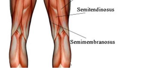
10 The Medial Femoral Muscles Gracilis Pectineus Adductor longus Adductor brevis Adductor magnus The Muscles of the Gluteal Region Glutæus maximus Glutæus medius Glutæus minimus Piriformis Tensor fasciæ latæ The Posterior Femoral Muscles (Hamstring Muscles) Biceps femoris Semitendinosus Semimembranosus
11 Tibialis anterior Extensor hallucis longus Extensor digitorum longus Peronæus tertius Tibialis posterior Gastrocnemius Soleus Popliteus Flexor hallucis longus Flexor digitorum longus Peronæus longus Peronæus brevis Abductor hallucis Abductor digiti quinti
12 Walking Theory Dr.Murali Krishna P.V Findings of the PET-CT analysis are usefull to analyse the walking in detail. Some of the questions I have raised previously were not answered in any of the textbooks and teachings. Lot of researchers have made their contributions in analysing the individual muscles. My walking theory is a culmination of all those dedicated researchers who have studied individual muscles in detail and my PET-CT analysis
13 Walking Theory Dr.Murali Krishna P.V. Human Walking is defined as the Propulsion of the body forward as a result of series of movement of lower limbs by the systematic contraction of the muscles of the lower limb over the adoptive bony frame work, in a specified way, with specific contraction of hip, thigh, leg and foot muscles to produce forward movement of the body, Maintaining stability, agility and speed.
14 Walking Theory Walking is divided into phases depending on the contraction of group of muscles and the action it brings in moving the body forward, Here I have described how body propels forward by individual set of action of muscles. Each group of muscles plays an important role in moving the body forward. 1. Movement of the Lower limb 2.Movement of the Trunk 3.Stabilisation Muscles
15 1. Movement of the Lower limb This phase is akin to swing phase, we explain the individual group of muscles activity to bring the movement of the lower limb forward Movement of the lower limb is divided into two phases 1. A. Movement of the lower limb from Toe off phase to Foot flat phase 1.B. Movement of the lower limb from Foot flat phase to Just before Heel strike phase
16 1. A. Movement of the lower limbfrom Toe off phase to Foot flat phase This phase extends from Toe off phase to Foot flat phase of walking in the swing Phase, Ileo Psoas Muscles Pull the Femur forward internally Tenosor fascia latae, Sartorius muscles pull the femur and tibia externally. Tenosor fascia latae with its unique attachment to the Ileo tibial band assist the pulling of the femur.


17

18 1.B. Movement of the lower limb from Foot flat phase to Just before Heel strike phase This phase extends from Foot flat phase to just before Heel strike Phase of walking in the swing Phase Quadriceps Muscles flexes the at the Hip and moves the lower limb forward
19 2.Movement of the Trunk Human body is designed extensively to move the body forward, every bone, muscle, joints and actions assist the forward movement of the trunk in a systematic way.. Foot is the only contact point of the body to the ground. Bony design provide sufficient leverage actions for efficiency of energy. the origin and insertion of the muscles assist specific activity and moves the body forward systematically effortlessly.
20 2.Movement of the Trunk This Stage is devided into several phases solely depending on the action of group of muscles. 2.A.- Heel strike Phase 2.B.- Heel strike to foot flat Phase 2.C.- Initial Heel off Phase 2.D- Late Heel off Phase 2.E- Extension of Knee 2.F-Pushing Phase of the Pelvis 2.G-Pulling Phase of the Pelvis
21 2.A.- Heel strike Phase Tibialis anterior, Extensor Hallusis Longus and Extensor digtorum Longus dorisflexes the ankle. Quadriceps muscles flexes at Hip and extneds at Knee dorsiflexion of the ankle fixes the foot to the ankle so much so that at the Heel strike phase the whole lower limb acts as a single Bony column, This facilitates the Heel strike. Striking the Heel by the contraction of the Dorsiflexors fixes the body at the Calcaneum, this Pulls the Pelvis at the top of the Bony column.


22
23 2.B.- Heel strike to foot flat Phase Heel strike Moves the Pelvis forward, but the Pelvis is heavy with all the organs of the body, Pelvis need to be pulled forward. Foot is brought down to the foot flat phase by Plantar flexors. Adductor group of muscles pull the pelvis forward Gracilis, Adductor Magnus, Adductor Brevis, Adductor longus, Pectineus muscles with their unique attachment along the Linea aspera of the femur to Pubic rami pulls the whole trunk smoothly. Adductor group of Muscles Pull the Pubic Rami forward, Lower limb glides over the Head of Smooth Talus.


24
25 2.C.- Initial Heel off Phase Plantar Flexors Gastro soleus Muscle plantar flexes the ankle. In reality the Heel lifts the whole body from the ground. Body weight shifts to the fore foot by the actions of the Tendo achilis, This lifting of the body at the ankle Lengthens the Lower limb this causes Passive flexion at Knee. This also raises the pelvis, raised Pelvis assist the free movement of the opposite limb.


26
27 2.D- Late Heel off Phase Action of the Tendoachilis do not lift the body sufficently, there is limitation of Plantar flexion because tendo achilis attaches to the calcaneum Tendo achilis can not lift the Mid foot, this needs an assistance to lift the hind foot and mid foot. Peroneus Longus and Peroneus Brevis attaches to the cuneifrom, and both ends of metatarsal bones, tendons runs in the Plantar surface of the foot, Thus the action of these muscles lifts the body further By the action of this muscles flexes the Knee further.


28
29 2.E-Extension of the Knee At the end of the Heel off phase, Knee is flexed Passively, Knee needs to be extended so that the whole weight of the body is shifted to the foot, this provides stability and agility. Hamstring Muscle pull the Knee back and extends the Knee, Gluteus Maximus attaches to the Ileo Tibial Band helps to extend the Knee


30
31 2.F-Pushing Phase of the Pelvis At the end of the Heel off phase after the Knee is extended, Lower limb is lengthened due to Full plantar flexion The lengthened Lower limb provides a lever mechnaism for the action of the Gluteus Maximus, Gluteus maximus extends the Hip by pulling the Ileo Tibial band. Lower limb is lengthened, Gluteus contraction results in Pelvis being pushed forward. Thus The whole trunk moves forward


32 Ileo Tibial Band Ileo tibial Band originates at the anterolateral Ileac tubercle portion of the external lip of the Iliac crest and inserts at the Lateral condyle of the Tibia. Gluteus Maximus attaches posteriorly and Tensor facia latae attaches anteriorly. ITB moves the Lower limb forward and backward.
33 2.G-Pulling Phase of the Pelvis Earth surface is not Plain it is uneven, walking on a hilly surface requires further pulling of the pelvis. Body needs to be upright at all times during walking. Gluteus Medius and minimus attaches from dorsal Ileum and greater trochanter, the muscle tissue are arranged in group of fibers. Each Groups of fibers pull one after the other which in turn pulls the Pelvis forward in a systematic way. This action Helps to walk in the slopy areas easily.


34 Martin Beck, John B. Sledge, Emmanuel Gautier, Claudio F. Dora The anatomy and function of the gluteus medius, minimus muscle: J Bone Joint Surg [Br] 2000;82-B:
35 Foot is the only contact point of the body Newton s Law of motion Walking in straight line provides least resistance. Hence Foot should move in a straight line from Heel to fore foot This achieves the goal of energy efficient movement This is achieved by fallowing the rules of Newton s Law of motion. logo drmkb.png
36 3.Stabilisation Muscles- stabilisation of all the joints are necessary for walking in straight line Newton s 1 st law- states Every object in a state of uniform motion tends to remain in that state of motion unless an external force is applied to it. In walking To reduce the resistance and wastage of muscular forces body needs to move in straight line. Foot moves should move in straight line, over which body moves, All joints of the lower limb requires to be stabilised to achieve this goal.
37 3.A Stabilisation at the Hip Obturator Internus, Obturator externus, Gamelli, Piriformis muscles stabilise the Hip during walking. Ligamentum teres Holds the femur in the Acetabulam


38 3.B.Stabilisation of the Femur and Knee Semi membranosus, semi tendinosus holds the femur in straight line. Popliteus muscle hold the knee joint firmly.
39 3.C Stabilisation of the Ankle Ankle is the most complex weight bearing joint, it has to withstand the rotational forces. Tibialis posterior Holds the foot in straight line during the whole of stance phase. Ankle is stabilised during Heel strike phase by Tibialis anterior, Extensor Hallusis longus and Extensor digitorum Longus muscles anteriorly. Posteriorly passive stretching of the Tendoachilis tendon provides support Eversion of the foot: strong Tibialis anterior Peroneus Brevis and Peroneus Longus muscles contract and bring the foot to normal level and prevent injury to the ankle.


40 Tibialis Posterior By its Unique attachment of Tibia, fibula, Intermuscualar septum Inserts into the navicualar, cuneiform, metatarsals. Holds the ankle in straight line.
41 3.D. Stabilisation at the foot Foot is stabilised In the Heel strike phase up to foot flat phase by the Adductor digiti minimi. The tough Plantaris fascia across the plantar aspect of the foot keeps integrity of the foot in carrying the load during the transition of the foot from dorsiflexion to plantar flexion, Plantaris fascia keeps the integrity of the shape of the foot at this high voltage phase.
42 Stabilisation at the foot In the Toe off phase stability of the foot is maintained by the Abductor Hallusis longus, abductor hallusis longus helps the foot to maintain the shape of full plantar flexion. other foot muscles firmly hold the foot, they not only assist and maintain the integrity of the foot in withstanding the tremendous load but also assist in smooth transition of the body.


43
44 Walking Theory Dr. P.V. Murali Krishna Walking is a complex phenomenon, each group of muscles play an important role, Each joint is adopted to the walking, Ligaments provide stability to each joint. Bones provide transmission of weight, levers to the action of the muscles. By walking in the way I have explained provides mechanical stimulus to all muscles of the lower limb. Walking is the only solution for the life style diseases.
45 Walking Theory- Future Walking is life, life is walking Walking the way explained provides mechanical stimulus to all groups of muscles. Muscle bulk of the body increases, Increased muscular activity increases the glucose and fat expenditure. Increased muscle bulk store the circulating glucose and fat. Right walking keeps the body into Virtuous cycle of Health
46 Walking Theory Questions Please Thank you
Muscles of the Hip 1. Tensor Fasciae Latae O: iliac crest I: lateral femoral condyle Action: abducts the thigh Nerve: gluteal nerve
 Muscles of the Hip 1. Tensor Fasciae Latae O: iliac crest I: lateral femoral condyle Action: abducts the thigh Nerve: gluteal nerve 2. Gluteus Maximus O: ilium I: femur Action: abduct the thigh Nerve:
Muscles of Lesson Five. Muscular Nomenclature and Kinesiology - Two. Muscles of Lesson Five, cont. Chapter 16
 Chapter 16 Muscular Nomenclature and Kinesiology - Two Lessons 5-6 Muscles of Lesson Five Iliopsoas (psoas major, iliacus) Hip outward rotators (piriformis, gemellus superior, gemellus inferior, obturator
The Muscular System. Chapter 10 Part D. PowerPoint Lecture Slides prepared by Karen Dunbar Kareiva Ivy Tech Community College
 Chapter 10 Part D The Muscular System Annie Leibovitz/Contact Press Images PowerPoint Lecture Slides prepared by Karen Dunbar Kareiva Ivy Tech Community College Table 10.14: Muscles Crossing the Hip and
Human Anatomy Biology 351
 Human Anatomy Biology 351 Lower Limb Please place your name on the back of the last page of this exam. You must answer all questions on this exam. Because statistics demonstrate that, on average, between
Muscles of the Gluteal Region
 Muscles of the Gluteal Region 1 Some of the most powerful in the body Extend the thigh during forceful extension Stabilize the iliotibial band and thoracolumbar fascia Related to shoulders and arms because
lesser trochanter of femur lesser trochanter of femur iliotibial tract (connective tissue) medial surface of proximal tibia
 medial surface of proximal tibia") LOWER LIMB MUSCLES OF THE APPENDICULAR SKELETON The muscles that act on the lower limb fall into three groups: those that move the thigh, those that move the lower leg, and those that move the ankle, foot,
LOWER LIMB MUSCLES OF THE APPENDICULAR SKELETON The muscles that act on the lower limb fall into three groups: those that move the thigh, those that move the lower leg, and those that move the ankle, foot,
Lower limb summary. Anterior compartment of the thigh. Done By: Laith Qashou. Doctor_2016
 Lower limb summary Done By: Laith Qashou Doctor_2016 Anterior compartment of the thigh Sartorius Anterior superior iliac spine Upper medial surface of shaft of tibia 1. Flexes, abducts, laterally rotates
Anatomy & Physiology. Muscles of the Lower Limbs.
 Anatomy & Physiology Muscles of the Lower Limbs http://www.ishapeup.com/musclecharts.html Muscles of the Lower Limbs Among the strongest muscles in the body. Because pelvic girdle is composed of heavy,
Human Anatomy Biology 351
 Human Anatomy Biology 351 Lower Limb Please place your name on the back of the last page of this exam. You must answer all questions on this exam. Because statistics demonstrate that, on average, between
Human Anatomy Biology 255
 Human Anatomy Biology 255 Exam #4 Please place your name and I.D. number on the back of the last page of this exam. You must answer all questions on this exam. Because statistics demonstrate that, on average,
MUSCLES OF THE LOWER LIMBS
 MUSCLES OF THE LOWER LIMBS Naming, location and general function Dr. Nabil khouri ROLES THAT SHOULD NOT BE FORGOTTEN Most anterior compartment muscles of the hip and thigh Flexor of the femur at the hip
Muscles of the lower extremities. Dr. Nabil khouri MD, MSc, Ph.D
 Muscles of the lower extremities Dr. Nabil khouri MD, MSc, Ph.D Posterior leg Popliteal fossa Boundaries Biceps femoris (superior-lateral) Semitendinosis and semimembranosis (superior-medial) Gastrocnemius
Main Menu. Joint and Pelvic Girdle click here. The Power is in Your Hands
 1 Hip Joint and Pelvic Girdle click here Main Menu K.6 http://www.handsonlineeducation.com/classes//k6entry.htm[3/23/18, 2:01:12 PM] Hip Joint (acetabular femoral) Relatively stable due to : Bony architecture
Lowe w r e r B ody Resistance Training
 Lower Body Resistance Training Tibialis Anterior Extensor Hallucis Longus Extensor Digitorum Longus Proneus Tertius AROM 25-40 degrees Extensor active-insufficiency Flexion & Eversion (Pronation) Flexion
CHAPTER 8: THE BIOMECHANICS OF THE HUMAN LOWER EXTREMITY
 CHAPTER 8: THE BIOMECHANICS OF THE HUMAN LOWER EXTREMITY _ 1. The hip joint is the articulation between the and the. A. femur, acetabulum B. femur, spine C. femur, tibia _ 2. Which of the following is
Muscles to know. Lab 21. Muscles of the Pelvis and Lower Limbs. Muscles that Position the Lower Limbs. Generally. Muscles that Move the Thigh
 Muscles to know Lab 21 Muscles of the Pelvis, Leg and Foot psoas major iliacus gluteus maximus gluteus medius sartorius quadriceps femoris (4) gracilus adductor longus biceps femoris semitendinosis semimembranosus
MUSCLES OF THE LOWER EXTREMITY
 MUSCLES OF THE LOWER EXTREMITY Muscles of the lower extremity are divisible into groups, corresponding with the different regions of the limb. I. Muscles of the Iliac Region II. Muscles of the Thigh III.
this makes sense, however this is lower order thinking and does not solve the lower leg
 Functional Knee Valgus in a Barbell Squat 1 One of the most common lower leg dysfunction we see in athletes, particularly general population is functional knee valgus, or better referred to as the knees
Topic 7: Hip and pelvis. Parts of the hip. Parts of the femur
 Topic 7: Hip and pelvis Parts of the hip Parts of the femur Classifying the hip joint Ball and socket Synovial Multiaxial Movements of the hip: Abduction/adduction Flexion/extension Medial/lateral rotation
The psoas minor is medial to the psoas major. The iliacus is a fan-shaped muscle that when contracted helps bring the swinging leg forward in walking
 1 p.177 2 3 The psoas minor is medial to the psoas major. The iliacus is a fan-shaped muscle that when contracted helps bring the swinging leg forward in walking and running. The iliopsoas and adductor
HUMAN BODY COURSE LOWER LIMB NERVES AND VESSELS
 HUMAN BODY COURSE LOWER LIMB NERVES AND VESSELS October 22, 2010 D. LOWER LIMB MUSCLES 2. Lower limb compartments ANTERIOR THIGH COMPARTMENT General lfunction: Hip flexion, knee extension, other motions
DISSECTION SCHEDULE. Session I - Hip (Front) & Thigh (Superficial)
 & Thigh (Superficial)") DISSECTION SCHEDULE Session I - Hip (Front) & Thigh (Superficial) Surface anatomy Inguinal region Gluteal region Thigh Leg Foot bones Hip bone Femur Superficial fascia Great saphenous vein Superficial
DISSECTION SCHEDULE Session I - Hip (Front) & Thigh (Superficial) Surface anatomy Inguinal region Gluteal region Thigh Leg Foot bones Hip bone Femur Superficial fascia Great saphenous vein Superficial
Lower Limb Dr. Robin Paudel
 Lower Limb n What is a limb? n Skeleton n Joints n Pelvis or limb girdle n Hip/Hip Muscles n Lumber and sacral plexus getting spinal nerves out onto limb n Muscles anterior and posterior compartments n
The Hip (Iliofemoral) Joint. Presented by: Rob, Rachel, Alina and Lisa
 Joint. Presented by: Rob, Rachel, Alina and Lisa") The Hip (Iliofemoral) Joint Presented by: Rob, Rachel, Alina and Lisa Surface Anatomy: Posterior Surface Anatomy: Anterior Bones: Os Coxae Consists of 3 Portions: Ilium Ischium Pubis Bones: Pubis Portion
The Hip (Iliofemoral) Joint Presented by: Rob, Rachel, Alina and Lisa Surface Anatomy: Posterior Surface Anatomy: Anterior Bones: Os Coxae Consists of 3 Portions: Ilium Ischium Pubis Bones: Pubis Portion
Muscles of the Thigh. 6.1 Identify, describe the attachments of and deduce the actions of the muscles of the thigh: Anterior group
 Muscles of the Thigh 6.1 Identify, describe the attachments of and deduce the actions of the muscles of the thigh: Anterior group Sartorius: This is a long strap like muscle with flattened tendons at each
BLUE SKY SCHOOL OF PROFESSIONAL MASSAGE AND THERAPEUTIC BODYWORK Musculoskeletal Anatomy & Kinesiology KNEE & ANKLE MUSCLES
 BLUE SKY SCHOOL OF PROFESSIONAL MASSAGE AND THERAPEUTIC BODYWORK Musculoskeletal Anatomy & Kinesiology KNEE & ANKLE MUSCLES MSAK201-I Session 3 1) REVIEW a) THIGH, LEG, ANKLE & FOOT i) Tibia Medial Malleolus
Gluteal region DR. GITANJALI KHORWAL
 Gluteal region DR. GITANJALI KHORWAL Gluteal region The transitional area between the trunk and the lower extremity. The gluteal region includes the rounded, posterior buttocks and the laterally placed
Lesson 24. A & P Hip
 Lesson 24 A & P Hip 1 Aims of the Session This session will allow candidates to have an understanding of the bony prominences and soft tissues of the hip 2 Learning Outcomes By the end of the lesson the
Hip joint and pelvic girdle. Lower Extremity. Pelvic Girdle 6/5/2017
 Hip joint and pelvic girdle Lower Extremity The relationship between the pelvic girdle and hip is similar to that between the shoulder girdle and shoulder joint. The lower limbs are attached to the axial
Lumbar Plexus. Ventral rami L1 L4 Supplies: Major nerves.. Abdominal wall External genitalia Anteromedial thigh
 Lower Limb Nerves Lectures Objectives Describe the structure and relationships of the plexuses of the lower limb. Describe the course, relationships and structures supplied for the major nerves of the
Sports Medicine 15. Unit I: Anatomy. The knee, Thigh, Hip and Groin. Part 4 Anatomies of the Lower Limbs
 Sports Medicine 15 Unit I: Anatomy Part 4 Anatomies of the Lower Limbs The knee, Thigh, Hip and Groin Anatomy of the lower limbs In Part 3 of this section we focused upon 11 of the 12 extrinsic muscles
Scapula Spine Lateral edge of clavicle. Medial border Scapula. Medial border of Scapula, between superior angle and root of spine. Scapula.
 Muscle attachments and actions answer sheet Muscle Origins insertions Movements Joints crossed Trapezius Base of skull Spinous process of C7 Thoracic Spine Lateral edge of clavicle Elevation Retraction
ANATYOMY OF The thigh
 ANATYOMY OF The thigh 1- Lateral cutaneous nerve of the thigh Ι) Skin of the thigh Anterior view 2- Femoral branch of the genitofemoral nerve 5- Intermediate cutaneous nerve of the thigh 1, 2 and 3 are
Lower Limb Nerves. Clinical Anatomy
 Lower Limb Nerves Clinical Anatomy Lumbar Plexus Ventral rami L1 L4 Supplies: Abdominal wall External genitalia Anteromedial thigh Major nerves.. Lumbar Plexus Nerves relation to psoas m. : Obturator n.
Anatomage Table Instructors Guide- Lower Limb
 The Lower Limb Anatomage Table Instructors Guide- Lower Limb Table of Contents Lower Limb 1- The Skeletal System...3 1: Hip Bone...3 2: Hip Joint and Femur...4 3: Patella and Knee Joint...7 4: Tibia, Fibula,
The thigh. Prof. Oluwadiya KS
 The thigh Prof. Oluwadiya KS www.oluwadiya.com The Thigh: Boundaries The thigh is the region of the lower limb that is approximately between the hip and knee joints Anteriorly, it is separated from the
musculoskeletal system anatomy nerves of the lower limb 2 done by: Dina sawadha & mohammad abukabeer
 musculoskeletal system anatomy nerves of the lower limb 2 done by: Dina sawadha & mohammad abukabeer #Sacral plexus : emerges from the ventral rami of the spinal segments L4 - S4 and provides motor and
Practical 1 Worksheet
 Practical 1 Worksheet ANATOMICAL TERMS 1. Use the word bank to fill in the missing words. reference side stand body arms palms anatomical forward All anatomical terms have a(n) point which is called the
Peripheral Nervous System: Lower Body
 Peripheral Nervous System: Lower Body MSTN121 - Neurophysiology Session 11 Department of Myotherapy Lumbar Plexus Iliohypogastric nerve (T12-L1) Motor: Transverse abdominis and internal obliques Sensory:
Human Anatomy and Physiology I Laboratory
 Human Anatomy and Physiology I Laboratory Gross Anatomy of the Muscular System (Two weeks) 1 This lab involves study of the laboratory exercise Gross Anatomy of the Muscular System. Complete the Review
In which arm muscle are intramuscular injections most often given? (not in text)
") AP1 Lab 9 - Muscles of the Arms and Legs Locate the following muscles on the models and on yourself. Recall anatomical position. Directional terms such as anterior, posterior, lateral, etc. all assume
AP1 Lab 9 - Muscles of the Arms and Legs Locate the following muscles on the models and on yourself. Recall anatomical position. Directional terms such as anterior, posterior, lateral, etc. all assume
Understanding Leg Anatomy and Function THE UPPER LEG
 Understanding Leg Anatomy and Function THE UPPER LEG The long thigh bone is the femur. It connects to the pelvis to form the hip joint and then extends down to meet the tibia (shin bone) at the knee joint.
The Hay is in the Barn
 Anatomy 1 Practical 1 Review Made by Forrest Allen (nerd) Edited by TJ Williamson (not nerd) The Hay is in the Barn 2019 Thunderbringers Too much to handle https://www.youtube.com/watch?v=glii-kaza d8
Organization of the Lower Limb Audrone Biknevicius, Ph.D. Dept. Biomedical Sciences, OU HCOM at Dublin Clinical Anatomy Immersion 2014
 Organization of the Lower Limb Audrone Biknevicius, Ph.D. Dept. Biomedical Sciences, OU HCOM at Dublin Clinical Anatomy Immersion 2014 www.thestudio1.co.za LIMB FUNCTION choco-locate.com blog.coolibar.com
5 Testing the Muscles of the Lower Extremity
 C H A P T E R 5 Testing the Muscles of the Lower Extremity Hip Flexion Hip Flexion, Abduction, and External Rotation with Knee Flexion Hip Extension Hip Abduction Hip Abduction from Flexed Position Hip
The Hip Joint. Shenequia Howard David Rivera
 The Hip Joint Shenequia Howard David Rivera Topics Of Discussion Movement Bony Anatomy Ligamentous Anatomy Muscular Anatomy Origin/Insertion/Action/Innervation Common Injuries MOVEMENT Flexion Extension
Lectures of Human Anatomy
 Lectures of Human Anatomy Lower Limb Gluteal Region and Hip Joint By DR. ABDEL-MONEM AWAD HEGAZY M.B. with honor 1983, Dipl."Gynecology and Obstetrics "1989, Master "Anatomy and Embryology" 1994, M.D.
Temporalis Elevates & retracts mandible. Masseter Elevates mandible. Sternocleidomastoid Neck flexion. Trapezius Elevates & depresses shoulders
 Anterior Posterior Temporalis Elevates & retracts mandible Masseter Elevates mandible Sternocleidomastoid Neck flexion Trapezius Elevates & depresses shoulders Masseter Elevates mandible Temporalis Elevates
Location Terms. Anterior and posterior. Proximal and Distal The term proximal (Latin proximus; nearest) describes where the appendage joins the body.
 describes where the appendage joins the body.") HUMAN ANAT OMY Location Terms Anterior and posterior In human anatomical usage, anterior refers to the front of the individual. Similarly, posterior refers to the back of the subject. In standard anatomical
HUMAN ANAT OMY Location Terms Anterior and posterior In human anatomical usage, anterior refers to the front of the individual. Similarly, posterior refers to the back of the subject. In standard anatomical
Baraa Ayed حسام أبو عوض. Ahmad Salman. 1 P a g e
 4 Baraa Ayed حسام أبو عوض Ahmad Salman 1 P a g e Today we are going to cover these concepts: Iliotibial tract Anterior compartment of the thigh and the hip Medial compartment of the thigh Femoral triangle
5.1 Identify, describe the attachments of and deduce the actions of the muscles of the thigh:
 5.1 Identify, describe the attachments of and deduce the actions of the muscles of the thigh: Anterior group Proximal attachment Distal attachment Sartorius ASIS» Upper part of shaft tibia (middle surface)»
Figure 1 - Hip and Pelvis
 Hip Figure 1 - Hip and Pelvis The terms hip and pelvis are frequently used interchangeably, but strictly speaking, the pelvis is a girdle of bones and the hip is a joint. The pelvis consists of The sacrum
Muscle Anatomy Review Chart
 Muscle Anatomy Review Chart BACK Superficial (5) Trapezius Transverse cervical a. Latissimus dorsi Thoracodorsal a. Rhomboideus major Dorsal scapular a. Rhomboideus minor Levator scapulae Intermediate
BIOL 4260 Human Evolu3onary Anatomy Lecture 12: Limb Development. Lecture 2: Fossil Record
 BIOL 4260 Human Evolu3onary Anatomy Lecture 12: Limb Development Lecture 2: Fossil Record Outline Limb Evolution Limb Development Limb Function A Few Definitions Appendicular skeleton girdles & limbs Pectoral
Synergist Muscles. Shoulder (glenohumeral joint) Flexion Deltoid (anterior fibers) Pectoralis major (upper fibers) Biceps Brachii Coracobrachialis
 Flexion Deltoid (anterior fibers) Pectoralis major (upper fibers) Biceps Brachii Coracobrachialis") Synergist Muscles Dr Gene Desepoli DrGeneLMT@gmail.com Shoulder (glenohumeral joint) Deltoid (anterior fibers) Pectoralis major (upper fibers) Biceps Brachii Coracobrachialis Deltoid (posterior fibers)
Synergist Muscles Dr Gene Desepoli DrGeneLMT@gmail.com Shoulder (glenohumeral joint) Deltoid (anterior fibers) Pectoralis major (upper fibers) Biceps Brachii Coracobrachialis Deltoid (posterior fibers)
Stretching the Major Muscle Groups of the Lower Limb
 Preface In 1964, at the beginning of my PhD studies at Southern Illinois University, I was first introduced to the theory and practice of proprioceptive neuromuscular facilitation (PNF). One specific application,
Head & Neck The muscle names are followed by the chapter number
 Head & Neck The muscle names are followed by the chapter number. Splenius capitis (9) 2. Occipitalis (2) Temporalis () 3. Temporalis () 4. Semispinalis capitis (9) Facial / Scalp (2) 5. Temporalis () Facial
Human Anatomy Lab #7: Muscles of the Cadaver
 Human Anatomy Lab #7: Muscles of the Cadaver Table of Contents: Expected Learning Outcomes.... 1 Introduction...... 1 Identifying Muscles on Yourself.... 2 Muscles of the Anterior Trunk and Arm.. 2 Muscles
Identify the muscles associated with the medial compartment of the thigh. Identify the attachment points of the medial thigh muscles.
 L 8 A B O R A T O R Y Thigh MEDIAL THIGH Identify the muscles associated with the medial compartment of the thigh. Identify the attachment points of the medial thigh muscles. Identify the actions of these
Main Menu. Ankle and Foot Joints click here. The Power is in Your Hands
 1 The Ankle and Foot Joints click here Main Menu Copyright HandsOn Therapy Schools 2009 K.8 http://www.handsonlineeducation.com/classes/k8/k8entry.htm[3/27/18, 1:40:03 PM] Ankle and Foot Joint 26 bones
rotation of the hip Flexion of the knee Iliac fossa of iliac Lesser trochanter Femoral nerve Flexion of the thigh at the hip shaft of tibia
 Anatomy of the lower limb Anterior & medial compartments of the thigh Dr. Hayder The fascia lata encloses the entire thigh like a sleeve/stocking. Three intramuscular fascial septa (lateral, medial, and
RN(EC) ENC(C) GNC(C) MN ACNP *** MECHANISM OF INJURY.. MOST IMPORTANT ***
 ENC(C) GNC(C) MN ACNP *** MECHANISM OF INJURY.. MOST IMPORTANT ***") HISTORY *** MECHANISM OF INJURY.. MOST IMPORTANT *** Age of patient - Certain conditions are more prevalent in particular age groups (Hip pain in children may refer to the knee from Legg-Calve-Perthes
HISTORY *** MECHANISM OF INJURY.. MOST IMPORTANT *** Age of patient - Certain conditions are more prevalent in particular age groups (Hip pain in children may refer to the knee from Legg-Calve-Perthes
BLUE SKY SCHOOL OF PROFESSIONAL MASSAGE AND THERAPEUTIC BODYWORK Musculoskeletal Anatomy & Kinesiology POSTURE & GAIT ASSESSMENT
 BLUE SKY SCHOOL OF PROFESSIONAL MASSAGE AND THERAPEUTIC BODYWORK Musculoskeletal Anatomy & Kinesiology POSTURE & GAIT ASSESSMENT MSAK201-I Session 4 LEARNING OBJECTIVES: By the end of session 4, the student
Myology of the Knee. PTA 105 Kinesiology
 Myology of the Knee PTA 105 Kinesiology Objectives Describe the planes of motion and axes of rotation of the knee joint Visualize the origins and insertions of the muscles about the knee List the innervations
Muscle Testing of Knee Extensors. Yasser Moh. Aneis, PhD, MSc., PT. Lecturer of Physical Therapy Basic Sciences Department
 Muscle Testing of Knee Extensors Yasser Moh. Aneis, PhD, MSc., PT. Lecturer of Physical Therapy Basic Sciences Department Muscle Testing of Knee Extensors othe Primary muscle Quadriceps Femoris -Rectus
ANATYOMY OF The thigh
 ANATYOMY OF The thigh 1- Lateral cutaneous nerve of the thigh Ι) Skin of the thigh Anterior view 2- Femoral branch of the genitofemoral nerve 5- Intermediate cutaneous nerve of the thigh 1, 2 and 3 are
Bony Anatomy. Femur. Femoral Head Femoral Neck Greater Trochanter Lesser Trochanter Intertrochanteric Crest Intertrochanteric Line Gluteal Tuberosity
 Hip Anatomy Bony Anatomy Femur Femoral Head Femoral Neck Greater Trochanter Lesser Trochanter Intertrochanteric Crest Intertrochanteric Line Gluteal Tuberosity Bony Anatomy Pelvic Girdle Acetabulum 3 bones
The Lower Limb II. Anatomy RHS 241 Lecture 3 Dr. Einas Al-Eisa
 The Lower Limb II Anatomy RHS 241 Lecture 3 Dr. Einas Al-Eisa Tibia The larger & medial bone of the leg Functions: Attachment of muscles Transfer of weight from femur to skeleton of the foot Articulations
LOWER LIMB. As we know the bony part of the body is divided into Axial and Appendicular (upper and lower Limbs)
") LOWER LIMB As we know the bony part of the body is divided into Axial and Appendicular (upper and lower Limbs) Bones of the Lower limb: 1-Pelvic Girdle: composed of: 1. Right hip bone : is formed by 3
LOWER LIMB As we know the bony part of the body is divided into Axial and Appendicular (upper and lower Limbs) Bones of the Lower limb: 1-Pelvic Girdle: composed of: 1. Right hip bone : is formed by 3
Anterior and Medial compartments of the thigh. Dr. Heba Kalbouneh Associate Professor of Anatomy and Histology
 Anterior and Medial compartments of the thigh Dr. Heba Kalbouneh Associate Professor of Anatomy and Histology Terms Related to Movements Movement Flexion Extension Abduction Adduction Medial (internal)
Chapter 3: Applied Kinesiology. ACE Personal Trainer Manual Third Edition
 Chapter 3: Applied Kinesiology ACE Personal Trainer Manual Third Edition Introduction Kinesiology is the study of the body s infinite number of movements, positions, and postures and is grounded in the
SKELETAL MUSCLE ANATOMY
 SKELETAL MUSCLE ANATOMY OUTLINE I. Anatomical Terms of Motion II. Head, Face & Neck Muscles III. Anterior Torso Muscles IV. Posterior Torso Muscles V. Arm & Shoulder Muscles VI. Leg & Hip Muscles 2 ANATOMICAL
MUSCULOSKELETAL LOWER LIMB
 MUSCULOSKELETAL LOWER LIMB Spinal Cord Lumbar and Sacral Regions Spinal cord Dorsal root ganglion Conus medullaris Cauda equina Dorsal root ganglion of the fifth lumbar nerve End of subarachnoid space
Cadaver Muscular System Practice Practical
 Cadaver Muscular System Practice Practical Station 1 Station 1 1. Specific structure 1. Rectus sheath 2. Red line 2. Linea alba Station 2 Station 2 3. Red muscle 1. Rectus abdominis 4. Red muscle actions
Acland's DVD Atlas of Human Anatomy. Transcript for Volume Robert D Acland
 Acland's DVD Atlas of Human Anatomy Transcript for Volume 2 2007 Robert D Acland This free downloadable pdf file is to be used for individual study only. It is not to be reproduced in any form without
Pelvic cavity. Gross anatomy of the lower limb. Walking. Sándor Katz M.D.,Ph.D.
 Pelvic cavity. Gross anatomy of the lower limb. Walking. Sándor Katz M.D.,Ph.D. Lower limb Pelvic girdle Free lower extremity Hip bone Definitive fusion of the Y- shaped growth plate occurs 16th -18th
STRETCHING. Benefits of stretching
 STRETCHING Benefits of stretching Most individuals and athletes, never take stretching seriously. They have what we refer to as a weight lifting mentality. The misconception is that if you do not feel
TABLE OF MUSCLES OF LOWER EXTREMITY 2018zillmusom ANTERIOR THIGH
 TABLE OF MUSCLES OF LOWER EXTREMITY 2018zillmusom ANTERIOR THIGH MUSCLE ORIGIN INSERTION ACTION NERVE Iliopsoas Ilium, vertebra Femur Flex hip joint Femoral nerve (T12-L5) Pectineus Pubis Femur Flex hip
The Leg. Prof. Oluwadiya KS
 The Leg Prof. Oluwadiya KS www.oluwadiya.sitesled.com Compartments of the leg 4 Four Compartments: 1. Anterior compartment Deep fibular nerve Dorsiflexes the foot and toes 2. Lateral Compartment Superficial
The University Of Jordan Faculty Of Medicine THE LOWER LIMB. Dr.Ahmed Salman Assistant Prof. of Anatomy. The University Of Jordan
 The University Of Jordan Faculty Of Medicine THE LOWER LIMB Dr.Ahmed Salman Assistant Prof. of Anatomy. The University Of Jordan Gluteal Region Cutaneous nerve supply of (Gluteal region) 1. Lateral cutaneous
The Human Muscular System Required reading before beginning this lab: Saladin, KS: Human Anatomy 5th ed (2017) Chapters 10, 11, 12 INTRODUCTION
 Chapters 10, 11, 12 INTRODUCTION") Biology 322: Human Anatomy The Human Muscular System Required reading before beginning this lab: Saladin, KS: Human Anatomy 5 th ed (2017) Chapters 10, 11, 12 INTRODUCTION We will use a number of lab periods
Biology 322: Human Anatomy The Human Muscular System Required reading before beginning this lab: Saladin, KS: Human Anatomy 5 th ed (2017) Chapters 10, 11, 12 INTRODUCTION We will use a number of lab periods
Where should you palpate the pulse of different arteries in the lower limb?
 Where should you palpate the pulse of different arteries in the lower limb? The femoral artery In the femoral triangle, its pulse is easily felt just inferior to the inguinal ligament midway between the
Human Muscles (Anterior View) Model 3-44
 Model 3-44") Human Muscles (Anterior View) Model 3-44 Temporalis Frontalis Orbicularis Occuli Orbicularis Oris Masseter Sternocleidomastoid Orbicularis Occuli Human Muscles (Anterior View) Model 3-65 Temporalis Masseter
Human Muscles (Anterior View) Model 3-44 Temporalis Frontalis Orbicularis Occuli Orbicularis Oris Masseter Sternocleidomastoid Orbicularis Occuli Human Muscles (Anterior View) Model 3-65 Temporalis Masseter
Anatomy of the lower limb
 Anatomy of the lower limb 1. Bones of the lower limb Pelvis Hip bone/coxal bone Acetabulum o Acetabular margin o Acetabular fossa o Acetabular notch o Lunate surface Ischiopubic ramus Obturator foramen
In-Depth Foundations: Anatomy Terms to Know
 Be familiar with / able to identify and define all the following parts. The Spine Cranium Vertebrae Cervical, Thoracic, Lumbar Sacrum Coccyx Bones of Upper Body Cranium Mastoid process; Occipital condyle,
UNIT 7 JOINTS. Knee and Ankle Joints DR. ABDEL-MONEM A. HEGAZY
 UNIT 7 JOINTS Knee and Ankle Joints BY DR. ABDEL-MONEM A. HEGAZY (Degree in Bachelor of Medicine and Surgery with honor 1983, Dipl."Gynaecology and Obstetrics "1989, Master "Anatomy and Embryology "1994,
Due in Lab weeks because of Thanksgiving Prelab #10. Homework #8. Both sides! Both sides!
 Lab 8 MUSCLES Due in Lab 10 2 weeks because of Thanksgiving Prelab #10 Both sides! Homework #8 Both sides! Refer to Muscles 22-23 Naming of muscles Origin Site of muscle attachment that doesn t move during
lower limb Anterior Compartment: lecture 3 The deep fascia ( fascia lata) divides the thigh into 3 compartments:
 divides the thigh into 3 compartments:") lower limb lecture 3 The deep fascia ( fascia lata) divides the thigh into 3 compartments: 1. Anterior Extensor compartment 2. Medial Adductor compartment 3. Posterior Flexor compartment Anterior Compartment:
lower limb lecture 3 The deep fascia ( fascia lata) divides the thigh into 3 compartments: 1. Anterior Extensor compartment 2. Medial Adductor compartment 3. Posterior Flexor compartment Anterior Compartment:
BIOH111. o Cell Module o Tissue Module o Skeletal system o Integumentary system o Muscle system o Nervous system o Endocrine system
 BIOH111 o Cell Module o Tissue Module o Skeletal system o Integumentary system o Muscle system o Nervous system o Endocrine system TEXTBOOK AND REQUIRED/RECOMMENDED READINGS o Principles of anatomy and
Anatomy images for MSS practical exam- 2019
 Anatomy images for MSS practical exam- 2019 Ilium Ischium Pubis Acetabulaum Iliac crest Iliac tubercle ASIS (muscle and ligament attached) AIIS (muscle attached) PSIS PIIS Ischial spine Ischial tuberosity
ANATYOMY OF The thigh
 ANATYOMY OF The thigh 1- Lateral cutaneous nerve of the thigh Ι) Skin of the thigh Anterior view 2- Femoral branch of the genitofemoral nerve 1, 2 and 3 are From the lumber plexus 5- Intermediate cutaneous
ANATOMY TEAM GLUTEAL REGION & BACK OF THIGH
 ANATOMY TEAM GLUTEAL REGION & BACK OF THIGH OBJECTIVES By the end of this lecture, the student should be able to identify and discuss: Contents of gluteal region: Groups of Glutei muscles and small muscles
Leg. Dr. Heba Kalbouneh Associate Professor of Anatomy and Histology
 Leg Dr. Heba Kalbouneh Associate Professor of Anatomy and Histology Skin of the Leg Cutaneous Nerves Medially: The saphenous nerve, a branch of the femoral nerve supplies the skin on the medial surface
The Muscular System Lab Power Point
 The Muscular System Lab Power Point Myoneural Junction Sarcoplasm Nucleus Myofibrils Sarcomere (black line to black line) Sarcolemma Myoneural space Nucleus Endomysium Motor Neuron Muscles of Facial Expression
Balanced Body Movement Principles
 Balanced Body Movement Principles How the Body Works and How to Train it. Module 3: Lower Body Strength and Power Developing Strength, Endurance and Power The lower body is our primary source of strength,
The Muscular System PART C. PowerPoint Lecture Slide Presentation by Patty Bostwick-Taylor, Florence-Darlington Technical College
 PowerPoint Lecture Slide Presentation by Patty Bostwick-Taylor, Florence-Darlington Technical College The Muscular System 6 PART C Five Golden Rules of Skeletal Muscle Activity Table 6.2 Muscles and Body
Contents. Preface xv. SECTION 1: Introduction to the Bodynamic System 1. SECTION 2: The Bodynamic Psycho-Motor Anatomy 29
 Contents Preface xv SECTION 1: Introduction to the Bodynamic System 1 Definitions in the Bodynamic System 3 Ego Formation through the Coding Elements 9 Examples of Formation of Coding 17 Using This Book
Chapter 9. The Muscular System
 1 Chapter 9 The Muscular System 2 Introduction Skeletal muscles: movement in environment Smooth muscles: intestines, ureters, veins and arteries Cardiac muscle: pumps blood through heart and blood vessels
THE LOWER LIMB NERVES VESSELS
 THE LOWER LIMB NERVES VESSELS LOWER LIMB: FEMORAL TRIANGLE FEMORAL TRIANGLE LOWER LIMB: FEMORAL TRIANGLE FEMORAL TRIANGLE is a triangular landmark useful in dissection and in understanding relationships
Muscles of the Cat. N Deltoid MUSCLES OF THE CHEST. Pectoralis major. (This muscle is superior to Pectoralis minor) MUSCLES OF THE CHEST
 MUSCLES OF THE CHEST") MUSCLES OF THE CHEST Pectoralis major (This muscle is superior to Pectoralis minor) 1. MUSCLES OF THE CHEST Pectoralis minor (This muscle is inferior to Pectoralis major) 2. MUSCLES OF THE ARM Deltoid
MUSCLES OF THE CHEST Pectoralis major (This muscle is superior to Pectoralis minor) 1. MUSCLES OF THE CHEST Pectoralis minor (This muscle is inferior to Pectoralis major) 2. MUSCLES OF THE ARM Deltoid