MRI KNEE WHAT TO SEE. Dr. SHEKHAR SRIVASTAV. Sr.Consultant KNEE & SHOULDER ARTHROSCOPY
|
|
|
- Bryce Lindsey
- 6 years ago
- Views:
Transcription

1 MRI KNEE WHAT TO SEE Dr. SHEKHAR SRIVASTAV Sr.Consultant KNEE & SHOULDER ARTHROSCOPY

2 MRI KNEE - WHAT TO SEE MRI is the most accurate and frequently used diagnostic tool for evaluation of internal derangement of knee. MRI examination of knee consists of spin echo sequences obtained in sagittal, coronal and usually axial plane.

3 Sagittal cuts most important

4 SAGITTAL CUTS Cruciates Meniscus Cartilage Extensor mechanism

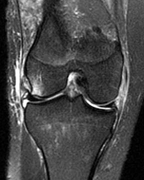
5 CORONAL CUTS Collaterals Meniscus Cartilage

6 AXIAL CUTS PATELLO-FEMORAL PROBLEMS

7 Meniscus ACL PCL Collateral ligaments Patello femoral Chondral defects Misc. MRI Knee

 appear as sharp points of the triangle facing")
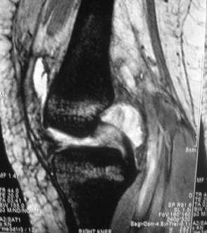
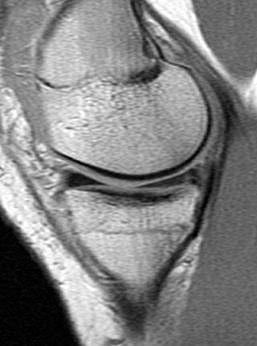
8 Meniscus Normal anatomy The normal meniscus shows uniform, low signal intensity (Black) Peripherally the meniscus have a bow- tie configuration Centrally, the normal meniscus is composed of 2 separate triangular structures, the anterior horn and the posterior horn. The apices (free edges or inner margins) appear as sharp points of the triangle facing each other.
9 MRI grading system for Meniscal Degeneration GRADE 1 & 2, NOT SIGNIFICANT GRADE 3, SIGNIFICANT MENISCUS TEAR
10 Types of Meniscal Tear Three basic meniscal tear Longitudinal Radial Horizontal


11 Longitudinal Tears Longitudinal tears separate the meniscus into inner and outer fragments and occur parallel to the outer margin of the meniscus are perpendicular to the tibial plateau and propagate parallel to the circumferential axis of the meniscus
")


12 Radial tears ( Transverse Tears) These are vertical tear and propagate perpendicular to the main axis of the meniscus

 portion and a bottom (")

13 Horizontal tear These tears are also called cleavage or fish- mouth tears They divide the meniscal tear into a top ( superior) portion and a bottom ( inferior) portion Most are degenerative, occurring in older patients with osteoarthritis.


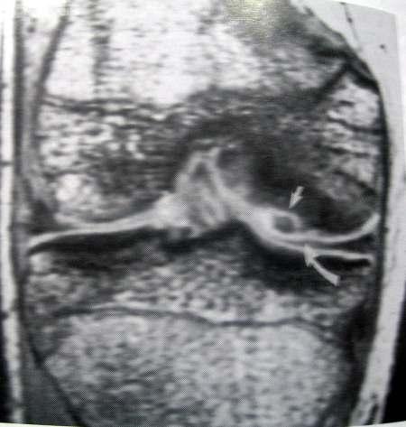
14 Bucket- Handle Tears These tears are displaced vertical longitudinal tears and usually involve the MM. The separated central ( inner) fragment, when viewed axially, resembles the handle of a bucket The remaining larger peripheral portion of the meniscus resembles the bucket. Typical locations of the displaced fragment include the intercondylar notch anterior and parallel to the PCL ( double PCL sign)


15 MRI criteria for meniscal tears Two MRI criteria have been established for diagnosing meniscal tears. Criteria 1 Criteria 1 is increased internal signal intensity in the meniscus The abnormal signal intensity must be in contact with one articular surface, either the superior or interior surface or at the tip ( free edge) of the meniscus Criteria 2 Criterion 2 is an abnormal meniscal shape
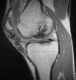
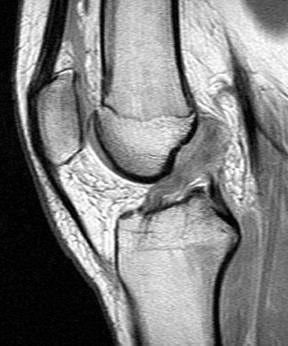
16 ACL The normal ACL appears on sagittal images as a solid band or as a striated band diverging slightly, ruler straight although mild sagging convex inferiorly can be present, especially with mild knee flexion. The ACL shows low- to intermediate signal intensity, higher than that of the PCL. ACL Tear Primary Signs Secondary Signs


17 Primary signs of the ACL tear Disruption, increased signal intensity in the substance of the ACL on T2 weighted images, abrupt angulation or wavy appearance and abnormal ACL axis. Nonvisualization or near nonvisualization with replacement be a cloud of focal edema and hemorrahage.
18 Secondary Signs of ACL Tear Pivot - shift bone bruises and osteochondral fracture Anterior translocation of the tibia Segond fracture Anterior tibial spine fractures Buckling of PCL


19 Secondary Signs of ACL Tear Pivot- Shift Bone Bruises and Fractures Anterior Translocation of the Tibia


20 Secondary Signs of ACL Tear Avulsion fracture of the tibia at ACL insertion Buckling PCL Segond Fracture


21 PCL Major stabilizer of knee Restrain against post. displacement Isolated 3% Combined 97% ACL injury - 65% MCL 50% Medial Meniscus 30% Posterior capsule and fibular collateral ligament


22 MRI Finding Intrasubstantial tear Partial tear Complete tear Avulsions

23 Collateral Ligaments Coronal images with anatomy defining and fluid-sensitive sequences demonstrate the medial and lateral supporting structures optimally. Additional useful information can be gleaned from sagittal and axial images of these structures


24 Chondral Defects Degenerative cartilage defects Osteochondritis dissecans Chondromalacia patellae
25 Osteochondritis Dissecans
26 Chondromalacia Patellae
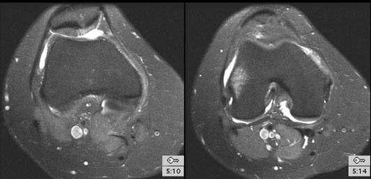
27 Meniscal cysts Ganglion cyst Discoid meniscus Infections PVNS Miscellaneous Extensor mechanism tear Quadriceps tendon tear Patellar tendon tear Tibial tuberosity avulsion

28 Meniscal Cysts Ganglion Cysts

29 Discoid Meniscus


30 INFECTION Tubercular Hypointense Synovial proliferation Marrow Edema Osteomyelitis Abcess Cortical erosion Sinuses


31 PVNS Low signal changes Synovial proliferation Hemosiderin deposition


32 Extensor Mechanism Tear Quadriceps Tendon Tear Patellar Tendon Tear Tibial Tuberosity Avulsion
33 MRI Very useful tool in hands of Radiologists & Orthopedicians Orthopedicians probably in a better position than Radiolologist to read MRI

34 Visit Thank You
35


36 MCL Tear Grade I Microscopic tear Grade II Partial tear Grade III Complete tear
37 LCL LCL tear is seen as a serpiginous or lax ligament with discontinuous fibers (or avulsed fibular head), often without significant thickening of the ligament. LCL tears rarely are isolated, and an LCL tear becomes more likely as associated PLC and cruciate ligament injuries increase in severity
38 Studies reported in diagnostic arthroscopy 51% (With use of MRI) MRI accuracy in Meniscus & ACL Pathology > 90% Difference in radiologist & arthroscopist opinion in meniscal injury because Different radiologist are at different level of the learning curve and different arthroscopist have different level of experience

39 Partial ACL Tear Partial tears of the ACL are common, accounting for 10-43% of all ACL tears Direct signs may include focal angulation of the ACL or partial thickness focal high signal intensity. T2- weighted images allow more confident identification of abnormal focal high signal intensity in the ACL substance ( stoller, 1997).

40 MRI grading system for Meniscal Degeneration Grade I Grade I is a nonarticular, focal or diffuse region of increased signal intensity within the substance of the meniscus. Grade 2 Grade 2 is a horizontal, linear area of increased signal intensity within the substance of the meniscus that extends to but does not involve the articular surface.. Patients are usually asymptomatic
41 Meniscal Degeneration Grade 3 Is a region of abnormal signal intensity within the meniscus extending to and communicating with at least 1 articular surface of the meniscus Clinically Significant
Imaging the Knee 17/10/2017. Friction syndrome Common in runners or cyclists Fluid between ITB and Lateral femoral condyle
 17/10/2017 Imaging the Knee Alicia M. Yochum RN, DC, DACBR, RMSK Iliotibial Band Syndrome Ligamentous Tears (ACL, PCL, MCL, LCL) Meniscal Tears Cartilage Degeneration Quadriceps/Patellar tendinosis Osteochondral
This presentation is the intellectual property of the author. Contact them for permission to reprint and/or distribute.
 MRI of the Knee Jennifer Swart, M.D. Musculoskeletal Radiology South Texas Radiology Group Outline Coils, Patient Positioning Acquisition Parameters, Planes and Pulse Sequences Knee Arthrography Normal
This presentation is the intellectual property of the author. Contact them at for permission to reprint and/or distribute.
 MRI of the Knee Jennifer Swart, M.D. Musculoskeletal Radiology South Texas Radiology Group Financial Disclosure Dr. Jennifer Swart has no relevant financial relationships with commercial interests to disclose.
Imaging the Athlete s Knee. Peter Lowry, MD Musculoskeletal Radiology University of Colorado
 Imaging the Athlete s Knee Peter Lowry, MD Musculoskeletal Radiology University of Colorado None Disclosures Knee Imaging: Radiographs Can be performed weight-bearing or non-weight-bearing View options
MRI of the Knee: Part 2 - menisci. Mark Anderson, M.D. University of Virginia Health System
 MRI of the Knee: Part 2 - menisci Mark Anderson, M.D. University of Virginia Health System Learning Objectives At the end of the presentation, each participant should be able to: describe the normal anatomy
Knee: Meniscus Back to Basics
 Knee: Meniscus Back to Basics Kyung Jin Suh kyungjin.suh@gmail.com Doctor Radiology, Daegu, KOREA Medial Lateral 7.7 10.2 11.6 9.6 10.6 mm Posterior > Anterior horn 10.6 mm Posterior = Anterior horn Medial
Knee MRI Update Case Review 2009 Russell C. Fritz, M.D. National Orthopedic Imaging Associates San Francisco, CA
 Knee MRI Update Case Review 2009 Russell C. Fritz, M.D. National Orthopedic Imaging Associates San Francisco, CA Meniscal Tears -linear increased signal extending to an articular surface is the hallmark
ACL AND PCL INJURIES OF THE KNEE JOINT
 ACL AND PCL INJURIES OF THE KNEE JOINT Dr.KN Subramanian M.Ch Orth., FRCS (Tr & Orth), CCT Orth(UK) Consultant Orthopaedic Surgeon, Special interest: Orthopaedic Sports Injury, Shoulder and Knee Surgery,
MENISCAL INJURY. Meniscus. Anterior Roots. Medial Meniscus. Lateral Meniscus. Posterior Roots. MRI and Arthroscopic Findings
 Meniscus Anterior Roots MENISCAL INJURY MRI and Arthroscopic Findings Medial Meniscus AH PH PH AH Lateral Meniscus Rawiwan Pattaweerakul Naresuan University Hospital Posterior Roots Meniscus Normal Meniscus
and K n e e J o i n t Is the most complicated joint in the body!!!!
 K n e e J o i n t K n e e J o i n t Is the most complicated joint in the body!!!! 1-Consists of two condylar joints between: A-The medial and lateral condyles of the femur and The condyles of the tibia
Knee Joint Anatomy 101
 Knee Joint Anatomy 101 Bone Basics There are three bones at the knee joint femur, tibia and patella commonly referred to as the thighbone, shinbone and kneecap. The fibula is not typically associated with
Anterior Cruciate Ligament Surgery
 Anatomy Anterior Cruciate Ligament Surgery Roger Ostrander, MD Andrews Institute Anatomy Anatomy Function Primary restraint to anterior tibial translation Secondary restraint to internal tibial rotation
The Knee. Two Joints: Tibiofemoral. Patellofemoral
 Evaluating the Knee The Knee Two Joints: Tibiofemoral Patellofemoral HISTORY Remember the questions from lecture #2? Girth OBSERVATION TibioFemoral Alignment What are the consequences of faulty alignment?
Knee Contusions and Stress Injuries. Laura W. Bancroft, M.D.
 Knee Contusions and Stress Injuries Laura W. Bancroft, M.D. Objectives Review 5 types of contusion patterns Pivot shift Dashboard Hyperextension Clip Lateral patellar dislocation Demonstrate various stress
MRI Evaluation of Internal derangement of the Knee Joint - A PICTORIAL REVIEW
 MRI Evaluation of Internal derangement of the Knee Joint - A PICTORIAL REVIEW Poster No.: C-1386 Congress: ECR 2015 Type: Educational Exhibit Authors: V. Nadaraja, F. Abubacker Sulaiman; Chennai/IN Keywords:
Meniscal Tears with Fragments Displaced: What you need to know.
 Meniscal Tears with Fragments Displaced: What you need to know. Poster No.: C-1339 Congress: ECR 2015 Type: Authors: Keywords: DOI: Educational Exhibit M. V. Ferrufino, A. Stroe, E. Cordoba, A. Dehesa,
Knee: Cruciate Ligaments
 72 Knee: Cruciate Ligaments R. Kent Sanders Sagittal oblique 2.5-mm sequences along the plane of the anterior cruciate ligament (ACL) typically yield three to four images of the ACL, with the first medial
ADVANCED IMAGING OF THE KNEE
 MENISCAL ANATOMY ADVANCED IMAGING OF THE KNEE MENISCAL ABNORMALITIES MENISCAL FUNCTION MENISCAL FUNCTION load transmission shock absorption stability The menisci DO NOT function as primary stabilizers
BASELINE QUESTIONNAIRE (SURGEON)
") SECTION A: STUDY INFORMATION Subject ID: - - Study Visit: Baseline Site Number: Date: / / Surgeon ID: SECTION B: INITIAL SURGEON HISTORY B1. Previous Knee Surgery: Yes No Not recorded B2. Number of Previous
SECTION A: STUDY INFORMATION Subject ID: - - Study Visit: Baseline Site Number: Date: / / Surgeon ID: SECTION B: INITIAL SURGEON HISTORY B1. Previous Knee Surgery: Yes No Not recorded B2. Number of Previous
Knee Injury Assessment
 Knee Injury Assessment Clinical Anatomy p. 186 Femur Medial condyle Lateral condyle Femoral trochlea Tibia Intercondylar notch Tibial tuberosity Tibial plateau Fibula Fibular head Patella Clinical Anatomy
ESMRMB. School of MRI Advanced MR Imaging of the Musculoskeletal System. November 10-12, 2016 Menton/FR
 School of MRI 2016 Advanced MR Imaging of the Musculoskeletal System November 10-12, 2016 Menton/FR Topic 5 Knee L. Sconfienza io@lucasconfienza.it THE KNEE Luca Maria Sconfienza, MD PhD Unit of Diagnostic
ASSESSMENT AND MANAGEMENT OF THE KNEE AND LOWER LIMB.
 ASSESSMENT AND MANAGEMENT OF THE KNEE AND LOWER LIMB www.fisiokinesiterapia.biz Overview History Examination X-rays Fractures and Dislocations. Soft Tissue Injuries Other Knee/Lower limb Problems Anatomy
Multi-ligamentous knee injuries - MRI injury patterns at a glance
 Multi-ligamentous knee injuries - MRI injury patterns at a glance Poster No.: P-0068 Congress: ESSR 2015 Type: Educational Poster Authors: A. Rastogi, D. Whelan, R. Martin, W. Mak, D. Pearce ; 1 1 1 2
ORIGINAL ARTICLE. ROLE OF MRI IN EVALUATION OF TRAUMATIC KNEE INJURIES Saurabh Chaudhuri, Priscilla Joshi, Mohit Goel
 ROLE OF MRI IN EVALUATION OF TRAUMATIC KNEE INJURIES Saurabh Chaudhuri, Priscilla Joshi, Mohit Goel 1. Associate Professor, Department of Radiodiagnosis & imaging, Bharati Vidyapeeth Medical College and
ACL Athletic Career. ACL Rupture - Warning Features Intensive pain Immediate swelling Locking Feel a Pop Dead leg Cannot continue to play
 FIMS Ambassador Tour to Eastern Europe, 2004 Belgrade, Serbia Montenegro Acute Knee Injuries - Controversies and Challenges Professor KM Chan OBE, JP President of FIMS Belgrade ACL Athletic Career ACL
MRI of ligaments. Ligament biomechanics Spine Shoulder Elbow Hand/wrist Pelvis/hip Knee Foot/ankle
 MRI of ligaments Chang Ho Kang M.D. Korea University Anam Hospital Spine Shoulder Elbow Hand/wrist Pelvis/hip Knee Foot/ankle Introduction Ligament Fibrous connective tissue Attaches bone to bone Holds
Role of magnetic resonance imaging in the evaluation of traumatic knee joint injuries
 Original Research Article Role of magnetic resonance imaging in the evaluation of traumatic knee joint injuries Dudhe Mahesh 1*, Rathi Varsha 2 1 Resident, 2 Professor, Department of Radio-Diagnosis, Grant
Unlocking the locked Knee
 Unlocking the locked Knee Poster No.: P-0027 Congress: ESSR 2013 Type: Scientific Exhibit Authors: J. P. SINGH, S. Srivastava, S. S. BAIJAL ; Gurgaon, Delhi 1 1 2 1 2 NCR/IN, LUCKNOW, UTTAR PRADESH/IN
Differential Diagnosis
 Case 31yo M who sustained an injury to L knee while playing Basketball approximately 2 weeks ago. He describes pivoting and hyperextending his knee, which swelled over the next few days. He now presents
Overview Ligament Injuries. Anatomy. Epidemiology Very commonly injured joint. ACL Injury 20/06/2016. Meniscus Tears. Patellofemoral Problems
 Overview Ligament Injuries Meniscus Tears Pankaj Sharma MBBS, FRCS (Tr & Orth) Consultant Orthopaedic Surgeon Manchester Royal Infirmary Patellofemoral Problems Knee Examination Anatomy Epidemiology Very
Meniscal tears on 3T MR: Patterns, pearls and pitfalls
 Meniscal tears on 3T MR: Patterns, pearls and pitfalls Poster No.: C-2221 Congress: ECR 2010 Type: Educational Exhibit Topic: Musculoskeletal Authors: J. C. Kandathil; Singapore/SG Keywords: Knee injuries,
The Impact of Age on Knee Injury Treatment
 The Impact of Age on Knee Injury Treatment Focus on the Meniscus Dr. Alvin J. Detterline, MD Sports Medicine and Orthopaedic Surgery Towson Orthopaedic Associates University of Maryland St. Joseph Medical
The Meniscus. History. Anatomy. Anatomy. Blood Supply. Attachments
 History The Meniscus W. Randall Schultz, MD, MS Austin, TX January 23, 2016 Meniscus originally thought to represent vestigial tissue 1883 first reported meniscal repair (Annandale) Total menisectomy treatment
Baqueira-Beret Medical Center STATISTICAL ANALYSIS OF ACL INJURIES IN SNOW SPORT ACCIDENTS
 Baqueira-Beret Medical Center STATISTICAL ANALYSIS OF ACL INJURIES IN SNOW SPORT ACCIDENTS Baqueira Beret 2005-2015 Main data Data From 2005 to 2015, Baqueira-Beret received more than 6,7 million visitors
40 th Annual Symposium on Sports Medicine. Knee Injuries In The Pediatric Athlete. Disclosure
 40 th Annual Symposium on Sports Medicine Travis Murray, MD Assistant Professor University of Texas Health Science Center San Antonio Knee Injuries In The Pediatric Athlete Disclosure Dr. Travis Murray
MRI SHOULDER WHAT TO SEE
 MRI SHOULDER WHAT TO SEE DR SHEKHAR SRIVASTAV Sr. Consultant- Knee & Shoulder Arthroscopy Sant Parmanand Hospital Normal Anatomy Normal Shoulder MRI Coronal Oblique Sagital Oblique Axial Cuts Normal Coronal
On Field Assessment and Management of Acute Knee Injuries: A Physiotherapist s Perspective
 On Field Assessment and Management of Acute Knee Injuries: A Physiotherapist s Perspective Jessica Condliffe Physiotherapist / Clinic Manager TBI Health Wellington Presentation Outline Knee anatomy review
Objectives. The BIG Joint. Case 1. Boney Architecture. Presenter Disclosure Information. Common Knee Problems
 3:30 4:15 pm Common Knee Problems SPEAKER Christopher J. Visco, MD Presenter Disclosure Information The following relationships exist related to this presentation: Christopher J. Visco, MD: Speaker s Bureau
SOFT TISSUE INJURIES OF THE KNEE: Primary Care and Orthopaedic Management
 SOFT TISSUE INJURIES OF THE KNEE: Primary Care and Orthopaedic Management Gauguin Gamboa Australia has always been a nation where emphasis on health and fitness has resulted in an active population engaged
42 nd Annual Symposium on Sports Medicine. Knee Injuries In The Pediatric Athlete. Disclosure
 42 nd Annual Symposium on Sports Medicine Travis Murray, MD Assistant Professor University of Texas Health Science Center San Antonio January 23, 2015 Knee Injuries In The Pediatric Athlete Disclosure
KNEE EXAMINATION. Tips & Tricks from an Emergency Physician Perspective. EM Physicians Less Exposed to MSK Medicine
 KNEE EXAMINATION Tips & Tricks from an Emergency Physician Perspective Dr P O CONNOR Emergency Medicine Physician EUSEM 10/09/2018 EM Physicians Less Exposed to MSK Medicine Musculoskeletal Medicine becoming
Role of Magnetic Resonance Imaging in Patients with Knee Trauma
 Original Research Article Role of Magnetic Resonance Imaging in Patients with Knee Trauma Bhautik Kapadia 1, Bhumika Suthar 2* 1 Associate Professor, 2 Assistant Professor, Department of Radiodiagnosis,
Original Report. The Reverse Segond Fracture: Association with a Tear of the Posterior Cruciate Ligament and Medial Meniscus
 Eva M. Escobedo 1 William J. Mills 2 John. Hunter 1 Received July 10, 2001; accepted after revision October 1, 2001. 1 Department of Radiology, University of Washington Harborview Medical enter, 325 Ninth
Biomechanics of the Knee. Valerie Nuñez SpR Frimley Park Hospital
 Biomechanics of the Knee Valerie Nuñez SpR Frimley Park Hospital Knee Biomechanics Kinematics Range of Motion Joint Motion Kinetics Knee Stabilisers Joint Forces Axes The Mechanical Stresses to which
The Knee. Prof. Oluwadiya Kehinde
 The Knee Prof. Oluwadiya Kehinde www.oluwadiya.sitesled.com The Knee: Introduction 3 bones: femur, tibia and patella 2 separate joints: tibiofemoral and patellofemoral. Function: i. Primarily a hinge joint,
Case study #11 Rt. knee
 The patient is a 55 year old female who presents with bilateral knee pain. Patient is a collegiate softball coach and has a very active lifestyle and career that is hampered by her chronic knee pain. She
The Egyptian Journal of Hospital Medicine (July 2018) Vol. 72 (10), Page
 Vol. 72 (10), Page") The Egyptian Journal of Hospital Medicine (July 2018) Vol. 72 (10), Page 5490-5494 The Role of Ultrasound in Evaluation of Meniscal Injury Nada Sayed Mahdy*, Hossam Mousa Sakr, Allam Elsayed Allam Radiology
The Egyptian Journal of Hospital Medicine (July 2018) Vol. 72 (10), Page 5490-5494 The Role of Ultrasound in Evaluation of Meniscal Injury Nada Sayed Mahdy*, Hossam Mousa Sakr, Allam Elsayed Allam Radiology
Do Not Fall on Your Knees - Recognizing Common and Uncommon Pitfalls that May Simulate Meniscal Tears
 Do Not Fall on Your Knees - Recognizing Common and Uncommon Pitfalls that May Simulate Meniscal Tears Poster No.: C-1146 Congress: ECR 2016 Type: Educational Exhibit Authors: P. Musa Aguiar, J. Goncalves,
Knee Joint Assessment and General View
 Knee Joint Assessment and General View Done by; Mshari S. Alghadier BSc Physical Therapy RHPT 366 m.alghadier@sau.edu.sa http://faculty.sau.edu.sa/m.alghadier/ Functional anatomy The knee is the largest
In the name of god. Knee. By: Tofigh Bahraminia Graduate Student of the Pathology Sports and corrective actions. Heat: Dr. Babakhani. Nov.
 In the name of god Knee By: Tofigh Bahraminia Graduate Student of the Pathology Sports and corrective actions Heat: Dr. Babakhani Nov. 2014 1 Anatomy-Bones Bones Femur Medial/lateral femoral condyles articulate
MENISCAL INJURIES. (copyright s h palmer 2009) MENISCAL FUNCTION
 MENISCAL FUNCTION") (copyright s h palmer 2009) MENISCAL FUNCTION MENISCAL INJURIES Menisci are important for weight bearing, load distribution, joint stability and proprioception. Figure 1: A normal medial meniscus Any load
(copyright s h palmer 2009) MENISCAL FUNCTION MENISCAL INJURIES Menisci are important for weight bearing, load distribution, joint stability and proprioception. Figure 1: A normal medial meniscus Any load
Hip joint Type: Articulating bones:
 Ana (242 ) Hip joint Type: Synovial, ball & socket Articulating bones: Formed between head of femur and lunate surface of acetabulum of hip bone. Capsule: it is a strong fibrous sleeve connecting the articulating
The Knee. Tibio-Femoral
 The Knee Tibio-Femoral Osteology Distal Femur with Proximal Tibia Largest Joint Cavity in the Body A modified hinge joint with significant passive rotation Technically, one degree of freedom (Flexion/Extension)
a - CM EARTICLE Clinics in diagnostic imaging (139) !Medical Education Singapore Med J 2012; 53(4) CASE PRESENTATION
 !Medical Education Singapore Med J 2012; 53(4) CASE PRESENTATION") !Medical Education Singapore Med J 2012; 53(4) 283 CM EARTICLE Clinics in diagnostic imaging (139) Wei Yang Liml, MBBS, FRCR, Nor Azam Mahmud2, MD, MMed, Wilfred CG Peh3, FRCP, FRCR a - - - Fig. 1 Sagittal
!Medical Education Singapore Med J 2012; 53(4) 283 CM EARTICLE Clinics in diagnostic imaging (139) Wei Yang Liml, MBBS, FRCR, Nor Azam Mahmud2, MD, MMed, Wilfred CG Peh3, FRCP, FRCR a - - - Fig. 1 Sagittal
MRI of LEFT KNEE. There is a fluid collection seen anterior to and inferior to the superiorly displaced patella.
 MRI of LEFT KNEE Protocol: Multiplanar MRI of the left knee joint performed in the sagittal, coronal and transverse planes using T1 weighted spin echo, T2 and proton-density weighted fast spin echo, fatsaturated
Ligamentous and Meniscal Injuries: Diagnosis and Management
 Ligamentous and Meniscal Injuries: Diagnosis and Management Daniel K Williams, MD Franciscan Physician Network Orthopedic Specialists September 29, 2017 No Financial Disclosures INTRODUCTION Overview of
Imaging the musculoskeletal system. An Introduction
 Imaging the musculoskeletal system An Introduction Objectives Discuss: commonly used imaging modalities in the musculoskeletal system normal imaging anatomy in the extremities fracture description Imaging
Post-injury painful and locked knee
 H R J Post-injury painful and locked knee, p. 54-59 Clinical Case - Test Yourself Musculoskeletal Imaging Post-injury painful and locked knee Ioannis I. Daskalakis 1, 2, Apostolos H. Karantanas 1, 2 1
Physical Examination of the Knee
 History: Pain Traumatic vs. atraumatic? Acute vs Chronic Previous procedures done on the knee? Swelling, catching, instability General Setup Examine standing, sitting and supine Evaluate gait Examine hip
Available online at
 Original Research Article Evaluation of knee joint by MRI in 65 patients Gulamus sibtain asad *, Himanshu Singla, Ankit vasoya, P. J. Jhala 2 2 nd year Resident, 2 Professor Radiology Department, SBKS
What is the most effective MRI specific findings for lateral meniscus posterior root tear in ACL injuries
 What is the most effective MRI specific findings for lateral meniscus posterior root tear in ACL injuries Kazuki Asai 1), Junsuke Nakase 1), Kengo Shimozaki 1), Kazu Toyooka 1), Hiroyuki Tsuchiya 1) 1)
UNIT 7 JOINTS. Knee and Ankle Joints DR. ABDEL-MONEM A. HEGAZY
 UNIT 7 JOINTS Knee and Ankle Joints BY DR. ABDEL-MONEM A. HEGAZY (Degree in Bachelor of Medicine and Surgery with honor 1983, Dipl."Gynaecology and Obstetrics "1989, Master "Anatomy and Embryology "1994,
Physical Examination of the Knee
 History: Pain Traumatic vs. atraumatic Acute vs Chronic Mechanism of injury Swelling, catching, instability Previous evaluation and treatment General Setup Examine standing, sitting and supine Evaluate
The Knee Joint By Prof. Dr. Muhammad Imran Qureshi
 The Knee Joint By Prof. Dr. Muhammad Imran Qureshi Structurally, it is the Largest and the most complex joint in the body because of the functions that it performs: Allows mobility (flexion/extension)
The examination of the painful knee. Maja K Artandi, MD, FACP Clinical Associate Professor of Medicine Stanford University
 The examination of the painful knee Maja K Artandi, MD, FACP Clinical Associate Professor of Medicine Stanford University Objectives of the talk By the end of this talk you will know The important anatomy
W. Dilworth Cannon, M.D. Professor of Clinical Orthopaedic Surgery University of California San Francisco
 Knee Pain And Injuries In Adults W. Dilworth Cannon, M.D. Professor of Clinical Orthopaedic Surgery University of California San Francisco Pain Control Overview Narcotics rarely necessary after 1 st 1-2
Meniscal Tears: Role of Axial MRI Alone and in Combination with Other Imaging Planes
 Nefise Cagla Tarhan 1,2 Christine. Chung 1 urea Valeria Rosa Mohana-orges 1 Tudor Hughes 1 Donald Resnick 1 Received September 30, 2003; accepted after revision February 2, 2004. 1 Department of Radiology,
ANTERIOR CRUCIATE LIGAMENT INJURY
 ANTERIOR CRUCIATE LIGAMENT INJURY WHAT IS THE ANTERIOR CRUCIATE LIGAMENT? The anterior cruciate ligament (ACL) is one of four major ligaments that stabilizes the knee joint. A ligament is a tough band
Medical Practice for Sports Injuries and Disorders of the Knee
 Sports-Related Injuries and Disorders Medical Practice for Sports Injuries and Disorders of the Knee JMAJ 48(1): 20 24, 2005 Hirotsugu MURATSU*, Masahiro KUROSAKA**, Tetsuji YAMAMOTO***, and Shinichi YOSHIDA****
Spectrum of meniscal lesions: An MR teaching atlas
 Spectrum of meniscal lesions: An MR teaching atlas Poster No.: C-2284 Congress: ECR 2010 Type: Educational Exhibit Topic: Musculoskeletal Authors: C. Leal, P. Alves, H. Tinto, J. Raposo, T. Bilhim, A.
ULTRASOUND EVALUATION OF NORMAL AND ABNORMAL POSTERIOR CRUCIATE LIGAMENT - A PROSPECTIVE STUDY
 ORIGINAL ARTICLE ULTRASOUND EVALUATION OF NORMAL AND ABNORMAL POSTERIOR CRUCIATE LIGAMENT - A PROSPECTIVE STUDY Palle Lalitha, 1 M. Ch. Balaji Reddy, 1 K. Jagannath Reddy, 1 Vijaya Kumari 2 1 Department
MY PATIENT HAS KNEE PAIN. David Levi, MD Chief, Division of Musculoskeletal l limaging Atlantic Medical Imaging
 MY PATIENT HAS KNEE PAIN David Levi, MD Chief, Division of Musculoskeletal l limaging Atlantic Medical Imaging Causes of knee pain Non traumatic Trauma Osteoarthritis Patellofemoral pain Menisci or ligaments
MRI of Bucket-Handle Te a rs of the Meniscus of the Knee 1
 MRI of ucket-handle Te a rs of the Meniscus of the Knee 1 Joon Yong Park, M.D., Young-uk Lee M.D., Eun-Chul Chung M.D., Hae-Won Park M.D., E u n - Kyung Youn M.D., Shin Ho Kook, M.D., Young Rae Lee, M.D.
Arthroscopy / MRI Correlation Conference. Department of Radiology, Section of MSK Imaging Department of Orthopedic Surgery 7/19/16
 Arthroscopy / MRI Correlation Conference Department of Radiology, Section of MSK Imaging Department of Orthopedic Surgery 7/19/16 Case 1: 29 YOM with recurrent shoulder dislocations Glenoid Axial T1FS
Spectrum of meniscal lesions: An MR teaching atlas
 Spectrum of meniscal lesions: An MR teaching atlas Poster No.: C-2284 Congress: ECR 2010 Type: Educational Exhibit Topic: Musculoskeletal - Joints Authors: C. Leal, P. Alves, H. A. M. R. Tinto, J. Raposo,
Anatomy and Sports Injuries of the Knee
 Anatomy and Sports Injuries of the Knee I. Anatomy II. Assessment III. Treatment IV. Case Study V. Dissection Anatomy Not a hinge joint 6 degrees of freedom Flexion/Extension Rotation Translation Anatomy
MRI grading of postero-lateral corner and anterior cruciate ligament injuries
 MRI grading of postero-lateral corner and anterior cruciate ligament injuries Poster No.: C-2533 Congress: ECR 2012 Type: Educational Exhibit Authors: J. Lopes Dias, J. A. Sousa Pereira, L. Fernandes,
FieldStrength. Achieva 3.0T enables cutting-edge applications, best-in-class MSK images
 FieldStrength Publication for the Philips MRI Community Issue 33 December 2007 Achieva 3.0T enables cutting-edge applications, best-in-class MSK images Palo Alto Medical Clinic Sports Medicine Center employs
Joints of the Lower Limb II
 Joints of the Lower Limb II Lecture Objectives Describe the components of the knee and ankle joint. List the ligaments associated with these joints and their attachments. List the muscles acting on these
JMSCR Vol 05 Issue 01 Page January
 www.jmscr.igmpublication.org Impact Factor 5.244 Index Copernicus Value: 83.27 ISSN (e)-2347-176x ISSN (p) 2455-0450 DOI: https://dx.doi.org/10.18535/jmscr/v5i1.28 Diagnostic Accuracy of Magnetic Resonance
The posterolateral corner of the knee: the normal and the pathological
 The posterolateral corner of the knee: the normal and the pathological Poster No.: P-0104 Congress: ESSR 2014 Type: Educational Poster Authors: M. Bartocci 1, C. Dell'atti 2, E. Federici 1, V. Martinelli
I have nothing to disclose
 Management of Common Knee Disorders: What You Knee d to Know UCSF Essentials of Women s Health July 8, 2015 Carlin Senter, M.D. I have nothing to disclose Learning objectives: in 1 hour you will be able
Standardised. knee. scanning of the. Basic pathology. Nemanja Damjanov. University of Belgrade Institute of Rheumatology
 Standardised scanning of the Nemanja Damjanov University of Belgrade Institute of Rheumatology knee Basic pathology Disclosure Lecturer: Pfizer, Abbvie, Roche, MSD, Boehringer-Ingelheim, Gedeon Richter,
CMEArticle Clinics in diagnostic imaging (141)
") Singapore Med J 2012; 53(9) : 625 CMEArticle Clinics in diagnostic imaging (141) Hollie MY Lim 1, MBBS, FRCR, Wilfred CG Peh 1, FRCP, FRCR 1a 1b 1c Fig. 1 (a & b) Contiguous sagittal fat-suppressed PD-W
Singapore Med J 2012; 53(9) : 625 CMEArticle Clinics in diagnostic imaging (141) Hollie MY Lim 1, MBBS, FRCR, Wilfred CG Peh 1, FRCP, FRCR 1a 1b 1c Fig. 1 (a & b) Contiguous sagittal fat-suppressed PD-W
Comparative study of high resolusion ultrasonography and magnetic resonance imaging in diagnosing traumatic knee injuries & pathologies
 Original article: Comparative study of high resolusion ultrasonography and magnetic resonance imaging in diagnosing traumatic knee injuries & pathologies Dr. Rakesh Gujjar*, Dr. R. P. Bansal, Dr. Sandeep
Ultrasound of the Knee
 Ultrasound of the Knee Jon A. Jacobson, M.D. Professor of Radiology Director, Division of Musculoskeletal Radiology University of Michigan Disclosures: Consultant: Bioclinica Book Royalties: Elsevier Advisory
Goals &Objectives. 1. Review the anatomy of the knee 2. Practice your hands-on skills 3. By the end of the workshop:
 Clinical Knee Exam Goals &Objectives 1. Review the anatomy of the knee 2. Practice your hands-on skills 3. By the end of the workshop: Be able to categorize knee injuries Understand the significance of
To describe he knee joint, ligaments, structure & To list the main features of other lower limb joints
 To describe he knee joint, ligaments, structure & neurovascular supply To demonstrate the ankle joint anatomy To list the main features of other lower limb joints To list main groups of lymph nodes in
RN(EC) ENC(C) GNC(C) MN ACNP *** MECHANISM OF INJURY.. MOST IMPORTANT *** - Useful in determining mechanism of injury / overuse
 ENC(C) GNC(C) MN ACNP *** MECHANISM OF INJURY.. MOST IMPORTANT *** - Useful in determining mechanism of injury / overuse") HISTORY *** MECHANISM OF INJURY.. MOST IMPORTANT *** Age of patient Sport / Occupation - Certain conditions are more prevalent in particular age groups (Osgood Schlaters in youth / Degenerative Joint Disease
HISTORY *** MECHANISM OF INJURY.. MOST IMPORTANT *** Age of patient Sport / Occupation - Certain conditions are more prevalent in particular age groups (Osgood Schlaters in youth / Degenerative Joint Disease
The Knee. Clarification of Terms. Osteology of the Knee 7/28/2013. The knee consists of: The tibiofemoral joint Patellofemoral joint
 The Knee Clarification of Terms The knee consists of: The tibiofemoral joint Patellofemoral joint Mansfield, p273 Osteology of the Knee Distal Femur Proximal tibia and fibula Patella 1 Osteology of the
A Patient s Guide to Knee Anatomy. Stephanie E. Siegrist, MD, LLC
 A Patient s Guide to Knee Anatomy Hands, shoulders, knees and toes (and elbows and ankles, too!) Most bone and joint conditions have several treatment options. The best treatment for you is based on your
Mucoid degeneration of the posterior cruciate ligament
 Mucoid degeneration of the posterior cruciate ligament Poster No.: C-2278 Congress: ECR 2010 Type: Educational Exhibit Topic: Musculoskeletal - Joints Authors: P. Papadopoulou, I. Kalaitzoglou, I. Tsifoundoudis,
LATERAL MENISCUS SLOPE AND ITS CLINICAL RELEVANCE IN PATIENTS WITH A COMBINED ACL TEAR AND POSTERIOR TIBIA COMPRESSION
 LATERAL MENISCUS SLOPE AND ITS CLINICAL RELEVANCE IN PATIENTS WITH A COMBINED ACL TEAR AND POSTERIOR TIBIA COMPRESSION R. ŚMIGIELSKI, B. DOMINIK, U, ZDANOWICZ, Z. GAJEWSKI, K. SKIERBISZEWSKA, K. SIEWRUK,
Exercise Science Section 4: Joint Mechanics and Joint Injuries
 Exercise Science Section 4: Joint Mechanics and Joint Injuries An Introduction to Health and Physical Education Ted Temertzoglou Paul Challen ISBN 1-55077-132-9 Types of Joints Fibrous joint Cartilaginous
Evaluation of Role of Magnetic Resonance Imaging in Knee Joint Injuries in Correlation with Arthroscopy
 Original Article Print ISSN: 2321-6379 Online ISSN: 2321-595X DOI: 10.17354/ijss/2017/442 Evaluation of Role of Magnetic Resonance Imaging in Knee Joint Injuries in Correlation with Arthroscopy T Sundara
Figure 3 Figure 4 Figure 5
 Figure 1 Figure 2 Begin the operation with examination under anesthesia to confirm whether there are any ligamentous instabilities in addition to the posterior cruciate ligament insufficiency. In particular
Stability of Post Traumatic Osteochondritis Dissecans of the Knee: MR Imaging Findings
 Chin J Radiol 2005; 30: 199-204 199 Stability of Post Traumatic Osteochondritis Dissecans of the Knee: MR Imaging Findings YU-CHUNG HUNG 1 JON-KWAY HUANG 1,2 Department of Radiology 1, Mackay Memorial
Anterior Cruciate Ligament (ACL)
") Anterior Cruciate Ligament (ACL) The anterior cruciate ligament (ACL) is one of the 4 major ligament stabilizers of the knee. ACL tears are among the most common major knee injuries in active people of
Anterior Cruciate Ligament (ACL) The anterior cruciate ligament (ACL) is one of the 4 major ligament stabilizers of the knee. ACL tears are among the most common major knee injuries in active people of
MUSCULOSKELETAL IMAGING FOR PHYSICAL THERAPISTS. COMBINED SECTIONS MEETING 2006 San Diego, CA February 1-5, 2006
 MUSCULOSKELETAL IMAGING FOR PHYSICAL THERAPISTS COMBINED SECTIONS MEETING 2006 San Diego, CA February 1-5, 2006 John Meyer, DPT, OCS University of Southern California Department of Athletic Medicine Los