Ligamentous and Meniscal Injuries: Diagnosis and Management
|
|
|
- Ethel Cain
- 6 years ago
- Views:
Transcription

1 Ligamentous and Meniscal Injuries: Diagnosis and Management Daniel K Williams, MD Franciscan Physician Network Orthopedic Specialists September 29, 2017
2 No Financial Disclosures
3 INTRODUCTION Overview of pertinent anatomy Osseous anatomy Muscular anatomy Ligamentous anatomy Meniscal anatomy Applied physical examination Common topics and pearls

4 KNEE ANATOMY--OVERVIEW Comprised of four bones Distal femur Proximal tibia Proximal fibula Patella Femur converges toward midline to meet nearly vertical tibia Anatomical axis Meet around 5-12 degrees


5 MUSCLE GROUPS Quadriceps muscle group Rectus femoris Vastus lateralis Vastus medialis Vastus intermedius Quadriceps tendon Superior portion of extensor mechanism

6 Hamstrings Biceps femoris Semimembranosus Semitendinosus Primary knee flexors Hamstrings Sartorius Gracilis Popliteus gastrocnemius MUSCLE GROUPS

7 KNEE LIGAMENTS Medial collateral ligament Lateral collateral ligament Anterior cruciate ligament Posterior cruciate ligament Medial patellofemoral ligament


8 MCL Composed of superficial/deep portions Superficial MCL Proximal: posterior aspect of medial femoral condyle Distal: metaphyseal region of tibia Primary valgus restraint depending on flexion At 25º flexion, 80% restraint At 5º flexion, 60% restraint Deep MCL Separated by bursa Attaches directly into edge of tibial plateau and meniscus Does not contribute to valgus restraint


9 LCL Proximal: posterior lateral condyle Distal: lateral base of fibular head Conjoint tendon BFT Primary restraint to varus stress at 5º and 25º of flexion At full extension, resists 55% of varus load Isolated injury uncommon


10 ANTERIOR CRUCIATE LIGAMENT Femoral side: arises from posteromedial corner of medial aspect of lateral femoral condyle Tibial side: in fossa anterior and lateral to tibial spine Two bundles: named for tibial insertion Anteromedial tight in flexion Posterolateral tight in extension Provides anterior and rotational restraint of the tibia in relation to the femur


11 POSTERIOR CRUCIATE LIGAMENT Technically extraarticular Broad femoral origin at PL medial condyle Insertion centrally on posterior tibia Primary restraint to posterior tibial translation Secondary restraint to external tibial rotation

12 MEDIAL PATELLOFEMORAL LIGAMENT Distinct condensation of capsular fibers Originates from medial epicondyle and MCL Deep to VMO to attach to superior aspect of patella Provides 50% to 80% restraining force to lateral patella dislocation


13 MENISCI Medial and lateral Semilunar fibrocartilaginous disks Interposed b/t femoral and tibial condyles Counteract hoop stress with weight bearing Dissipates load to articular cartilage and subchondral bone Knee extension: 50% of axial load Knee flexion: 85% of axial load Joint stability/congruity Only 10% to 30% peripheral vascularized: lateral and medial genicular arteries
14 PHYSICAL EXAM Observation/alignment ROM Active Passive Normal ROM -5º to 143º in women Normal ROM -6º to 140º in men Palpation Tendonitis Joint line pain etc Strength Gait Specific physical exam maneuvers

15 OBSERVATION
16 ANTERIOR/POSTERIOR INSTABILITY Lachman s Test Anterior drawer Tibial drop back test Posterior drawer test


17 LACHMAN S TEST Assess ACL Knee 15º flexion Stabilize distal femur, anteriorly translate proximal tibia Quantify displacement in mm Soft vs hard endpoint Clinically Mild 0-5mm displacement Moderate 6-10 mm displacement Severe mm displacement


18 Assess ACL ANTERIOR DRAWER Flex knee to 80º Stabilize foot in neutral rotation Anterior force applied to tibia Compare to nl side

19 TIBIAL DROP BACK TEST Compare prominence of proximal tibia to femoral condyles with knees flexed 80º In nl knee, tibia 1 cm anterior to condyles Assess PCL


20 POSTERIOR DRAWER Most accurate for PCL integrity Knee flexed to 90 w/ posterior force on tibia Normal plateau 1 cm anterior to condyle Grade I: excessive translation but maintenance of anterior step-off Grade II: 5-10mm translation but not posterior to condyle Grade III: >10mm translation/posterior to condyle
21 MCL COMPLEX INSTABILITY Valgus stress test at 0º Valgus stress test at 30º


22 VALGUS STRESS AT 0º Assess MCL/ multiligamentous injury Start with knee in extension Fingers over joint line Apply valgus stress Compare to nl side

23 VALGUS STRESS AT 30º Isolates MCL Similar technique as before

24 PATELLOFEMORAL TESTS Patellar grind test (Clarke s Sign) Zohler s Sign Lateral patellar apprehension test Medial patellar apprehension

25 PATELLAR GRIND TEST Patient relaxed with knee extended Examiner presses down onto patella with palm of hand Patient actively contracts quadriceps Test negative if no pain Repeat several times with increasing force


26 LATERAL PATELLAR APPREHENSION Knee fully relaxed, 45º flexion One hand stabilizes leg Lateral force on patella Amount of subluxation/translation graded based on 4 quadrants of patella Hypermobility >3 quadrants of translation
27 MENISCAL TESTS Palpation/joint line tenderness McMurray s Apley s Steinman s Spring Sign


28 MCMURRAY S TEST Patient s knee acutely/forcibly flexed Fingers placed on medial lateral joint line Tibia interanally/externally rotated and extended Positive if pain or feeling of a click

29 APLEY S TEST Patient prone with knee flexed to 90º Examiner stabilizes thigh Tibia compressed into knee with external rotation

30 STEINMANN S TEST Patient sitting at end of exam table Knee flexed to 90º Examiner internally/externally rotates foot Positive if internal rotation=lateral pain or external rotation=medial pain Repeat in various degrees of knee flexion
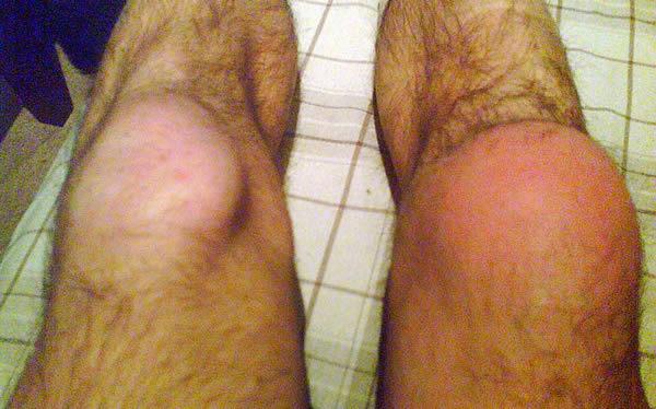
31 SPRING SIGN Patient supine on table Relaxed leg/knee in extension Examiner holds ankle with knee in extension Holds knee slightly flexed with other hand Release hold on knee and allow knee to fall into extension Positive if pain or knee springs back

32 Common Knee Pathology From PCPs My Experience and Personal Pearls Patellofemoral Pain ACL tears Meniscus Tears MCL Sprains Patella Instability/Dislocation Bursitis

33 Patellofemoral Pain My personal pain 2 finger sign Pain with stairs, sitting for long time, driving, etc. Sunrise view on plain xrays can be beneficial Usually from a kinematic imbalance/muscular group weakness Look at their feet For me, usually nonoperative PT, PT, and more PT Knee braces Shoe inserts Anti-inflammatories injections

34 ACL Tears Could have entire week of talks dedicated to this injury If you suspect it or have diagnosed it, then refer it Do all ACL tears need to be fixed? Nope Patient life style, activity level, amount of OA can help decide


35 Meniscus Tears Acute vs Chronic Joint Line Pain Sharp, mechanical pain Catching, giving, out Worse with pivoting/twisting Xrays, xrays, xrays!!!! MRI for some Conservative vs operative treatment surgeon/patient specific

36 MCL Injuries Hit from the lateral side of the knee Can be noncontact esp. when in combination with other injuries Xrays, Xrays, Xrays!!! MRI to confirm and r/o other injuries Grades 1 to 3 Most all isolated can be treated conservatively Hinged knee brace PT Few need surgical intervention


37 Patella Dislocation Twist with a clunk Some have to be reduced in ER Xrays, xrays, xrays!!!...esp sunrise view MRI As a general rule, most first time patella dislocators can be treated conservatively Lateral J brace PT


38 Prepatellar Bursitis Swelling anterior to patella Common in people on knees for work Conservative Treatment Rest, ice, anti-inflammatories Avoid kneeling Cushioned knee brace/pad Aspirate/injection Pes Anserine Bursitis Easy diagnosis Point tender over pes Conservative Treatment Ice, anti-inflammatories Injection PT
39 CONCLUSION Knee complicated anatomy Knowing anatomy=knowing diagnosis Multiple physical exam maneuvers Choose those most comfortable with and have been reliable in the past Always get xrays first!!!!
40 THANK YOU
RN(EC) ENC(C) GNC(C) MN ACNP *** MECHANISM OF INJURY.. MOST IMPORTANT *** - Useful in determining mechanism of injury / overuse
 ENC(C) GNC(C) MN ACNP *** MECHANISM OF INJURY.. MOST IMPORTANT *** - Useful in determining mechanism of injury / overuse") HISTORY *** MECHANISM OF INJURY.. MOST IMPORTANT *** Age of patient Sport / Occupation - Certain conditions are more prevalent in particular age groups (Osgood Schlaters in youth / Degenerative Joint Disease
HISTORY *** MECHANISM OF INJURY.. MOST IMPORTANT *** Age of patient Sport / Occupation - Certain conditions are more prevalent in particular age groups (Osgood Schlaters in youth / Degenerative Joint Disease
Prevention and Treatment of Injuries. Anatomy. Anatomy. Chapter 20 The Knee Westfield High School Houston, Texas
 Prevention and Treatment of Injuries Chapter 20 The Knee Westfield High School Houston, Texas Anatomy MCL, Medial Collateral Ligament LCL, Lateral Collateral Ligament PCL, Posterior Cruciate Ligament ACL,
Knee Injury Assessment
 Knee Injury Assessment Clinical Anatomy p. 186 Femur Medial condyle Lateral condyle Femoral trochlea Tibia Intercondylar notch Tibial tuberosity Tibial plateau Fibula Fibular head Patella Clinical Anatomy
Physical Examination of the Knee
 History: Pain Traumatic vs. atraumatic? Acute vs Chronic Previous procedures done on the knee? Swelling, catching, instability General Setup Examine standing, sitting and supine Evaluate gait Examine hip
Knee Joint Assessment and General View
 Knee Joint Assessment and General View Done by; Mshari S. Alghadier BSc Physical Therapy RHPT 366 m.alghadier@sau.edu.sa http://faculty.sau.edu.sa/m.alghadier/ Functional anatomy The knee is the largest
Physical Examination of the Knee
 History: Pain Traumatic vs. atraumatic Acute vs Chronic Mechanism of injury Swelling, catching, instability Previous evaluation and treatment General Setup Examine standing, sitting and supine Evaluate
The Knee. Two Joints: Tibiofemoral. Patellofemoral
 Evaluating the Knee The Knee Two Joints: Tibiofemoral Patellofemoral HISTORY Remember the questions from lecture #2? Girth OBSERVATION TibioFemoral Alignment What are the consequences of faulty alignment?
In the name of god. Knee. By: Tofigh Bahraminia Graduate Student of the Pathology Sports and corrective actions. Heat: Dr. Babakhani. Nov.
 In the name of god Knee By: Tofigh Bahraminia Graduate Student of the Pathology Sports and corrective actions Heat: Dr. Babakhani Nov. 2014 1 Anatomy-Bones Bones Femur Medial/lateral femoral condyles articulate
Musculoskeletal Examination Benchmarks
 Musculoskeletal Examination Benchmarks _ The approach to examining the musculoskeletal system is the same no matter what joint or limb is being examined. The affected and contralateral region should both
The Knee. Prof. Oluwadiya Kehinde
 The Knee Prof. Oluwadiya Kehinde www.oluwadiya.sitesled.com The Knee: Introduction 3 bones: femur, tibia and patella 2 separate joints: tibiofemoral and patellofemoral. Function: i. Primarily a hinge joint,
The Lower Limb II. Anatomy RHS 241 Lecture 3 Dr. Einas Al-Eisa
 The Lower Limb II Anatomy RHS 241 Lecture 3 Dr. Einas Al-Eisa Tibia The larger & medial bone of the leg Functions: Attachment of muscles Transfer of weight from femur to skeleton of the foot Articulations
Joints of the Lower Limb II
 Joints of the Lower Limb II Lecture Objectives Describe the components of the knee and ankle joint. List the ligaments associated with these joints and their attachments. List the muscles acting on these
Checklist for Physical Examination of the Knee Muscuoskeletal Block -- Chris McGrew MD, Andrew Ashbaugh DO
 Checklist for Physical Examination of the Knee Muscuoskeletal Block -- Chris McGrew MD, Andrew Ashbaugh DO This handout is for use as a rough guide and study aid. Your instructor may perform certain maneuvers
Recognizing common injuries to the lower extremity
 Recognizing common injuries to the lower extremity Bones Femur Patella Tibia Tibial Tuberosity Medial Malleolus Fibula Lateral Malleolus Bones Tarsals Talus Calcaneus Metatarsals Phalanges Joints - Knee
The examination of the painful knee. Maja K Artandi, MD, FACP Clinical Associate Professor of Medicine Stanford University
 The examination of the painful knee Maja K Artandi, MD, FACP Clinical Associate Professor of Medicine Stanford University Objectives of the talk By the end of this talk you will know The important anatomy
and K n e e J o i n t Is the most complicated joint in the body!!!!
 K n e e J o i n t K n e e J o i n t Is the most complicated joint in the body!!!! 1-Consists of two condylar joints between: A-The medial and lateral condyles of the femur and The condyles of the tibia
THE LOWER EXTREMITY EXAM FOR THE FAMILY PRACTITIONER
 THE LOWER EXTREMITY EXAM FOR THE FAMILY PRACTITIONER Melinda A. Scott, D.O. Orthopedic Associates of Dayton Board Certified in Primary Care Sports Medicine GOALS Identify landmarks necessary for exam of
On Field Assessment and Management of Acute Knee Injuries: A Physiotherapist s Perspective
 On Field Assessment and Management of Acute Knee Injuries: A Physiotherapist s Perspective Jessica Condliffe Physiotherapist / Clinic Manager TBI Health Wellington Presentation Outline Knee anatomy review
Please differentiate an internal derangement from an external knee injury.
 Knee Orthopaedic Tests Sports and Knee Injuries James J. Lehman, DC, MBA, DABCO University of Bridgeport College of Chiropractic Knee Injury Strain, Sprain, Internal Derangement Anatomy of the Knee Please
CHAPTER 8: THE BIOMECHANICS OF THE HUMAN LOWER EXTREMITY
 CHAPTER 8: THE BIOMECHANICS OF THE HUMAN LOWER EXTREMITY _ 1. The hip joint is the articulation between the and the. A. femur, acetabulum B. femur, spine C. femur, tibia _ 2. Which of the following is
Differential Diagnosis
 Case 31yo M who sustained an injury to L knee while playing Basketball approximately 2 weeks ago. He describes pivoting and hyperextending his knee, which swelled over the next few days. He now presents
The Knee. Tibio-Femoral
 The Knee Tibio-Femoral Osteology Distal Femur with Proximal Tibia Largest Joint Cavity in the Body A modified hinge joint with significant passive rotation Technically, one degree of freedom (Flexion/Extension)
Muscle Testing of Knee Extensors. Yasser Moh. Aneis, PhD, MSc., PT. Lecturer of Physical Therapy Basic Sciences Department
 Muscle Testing of Knee Extensors Yasser Moh. Aneis, PhD, MSc., PT. Lecturer of Physical Therapy Basic Sciences Department Muscle Testing of Knee Extensors othe Primary muscle Quadriceps Femoris -Rectus
Sports Medicine 15. Unit I: Anatomy. The knee, Thigh, Hip and Groin. Part 4 Anatomies of the Lower Limbs
 Sports Medicine 15 Unit I: Anatomy Part 4 Anatomies of the Lower Limbs The knee, Thigh, Hip and Groin Anatomy of the lower limbs In Part 3 of this section we focused upon 11 of the 12 extrinsic muscles
Myology of the Knee. PTA 105 Kinesiology
 Myology of the Knee PTA 105 Kinesiology Objectives Describe the planes of motion and axes of rotation of the knee joint Visualize the origins and insertions of the muscles about the knee List the innervations
KNEE EXAMINATION. Tips & Tricks from an Emergency Physician Perspective. EM Physicians Less Exposed to MSK Medicine
 KNEE EXAMINATION Tips & Tricks from an Emergency Physician Perspective Dr P O CONNOR Emergency Medicine Physician EUSEM 10/09/2018 EM Physicians Less Exposed to MSK Medicine Musculoskeletal Medicine becoming
Mastering the Musculoskeletal Exam UCSF Essentials of Women s Health July 7, 2016 Carlin Senter, M.D. Henry Crevensten, M.D.
 Mastering the Musculoskeletal Exam UCSF Essentials of Women s Health July 7, 2016 Carlin Senter, M.D. Henry Crevensten, M.D. I have nothing to disclose Outline Knee exam Shoulder exam Knee Anatomy The
Knee Injuries. PSK 4U Mr. S. Kelly North Grenville DHS. Medial Collateral Ligament Sprain
 Knee Injuries PSK 4U Mr. S. Kelly North Grenville DHS Medial Collateral Ligament Sprain Result from either a direct blow from the lateral side in a medial direction or a severe outward twist Greater injury
Financial Disclosure. Medial Collateral Ligament
 Matthew Murray, M.D. UTHSCSA Sports Medicine Financial Disclosure Dr. Matthew Murray has no relevant financial relationships with commercial interests to disclose. Medial Collateral Ligament Most commonly
Anatomy. ACL PCL MCL LCL Meniscus. Medial Lateral
 Skis for Knees Anatomy ACL PCL MCL LCL Meniscus Medial Lateral Knee Anatomy THE KNEE HISTORY Pain (PQRST) Contact vs noncontact Effusions Mechanical symptoms Locking Instability (falls) Initial treatment
Overview Ligament Injuries. Anatomy. Epidemiology Very commonly injured joint. ACL Injury 20/06/2016. Meniscus Tears. Patellofemoral Problems
 Overview Ligament Injuries Meniscus Tears Pankaj Sharma MBBS, FRCS (Tr & Orth) Consultant Orthopaedic Surgeon Manchester Royal Infirmary Patellofemoral Problems Knee Examination Anatomy Epidemiology Very
The Knee. Clarification of Terms. Osteology of the Knee 7/28/2013. The knee consists of: The tibiofemoral joint Patellofemoral joint
 The Knee Clarification of Terms The knee consists of: The tibiofemoral joint Patellofemoral joint Mansfield, p273 Osteology of the Knee Distal Femur Proximal tibia and fibula Patella 1 Osteology of the
DIAGNOSIS AND EARLY MANAGEMENT OF KNEE INJURIES
 DIAGNOSIS AND EARLY MANAGEMENT OF KNEE INJURIES INTRODUCTION: The knee is a common site of athletic injury. The knee injuries can be classified either into traumatic or acute and chronic, with overuse
W. Dilworth Cannon, M.D. Professor of Clinical Orthopaedic Surgery University of California San Francisco
 Knee Pain And Injuries In Adults W. Dilworth Cannon, M.D. Professor of Clinical Orthopaedic Surgery University of California San Francisco Pain Control Overview Narcotics rarely necessary after 1 st 1-2
American College of Physicians 2013 Ohio Chapter Scientific Meeting Columbus, OH October 11, 2013
 American College of Physicians 2013 Ohio Chapter Scientific Meeting Columbus, OH October 11, 2013 Paul J. Gubanich, MD, MPH Assistant Professor of Internal Medicine/Sports Medicine Team Physician, Ohio
To describe he knee joint, ligaments, structure & To list the main features of other lower limb joints
 To describe he knee joint, ligaments, structure & neurovascular supply To demonstrate the ankle joint anatomy To list the main features of other lower limb joints To list main groups of lymph nodes in
UNIT 7 JOINTS. Knee and Ankle Joints DR. ABDEL-MONEM A. HEGAZY
 UNIT 7 JOINTS Knee and Ankle Joints BY DR. ABDEL-MONEM A. HEGAZY (Degree in Bachelor of Medicine and Surgery with honor 1983, Dipl."Gynaecology and Obstetrics "1989, Master "Anatomy and Embryology "1994,
Ultrasound of the Knee Joint. Jun Sung Park,M.D. Bundang General Hospital Dept. of Rehabilitation Medicine
 Ultrasound of the Knee Joint Jun Sung Park,M.D. Bundang General Hospital Dept. of Rehabilitation Medicine Clinical History and P/E Chronic or Acute Symptoms Chronic Sx. : possible of systemic articular
Copyright 2012 by The McGraw-Hill Companies, Inc. All rights reserved. McGraw-Hill/Irwin
 CHAPTER 8: THE LOWER EXTREMITY: KNEE, ANKLE, AND FOOT KINESIOLOGY Scientific Basis of Human Motion, 12 th edition Hamilton, Weimar & Luttgens Presentation Created by TK Koesterer, Ph.D., ATC Humboldt State
FUNCTIONAL ANATOMY: Knee and Leg
 ACSM Team Physician Course San Antonio Feb 2015 FUNCTIONAL ANATOMY: Knee and Leg Marlene DeMaio, MD Professor, Orthopaedic Surgery Marshall University VAMC Huntington, WV Mary Lloyd Ireland, MD Professor
Arthritic history is similar to that of the hip. Add history of give way and locking, swelling
 KNEE VASU PAI Arthritic history is similar to that of the hip. Add history of give way and locking, swelling INJURY MECHANISM When How Sequence Progress Disability IKDC Activity I - Strenuous activity
Goals &Objectives. 1. Review the anatomy of the knee 2. Practice your hands-on skills 3. By the end of the workshop:
 Clinical Knee Exam Goals &Objectives 1. Review the anatomy of the knee 2. Practice your hands-on skills 3. By the end of the workshop: Be able to categorize knee injuries Understand the significance of
ACL AND PCL INJURIES OF THE KNEE JOINT
 ACL AND PCL INJURIES OF THE KNEE JOINT Dr.KN Subramanian M.Ch Orth., FRCS (Tr & Orth), CCT Orth(UK) Consultant Orthopaedic Surgeon, Special interest: Orthopaedic Sports Injury, Shoulder and Knee Surgery,
SOFT TISSUE INJURIES OF THE KNEE: Primary Care and Orthopaedic Management
 SOFT TISSUE INJURIES OF THE KNEE: Primary Care and Orthopaedic Management Gauguin Gamboa Australia has always been a nation where emphasis on health and fitness has resulted in an active population engaged
Examination of the Knee
 Examination of the Knee Wash your hands & Introduce the exam to the patient Positioning & Draping With the patient supine, make sure both legs are exposed in order to compare each side be sure to use draping
Diagnosis and Management of Knee Conditions. Jenny Love / Lynn Robertson AFLAR Oct 2009
 Diagnosis and Management of Knee Conditions Jenny Love / Lynn Robertson AFLAR Oct 2009 AIMS Review 4 common Knee Conditions: Anterior knee pain Meniscal Injuries Ligament injuries ACL Osteoarthritis Discuss
Chapter 10. The Knee Joint. The Knee Joint. Bones. Bones. Bones. Bones. Knee joint. Manual of Structural Kinesiology R.T. Floyd, EdD, ATC, CSCS
 The Knee Joint Chapter 10 The Knee Joint Manual of Structural Kinesiology R.T. Floyd, EdD, ATC, CSCS 2007 McGraw-Hill Higher Education. All rights reserved. 10-1 Knee joint largest joint in body very complex
Exam of the Knee and Ankle I HAVE NO FINANCIAL DISCLOSURES RELEVANT TO THIS PRESENTATION
 Exam of the Knee and Ankle I HAVE NO FINANCIAL DISCLOSURES RELEVANT TO THIS PRESENTATION Disclosures I have no relevant financial relationships with the manufacturers of any commercial products and or
Evaluation of Knee Problems
 Evaluation of Knee Problems OBJECTIVES Review knee anatomy Explain tests to look for pathology Briefly introduce knee problems Only by a thorough knowledge of anatomy and functional testing can one make
ACL Athletic Career. ACL Rupture - Warning Features Intensive pain Immediate swelling Locking Feel a Pop Dead leg Cannot continue to play
 FIMS Ambassador Tour to Eastern Europe, 2004 Belgrade, Serbia Montenegro Acute Knee Injuries - Controversies and Challenges Professor KM Chan OBE, JP President of FIMS Belgrade ACL Athletic Career ACL
Chapter 20 The knee and related structures
 Chapter 20 The knee and related structures Athletic Training Spring 2014 Jihong Park Bones & joints Femur, tibia, fibula, & patella Femur & tibia Weight bearing & muscle attachment Patella functions Anterior
Imaging the Knee 17/10/2017. Friction syndrome Common in runners or cyclists Fluid between ITB and Lateral femoral condyle
 17/10/2017 Imaging the Knee Alicia M. Yochum RN, DC, DACBR, RMSK Iliotibial Band Syndrome Ligamentous Tears (ACL, PCL, MCL, LCL) Meniscal Tears Cartilage Degeneration Quadriceps/Patellar tendinosis Osteochondral
BATES VISUAL GUIDE TO PHYSICAL EXAMINATION. OSCE 4: Knee Pain
 BATES VISUAL GUIDE TO PHYSICAL EXAMINATION OSCE 4: Knee Pain This video format is designed to help you prepare for objective structured clinical examinations, or OSCEs. You are going to observe and participate
ASSESSMENT AND MANAGEMENT OF THE KNEE AND LOWER LIMB.
 ASSESSMENT AND MANAGEMENT OF THE KNEE AND LOWER LIMB www.fisiokinesiterapia.biz Overview History Examination X-rays Fractures and Dislocations. Soft Tissue Injuries Other Knee/Lower limb Problems Anatomy
Practical 1 Worksheet
 Practical 1 Worksheet ANATOMICAL TERMS 1. Use the word bank to fill in the missing words. reference side stand body arms palms anatomical forward All anatomical terms have a(n) point which is called the
Imaging the Athlete s Knee. Peter Lowry, MD Musculoskeletal Radiology University of Colorado
 Imaging the Athlete s Knee Peter Lowry, MD Musculoskeletal Radiology University of Colorado None Disclosures Knee Imaging: Radiographs Can be performed weight-bearing or non-weight-bearing View options
Muscles of the Thigh. 6.1 Identify, describe the attachments of and deduce the actions of the muscles of the thigh: Anterior group
 Muscles of the Thigh 6.1 Identify, describe the attachments of and deduce the actions of the muscles of the thigh: Anterior group Sartorius: This is a long strap like muscle with flattened tendons at each
Human Anatomy Biology 351
 Human Anatomy Biology 351 Lower Limb Please place your name on the back of the last page of this exam. You must answer all questions on this exam. Because statistics demonstrate that, on average, between
SOFT TISSUE KNEE INJURIES
 SOFT TISSUE KNEE INJURIES Soft tissue injuries of the knee commonly occur in all sports or in any activity that requires sudden changes in activity or movement. The knee is a complex joint and any injury
Evaluation of the Knee and Shoulder
 Evaluation of the Knee and Shoulder Karen J. Boselli, MD Northeast Regional Nurse Practitioner Conference May 2018 Knee Overview History Examination Top 5 diagnoses When to image When to refer Pain most
Knee Contusions and Stress Injuries. Laura W. Bancroft, M.D.
 Knee Contusions and Stress Injuries Laura W. Bancroft, M.D. Objectives Review 5 types of contusion patterns Pivot shift Dashboard Hyperextension Clip Lateral patellar dislocation Demonstrate various stress
Human Anatomy Biology 351
 Human Anatomy Biology 351 Lower Limb Please place your name on the back of the last page of this exam. You must answer all questions on this exam. Because statistics demonstrate that, on average, between
PRIMARY CARE EXAMINATION OF KEY JOINTS. Thomas M. Howard, MD, FACSM FFPC Sports Medicine
 PRIMARY CARE EXAMINATION OF KEY JOINTS Thomas M. Howard, MD, FACSM FFPC Sports Medicine General exam principles: Expose entire joint and opposite limb for comparison Have a Differential Diagnosis Exam
KNEE INJURIES IN FEMALE SOCCER PLAYERS: A FOCUS ON THE ACL
 KNEE INJURIES IN FEMALE SOCCER PLAYERS: A FOCUS ON THE ACL Item Type text; Electronic Thesis Authors PEÑA, VANESSA NICOLE Publisher The University of Arizona. Rights Copyright is held by the author. Digital
The Knee Joint By Prof. Dr. Muhammad Imran Qureshi
 The Knee Joint By Prof. Dr. Muhammad Imran Qureshi Structurally, it is the Largest and the most complex joint in the body because of the functions that it performs: Allows mobility (flexion/extension)
AAP Boot Camp KNEE AND ANKLE EXAM
 AAP Boot Camp KNEE AND ANKLE EXAM Disclosures I have no relevant financial relationships with the manufacturers of any commercial products and or providers of commercial services discussed in this CME
9/24/2012. Greg Bennett, PT, DSc Excel Physical Therapy Marymount University
 Greg Bennett, PT, DSc Excel Physical Therapy Marymount University Hx often diagnostic Least to most threatening Sx trump exam Develop consistent routine Don t inflame inflamed tissue 1 1. ESTABLISH OR
Biomechanics of the Knee. Valerie Nuñez SpR Frimley Park Hospital
 Biomechanics of the Knee Valerie Nuñez SpR Frimley Park Hospital Knee Biomechanics Kinematics Range of Motion Joint Motion Kinetics Knee Stabilisers Joint Forces Axes The Mechanical Stresses to which
Knee Joint Anatomy 101
 Knee Joint Anatomy 101 Bone Basics There are three bones at the knee joint femur, tibia and patella commonly referred to as the thighbone, shinbone and kneecap. The fibula is not typically associated with
Muscles of the Hip 1. Tensor Fasciae Latae O: iliac crest I: lateral femoral condyle Action: abducts the thigh Nerve: gluteal nerve
 Muscles of the Hip 1. Tensor Fasciae Latae O: iliac crest I: lateral femoral condyle Action: abducts the thigh Nerve: gluteal nerve 2. Gluteus Maximus O: ilium I: femur Action: abduct the thigh Nerve:
Anatomy and Sports Injuries of the Knee
 Anatomy and Sports Injuries of the Knee I. Anatomy II. Assessment III. Treatment IV. Case Study V. Dissection Anatomy Not a hinge joint 6 degrees of freedom Flexion/Extension Rotation Translation Anatomy
HANDS ON: Knee Evaluation J. Scott Delaney MD, FRCPC, FACEP, CSPQ
 HANDS ON: Knee Evaluation J. Scott Delaney MD, FRCPC, FACEP, CSPQ FACULTY DISCLOSURE Dr. Delaney has no affiliation with the manufacturer of any commercial product or provider of any commercial service
Objectives. The BIG Joint. Case 1. Boney Architecture. Presenter Disclosure Information. Common Knee Problems
 3:30 4:15 pm Common Knee Problems SPEAKER Christopher J. Visco, MD Presenter Disclosure Information The following relationships exist related to this presentation: Christopher J. Visco, MD: Speaker s Bureau
Lateral knee injuries
 Created as a free resource by Clinical Edge Based on Physio Edge podcast episode 051 with Matt Konopinski Get your free trial of online Physio education at Orthopaedic timeframes Traditionally Orthopaedic
PRINCIPLES OF EXAMNINIG THE KNEE
 Welcome! Pignon, Haiti IS IT. GOOD MORNING LORD! OR GOOD LORD, MORNING! PRINCIPLES OF EXAMNINIG THE KNEE Greg Bennett, PT, DSc Excel Physical Therapy Marymount University Rules Hx often diagnostic Least
POSTEROLATERAL CORNER RECONSTRUCTION WHEN AND HOW?
 OTHER KNEE SURGERIES POSTEROLATERAL CORNER RECONSTRUCTION WHEN AND HOW? Written by Jacques Ménétrey, Eric Dromzée and Philippe M. Tscholl, Switzerland Injury of the posterolateral corner (PLC) is relatively
SAMPLE ONLY. NOT FOR DISTRIBUTION. The Knee
 SAMPLE ONLY. NOT FOR DISTRIBUTION. 1 The Knee 2 CHAPTER 1 SAMPLE ONLY. NOT FOR DISTRIBUTION. THEY ARE NOT CUSHIONS! I can t tell you how many times I ve heard it said that the meniscus cartilages are the
Posterolateral Corner Injuries of the Knee: Pearls and Pitfalls
 Posterolateral Corner Injuries of the Knee: Pearls and Pitfalls Robert A. Arciero,MD,Col,ret Professor, Orthopaedics University of Connecticut Incidence of PLC Injuries with ACL Tears Fanelli, 1995 12%
Knee Movement Coordination Deficits. ICD-9-CM: Sprain of cruciate ligament of knee
 1 Knee Movement Coordination Deficits Anterior Cruciate Ligament ACL Tear ICD-9-CM: 844.2 Sprain of cruciate ligament of knee ACL Insufficiency ICD-9-CM: 717.83 Old disruption of anterior cruciate ligament
Learning IRM. The Knee: lateral ligaments and anatomical quadrants.
 Learning IRM. The Knee: lateral ligaments and anatomical quadrants. Poster No.: C-1733 Congress: ECR 2014 Type: Educational Exhibit Authors: A. Amador Gil, M. D. C. Jurado Gómez, V. de Lara Bendahan ;
Patellofemoral Pathology
 Patellofemoral Pathology Matthew Murray, MD UT Health Science Center/UT Medicine Sports Medicine and Arthroscopic Surgery I have disclosed that I am a consultant for Biomet Orthopaedics. Anterior Knee
Disclosures. Outline. The Posterior Cruciate Ligament 5/3/2016
 The Posterior Cruciate Ligament Christopher J. Utz, MD Assistant Professor of Orthopaedic Surgery University of Cincinnati Disclosures I have no disclosures relevant to this topic. Outline 1. PCL Basic
Other Culprits in Knee Dysfunction
 Unraveling the Mystery of Knee Pain #6: Other Culprits in Knee Dysfunction 1 Webinar Goals Explore the assessment and treatment of other culprits in knee dysfunction. 2 Time: 60 minutes Schedule: Logistics
MCL Injuries: When and How to Repair Scott D. Mair, MD
 MCL Injuries: When and How to Repair Scott D. Mair, MD Professor and Team Physician: Orthopaedic Surgery University of Kentucky School of Medicine Disclosure Institution: Research/Education Smith-Nephew
The Muscular System. Chapter 10 Part D. PowerPoint Lecture Slides prepared by Karen Dunbar Kareiva Ivy Tech Community College
 Chapter 10 Part D The Muscular System Annie Leibovitz/Contact Press Images PowerPoint Lecture Slides prepared by Karen Dunbar Kareiva Ivy Tech Community College Table 10.14: Muscles Crossing the Hip and
emoryhealthcare.org/ortho
 COMMON SOCCER INJURIES Oluseun A. Olufade, MD Assistant Professor, Department of Orthopedics and PM&R 1/7/18 GOALS Discuss top soccer injuries and treatment strategies Simplify hip and groin injuries in
I have nothing to disclose
 Management of Common Knee Disorders: What You Knee d to Know UCSF Essentials of Women s Health July 8, 2015 Carlin Senter, M.D. I have nothing to disclose Learning objectives: in 1 hour you will be able
Anterior Cruciate Ligament Surgery
 Anatomy Anterior Cruciate Ligament Surgery Roger Ostrander, MD Andrews Institute Anatomy Anatomy Function Primary restraint to anterior tibial translation Secondary restraint to internal tibial rotation
MUSCULOSKELETAL PROBLEMS are among the most
 592 REVIEW ARTICLE Physical Examination of the Knee: A Review of the Original Test Description and Scientific Validity of Common Orthopedic Tests Gerard A. Malanga, MD, Steven Andrus, MD, Scott F. Nadler,
Knee Evaluation
 www.fisiokinesiterapia.biz Knee Evaluation Quick Facts Tibiofemoral Joint (TFJ) Normal ROM Flexion 135-140 140 degrees Extension 0 degrees Closed Pack Position Full extension with ER Loose Packed Position
Contents of the Posterior Fascial Compartment of the Thigh
 Contents of the Posterior Fascial Compartment of the Thigh 1-Muscles: B i c e p s f e m o r i s S e m i t e n d i n o s u s S e m i m e m b r a n o s u s a small part of the adductor magnus (h a m s t
ABSTRACT 2 Key Words: meniscus, tibiofemoral joint, osteoarthritis
 ABSTRACT In the United States, meniscal lesions represent the most common intra-articular knee injury. 1 In fact, the mean annual incidence of meniscal tears is approximately 60 to 70 per 100,000 patients.
Anatomage Table Instructors Guide- Lower Limb
 The Lower Limb Anatomage Table Instructors Guide- Lower Limb Table of Contents Lower Limb 1- The Skeletal System...3 1: Hip Bone...3 2: Hip Joint and Femur...4 3: Patella and Knee Joint...7 4: Tibia, Fibula,
Exercise Science Section 4: Joint Mechanics and Joint Injuries
 Exercise Science Section 4: Joint Mechanics and Joint Injuries An Introduction to Health and Physical Education Ted Temertzoglou Paul Challen ISBN 1-55077-132-9 Types of Joints Fibrous joint Cartilaginous
HUMAN BODY COURSE LOWER LIMB NERVES AND VESSELS
 HUMAN BODY COURSE LOWER LIMB NERVES AND VESSELS October 22, 2010 D. LOWER LIMB MUSCLES 2. Lower limb compartments ANTERIOR THIGH COMPARTMENT General lfunction: Hip flexion, knee extension, other motions
Outline of Session. Evaluation and Treatment of Common Musculoskeletal Complaints. Katherine Julian July 2008
 Outline of Session Evaluation and Treatment of Common Musculoskeletal Complaints Katherine Julian July 2008 Joint Anatomy Exam Demonstration: HIT ME NOT 1 History Inspect Touch Move Extra maneuvers Things
42 nd Annual Symposium on Sports Medicine. Knee Injuries In The Pediatric Athlete. Disclosure
 42 nd Annual Symposium on Sports Medicine Travis Murray, MD Assistant Professor University of Texas Health Science Center San Antonio January 23, 2015 Knee Injuries In The Pediatric Athlete Disclosure
40 th Annual Symposium on Sports Medicine. Knee Injuries In The Pediatric Athlete. Disclosure
 40 th Annual Symposium on Sports Medicine Travis Murray, MD Assistant Professor University of Texas Health Science Center San Antonio Knee Injuries In The Pediatric Athlete Disclosure Dr. Travis Murray
Knee Ultrasonography step by step
 Knee Ultrasonography step by step Poster No.: C-2809 Congress: ECR 2018 Type: Educational Exhibit Authors: J. A. Torres de Abreu Macedo, N. Pereira da Silva, A. I. Aguiar, F. Alves, F. Caseiro Alves; Coimbra/PT