Technique Guide. Rotation Correction Plates 1.5 and 2.0. Reposition plates for fractures and osteotomies at the metacarpals and phalanges.
|
|
|
- Shana Newman
- 5 years ago
- Views:
Transcription

1 Technique Guide Rotation Correction Plates 1.5 and 2.0. Reposition plates for fractures and osteotomies at the metacarpals and phalanges.
2
3 Table of Contents Introduction Rotation Correction Plates 1.5 and Indications and Contraindications 4 Case Studies 5 Surgical Technique Implantation 7 Implant Removal 15 Insert Cortex Screws 16 Insert Locking Screws 19 Product Information Implants 22 Instruments 23 Bibliography 24 Warning This description is not sufficient for immediate application of the instrumentation. Instruction by a surgeon experienced in handling these instruments is highly recommended. Synthes 1


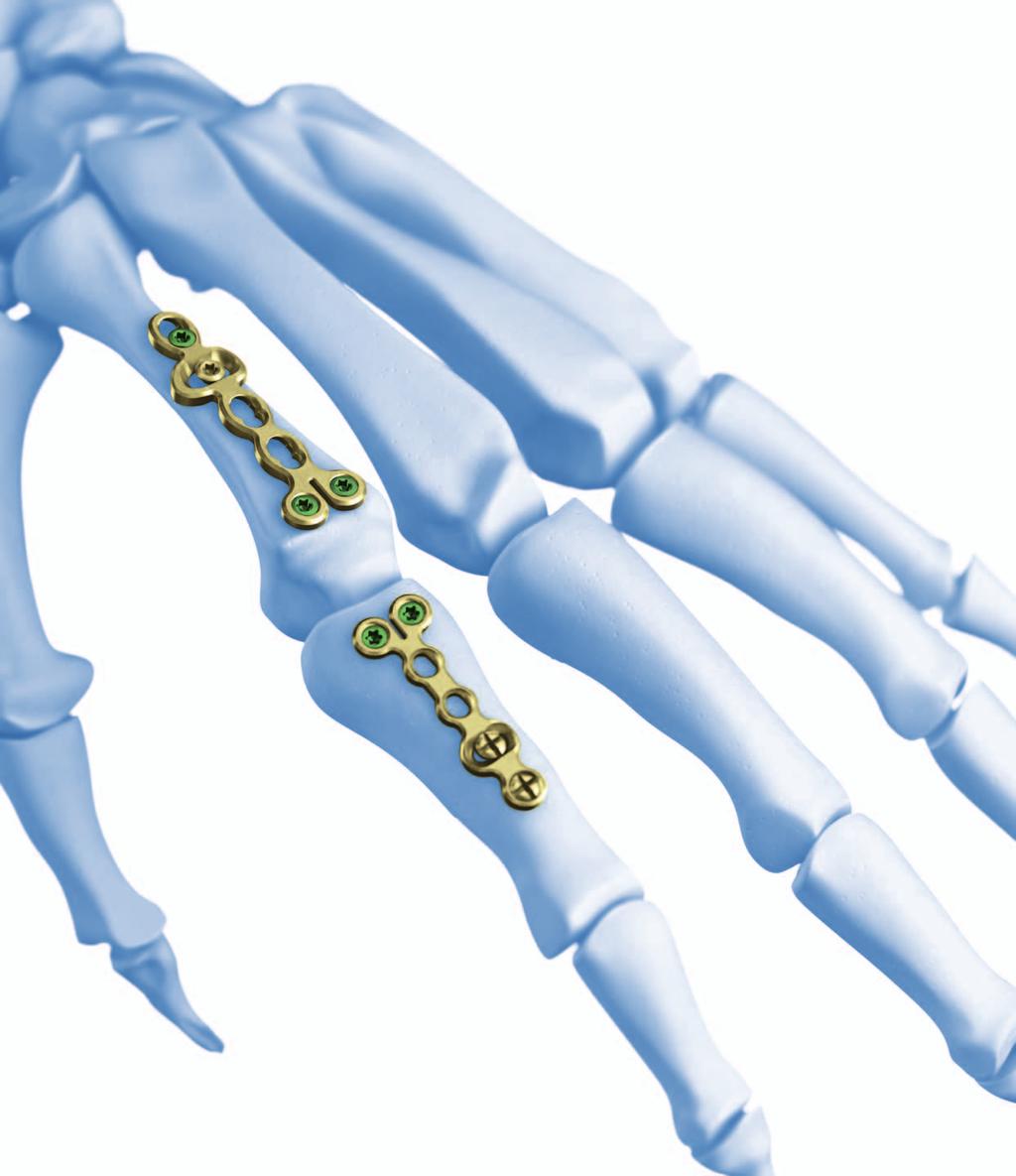
4 Rotation Correction Plates 1.5 and 2.0. Reposition plates for fractures and osteotomies at the metacarpals and phalanges. Anatomically precontoured Minimal irritation of ligaments and soft tissue thanks to a flat plate and screw profile, rounded edges and polished surfaces. Transversally elongated holes An elongated hole transversal to the axis of the plate shaft allows intraoperative inspection of the reduction and allows the surgeon to correct the rotational axis of the bone if necessary Angular stability 2.0 mm locking screws can be used in the head. Cortex or locking screws can be used in the shaft, depending on the plate size. The design of the plate head makes it easy to adjust to the anatomy of the condyles. Two shaft lengths Two different shaft lengths of the plates allow fragment-specific treatment of fractures of the metacarpals and proximal phalanges. 2 Synthes Rotation Correction Plates 1.5 and 2.0 Technique Guide
5
6 Indications and Contraindications Indications 1. All fractures of the phalanges and metacarpals, where the exact reposition is difficult or where a rotational error can easily occur. subcapital fractures of the metacarpals (in particular impacted compression fractures) transverse fractures short oblique fractures comminuted fractures defect fractures (circular saw injuries) amputation injuries to the fingers (with primary shortening) Winterstein fracture, Rolando fracture 2. Corrective osteotomies for fractures of the phalanges or metacarpals that have healed with axial errors and/or rational errors. In the case of metaphyseal fractures, comminuted fractures and osteoporotic bone, the clinical results can be improved by the angular-stable screw/plate connection. Contraindications Easily reduced oblique or spiral fractures that can be precisely and firmly stabilized using screw osteosynthesis. 4 Synthes Rotation Correction Plates 1.5 and 2.0 Technique Guide


7 Case Studies Case 1 Subcapital comminuted fracture of the 2nd metacarpal bone 42-year-old man, fall from bike. Compressed fracture of the metacarpal head with 4 mm shortening and 20 ulnar malangulation. Instable, subcapital impacted compression zone with bony collateral ligament tears. Primary treatment with plaster splint with only 30 flexion of the metacarpophalangeal joints. Intraoperative far distal angular stable fixation of the head, compensating for errors in the length and rotation. Immediate active exercise treatment. Preoperative, dorsopalmar and oblique Postoperative, dorsopalmar and lateral Synthes 5


8 Case studies Case 2 Pseudoarthrosis of the 4th metacarpal bone following failed screw osteosynthesis Preoperative with pseudoarthrosis and 6 mm shortening, dorsopalmar 32-year-old woman with screw osteosynthis treated elsewhere. The patient presented with pain, swelling and rotational error 6 months after the primary treatment. Intraoperatively, an anatomical reposition is no longer possible. Angularly stable fixation close to joint. Proximal with cortex screws. Immediate active exercise treatment. Postoperative following compensation of length and rotational error, dorsopalmar Postoperative, laterale 6 Synthes Rotation Correction Plates 1.5 and 2.0 Technique Guide
9 Implantation If fractures to the metacarpals or the proximal phalanges are misaligned, the function of the hand can be severely affected. Especially rotational misalignments are problematic because of crossing and scissoring digits whenever full flexion of the fingers to a fist is attempted. Even minor rotational errors in the fingers frequently have to be surgically corrected after a fracture has healed. For metacarpal fractures and fractures of the proximal phalanges, optimal recovery of hand function includes the meticulous restoration of alignment, length and rotation of digits and metacarpals. A careful approach needs to be chosen in order not to further compromise the soft tissue situation which again requires proper reconstruction. The fixation has to be stable enough in order to facilitate immediate active and passive finger exercises. Approaches For the metacarpal bones, the 2nd ray may be approached dorso-radially above the palpable bone, the 5th ray dorsoulnar above the bone, and the 3rd and 4th metacarpal either directly above the respective bone or between both of them when two adjacent bones are affected. The proximal phalanges are usually approached through the median line of the respective bone. Synthes 7

10 Implantation 1 Temporary fixation of fractures with Kirschner wires Reduction can be preliminary held with 1.0 mm K-wires not protruding the articular surface. Rotational alignment should be less than 10 when carefully checked in full extension, and flexion to a fist. 2 Select plate Rotation correction plates are available in various sizes and lengths, allowing fragment-specific treatment of fractures to the metacarpalia and the proximal phalanges. Determine the approach and select the plates according to the fracture pattern and the anatomical situation. 8 Synthes Rotation Correction Plates 1.5 and 2.0 Technique Guide


11 3 Bending the plate head and plate shaft 1 Instruments Bending Pin for LCP Plates 2.0, with thread Pliers, flat-nosed, pointed, for Plates 1.0 to 2.4 If necessary, bend the plate to suit the anatomical conditions. 1. Preferably bend the plate head with two bending pins. 2. Ensure that the shaft is bent between the combination holes, as it will otherwise be difficult to insert the locking screws. We recommend using two pairs of pliers. Note: The design of the plate holes allows a certain degree of deformation tolerance. Locking is, however, not as efficient if the thread holes are significantly deformed. For this reason the surgeon should avoid bending the plate by inserting the bending pins into a combination hole. 2 Warning: The plate should ideally not be cut. The sharp cut edges can damage the radiodorsal tendons and can irritate the soft tissue. Synthes 9

12 Implantation 4 Position plate Position the plate near the joint. The transverse elongated hole must be on the far side of the fracture on a section of bone that is intact. First fix the head or condyle region near the joint using two 2.0 mm locking screws to support the joint surfaces and to prevent repositioning loss. Please refer to page 19 for information on inserting the locking screws. Caution: Ensure that there is no dorsal or palmar damage to the cartilaginous joint surfaces by either the plate or the screws and that the collateral ligaments are not unintentionally fixed. Tip: If the axis and the length of the bone are not yet exactly set the plate can be fixed to the bone with plate holding forceps and the Kirschner wire can be removed. The length and the axis of the bone can now be corrected using the plate and the plate holding forceps. The Kirschner wire can be left in place if it is already fixing the exact position and is not blocking the transverse elongated hole. 10 Synthes Rotation Correction Plates 1.5 and 2.0 Technique Guide

13 5 Insert screw into elongated hole Insert and carefully tighten a standard cortex screw in the exact position in the central part of the transverse elongated hole. Either select 1.5 mm or 2.0 mm cortex screws depending on the plate size. Please refer to page 16 for information on inserting the cortex screws. Synthes 11


14 Implantation 6 Check result of repositioning Prop up the forearm with the elbow supported on the operating table while the wrist is at maximum flexion. In this position the finger joints are straightened by the tenodesis effect of the extensor tendons. Check the axis and the rotation of the fingers. Place the wrist passively in the maximum extension position while compressing the forearm anteriorly in the middle of its shaft. In this position, the tenodesis effect of the flexor tendons and the compression of the forearm muscles will cause the fingers to flex to nearly form a fist. This way, rotational errors of the digits are easily detected. Check the repositioning result with the wrist at maximum extension Releasing and tightening the cortex screw in the transverse longitudinal hole ensures that the optimum position of the finger can be identified Set the optimum finger positions by releasing and tightening the elongated hole screw 12 Synthes Rotation Correction Plates 1.5 and 2.0 Technique Guide

15 7 Final fixation Fix a screw in the optimum position distal or proximal to the transverse elongated hole. The other holes may remain free or can be fitted with additional cortex screws. The hole above the fracture line generally remains free. Please refer to page 16 for information on inserting the cortex screws. Note: For 2.0 correction plates either 2.0 mm locking screws or 2.0 mm cortex screws may be used in the shaft. Only 1.5 mm cortex screws are used in the stem of 1.5/2.0 correction plates. The synovial sheath above the plate should be closed with a resorbable 6/0 suture as far as possible. The extensor tendon is readapted in the median line with a resorbable 5/0 suture. After fitting a «size 6» Redon Drain, close the skin with a single button suture and then apply an elasto-compressive bandage. Synthes 13
16 Implantation 8 Postoperative treatment Post-operatively, the hand is consequently put upright. Remove the Redon Drain one day post-operatively. Subsequently, active and passive finger exercises should be started with the aim of full extension and flexion within a week. 14 Synthes Rotation Correction Plates 1.5 and 2.0 Technique Guide
17 Implant removal To remove locking screws, first unlock all screws from the plate; then remove the screws completely from the bone. This prevents rotation of the plate when removing the last locking screw. Synthes 15
18 Insert cortex screws Instruments for correction plates Handle, medium, with Mini Quick Coupling Screwdriver Shaft Stardrive 2.0, with Holding Sleeve, length 66 mm, for Mini Quick Coupling Universal Drill Guide Depth Gauge for Screws 2.0 and 2.4 mm, measuring range up to 40 mm Drill Bit 1.5 mm with marking, length 96/82 mm, 2-flute, for Mini Quick Coupling Drill Bit 2.0 mm, length 67/55 mm, 2-flute, for Mini Quick Coupling Additional instruments required for correction plates 1.5/ Screwdriver Shaft 1.5, cruciform, with Holding Sleeve, length 66 mm, with Mini Quick Coupling Double Drill Guide 1.5/ Depth Gauge for Screws 1.3 to 1.5 mm, measuring range up to 24 mm Drill Bit 1.1 mm, length 45/33 mm, 2-flute, for Mini Quick Coupling 16 Synthes Rotation Correction Plates 1.5 and 2.0 Technique Guide


19 1 Predrill screw hole Predrill the holes for the combination screws either neutrally (support) or eccentrically (compression) into the non-thread bearing part of the combination hole using the universal drill guide that is appropriate for the screw diameter. Cortex screw 2.0 mm: Use the 1.5 mm drill bit for a threaded hole and the 2.0 mm drill bit for a gliding hole. Note: For rotation correction plates 1.5/2.0 use the universal drill guide 1.5/1.1 for 1.5 mm cortex screws. Use the 1.1 mm drill bit for the threaded hole and a 1.5 mm drill bit for the gliding hole. Neutral pre-drilling Eccentric pre-drilling 2 Determine screw length Determine the screw length using the 2.0 mm depth gauge. Note: Use the 1.5 mm depth gauge for 1.5 mm cortex screws. Synthes 17
 Stardrive 2.")

20 Insert cortex screws 3 Pick up screw Pick up the selected 2.0 mm cortex screw with the screwdriver shaft Stardrive 2.0 with holding sleeve and the corresponding handle. Notes A self-holding screwdriver (such as / ) Stardrive 2.0 can also be used. If using 1.5 mm cortex screws the cruciform screwdriver shaft with holding sleeve should be used. 4 Insert self-tapping standard screws Insert the self-tapping standard screws with the screwdriver. 18 Synthes Rotation Correction Plates 1.5 and 2.0 Technique Guide

21 Insert locking screw Instruments Handle, medium, with Mini Quick Coupling Screwdriver Shaft Stardrive 2.0, with Holding Sleeve, length 66 mm, for Mini Quick Coupling LCP Drill Sleeve 2.0, with Scale, for Drill Bits 1.5 mm with marking Depth Gauge for Screws 2.0 and 2.4 mm, measuring range up to 40 mm Drill Bit 1.5 mm with marking, length 96/82 mm, 2-flute, for Mini Quick Coupling 1 Use LCP drill sleeve Screw and lock the drill sleeve vertically into the thread of the selected hole. Synthes 19

 Determine the screw length using the 2.")
22 Insert locking screw 2 Predrill screw hole Predrill the screw hole with the 1.5 mm drill bit through the drill sleeve for locking screws 2.0 mm to the required depth and then read the screw length directly from the drill sleeve scale. 3 Determine screw length (optional) Determine the screw length using the 2.0 mm depth gauge for screws, as shown on page Synthes Rotation Correction Plates 1.5 and 2.0 Technique Guide
 Stardrive 2.")

23 4 Pick up screw Pick up the selected screw with the screwdriver shaft Stardrive with holding sleeve and the corresponding handle. Note: A self-holding screwdriver (such as / ) Stardrive 2.0 can also be used. 5 Insert self-tapping locking screws Manually insert the locking screws with the screwdriver. Carefully tighten the locking screw, as excessive force is not necessary to effectively lock the screws. Synthes 21

24 Implants Plates X Rotation Correction Plate 1.5/ 2.0, shaft 4 holes, head 2 holes, length 27 mm X Rotation Correction Plate 1.5/ 2.0, shaft 5 holes, head 2 holes, length 32 mm X LCP Rotation Correction Plate 2.0, shaft 4 holes, head 2 holes, length 34 mm X LCP Rotation Correction Plate 2.0, shaft 5 holes, head 2 holes, length 40 mm Locking screws X Locking Screw 2.0 mm, self-tapping Standard screws X X Cortex Screw 2.0 mm, self-tapping Cortex Screw 1.5 mm, self-tapping All screws 2.0 mm with Stardrive T6 drive. All screws 1.5 mm with cruciform drive. X=2: Steel (SSt) X=4: Titanium (plates) Titanium alloy TAN (screws) 22 Synthes Rotation Correction Plates 1.5 and 2.0 Technique Guide
25 Instruments Handle, medium, with Mini Quick Coupling Handle with Quick Coupling, length 110 mm Screwdriver Shaft 1.5, cruciform, with Holding Sleeve, length 66 mm for Mini Quick Coupling Screwdriver Shaft Stardrive 2.0, with Holding Sleeve, length 66 mm for Mini Quick Coupling LCP Drill Sleeve 2.0, with Scale, for Drill Bits 1.5 mm with marking Double Drill Guide 1.5/ Universal Drill Guide Drill Bit 1.1 mm, length 45/33 mm, 2-flute, for Mini Quick Coupling Drill Bit 1.5 mm with Stop, length 96/82 mm, 2-flute, for Mini Quick Coupling Drill Bit 2.0 mm, length 67/55 mm, 2-flute, for Mini Quick Coupling Depth Gauge for Screws 1.3 to 1.5 mm, measuring range up to 24 mm Depth Gauge for Screws 2.0 to 2.4 mm, measuring range up to 40 mm Bending Pin for LCP Plates 2.0, with thread All listed instruments are part of the LCP Compact Hand. Synthes 23
26 Bibliography Büchler U, Gupta A, Ruf S (1996) Corrective Osteotomy for Post-traumatic Malunion of the Phalanges of the Hand. J Hand Surg 21B:33 42 Freeland AE, Geissler WB, Weiss APC (2001) Operative Treatment of Common Displaced and Unstable Fractures of the Hand. JBJS 83A (6): Gollamudi S, Jones WA (2000) Corrective Osteotomy of Malunited Fractures of Phalanges and Metacarpals. J Hand Surg [Br] 25(5): Green DP (1986) Complications of Phalangeal and Metacarpal Fractures. Hand Clin 2: Gross MS, Gelberman RH (1985) Metacarpal Rotational Osteotomy. J Hand Surg [Am] 10:105 8 Ring D (2005) Malunion and Nonunion of the Metacarpals and Phalanges. JBJS 87A: Schaefer M, Siebert HR (2000) Finger- und Mittelhandfrakturen. Unfallchirurg 103: Strauch RJ, Rosenwasser MP, Lunt JG (1998) Metacarpal Shaft Fractures: The Effect of Shortening on the Extensor Tendon Mechanism. J Hand Surg [Am] 23: Trumble T, Gilbert M (1998) In Situ Osteotomy for Extra-articular Malunion of the Proximal Phalanx. J Hand Surg 23A: Van der Lei B, Robinson PH, Klasen HJ (1993) Correction Osteotomies of Phalanges and Metacarpals for Rotational and Angular Malunion: A Long-term Follow-up and a Review of the Literature. J Trauma 35(6): Synthes Rotation Correction Plates 1.5 and 2.0 Technique Guide
27
28 Synthes GmbH Eimattstrasse 3 CH-4436 Oberdorf Presented by: SE_ AA Synthes 2007 LCP and Stardrive are trademarks of Synthes Subject to modifications
Rotation Correction Plates 1.5 and 2.0. Reposition plates for fractures and osteotomies at the metacarpals and phalanges.
 Rotation Correction Plates 1.5 and 2.0. Reposition plates for fractures and osteotomies at the metacarpals and phalanges. Surgical Technique This publication is not intended for distribution in the USA.
LCP Proximal Radius Plates 2.4. Plates for radial head rim and for radial head neck address individual fracture patterns of the proximal radius.
 Technique Guide LCP Proximal Radius Plates 2.4. Plates for radial head rim and for radial head neck address individual fracture patterns of the proximal radius. Table of Contents Introduction LCP Proximal
2.4 mm Variable Angle LCP Volar Extra-Articular Distal Radius System. For fragment-specific fracture fixation with variable angle locking technology.
 Technique Guide 2.4 mm Variable Angle LCP Volar Extra-Articular Distal Radius System. For fragment-specific fracture fixation with variable angle locking technology. Table of Contents Introduction 2.4
LCP Medial Distal Tibia Plate, without Tab. The Low Profile Anatomic Fixation System with Angular Stability and Optimal Screw Orientation.
 LCP Medial Distal Tibia Plate, without Tab. The Low Profile Anatomic Fixation System with Angular Stability and Optimal Screw Orientation. Technique Guide LCP Small Fragment System Table of Contents Introduction
2.4 mm LCP Radial Head Plates. Part of the Synthes LCP Distal Radius Plate System.
 2.4 mm LCP Radial Head Plates. Part of the Synthes LCP Distal Radius Plate System. Technique Guide Instruments and Implants approved by the AO Foundation Table of Contents Introduction 2.4 mm LCP Radial
LCP Distal Tibia Plate
 Surgical Technique LCP Locking Compression Plate Original Instruments and Implants of the Association for the Study of Internal Fixation AO/ASIF Table of contents Indications 3 Implants/Instruments 5 Surgical
LCP Distal Humerus Plates
 The anatomic fixation system for the distal humerus with angular stability Surgical technique LCP Locking Compression Plate Contents Indications and contraindications 2 Implants 3 Instruments 5 Preparation
Instrument and Implant for wrist fracture
 Instrument and Implant for wrist fracture Jansri Janpanya Product specialist The Bangkok Unitrade Co,.ltd. Objectives Type of LCP for distal radius Fx. The new LCP design for distal radius Fx. Have knowledge
LCP Proximal Radius Plates 2.4. Plates for radial head rim and for radial head neck address individual fracture patterns of the proximal radius.
 LCP Proximal Radius Plates 2.4. Plates for radial head rim and for radial head neck address individual fracture patterns of the proximal radius. Surgical Technique This publication is not intended for
Technique Guide. 3.5 mm LCP Olecranon Plates. Part of the Synthes locking compression plate (LCP) system.
 system.") Technique Guide 3.5 mm LCP Olecranon Plates. Part of the Synthes locking compression plate (LCP) system. Table of Contents Introduction 3.5 mm LCP Olecranon Plates 2 AO Principles 3 Indications 3 Clinical
Technique Guide 3.5 mm LCP Olecranon Plates. Part of the Synthes locking compression plate (LCP) system. Table of Contents Introduction 3.5 mm LCP Olecranon Plates 2 AO Principles 3 Indications 3 Clinical
Distal Ulnar Locking Plate
 INDEX Indications Patient Position Surgical Technique - Step 1 Approach - Step 2 Plate Contouring - Step 3 Fracture Reduction - Step 4 Distal Plate Fixation - Step 5 Confirm Proper Reconstruction - Step
Technique Guide. 2.4 mm Variable Angle LCP Distal Radius System. For fragment-specific fracture fixation with variable angle locking technology.
 Technique Guide 2.4 mm Variable Angle LCP Distal Radius System. For fragment-specific fracture fixation with variable angle locking technology. Table of Contents Introduction 2.4 mm Variable Angle LCP
Variable Angle LCP Volar Rim Distal Radius Plate 2.4. For fragment-specific fracture fixation with variable angle locking technology.
 Technique Guide Variable Angle LCP Volar Rim Distal Radius Plate 2.4. For fragment-specific fracture fixation with variable angle locking technology. Image intensifier control Warning This description
Distal Radius Plate 2.4/2.7 dorsal and volar
 Distal Radius Plate 2.4/2.7 dorsal and volar Surgical Technique This publication is not intended for distribution in the USA. Instruments and implants approved by the AO Foundation. Distal Radius Plate
LCP Proximal Radius Plates 2.4. Plates for radial head rim and for radial head neck address individual fracture patterns of the proximal radius.
 LCP Proximal Radius Plates 2.4. Plates for radial head rim and for radial head neck address individual fracture patterns of the proximal radius. Surgical Technique This publication is not intended for
LCP Medial Proximal Tibial Plate 3.5. Part of the Synthes small fragment Locking Compression Plate (LCP) system.
 system.") LCP Medial Proximal Tibial Plate 3.5. Part of the Synthes small fragment Locking Compression Plate (LCP) system. Technique Guide This publication is not intended for distribution in the USA. Instruments
LCP Medial Proximal Tibial Plate 3.5. Part of the Synthes small fragment Locking Compression Plate (LCP) system. Technique Guide This publication is not intended for distribution in the USA. Instruments
Surgical Technique. Cannulated Angled Blade Plate 3.5 and 4.5, 90
 Surgical Technique Cannulated Angled Blade Plate 3.5 and 4.5, 90 Cannulated Angled Blade Plate 3.5 and 4.5, 90 Table of contents Indications/Contraindications 2 Implants 3 Surgical technique 5 Implant
Technique Guide. 2.7 mm/3.5 mm LCP Distal Fibula Plates. Part of the Synthes locking compression plate (LCP) system.
 system.") Technique Guide 2.7 mm/3.5 mm LCP Distal Fibula Plates. Part of the Synthes locking compression plate (LCP) system. Table of Contents Introduction 2.7 mm/3.5 mm LCP Distal Fibula Plates 2 AO Principles
Technique Guide 2.7 mm/3.5 mm LCP Distal Fibula Plates. Part of the Synthes locking compression plate (LCP) system. Table of Contents Introduction 2.7 mm/3.5 mm LCP Distal Fibula Plates 2 AO Principles
Technique Guide. 3.5 mm LCP Low Bend Medial Distal Tibia Plates. Part of the Synthes locking compression plate (LCP) system.
 system.") Technique Guide 3.5 mm LCP Low Bend Medial Distal Tibia Plates. Part of the Synthes locking compression plate (LCP) system. Table of Contents Introduction 3.5 mm LCP Low Bend Medial Distal Tibia Plates
Technique Guide 3.5 mm LCP Low Bend Medial Distal Tibia Plates. Part of the Synthes locking compression plate (LCP) system. Table of Contents Introduction 3.5 mm LCP Low Bend Medial Distal Tibia Plates
Technique Guide. LCP Distal Fibula Plates. Part of the Synthes locking compression plate (LCP) system.
 system.") Technique Guide LCP Distal Fibula Plates. Part of the Synthes locking compression plate (LCP) system. Table of Contents Introduction LCP Distal Fibula Plates 2 AO Principles 4 Indications 5 Surgical Technique
Technique Guide LCP Distal Fibula Plates. Part of the Synthes locking compression plate (LCP) system. Table of Contents Introduction LCP Distal Fibula Plates 2 AO Principles 4 Indications 5 Surgical Technique
LCP Extra-articular Distal Humerus Plate.
 Technique Guide LCP Extra-articular Distal Humerus Plate. The anatomically shaped and angular stable fixation system for extraarticular fractures of the distal humerus. Table of Contents Introduction
Long Volar Plates for Diaphyseal-Metaphyseal Radius Fractures LCP. Dia-Meta Volar Distal Radius Plates. Surgical Technique
 Long Volar Plates for Diaphyseal-Metaphyseal Radius Fractures LCP Dia-Meta Volar Distal Radius Plates Surgical Technique Table of Contents Introduction LCP Dia-Meta Volar Distal Radius Plates 2 AO Principles
VA-LCP Olecranon Plates 2.7/3.5. The fracture-specific low-profile fixation system with variable angle locking technology.
 VA-LCP Olecranon Plates 2.7/3.5. The fracture-specific low-profile fixation system with variable angle locking technology. Surgical Technique This publication is not intended for distribution in the USA.
2.4 mm Variable Angle LCP Volar Extra-Articular Distal Radius System. For fragment-specific fracture fixation with variable angle locking technology.
 2.4 mm Variable Angle LCP Volar Extra-Articular Distal Radius System. For fragment-specific fracture fixation with variable angle locking technology. Surgical Technique This publication is not intended
Technique Guide. VA-Locking Intercarpal Fusion System. Variable angle locking technology for mediocarpal partial arthrodesis.
 Technique Guide VA-Locking Intercarpal Fusion System. Variable angle locking technology for mediocarpal partial arthrodesis. Table of Contents Introduction VA-Locking Intercarpal Fusion System 2 Indications
LCP Medial Proximal Tibial Plate 4.5/5.0. Part of the Synthes LCP periarticular plating system.
 LCP Medial Proximal Tibial Plate 4.5/5.0. Part of the Synthes LCP periarticular plating system. Technique Guide This publication is not intended for distribution in the USA. Instruments and implants approved
3.5 mm LCP Distal Humerus Plates
 Part of the DePuy Synthes Locking Compression Plate (LCP ) System 3.5 mm LCP Distal Humerus Plates Surgical Technique Table of Contents Introduction 3.5 mm LCP Distal Humerus Plates 2 AO Principles 4 Indications
3.5 mm LCP Olecranon Plates
 Part of the DePuy Synthes Locking Compression Plate (LCP ) System 3.5 mm LCP Olecranon Plates Surgical Technique Table of Contents Introduction 3.5 mm LCP Olecranon Plates 2 AO Principles 3 Indications
Technique Guide. VA-LCP Distal Humerus Plates 2.7/3.5. The low-profile fixation system with variable angle locking technology.
 Technique Guide VA-LCP Distal Humerus Plates 2.7/3.5. The low-profile fixation system with variable angle locking technology. Image intensifier control Warning This description alone does not provide sufficient
Elbow Hinge Fixator. Guided Flexion/Extension for Unstable Elbow Fractures.
 Elbow Hinge Fixator. Guided Flexion/Extension for Unstable Elbow Fractures. Surgical Technique MR Safe Radiolucent Table of Contents System Description 3 Indications and Contraindications 4 Fixation Components
LCP Metaphyseal Plates. For extra-articular fractures.
 LCP Metaphyseal Plates. For extra-articular fractures. Surgical Technique This publication is not intended for distribution in the USA. Instruments and implants approved by the AO Foundation. Image intensifier
VA-LCP Distal Humerus Plates 2.7/3.5. The low-profile fixation system with variable angle locking technology.
 VA-LCP Distal Humerus Plates 2.7/3.5. The low-profile fixation system with variable angle locking technology. Technique Guide This publication is not intended for distribution in the USA. Instruments and
LCP Low Bend Medial Distal Tibia Plates 3.5 mm. Anatomic plates with low profile head for intra- and extraarticular fractures.
 LCP Low Bend Medial Distal Tibia Plates 3.5 mm. Anatomic plates with low profile head for intra- and extraarticular fractures. Surgical Technique This publication is not intended for distribution in the
2.4 mm LCP Volar Column Distal Radius Plates. Part of the 2.4 mm LCP Distal Radius System.
 2.4 mm LCP Volar Column Distal Radius Plates. Part of the 2.4 mm LCP Distal Radius System. 12 anatomically shaped volar plates Multiple screw options for fixedangle support to articular surface Combi holes
Technique Guide. Compact 2.0 LOCK Mandible. The locking system for the mandible.
 Technique Guide Compact 2.0 LOCK Mandible. The locking system for the mandible. Table of Contents Introduction Compact 2.0 LOCK Mandible 2 AO Principles 4 Indications and Contraindications 5 Surgical
OBSOLETED. LCP Medial Distal Tibia Plate, without Tab. The Low Profile Anatomic Fixation System with Angular Stability and Optimal Screw Orientation.
 LCP Medial Distal Tibia Plate, without Tab. The Low Profile Anatomic Fixation System with Angular Stability and Optimal Screw Orientation. Surgical Technique LCP Small Fragment System This publication
LCP Superior Clavicle Plate. The anatomically precontoured fixation system with angular stability for clavicle shaft and lateral clavicle.
 Technique Guide LCP Superior Clavicle Plate. The anatomically precontoured fixation system with angular stability for clavicle shaft and lateral clavicle. Table of Contents Introduction LCP Superior Clavicle
Surgical technique. Angular Stable X-Plate and 2-Hole Plate. For osteotomies, arthrodeses and fractures of the foot.
 Surgical technique Angular Stable X-Plate and 2-Hole Plate. For osteotomies, arthrodeses and fractures of the foot. Table of Contents Indications 4 Implants 5 X-plate: Crescentic osteotomy 6 X-plate:
Surgical technique. LCP Distal Radius System 2.4. Dorsal and volar plates for fractures and osteotomies of the distal radius.
 Surgical technique LCP Distal Radius System 2.4. Dorsal and volar plates for fractures and osteotomies of the distal radius. Contents AO ASIF Principles of Internal Fixation 4 Indications 5 Case Studies
3.5 mm LCP Hook Plate
 Part of the DePuy Synthes Locking Compression Plate (LCP ) System 3.5 mm LCP Hook Plate Surgical Technique Table of Contents Introduction 3.5 mm LCP Hook Plate 2 AO Principles 4 Indications 5 Clinical
Zimmer Small Fragment Universal Locking System. Surgical Technique
 Zimmer Small Fragment Universal Locking System Surgical Technique Zimmer Small Fragment Universal Locking System 1 Zimmer Small Fragment Universal Locking System Surgical Technique Table of Contents Introduction
Humerus Block. Discontinued December 2016 DSEM/TRM/0115/0296(1) Surgical Technique. This publication is not intended for distribution in the USA.
 Surgical Technique. This publication is not intended for distribution in the USA.") Humerus Block Surgical Technique Discontinued December 2016 DSEM/TRM/0115/0296(1) This publication is not intended for distribution in the USA. Instruments and implants approved by the AO Foundation. Contents
Humerus Block Surgical Technique Discontinued December 2016 DSEM/TRM/0115/0296(1) This publication is not intended for distribution in the USA. Instruments and implants approved by the AO Foundation. Contents
Distal Radius Plate Instrument and Implant Set. Discontinued December 2017 DSUS/TRM/0916/1063(1)
") Distal Radius Plate Instrument and Implant Set Surgical Technique Discontinued December 2017 DSUS/TRM/0916/1063(1) The Distal Radius Plates Indications For fixation of fractures and osteotomies, including
Distal Radius Plate Instrument and Implant Set Surgical Technique Discontinued December 2017 DSUS/TRM/0916/1063(1) The Distal Radius Plates Indications For fixation of fractures and osteotomies, including
Surgical technique. IMF Screw Set. For temporary, peri opera tive stabilisation of the occlusion in adults.
 Surgical technique IMF Screw Set. For temporary, peri opera tive stabilisation of the occlusion in adults. Table of contents Features and benefits 2 Indications and contraindications 3 Surgical technique
LCP Superior Clavicle Plate. The anatomically precontoured fixation system with angular stability for clavicle shaft and lateral clavicle.
 LCP Superior Clavicle Plate. The anatomically precontoured fixation system with angular stability for clavicle shaft and lateral clavicle. Surgical Technique This publication is not intended for distribution
Olecranon Locking Plate II
 INDEX Indications Patient Position Fracture Reduction and Fixation Surgical Technique Step 1 Surgical Approach Step 2 Implantation Step 3 Proximal Locking Screw Insertion Step 4 Distal Screw Insertion
Technique Guide. LCP Proximal Femoral Hook Plate 4.5/5.0. Part of the LCP Periarticular Plating System.
 Technique Guide LCP Proximal Femoral Hook Plate 4.5/5.0. Part of the LCP Periarticular Plating System. Table of Contents Introduction Features and Benefits 2 AO ASIF Principles 4 Indications 5 Surgical
Technique Guide. Small Fragment Locking Compression Plate (LCP) System. Stainless steel and titanium.
 System. Stainless steel and titanium.") Technique Guide Small Fragment Locking Compression Plate (LCP) System. Stainless steel and titanium. Table of Contents Introduction Small Fragment Locking Compression Plate (LCP) System 2 AO Principles
Technique Guide Small Fragment Locking Compression Plate (LCP) System. Stainless steel and titanium. Table of Contents Introduction Small Fragment Locking Compression Plate (LCP) System 2 AO Principles
Locking Proximal Humerus Plate. For complex and unstable fractures.
 Locking Proximal Humerus Plate. For complex and unstable fractures. Features and Benefits Anatomic design & low profile (2.2mm) 95 No bending required Minimised soft tissue irritation Low risk of subacromial
Technique Guide. Locking Attachment Plate. For treatment of periprosthetic fractures.
 Technique Guide Locking Attachment Plate. For treatment of periprosthetic fractures. Table of Contents Introduction Locking Attachment Plate 2 Indications 4 Surgical Technique Patient Positioning 5 Preparation
2.4 mm Variable Angle LCP Intercarpal Fusion System. Variable angle locking technology for mediocarpal partial arthrodesis.
 2.4 mm Variable Angle LCP Intercarpal Fusion System. Variable angle locking technology for mediocarpal partial arthrodesis. Technique Guide Instruments and implants approved by the AO Foundation Table
3.5 MM VA-LCP PROXIMAL TIBIA PLATE SYSTEM
 3.5 MM VA-LCP PROXIMAL TIBIA PLATE SYSTEM Part of the DePuy Synthes Variable Angle Periarticular Plating System SURGICAL TECHNIQUE TABLE OF CONTENTS INTRODUCTION 3.5 mm VA-LCP Proximal Tibial Plate 2 AO
3.5 mm LCP Extra-articular Distal Humerus Plate
 Part of the DePuy Synthes Locking Compression Plate (LCP ) System 3.5 mm LCP Extra-articular Distal Humerus Plate Surgical Technique Table of Contents Introduction 3.5 mm LCP Extra-articular Distal Humerus
LCP Anterolateral Distal Tibia Plate 3.5. The low profile anatomic fixation system with optimal plate placement and angular stability.
 LCP Anterolateral Distal Tibia Plate 3.5. The low profile anatomic fixation system with optimal plate placement and angular stability. Technique Guide LCP Small Fragment System Table of Contents Introduction
VA-LCP Anterior Clavicle Plate. The anatomically precontoured fixation system with angular stability for clavicle shaft and lateral clavicle.
 Technique Guide VA-LCP Anterior Clavicle Plate. The anatomically precontoured fixation system with angular stability for clavicle shaft and lateral clavicle. Table of Contents Introduction VA-LCP Anterior
Technique Guide. DHS Blade. For osteoporotic bone.
 Technique Guide DHS Blade. For osteoporotic bone. Table of Contents Introduction Features and Benefits 2 Indications and Contraindications 4 Clinical Cases 5 Surgical Technique Implantation 6 Implant
Surgical Technique. This publication is not intended for distribution in the USA. Instruments and implants approved by the AO Foundation.
 LCP Extra-articular Distal Humerus Plate. The anatomically shaped and angular stable fixation system for extraarticular fractures of the distal humerus. Surgical Technique This publication is not intended
Technique Guide. 3.5 mm LCP Periarticular Proximal Humerus Plate. Part of the Synthes locking compression plate (LCP) system.
 system.") Technique Guide 3.5 mm LCP Periarticular Proximal Humerus Plate. Part of the Synthes locking compression plate (LCP) system. Table of Contents Introduction 3.5 mm LCP Proximal Humerus Plate 2 AO Principles
Technique Guide 3.5 mm LCP Periarticular Proximal Humerus Plate. Part of the Synthes locking compression plate (LCP) system. Table of Contents Introduction 3.5 mm LCP Proximal Humerus Plate 2 AO Principles
LCP Wrist Fusion Set. Anatomic plates for total wrist fusion.
 LCP Wrist Fusion Set. Anatomic plates for total wrist fusion. Technique Guide This publication is not intended for distribution in the USA. Instruments and implants approved by the AO Foundation. Table
WINSTA-R. Distal Radius System
 Distal Radius System Table of Contents Introduction WINSTA-R System 2 Indication 2 Surgical Technique Palmar Access for Radius Plate 3 Dorsal Access for Radius Plate 3 Positioning of the Radius Plate
SpeedTip CCS 2.2, 3.0
 PRODUCT INFORMATION SpeedTip CCS 2.2, 3.0 Cannulated Compression Screws APTUS 2 SpeedTip CCS 2.2, 3.0 Cannulated Compression Screws SpeedTip CCS * 2.2, 3.0 Cannulated Compression Screws A new generation
Technique Guide. 4.5 mm LCP Proximal Tibia Plates. Part of the Synthes LCP Periarticular Plating System.
 Technique Guide 4.5 mm LCP Proximal Tibia Plates. Part of the Synthes LCP Periarticular Plating System. Table of Contents Introduction 4.5 mm LCP Proximal Tibia Plates 2 AO Principles 4 Indications 5 Surgical
LCP Distal Fibula Plates. Part of the Synthes locking compression plate (LCP) system.
 system.") LCP Distal Fibula Plates. Part of the Synthes locking compression plate (LCP) system. Surgical Technique This publication is not intended for distribution in the USA. Instruments and implants approved
LCP Distal Fibula Plates. Part of the Synthes locking compression plate (LCP) system. Surgical Technique This publication is not intended for distribution in the USA. Instruments and implants approved
LCP Medial Proximal Tibial Plate 3.5. Part of the Synthes small fragment Locking Compression Plate (LCP) system.
 system.") LCP Medial Proximal Tibial Plate 3.5. Part of the Synthes small fragment Locking Compression Plate (LCP) system. Surgical Technique This publication is not intended for distribution in the USA. Instruments
LCP Medial Proximal Tibial Plate 3.5. Part of the Synthes small fragment Locking Compression Plate (LCP) system. Surgical Technique This publication is not intended for distribution in the USA. Instruments
LCP Superior Anterior Clavicle Plate. The anatomically precontoured fixation system with angular stability for clavicle shaft and lateral clavicle.
 LCP Superior Anterior Clavicle Plate. The anatomically precontoured fixation system with angular stability for clavicle shaft and lateral clavicle. Surgical Technique This publication is not intended for
Ordering information. LCP Locking Compression Plate. Combine without compromise.
 Ordering information LCP Locking Compression Plate. Combine without compromise. Table of Contents Small Fragment System Large Fragment System LCP Instruments Emergency Instruments Implants Humerus Plates,
LCP Locking Compression Plate. Surgical Technique
 LCP Locking Compression Plate Surgical Technique Image intensifier control This description alone does not provide sufficient background for direct use of DePuy Synthes products. Instruction by a surgeon
LCP Anterolateral Distal Tibia Plate 3.5. The low profile anatomic fixation system with optimal plate placement and angular stability.
 LCP Anterolateral Distal Tibia Plate 3.5. The low profile anatomic fixation system with optimal plate placement and angular stability. Technique Guide LCP Small Fragment System Table of Contents Introduction
Technique Guide. 3.5 mm LCP Low Bend Medial Distal Tibia Plate Aiming Instruments. Part of the 3.5 mm LCP Percutaneous Instrument System.
 Technique Guide 3.5 mm LCP Low Bend Medial Distal Tibia Plate Aiming Instruments. Part of the 3.5 mm LCP Percutaneous Instrument System. Table of Contents Introduction 3.5 mm LCP Low Bend Medial Distal
LCP Ulna Osteotomy System 2.7. Low profile angular stable fixation for ulna shortening osteotomies.
 LCP Ulna Osteotomy System 2.7. Low profile angular stable fixation for ulna shortening osteotomies. Surgical Technique This publication is not intended for distribution in the USA. Instruments and implants
Technique Guide. PHILOS and PHILOS Long. The anatomic fixation system for the proximal humerus.
 Technique Guide PHILOS and PHILOS Long. The anatomic fixation system for the proximal humerus. Table of Contents Introduction PHILOS and PHILOS Long 2 AO Principles 4 Indications 5 Surgical Technique
Low Bend Distal Tibia Plates
 Part of the DePuy Synthes Locking Compression Plate (LCP ) System 3.5 mm LCP Low Bend Medial Distal Tibia Plates Surgical Technique Table of Contents Introduction 3.5 mm LCP Low Bend Medial Distal Tibia
2.7 mm/3.5 mm LCP Distal Fibula Plate
 Part of the DePuy Synthes Locking Compression Plate (LCP ) System 2.7 mm/3.5 mm LCP Distal Fibula Plate Surgical Technique Table of Contents Introduction 2.7 mm/3.5 mm LCP Distal Fibula Plates 2 AO Principles
LCP Distal Fibula Plates. Part of the Synthes locking compression plate (LCP) system.
 system.") LCP Distal Fibula Plates. Part of the Synthes locking compression plate (LCP) system. Surgical Technique This publication is not intended for distribution in the USA. Instruments and implants approved
LCP Distal Fibula Plates. Part of the Synthes locking compression plate (LCP) system. Surgical Technique This publication is not intended for distribution in the USA. Instruments and implants approved
2.7 mm/3.5 mm LCP Distal Fibula Plate System. Part of the Synthes locking compression plate (LCP) system.
 system.") 2.7 mm/3.5 mm LCP Distal Fibula Plate System. Part of the locking compression plate (LCP) system. Anatomically contoured Multiple screw options for fixedangle support Coaxial holes minimize screw head
2.7 mm/3.5 mm LCP Distal Fibula Plate System. Part of the locking compression plate (LCP) system. Anatomically contoured Multiple screw options for fixedangle support Coaxial holes minimize screw head
2.7 mm/3.5 mm Variable Angle LCP Elbow System DJ9257-B 1
 2.7 mm/3.5 mm Variable Angle LCP Elbow System DJ9257-B 1 System overview Simply complete: A comprehensive system, consisting of five (5) distal humerus plates and three (3) types of olecranon plates Implant
3.5 mm Locking Attachment Plate
 For Treatment of Periprosthetic Fractures 3.5 mm Locking Attachment Plate Surgical Technique Table of Contents Introduction 3.5 mm Locking Attachment Plate 2 Indications 4 Surgical Technique Preparation
LCP DISTAL TIBIA PLATE
 LCP DISTAL TIBIA PLATE Instruments and implants approved by the AO Foundation. This publication is not intended for distribution in the USA. SURGICAL TECHNIQUE Image intensifier control This description
3.5 mm LCP Clavicle Hook Plates
 Part of the Synthes Locking Compression Plate (LCP ) System 3.5 mm LCP Clavicle Hook Plates Surgical Technique Table of Contents Introduction 3.5 mm LCP Clavicle Hook Plates 2 AO Principles 4 Indications
Technique Guide. 3.5 mm LCP Proximal Tibia Plate. Part of the Synthes Small Fragment LCP System.
 Technique Guide 3.5 mm LCP Proximal Tibia Plate. Part of the Synthes Small Fragment LCP System. Table of Contents AO ASIF Principles of Internal Fixation 4 Indications/Contraindications 5 Surgical Technique
Olecranon Osteotomy Nail. For simple fractures and osteotomies of the olecranon.
 Olecranon Osteotomy Nail. For simple fractures and osteotomies of the olecranon. Technique Guide Discontinued June 2016; AVAILABLE FOR IMPLANT REMOVAL PURPOSES ONLY DSEM/TRM/0517/0843 Table of Contents
VA-LCP Distal Humerus Plates 2.7/3.5. The low-profile fixation system with variable angle locking technology.
 VA-LCP Distal Humerus Plates 2.7/3.5. The low-profile fixation system with variable angle locking technology. Surgical Technique This publication is not intended for distribution in the USA. Instruments
Mini External Fixator.
 Mini External Fixator. Assembly and Surgical Technique This publication is not intended for distribution in the USA. Instruments and implants approved by the AO Foundation. Image intensifier control Warning
DOUBLE/TRIPLE PELVIC OSTEOTOMY PLATES For Treating Coxofemoral Joint Instability and Subluxation in Immature Dogs
 DOUBLE/TRIPLE PELVIC OSTEOTOMY PLATES For Treating Coxofemoral Joint Instability and Subluxation in Immature Dogs Instruments and implants approved by the AO Foundation. This publication is not intended
Cannulated Angled Blade Plate 3.5 and 4.5, 90.
 Cannulated Angled Blade Plate 3.5 and 4.5, 90. Technique Guide This publication is not intended for distribution in the USA. Instruments and implants approved by the AO Foundation. Table of Contents Introduction
LCP Distal Radius System 2.4. Dorsal and volar plates for fractures and osteotomies of the distal radius.
 LCP Distal Radius System 2.4. Dorsal and volar plates for fractures and osteotomies of the distal radius. Surgical technique This publication is not intended for distribution in the USA. Instruments and
Technique Guide. Orthodontic Bone Anchor (OBA) System. Skeletal implants for the orthodontic movement of the teeth.
 System. Skeletal implants for the orthodontic movement of the teeth.") Technique Guide Orthodontic Bone Anchor (OBA) System. Skeletal implants for the orthodontic movement of the teeth. Table of Contents Introduction Orthodontic Bone Anchor (OBA) System 2 Indications and
Technique Guide Orthodontic Bone Anchor (OBA) System. Skeletal implants for the orthodontic movement of the teeth. Table of Contents Introduction Orthodontic Bone Anchor (OBA) System 2 Indications and
3.5 mm LCP Distal Tibia T-Plates
 Part of the DePuy Synthes Locking Compression Plate (LCP ) System 3.5 mm LCP Distal Tibia T-Plates Surgical Technique Table of Contents Introduction 3.5 mm LCP Distal Tibia T-Plates 2 AO Principles 4 Indications
2.4 mm Variable Angle LCP Volar Rim Distal Radius Plates
 For Fragment-Specific Fracture Fixation With Variable Angle Locking Technology 2.4 mm Variable Angle LCP Volar Rim Distal Radius Plates Surgical Technique Table of Contents Introduction 2.4 mm Variable
Small Fragment Locking Compression Plate (LCP ) System
 System") Stainless Steel and Titanium Small Fragment Locking Compression Plate (LCP ) System Surgical Technique Table of Contents Introduction Small Fragment Locking Compression Plate (LCP) System 2 AO Principles
Stainless Steel and Titanium Small Fragment Locking Compression Plate (LCP ) System Surgical Technique Table of Contents Introduction Small Fragment Locking Compression Plate (LCP) System 2 AO Principles
Technique Guide. Small Fragment Locking Compression Plate (LCP) System. Stainless Steel and Titanium.
 System. Stainless Steel and Titanium.") Technique Guide Small Fragment Locking Compression Plate (LCP) System. Stainless Steel and Titanium. Table of Contents Introduction Small Fragment Locking Compression Plate (LCP) System 2 AO Principles
Technique Guide Small Fragment Locking Compression Plate (LCP) System. Stainless Steel and Titanium. Table of Contents Introduction Small Fragment Locking Compression Plate (LCP) System 2 AO Principles
Distal Radius and Distal Ulna Plates System Self-Tapping Spherical Locking Screw Self-Tapping Conical Locking Screw Cortex Screw
 DISTAL RADIUS AND ULNA LOCKING PLATE SYSTEM Surgical Technique Distal Radius and Distal Ulna Plates System Self-Tapping Spherical Locking Screw Self-Tapping Conical Locking Screw Cortex Screw Approved
Fractures of the Hand in Children Which are simple? And Which have pitfalls??
 Fractures of the Hand in Children Which are simple? And Which have pitfalls?? Kaye E Wilkins DVM, MD Professor of Orthopedics and Pediatrics Departments of Orthopedics and Pediatrics University of Texas
RapidSorb Resorbable Tacks. Resorbable Fixation System.
 RapidSorb Resorbable Tacks. Resorbable Fixation System. Fast Safe Resorbable Drill Press Fixed Table of Contents Introduction Overview 2 Indications and Contraindications 4 RapidSorb 5 Surgical Technique
Hand Fracture System. Surgical Technique
 Hand Fracture System Surgical Technique Acumed is a global leader of innovative orthopaedic and medical solutions. We are dedicated to developing products, service methods, and approaches that improve
CSLP-Cervical Spine Locking Plate
 For anterior, cervical fixation CSLP-Cervical Spine Locking Plate Surgical Technique Image intensifier control This description alone does not provide sufficient background for direct use of DePuy Synthes
Open reduction; plate fixation 1 Principles
 Executive Editor: Peter Trafton Authors: Martin Jaeger, Frankie Leung, Wilson Li Proximal humerus 11-A2 Open reduction, plate fixation Search search... Shortcuts All Preparations All Approaches All Reductions