Insults to the Term Brain
|
|
|
- Miles Gilmore
- 5 years ago
- Views:
Transcription


1 Insults to the Term Brain Monica Epelman, MD
2


3
4 Literature comparing US and MR has deficiencies Retrospective studies Long time interval between US and MR exams in the same patient US exams not state of the art No use of linear transducers No Doppler information US images frequently not shown in articles! Sie 2000, Maaluf 2001, Childs 2001, Debillon 2003, Miller 2003, etc etc


5 BUT.it is generally believed that US is less good at imaging of the periphery of the brain and that in these areas, CT or MRI may provide better assessment Neonatal encephalopathy (NE) is a major cause of mortality and morbidity in newborns. NE occurs in 1 to 6 per 1000 live full-term births and is the most important cause of brain damage in the newborn.
6 Group of disorders that present after birth characterized by abnormal control of movement or posture absence of recognized underlying progressive disease Not a single disease, but group of conditions different parts of body involved other associated disabilities
7 4,000,000 live births / year 2-3/1,000 live births = cerebral palsy 8,000-12,000 cerebral palsy/year 10-15% of CP is acquired through known brain injury, infection or trauma after first month of life


8 Athetoid Spastic quadriplegia Spastic diplegia Spastic hemiplegia Non progressive disorder of posture and movement caused by a defect or insult to the central nervous system Static encephalopathy with a delayed developmental presentation it may appear to worsen by 2 y.o. of age However, changes are the result of the deficits becoming more obvious as the child grows and matures
 Mild-Moderate Hypotension Severe")
 injury (most")

9 PRETERM NEONATES TERM NEONATES long penetrators in this 23 weeks premature (red arrows) Mild-Moderate Hypotension Severe Hypotension Mild-Moderate Hypotension Severe Hypotension PV white matter injury Deep gray matter (thalami) injury (most metabolically active) Subcortical white matter parasagittal cortex Thalami, BG, C-S tracts, peri-rolandic cortex injury (most metabolically active)
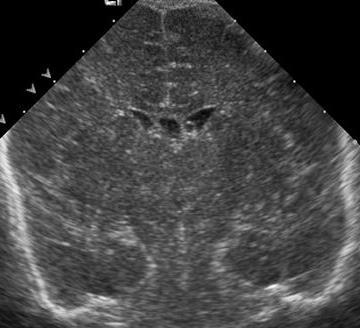
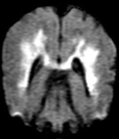
10 Focal or diffuse increase in parenchymal echogenicity Slit-like ventricles Obliteration of the extra-axial spaces
11 HII: Less recognized US findings 1. Peripheral brain findings 2. Central brain findings 3. Doppler findings
12 HII: Less recognized US findings 1. Peripheral brain findings 2. Central brain findings a. Gray-white matter differentiation b. Cortical abnormalities c. Subcortical white matter d. Extra-axial spaces abnormalities 3. Doppler findings
13 HII: Less recognized US findings 1. Peripheral brain findings 2. Central brain findings 3. Doppler findings a. Basal ganglia evaluation b. Periventricular white matter incl. medullary veins c. Active hemorrhage evaluation d. Ventricular size evaluation e. Brainstem evaluation f. Corpus Callosum evaluation
14 HII: Less recognized US findings 1. Peripheral brain findings 2. Central brain findings 3. Doppler findings a. Resistive indices (RI) fluctuation b. Hyperemia c. Sinus vein patency evaluation
15 1. HII: Peripheral US findings 1a. Gray white matter differentiation a. Accentuation b. Loss c. Mixed pattern 1b. Cortical abnormalities a. Abnormal cortical thickness and echogenicity 1c. Subcortical white matter a. Focal peripheral echogenicities 1d. Extra-axial spaces abnormalities a. Subdural collections b. Blurring interhemispheric fissure
16 1a. Gray white matter differentiation In infants with extensive white matter disease corticomedullary differentiation is considerably enhanced P. Winkler, European Journal of Radiology, 1998;26:


")
17 1a. Gray white matter differentiation Normal G-W differentiation Coronal Sagittal These images show the normal G-W differentiation, The sulci are of medium echogenecity (green line) The cortex is slightly hypoechogenic (yellow) when compared to adjacent subcortical white matter ( ) Peripheral-angled views


18 1a. Gray white matter differentiation Accentuation G-W differentiation Coronal Coronal Ax DWI DWI axial Coronal Hypoechogenic cortex Hyperechogenic sulcus Hyperechogenic white matter Sagittal Peripheral angled sagittal view in region of restricted diffusion These images show abnormal accentuation of the G-W differentiation. The sulci are thick and hyperechogenic, the cortex is hypoechogenic and the subcortical white matter is hyperechogenic in the regions of restricted diffusion


19 1a. Gray white matter differentiation Loss G-W differentiation Coronal Coronal Coronal Peripheral angled view DWI axial DWI coronal These images show loss of the G-W differentiation on both, US and DWI


20 Normal GWMD Mild Accentuation of the GWMD Accentuation GWMD + thick, bright gyri & sulci Accentuation + loss of the GWMD Loss of the GWMD Normal Worst


21 1b. Cortical abnormalities Abnormal cortical thickness and echogenicity in cortical laminar necrosis Ax T1 Cor DWI Sag T1


22 1b. Cortical abnormalities Ax T1 24 h later 24 h later Corresponding axial noncontrast T1W image shows laminar hyperintensities in the bilateral parietooccipital cortex consistent with cortical laminar necrosis Neonate with HSV encephalitis. Initial US images show mild accentuation of the G-W differentiation. Follow up US, 24 hs later show diffuse blurring of the sylvian fissures (green arrows) and abnormal thickness and echogenicity of the parietooccipital cortex (red arrows)


23 1c. Subcortical white matter abnormalities US images show patchy abnormal subcortical areas of increased echogenicity in the left perisylvian region (red arrows) Corresponding to the left perisylvian infarct with hemorrhagic conversion /hematoma seen on MRI Ax DWI Ax DWI Sag T1 Cor T2


24


25 1d. Extra-axial spaces abnormalities: Subdural collections CT wo C CT wo C Meconium aspiration. Evaluation prior to ECMO. US images show abnormal fluid collection encircling the right cerebral hemisphere (red arrows). Corresponding CT images show bilateral subdural hemorrhages.


26 1d. Extra-axial spaces abnormalities: Blurring of the inter-hemispheric fissure
27 common, partially preventable if maternal cervical colonization, 10% systemic GBS (1-5 per 1000 births meningitis in 5-10% risk in: PROM, prematurity, maternal chorioamnionitis VLBW 70% mortality full term: insidious onset, meningitis/osteomyelitis 5% mortality significant hydrocephalus, developmental delay, seizures


28 organizing exudate with fibroblast proliferation fills sulci, extends along (and bathes) vessels... leading to vascular occlusion Courtesy Dr Susan Blaser


29 GBS+


30 12/24/09


31 12/29/09 12/30/09


32 1/4/2010


33 7/15/2010
34 2. HII: Central US findings 2a. Basal Ganglia evaluation Echogenicity pattern important 2b. Periventricular white matter incl. medullary veins 2c. Active IVH 2d. Ventricular size evaluation Easiest to assess, but least useful 2e. Brainstem evaluation Less common still important 2e. Corpus Callosum evaluation

 and slitlike ventricles.")
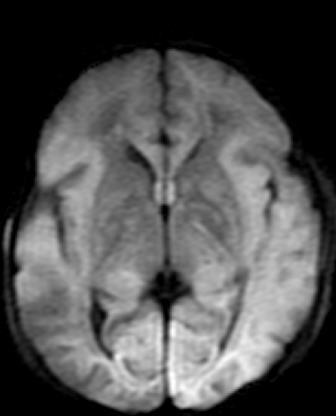
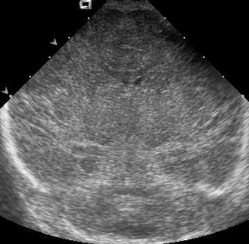
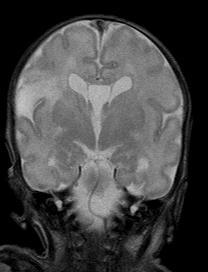
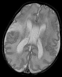
35 2a. Basal ganglia abnormalities Ax DWI Fullterm neonate with a history of profound HII. Coronal US image shows diffuse increased echogenicity of the basal ganglia (red arrows) and slitlike ventricles. Axial DWI shows restricted diffusion in the bilateral posterior putamen (green arrows) and ventrolateral thalami (blue arrows).


36 2a. Basal ganglia abnormalities


37
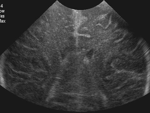
.")


38 2a. Basal ganglia abnormalities Ax T2W Coronal US image shows bilateral areas of abnormally increased echogenicity involving the thalami (red arrows). Note blurring of the interhemispheric fissure (blue arrows). Corresponding axial T2-weighted image reveals extensive edema in the bilateral thalami (green arrows), periventricular white matter and within both frontal regions. These findings are characteristic of deep cerebral venous thrombosis involving the bilateral thalamoperforating veins and bilateral internal cerebral veins.


 when")


39 2a. Basal ganglia abnormalities There is subtle increased echogenicity on the right basal ganglia (yellow arrow) when compared to the left (green arrow) The MR images confirm the areas of abnormality on the left and show this more floridly than the US Ax DWI Ax T2 Ax T1 Cor T1


40 2b. Periventricular white matter abnormalities incl. medullary veins

41 Evaluation of medullary veins
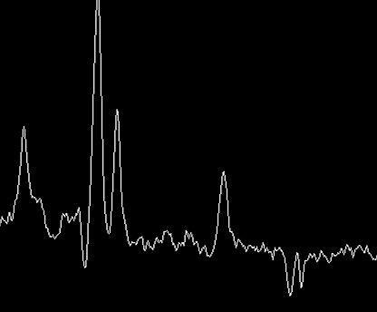
 can")


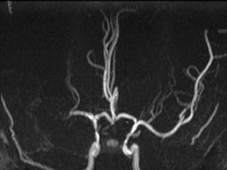
42 Fetal MRI. 20 weeks gestation. Normal medullary veins(arrows) can be seen in HASTE and EPI sequences Fetal MRI. 28 weeks gestation. Normal medullary veins (arrows) can be seen in EPI sequences. Courtesy Dr. Tamara Feygin


43 HII: Hypoperfusion Medullary venous congestion in twintwin transfusion Courtesy Susan Blaser MD


44 Courtesy Dr. Alan Daneman


45 It is important to differentiate GM-IVH from bilateral hemorrhagic PVL. These two entities are distinct in their neurodevelopmental outcome, which is more favorable for the GM-IVH


46 Courtesy Dr. Tamara Feygin


47 2b. Periventricular white matter abnormalities incl. medullary veins Sagittal US images show increased periventricular echogenicity consistent with PVL (red arrows). These findings are most conspicuous on the image on the left obtained with a linear transducer and reflect small areas of ischemia and/or hemorrhage in medullary vein distribution (green arrows)


48 2b. Periventricular white matter abnormalities incl. medullary veins 6-day-old with large SCT and acute drop in hemoglobine. Head US shows a left grade 1 IVH and abnormal accentuation of the periventicular medullary veins, noted as periventricular linear echogenicities (yellow arrows). These are believed to play an important role in the physiopathology of PVL


49 2c. Active IVH Us movies depict active left grade 1 IVH
 Asymmetry with heterogenous")
 peduncle Ax DWI Brainstem axial view")

50 2d. Brainstem evaluation Normal brainstem echo pattern (green arrows) Asymmetry with heterogenous echotexture of the abnormal left cerebral (yellow arrow) peduncle Ax DWI Brainstem axial view Brainstem axial view Corresponding abnormal brainstem restricted diffusion in the left cerebral peduncle (yellow arrow)


51 2d. Brainstem evaluation Focused axial view of the cerebral peduncles Brainstem axial view Ax DWI Cor DWI Neonate with HII. Asymmetry of the cerebral peduncles with abnormal increase in echotexture on the left (yellow arrows) noted on axial US scans through the mendosal suture. Corresponding DWI confirms restricted diffusion within the left cerebral peduncle (blue arrows).


52 2e. Ventricular size evaluation Least useful, may be normal in severe cases Full-term baby with home delivery and abruptio placenta. PPHN, hypoxemia, respiratory distress and abnormal EEG. US shows normal ventricular size. Note moderate accentuation of the GW differentiation, abnormal basal ganglia echogenicity and blurring of the interhemispheric fissure Corresponding MRI shows extensive bilateral restricted diffusion involving the cerebral hemispheres bilaterally with sparing of the unmyelinated white matter. The ventricles are normal in size, shape, and configuration.


53 2f. Corpus callosum evaluation Newborn with severe HII. The corpus callosum (red arrows) is thickened and shows abnormal increase in echogenicity
54 3. HII: Doppler US findings 3a. Resistive indices fluctuation 3b. Assessment of hyperemia Dr Faingold 2c. Sinus vein patency evaluation
 fluctuation 11:10 25")


55 3a. Resistive indices (Ris) fluctuation 11:10 25 PM 11:12 04 PM In this patient the Doppler study of the ACA shows fluctuation of the resistive index in images taken 2 minutes apart FLUCTUATION of the RI s during the same exam, probably due to loss of autoregulation


56 3b. Assessment of hyperemia Prominent lenticulostriate vessels Prominent lenticulostriate vessels MRA Ax T2W Prominent lenticulostriate vessels Prominent lenticulostriate vessels Severe DWI restriction In this patient hyperemia is noted in the basal ganglia bilaterally on Color and Power Doppler and this was confirmed on the MRA. In addition prominent lenticulostriate vessels are appreciated on T2 weighted images as flow voids


57 Dynamic Cerebral CDS - CPI Courtesy Dr R. Faingold, Montreal, QC, Canada


58 Superior sagittal sinus Superior sagittal sinus Metabolic disorder


 Neonatal Cerebral")

59 3c. Sinus vein patency evaluation US and MRI images show extensive superior sagittal vein thrombosis. No flow is elicited in the superior sagittal sinus (arrows) on color Doppler imaging despite adequate low settings (as low as 2.2 cm/s) Neonatal Cerebral Sinovenous Thrombosis - Transient abnormal protein C with infection and birth - Poor outcome
60 Real time imaging Multiple transducers High frequency (~ 8 MHz) Vector, curved array, linear Multiple windows and focused views Angled views to include the periphery of the brain; Central focused views; Posterior fossa Doppler - spectral, color, power Implementation of rounds including: Sonographers Neuroradiologists neonatologists Implementation of these practices and techniques will improve the reliability of HUS to depict HII related changes


61 Special Thanks: Drs. Alan Daneman and Susan Blaser, Toronto, Sick Kids, Canada Drs. Ricardo Faingold and Natalia Gorelik, Montreal Children s Hospital, Canada Drs. Tamara Feygin, Zimmerman, Nancy Chauvin & Tedi Victoria, CHOP, Philadelphia, USA
Enhancement of Cranial US: Utility of Supplementary Acoustic Windows and Doppler Harriet J. Paltiel, MD
 Enhancement of Cranial US: Utility of Supplementary Acoustic Windows and Doppler Harriet J. Paltiel, MD Boston Children s Hospital Harvard Medical School None Disclosures Conventional US Anterior fontanelle
Neurosonography: State of the art
 Neurosonography: State of the art Lisa H Lowe, MD, FAAP Professor and Academic Chair, University MO-Kansas City Pediatric Radiologist, Children s Mercy Hospitals and Clinics Learning objectives After this
NEURO IMAGING 2. Dr. Said Huwaijah Chairman of radiology Dep, Damascus Univercity
 NEURO IMAGING 2 Dr. Said Huwaijah Chairman of radiology Dep, Damascus Univercity I. EPIDURAL HEMATOMA (EDH) LOCATION Seventy to seventy-five percent occur in temporoparietal region. CAUSE Most likely caused
Ultrasound examination of the neonatal brain
 Ultrasound examination of the neonatal brain Guideline for the performance and reporting of neonatal and preterm brain ultrasound examination, by the Finnish Perinatology Society and the Paediatric Radiology
IMAGING OF HYPOXIC ISCHEMIC INJURY IN A NEONATE FN3 STATE MEETING NEMOURS CHILDREN'S HOSPITAL ORLANDO,FL 08/04/18
 IMAGING OF HYPOXIC ISCHEMIC INJURY IN A NEONATE FN3 STATE MEETING NEMOURS CHILDREN'S HOSPITAL ORLANDO,FL 08/04/18 Dhanashree Rajderkar,MD Assistant Professor Department of Radiology University of Florida
ISCHEMIC STROKE IMAGING
 ISCHEMIC STROKE IMAGING ผศ.พญ พญ.จ ร ร ตน ธรรมโรจน ภาคว ชาร งส ว ทยา คณะแพทยศาสตร มหาว ทยาล ยขอนแก น A case of acute hemiplegia Which side is the abnormality, right or left? Early Right MCA infarction
Neonatal Hypoxic-Ischemic Injury: Ultrasound and Dynamic Color Doppler Sonography perfusion of the Brain and Abdomen with pathologic correlation.
 Neonatal Hypoxic-Ischemic Injury: Ultrasound and Dynamic Color Doppler Sonography perfusion of the Brain and Abdomen with pathologic correlation. Ricardo Faingold,MD Montreal Children s Hospital Medical
Neonatal hypoxic-ischemic brain injury imaging: A pictorial review
 Neonatal hypoxic-ischemic brain injury imaging: A pictorial review Poster No.: C-1425 Congress: ECR 2014 Type: Educational Exhibit Authors: E. Alexopoulou 1, A. Mazioti 1, D. K. Filippiadis 2, C. Chrona
Pearls and Pitfalls in Neuroradiology of Cerebrovascular Disease The Essentials with MR and CT
 Pearls and Pitfalls in Neuroradiology of Cerebrovascular Disease The Essentials with MR and CT Val M. Runge, MD Wendy R. K. Smoker, MD Anton Valavanis, MD Control # 823 Purpose The focus of this educational
Hypoxic ischemic brain injury in neonates - early MR imaging findings
 Hypoxic ischemic brain injury in neonates - early MR imaging findings Poster No.: C-1208 Congress: ECR 2015 Type: Authors: Keywords: DOI: Educational Exhibit E.-M. Heursen, R. Reina Cubero, T. Guijo Hernandez,
Essentials of Clinical MR, 2 nd edition. 14. Ischemia and Infarction II
 14. Ischemia and Infarction II Lacunar infarcts are small deep parenchymal lesions involving the basal ganglia, internal capsule, thalamus, and brainstem. The vascular supply of these areas includes the
Cerebro-vascular stroke
 Cerebro-vascular stroke CT Terminology Hypodense lesion = lesion of lower density than the normal brain tissue Hyperdense lesion = lesion of higher density than normal brain tissue Isodense lesion = lesion
HEAD AND NECK IMAGING. James Chen (MS IV)
") HEAD AND NECK IMAGING James Chen (MS IV) Anatomy Course Johns Hopkins School of Medicine Sept. 27, 2011 OBJECTIVES Introduce cross sectional imaging of head and neck Computed tomography (CT) Review head
HEAD AND NECK IMAGING James Chen (MS IV) Anatomy Course Johns Hopkins School of Medicine Sept. 27, 2011 OBJECTIVES Introduce cross sectional imaging of head and neck Computed tomography (CT) Review head
Quick practical guide to Cranial Ultrasound in the newborn
 Quick practical guide to Cranial Ultrasound in the newborn Introduction A standard set of views is taken to assist with consistent visualisation of structures and in the interpretation of possible abnormalities.
Head CT Scan Interpretation: A Five-Step Approach to Seeing Inside the Head Lawrence B. Stack, MD
 Head CT Scan Interpretation: A Five-Step Approach to Seeing Inside the Head Lawrence B. Stack, MD Five Step Approach 1. Adequate study 2. Bone windows 3. Ventricles 4. Quadrigeminal cistern 5. Parenchyma
Insults to the Developing Brain & Effect on Neurodevelopmental Outcomes
 Insults to the Developing Brain & Effect on Neurodevelopmental Outcomes Ira Adams-Chapman, MD Assistant Professor of Pediatrics Director, Developmental Progress Clinic Emory University School of Medicine
I t is increasingly recognised that arterial cerebral infarction
 F252 ORIGINAL ARTICLE Does cranial ultrasound imaging identify arterial cerebral infarction in term neonates? F Cowan, E Mercuri, F Groenendaal, L Bassi, D Ricci, M Rutherford, L de Vries... See end of
Term Hypoxic Ischemic Injury Joseph Junewick, MD FACR
 Term Hypoxic Ischemic Injury Joseph Junewick, MD FACR 08/11/2010 History Term infant with perinatal distress and attempted forceps delivery. Diagnosis Term Hypoxic Ischemic Injury Discussion Encephalopathy
Original Articles NEUROSONOGRAPHIC ABNORMALITIES IN NEONATES WITH HYPOXIC ISCHEMIC ENCEPHALOPATHY
 Original Articles NEUROSONOGRAPHIC ABNORMALITIES IN NEONATES WITH HYPOXIC ISCHEMIC ENCEPHALOPATHY N.K. Anand A.K. Gupta I.M.S. Lamba ABSTRACT Pattern of neurosonographic (NSG) abnormalities in 150 term
State-of-the-Art Cranial Sonography: Part 1, Modern Techniques and Image Interpretation
 Pediatric Imaging Review Lowe and ailey Cranial Sonography Pediatric Imaging Review Downloaded from www.ajronline.org by 37.44.206.98 on 02/10/18 from IP address 37.44.206.98. Copyright RRS. For personal
SWISS SOCIETY OF NEONATOLOGY. Neonatal cerebral infarction
 SWISS SOCIETY OF NEONATOLOGY Neonatal cerebral infarction May 2002 2 Mann C, Neonatal and Pediatric Intensive Care Unit, Landeskrankenhaus und Akademisches Lehrkrankenhaus Feldkirch, Austria Swiss Society
Imaging findings in neonates with hypoxic-ischaemic encephalopathy and terapeutic hypothermia.
 Imaging findings in neonates with hypoxic-ischaemic encephalopathy and terapeutic hypothermia. Poster No.: C-1577 Congress: ECR 2014 Type: Scientific Exhibit Authors: S. Manso Garcia, M. J. Velasco Marcos,
Original article: Evaluation of hypoxic-ischaemic events in preterm neonates using trans cranial ultrasound
 Original article: Evaluation of hypoxic-ischaemic events in preterm neonates using trans cranial ultrasound Priyanka Upadhyay *, Ketki U Patil 1, Rajesh Kuber 2, Vilas Kulkarni 3, Amarjit Singh 4 * Chief
Chapter 3. Neonatal cranial ultrasonography: how to optimize its performance
 Chapter 3 Neonatal cranial ultrasonography: how to optimize its performance Sylke J. Steggerda Lara M. Leijser Frans J. Walther Gerda van Wezel-Meijler Early Human Development 2009; 85(2): 93-99 Chapter
brain MRI for neuropsychiatrists: what do you need to know
 brain MRI for neuropsychiatrists: what do you need to know Christoforos Stoupis, MD, PhD Department of Radiology, Spital Maennedorf, Zurich & Inselspital, University of Bern, Switzerland c.stoupis@spitalmaennedorf.ch
Imaging findings in neonates with hypoxic-ischaemic encephalopathy and terapeutic hypothermia.
 Imaging findings in neonates with hypoxic-ischaemic encephalopathy and terapeutic hypothermia. Poster No.: C-1577 Congress: ECR 2014 Type: Scientific Exhibit Authors: S. Manso Garcia, M. J. Velasco Marcos,
1 MS Lesions in T2-Weighted Images
 1 MS Lesions in T2-Weighted Images M.A. Sahraian, E.-W. Radue 1.1 Introduction Multiple hyperintense lesions on T2- and PDweighted sequences are the characteristic magnetic resonance imaging (MRI) appearance
41 year old female with headache. Elena G. Violari MD and Leo Wolansky MD
 41 year old female with headache Elena G. Violari MD and Leo Wolansky MD ? Dural Venous Sinus Thrombosis with Hemorrhagic Venous Infarct Acute intraparenchymal hematoma measuring ~3 cm in diameter centered
V. CENTRAL NERVOUS SYSTEM TRAUMA
 V. CENTRAL NERVOUS SYSTEM TRAUMA I. Concussion - Is a clinical syndrome of altered consiousness secondary to head injury - Brought by a change in the momentum of the head when a moving head suddenly arrested
Surgical Options in Post Haemorrhagic Ventricular Dilation
 Surgical Options in Post Haemorrhagic Ventricular Dilation Benedetta Pettorini Consultant Paediatric Neurosurgeon Alder Hey Childrens Hospital Liverpool, UK Risk Factors for IVH 1. Prematurity: Occurs
ECMUS The Safety Committee of EFSUMB : Tutorial
 Neonatal cranial ultrasound Safety Aspects (2013) Prepared for ECMUS by B.J. van der Knoop, M.D. 1, J.I.P. de Vries, M.D., PhD 1, I.A. Zonnenberg, M.D. 2, J.I.M.L. Verbeke, M.D. 3 R.J. Vermeulen, M.D.,
The child with hemiplegic cerebral palsy thinking beyond the motor impairment. Dr Paul Eunson Edinburgh
 The child with hemiplegic cerebral palsy thinking beyond the motor impairment Dr Paul Eunson Edinburgh Content Coming to a diagnosis The importance of understanding the injury MRI scans Role of epilepsy
Neonatal Intracranial Ultrasound Imaging - A Pictorial Review from The Royal Liverpool Children's Hospital, Alder Hey, Liverpool.
 Neonatal Intracranial Ultrasound Imaging - A Pictorial Review from The Royal Liverpool Children's Hospital, Alder Hey, Liverpool. Poster No.: C-1115 Congress: ECR 2012 Type: Educational Exhibit Authors:
Neuroradiological Findings in Non- Accidental Trauma Educational Pictorial Review
 Neuroradiological Findings in Non- Accidental Trauma Educational Pictorial Review M B Moss, MD; L Lanier, MD; R Slater; C L Sistrom, MD; R G Quisling, MD; I M Schmalfuss, MD; and D Rajderkar, MD Contact:
Stroke School for Internists Part 1
 Stroke School for Internists Part 1 November 4, 2017 Dr. Albert Jin Dr. Gurpreet Jaswal Disclosures I receive a stipend for my role as Medical Director of the Stroke Network of SEO I have no commercial
The rationale for routine cerebral ultrasound in premature infants
 Pediatr Radiol (2015) 45:646 650 DOI 10.1007/s00247-014-2985-1 RESEARCH FORUM The rationale for routine cerebral ultrasound in premature infants Maria I. Argyropoulou & Corinne Veyrac Received: 4 October
SWI including phase and magnitude images
 On-line Table: MRI imaging recommendation and summary of key features Sequence Pathologies Visible Key Features T1 volumetric high-resolution whole-brain reformatted in axial, coronal, and sagittal planes
Acute stroke. Ischaemic stroke. Characteristics. Temporal classification. Clinical features. Interpretation of Emergency Head CT
 Ischaemic stroke Characteristics Stroke is the third most common cause of death in the UK, and the leading cause of disability. 80% of strokes are ischaemic Large vessel occlusive atheromatous disease
SOP: Cerebral Ultrasound
 SOP: Cerebral Ultrasound Version Author(s) Date Changes Approved by 1.0 Cornelia Hagmann Manon Benders 29.5.2012 Initial Version Gorm Greisen 1.1 Cornelia Hagmann 18.6.2012 Minor changes Gorm Greisen 1.2
Advanced magnetic resonance imaging for monitoring brain development and injury
 Advanced magnetic resonance imaging for monitoring brain development and injury Stéphane Sizonenko, MD-PhD Division of Development and Growth Department of Child and Adolescent Medicine Geneva University
MR imaging of neonatal brain infections: Germ and specific signs
 MR imaging of neonatal brain infections: Germ and specific signs Poster No.: C-1646 Congress: ECR 2016 Type: Educational Exhibit Authors: N. Mama 1, A. Cherif 1, Y. BEN CHEIKH 2, A. Berrich 1, M. Gaha
2016 SPR Pediatric Ultrasound Course Neurosonography Session Friday, November 4, SAM Reference Document
 2016 SPR Pediatric Ultrasound Course Neurosonography Session Friday, November 4, 2016 SAM Reference Document Neurosonography - Assessing the Premature Neonate Harris L. Cohen, MD, FACR 1. Regarding the
Transfontanelar Ultrasound Technique, Normal Anatomy, Anatomic Variants and Classification Review
 Transfontanelar Ultrasound Technique, Normal Anatomy, Anatomic Variants and Classification Review Poster No.: C-2615 Congress: ECR 2013 Type: Educational Exhibit Authors: S. E. Vazquez, R. E. Ochoa Albíztegui
In utero and perinatal hypoxic brain damage
 In utero and perinatal hypoxic brain damage G Dekker, MB ChB, MFam Med H B Louw, S Andronikou, MB BCh, FC Rad (SA) M Pienaar, BSc, MB ChB S Hlongwane, MB ChB A Brandt, MB ChB D van der Merwe, MB ChB Department
C. Douglas Phillips, MD FACR Director of Head and Neck Imaging Weill Cornell Medical College NewYork-Presbyterian Hospital
 C. Douglas Phillips, MD FACR Director of Head and Neck Imaging Weill Cornell Medical College NewYork-Presbyterian Hospital I have no financial disclosures Understand range of pathology that may present
CT and MR findings of systemic lupus erythematosus involving the brain: Differential diagnosis based on lesion distribution
 CT and MR findings of systemic lupus erythematosus involving the brain: Differential diagnosis based on lesion distribution Poster No.: C-2723 Congress: ECR 2010 Type: Educational Exhibit Topic: Neuro
Pediatric MS MRI Study Methodology
 General Pediatric MS MRI Study Methodology SCAN PREPARATION axial T2-weighted scans and/or axial FLAIR scans were obtained for all subjects when available, both T2 and FLAIR scans were scored. In order
RINGS N THINGS: Imaging Patterns in Differential Diagnosis. Anne G. Osborn, M.D.
 RINGS N THINGS: Imaging Patterns in Differential Diagnosis Anne G. Osborn, M.D. ExpDDxs: Intra-axial (Parenchymal) Lesions Ring-enhancing lesions, solitary 1 Ring-enhancing lesion crossing corpus callosum
Perinatal cerebral white matter injuries influence early communication and language development
 Perinatal cerebral white matter injuries influence early communication and language development Blazenka Brozovic University of Zagreb Department of Speech and Language Pathology Developmental Neurolinguistic
intracranial anomalies
 Chapter 5: Fetal Central Nervous System 84 intracranial anomalies Hydrocephaly Dilatation of ventricular system secondary to an increase in the amount of CSF. Effects of hydrocephalus include flattening
Neonatal Hypoxic ischemic Encephalopathy: A Radiological Review
 Review Article Neonatal Hypoxic ischemic Encephalopathy: A Radiological Review Shahina Bano, Vikas Chaudhary 1, Umesh Chandra Garga Department of Radiodiagnosis, PGIMER, Dr. RML Hospital, 1 Department
National follow-up program CPUP Pediatric Neurology paper form
 National follow-up program CPUP Pediatric Neurology paper form 110206 1 National Follow-Up program- CPUP Pediatric Neurology Personal nr (unique identifier): Last name: First name: Region child belongs
NEURORADIOLOGY DIL part 3
 NEURORADIOLOGY DIL part 3 Bleeds and hemorrhages K. Agyem MD, G. Hall MD, D. Palathinkal MD, Alexandre Menard March/April 2015 OVERVIEW Introduction to Neuroimaging - DIL part 1 Basic Brain Anatomy - DIL
Prognosis in. Encephalopathy. Hypoxic-Ischemic. Özge Aydemİr MD
 Prognosis in Hypoxic-Ischemic Encephalopathy Özge Aydemİr MD Major problems we have to face while caring infants with HIE are; Øto provide families with reliable information about outcome. Øto decide how
Automated Identification of Neoplasia in Diagnostic Imaging text reports
 Automated Identification of Neoplasia in Diagnostic Imaging text reports "This work has been funded in whole or in part with Federal funds from the National Cancer Institute, National Institutes of Health,
STROKE - IMAGING. Dr RAJASEKHAR REDDY 2nd Yr P.G. RADIODIAGNOSIS KIMS,Narkatpalli.
 STROKE - IMAGING Dr RAJASEKHAR REDDY 2nd Yr P.G. RADIODIAGNOSIS KIMS,Narkatpalli. STROKE Describes a clinical event that consists of sudden onset of neurological symptoms Types Infarction - occlusion of
Attenuation value in HU From -500 To HU From -10 To HU From 60 To 90 HU. From 200 HU and above
 Brain Imaging Common CT attenuation values Structure Air Fat Water Brain tissue Recent hematoma Calcifications Bone Brain edema and infarction Normal liver parenchyma Attenuation value in HU From -500
Blood Supply. Allen Chung, class of 2013
 Blood Supply Allen Chung, class of 2013 Objectives Understand the importance of the cerebral circulation. Understand stroke and the types of vascular problems that cause it. Understand ischemic penumbra
Fundamental Clinical Brain MR Imaging Applications and Protocols
 Continuing Education Seminar for Radiologic Technologists Fundamental Clinical Brain MR Imaging Applications and Protocols Darren P. O Neill, MD Indiana University Neuroradiology Objectives Review fundamental
Anoxic brain injury CT and MRI patterns - quick pictoral quide for junior radiologists.
 Anoxic brain injury CT and MRI patterns - quick pictoral quide for junior radiologists. Poster No.: C-1844 Congress: ECR 2017 Type: Educational Exhibit Authors: A. Kecler - Pietrzyk, W. Torreggiani ; Dublin/IE,
Non-Traumatic Neuro Emergencies
 Department of Radiology University of California San Diego Non-Traumatic Neuro Emergencies John R. Hesselink, M.D. Nontraumatic Neuroemergencies 1. Acute focal neurological deficit 2. Worst headache of
Cerebral malaria: MR imaging spectrum
 Cerebral malaria: MR imaging spectrum Poster No.: C-2705 Congress: ECR 2010 Type: Educational Exhibit Topic: Neuro Authors: P. S. Naphade, M. D. Agrawal, S. S. Sankhe, K. M. Siva, B. K. Jain; Mumbai/IN
[(PHY-3a) Initials of MD reviewing films] [(PHY-3b) Initials of 2 nd opinion MD]
![[(PHY-3a) Initials of MD reviewing films] [(PHY-3b) Initials of 2 nd opinion MD]](/thumbs/89/98619893.jpg "[(PHY-3a) Initials of MD reviewing films] [(PHY-3b) Initials of 2 nd opinion MD]") 2015 PHYSICIAN SIGN-OFF (1) STUDY NO (PHY-1) CASE, PER PHYSICIAN REVIEW 1=yes 2=no [strictly meets case definition] (PHY-1a) CASE, IN PHYSICIAN S OPINION 1=yes 2=no (PHY-2) (PHY-3) [based on all available
2015 PHYSICIAN SIGN-OFF (1) STUDY NO (PHY-1) CASE, PER PHYSICIAN REVIEW 1=yes 2=no [strictly meets case definition] (PHY-1a) CASE, IN PHYSICIAN S OPINION 1=yes 2=no (PHY-2) (PHY-3) [based on all available
Natalia Gorelik 1, Ricardo Faingold 2, Alan Daneman 3, Monica Epelman 3,4. Original Article
 Original Article Intraventricular hemorrhage in term neonates with hypoxicischemic encephalopathy: a comparison study between neonates treated with and without hypothermia Natalia Gorelik 1, Ricardo Faingold
The central nervous system
 Sectc.qxd 29/06/99 09:42 Page 81 Section C The central nervous system CNS haemorrhage Subarachnoid haemorrhage Cerebral infarction Brain atrophy Ring enhancing lesions MRI of the pituitary Multiple sclerosis
RESEARCH ARTICLE EVALUATION OF NEUROIMAGING IN CEREBRAL PALSY. S.H. Hasanpour avanji MD
 RESEARCH ARTICLE EVALUATION OF NEUROIMAGING IN CEREBRAL PALSY S.H. Hasanpour avanji MD Assistant Professor of Child Neurology, Iran University of Medical Sciences Corresponding Author: S.H. Hasanpour avanji
Neonatal Periventricular Leukomalacia: Real-Time
 383 Neonatal Periventricular Leukomalacia: Real-Time Sonographic Diagnosis with CT Correlation Peter P. Chow 1. 2 J. Gerard Horgan 1, 3 Kenneth J. W. Taylor 1 The utility of real-time sonography in the
2. Subarachnoid Hemorrhage
 Causes: 2. Subarachnoid Hemorrhage A. Saccular (berry) aneurysm - Is the most frequent cause of clinically significant subarachnoid hemorrhage is rupture of a saccular (berry) aneurysm. B. Vascular malformation
Difficulties at Birth: Long Term Developmental Outcomes
 Difficulties at Birth: Long Term Developmental Outcomes Alan D. Bedrick MD Division of Neonatology and Developmental Biology Department of Pediatrics University of Arizona Tucson, Arizona DISCLOSURE I
3T MRI imaging approach to pediatric epileptic seizures:
 3T MRI imaging approach to pediatric epileptic seizures: Poster No.: C-1886 Congress: ECR 2016 Type: Educational Exhibit Authors: J. S. Alaín, E. M. DE LUCAS, J. C. Quintero Rivera, C. Pérez 1 2 3 3 3
Study of role of MRI brain in evaluation of hypoxic ischemic encephalopathy
 Original article: Study of role of MRI brain in evaluation of hypoxic ischemic encephalopathy *Dr Harshad Bhagat, ** Dr Ravindra Kawade, ***Dr Y.P.Sachdev *Junior Resident, Department Of Radiodiagnosis,
Cerebellar Vermian Atrophy after Neonatal Hypoxic-Ischemic Encephalopathy
 AJNR Am J Neuroradiol 25:1008 1015, June/July 2004 Vermian Atrophy after Neonatal Hypoxic-Ischemic Encephalopathy Michael A. Sargent, Kenneth J. Poskitt, Elke H. Roland, Alan Hill, and Glenda Hendson BACKGROUND
Differentiation between peritrigonal terminal zones and hypoxic-ischemic white matter injury on MRI
 Chapter 4 Differentiation between peritrigonal terminal zones and hypoxic-ischemic white matter injury on MRI Lishya Liauw Jeroen van der Grond Valerie Slooff Francisca Wiggers-de Bruïne Laura Laan Saskia
L M Thornton, MD; L Lanier, MD; C L Sistrom, MD; D Rajderkar, MD; A Mancuso, MD; IM Schmalfuss, MD University of Florida, Gainesville Department of
 L M Thornton, MD; L Lanier, MD; C L Sistrom, MD; D Rajderkar, MD; A Mancuso, MD; IM Schmalfuss, MD University of Florida, Gainesville Department of Radiology RSNA Annual Meeting 2016 Trainee call readiness
High spatial resolution reveals excellent detail in pediatric neuro imaging
 Publication for the Philips MRI Community Issue 46 2012/2 High spatial resolution reveals excellent detail in pediatric neuro imaging Achieva 3.0T with 32-channel SENSE Head coil has become the system
An Approach to Cystic White Matter Diseases of the Paediatric Brain
 An Approach to Cystic White Matter Diseases of the Paediatric Brain Poster No.: C-0239 Congress: ECR 2017 Type: Educational Exhibit Authors: S. Culleton, J. P. Donnellan, E. Laffan, I. Robinson, E. L.
An Introduction to Imaging the Brain. Dr Amy Davis
 An Introduction to Imaging the Brain Dr Amy Davis Common reasons for imaging: Clinical scenarios: - Trauma (NICE guidelines) - Stroke - Tumours - Seizure - Neurological degeneration memory, motor dysfunction,
Neuropathology Specialty Conference
 Neuropathology Specialty Conference March 22, 2010 Case 2 Rebecca Folkerth, MD Brigham and Women s Hospital Children s Hospital Harvard Medical School Clinical History 18-gestational-week fetus found on
CENTRAL NERVOUS SYSTEM TRAUMA and Subarachnoid Hemorrhage. By: Shifaa AlQa qa
 CENTRAL NERVOUS SYSTEM TRAUMA and Subarachnoid Hemorrhage By: Shifaa AlQa qa Subarachnoid Hemorrhage Causes: Rupture of a saccular (berry) aneurysm Vascular malformation Trauma Hematologic disturbances
Discovering the hippocampus with cranial-ct.
 Discovering the hippocampus with cranial-ct. Poster No.: C-0378 Congress: ECR 2018 Type: Educational Exhibit Authors: F. Pozo Piñon, A. B. Barba Arce, E. herrera romero, V. 1 2 3 1 3 3 Fernández Lobo,
MRI OF THE THALAMUS. Mohammed J. Zafar, MD, FAAN Kalamazoo, MI
 1 MRI OF THE THALAMUS Mohammed J. Zafar, MD, FAAN Kalamazoo, MI Objectives: The thalamic nuclei can be involved in a wide variety of conditions. A systematic imaging approach would be useful for narrowing
Correlation of Neurodevelopmental Outcome and brain MRI/EEG findings in term HIE infants
 Correlation of Neurodevelopmental Outcome and brain MRI/EEG findings in term HIE infants Ajou University School of Medicine Department of Pediatrics Moon Sung Park M.D. Hee Cheol Jo, M.D., Jang Hoon Lee,
Intracranial spontaneous hemorrhage mechanisms, imaging and management
 Intracranial spontaneous hemorrhage mechanisms, imaging and management Dora Zlatareva Department of Diagnostic Imaging Medical University, Sofia, Bulgaria Intracranial hemorrhage (ICH) ICH 15% of strokes
Index. aneurysm, 92 carotid occlusion, 94 ICA stenosis, 95 intracranial, 92 MCA, 94
 A ADC. See Apparent diffusion coefficient (ADC) Aneurysm cerebral artery aneurysm, 93 CT scan, 93 gadolinium, 93 Angiography, 13 Anoxic brain injury, 25 Apparent diffusion coefficient (ADC), 7 Arachnoid
Necrotizing Enterocolitis: the role of ultrasound in the assessment of bowel viability
 Necrotizing Enterocolitis: the role of ultrasound in the assessment of bowel viability Ricardo Faingold, MD. Department of Medical Imaging The Montreal Children s Hospital McGill University SPR Vancouver
Newborn Hypoxic Ischemic Brain Injury. Hisham Dahmoush, MBBCh FRCR Lucile Packard Children s Hospital at Stanford
 Newborn Hypoxic Ischemic Brain Injury Hisham Dahmoush, MBBCh FRCR Lucile Packard Children s Hospital at Stanford NO DISCLOSURES INTRODUCTION Neonatal hypoxic-ischemic encephalopathy (HIE) is a major cause
Regional and Lobe Parcellation Rhesus Monkey Brain Atlas. Manual Tracing for Parcellation Template
 Regional and Lobe Parcellation Rhesus Monkey Brain Atlas Manual Tracing for Parcellation Template Overview of Tracing Guidelines A) Traces are performed in a systematic order they, allowing the more easily
NEURO IMAGING OF ACUTE STROKE
 1 1 NEURO IMAGING OF ACUTE STROKE ALICIA RICHARDSON, MSN, RN, ACCNS-AG, ANVP-BC WENDY SMITH, MA, RN, MBA, SCRN, FAHA LYNN HUNDLEY, APRN, CNRN, CCNS, ANVP-BC 2 2 1 DISCLOSURES Alicia Richardson: Stryker
Brain anatomy tutorial. Dr. Michal Ben-Shachar 459 Neurolinguistics
 Brain anatomy tutorial Dr. Michal Ben-Shachar 459 Neurolinguistics The human brain Left hemisphere Right hemisphere http://www.brainmuseum.org/ Zoom out Zoom in Types of Brain Tissue Gray Matter: Cell
Prevalence of "Compressed" and Asymmetric Lateral Ventricles in Healthy Full Term Neonates: Sonographic
 Prevalence of "Compressed" and symmetric Lateral Ventricles in Healthy Full Term Neonates: Sonographic Study 149 Patricia Winchester 1 Paula W. rill1 Rebecca Cooper2 lfred N. Krauss 2 Hart dec Peterson
P rominent Extraaxial CSF Space on Cranial Ultra s o u n d in Infants: C o r relation with Neuro d evelopmental Outc o m e 1
 P rominent Extraaxial CSF Space on Cranial Ultra s o u n d in Infants: C o r relation with Neuro d evelopmental Outc o m e 1 Bokyung Kim Han, M.D., Mu n hyang Lee, M.D. 2, Hye - Kyung Yoon, M.D., Kyung-Jae
TOXIC AND NUTRITIONAL DISORDER MODULE
 TOXIC AND NUTRITIONAL DISORDER MODULE Objectives: For each of the following entities the student should be able to: 1. Describe the etiology/pathogenesis and/or pathophysiology, gross and microscopic morphology
Epilepsy in children with cerebral palsy
 Seizure 2003; 12: 110 114 doi:10.1016/s1059 1311(02)00255-8 Epilepsy in children with cerebral palsy A.K. GURURAJ, L. SZTRIHA, A. BENER,A.DAWODU & V. EAPEN Departments of Paediatrics, Community Medicine
Anatomy & Physiology Central Nervous System Worksheet
 1. What are the two parts of the CNS? 2. What are the four functions of the CNS Anatomy & Physiology Central Nervous System Worksheet 3. What are the four functions of the meninges? (p430) 4. Starting
Atypical Unilateral Posterior Reversible Encephalopathy Syndrome Mimicking a Middle Cerebral Artery Infarction
 Case Report Neuroimaging and Head & Neck http://dx.doi.org/10.3348/kjr.2015.16.5.1104 pissn 1229-6929 eissn 2005-8330 Korean J Radiol 2015;16(5):1104-1108 Atypical Unilateral Posterior Reversible Encephalopathy
Benign brain lesions
 Benign brain lesions Diagnostic and Interventional Radiology Hung-Wen Kao Department of Radiology, Tri-Service General Hospital, National Defense Medical Center Computed tomography Hounsfield unit (HU)
Chapter 5: Fetal Central Nervous System 71
 71 Chapter 5 Fetal Central Nervous System Embryology NEURULATION begins with the formation of the neural plate, the neural folds and their ultimate fusion and closure as the NEURAL TUBE. NEURAL PLATE -
NEURORADIOLOGY DIL part 4
 NEURORADIOLOGY DIL part 4 Strokes and infarcts K. Agyem MD, G. Hall MD, D. Palathinkal MD, Alexandre Menard March/April 2015 OVERVIEW Introduction to Neuroimaging - DIL part 1 Basic Brain Anatomy - DIL
HYPOXIC ISCHEMIC ENCEPHALOPATHY AND THE OBSTETRICIAN
 HYPOXIC ISCHEMIC ENCEPHALOPATHY AND THE OBSTETRICIAN DISCLOSURE I have nothing to disclose and have no real or potential conflicts with this presentation and its content. Michael P. Nageotte, M.D. CASE:
White matter diseases affecting the corpus callosum; demyelinating and metabolic diseases
 White matter diseases affecting the corpus callosum; demyelinating and metabolic diseases Poster No.: C-0199 Congress: ECR 2011 Type: Educational Exhibit Authors: J. H. Yoo; Seoul/KR Keywords: Neuroradiology