06 Tooth Development and Eruption
|
|
|
- Noel Green
- 5 years ago
- Views:
Transcription
1 + 06 Tooth Development and Eruption Tooth development Root development PDL and alveolar bone development Primary tooth eruption and shedding Permanent tooth eruption


2 Q. Where and how tooth starts to form? Primitive oral cavity
3 + Tooth development (Odontogenesis) Primary epithelial band Primary epithelial band Tooth germ


4 Midsagittal section of embryo at 4 weeks Future Maxilla ectomesenchyme Future Tooth epithelium Stomodeum Future Tongue Future Mandible Future Tooth


5
6 + Tooth development (Odontogenesis) A continuous process Be divided into 4 stages based on the appearance of the developing structures Initiation, bud, cap, bell, apposition, and maturation stage Physiological processes: induction, proliferation, differentiation, morphogenesis, and maturation Not all the teeth begin to develop at the same time. Teeth have the longest developmental period.


7 Table 6-1 Stages of Tooth Development Initiation stage/sixth to seventh week Bud stage/eighth week Induction Proliferation Ectoderm lining stomodeum gives rise to oral epithelium and then to dental lamina; adjacent to deeper ectomesenchyme, which is influenced by the neural crest cells. Both tissue types are separated by a basement membrane Growth of dental lamina into bud shape that penetrates growing ectomesenchyme Cap stage/ninth to tenth week Proliferation, differentiation, morphogenesis Formation of tooth germ as enamel organ forms into cap shape that surrounds inside mass of dental papilla, with an outside mass of dental sac, both from the ectomesenchyme. Bell stage/eleventh to twelfth week Proliferation, differentiation, morphogenesis Differentiation of enamel organ into bell shape with four cell types and dental papilla into two cell types Apposition stage/varies per tooth Induction, proliferation Dental tissue types secreted in successive layers as matrix Maturation stage/varies per tooth Maturation Dental tissue types fully mineralize to mature form
8 + Tooth development (Odontogenesis) 1. Initiation stage 2. Bud stage 5. Apposition stage 6. Maturation stage 3. Cap stage 4. Bell stage

9 + Tooth development (Odontogenesis) Initiation stage Begins between 6 th to 7 th weeks, FIRST stage Induction :the mesenchymal tissue must influence the ectodermal tissue to initiate odotogenesis Ectoderm lining stomodeum gives rise to oral epithelium and dental lamina

10 + Tooth development (Odontogenesis) Bud stage 8 th weeks Growth of dental lamina into bud that penetrates growing ectomesenchyme Condensation of the ectomesenchyme Epithelial bud Ectomesenchyme Basement membrane ** Tooth germ : epithelial bud + ectomesenchyme


11 + Tooth development (Odontogenesis) Cap stage 9 th to 10 th weeks Proliferation Differentiation : cytodifferentiation histodifferentiation morphodifferentiation Unequal growth in different parts of the tooth bud, leads to concave surface forming cap-like structure. Morphogenesis


12 Tooth germ 1. dental organ Formation of tooth bud in a cap shape with deep central depression Derived from ectoderm Enamel 2. dental papilla Condensed mass within the concavity of the enamel organ Derived form ectomesenchyme Dentin and pulp 3. dental follicle or dental sac Condensed mass of ectomesenchyme surrounding outside of the enamel organ Cementum, periodontal ligament, alveolar bone Basement membrane dentinoenamel junction (DEJ) Tooth germ enamel organ dental papilla dental follicle

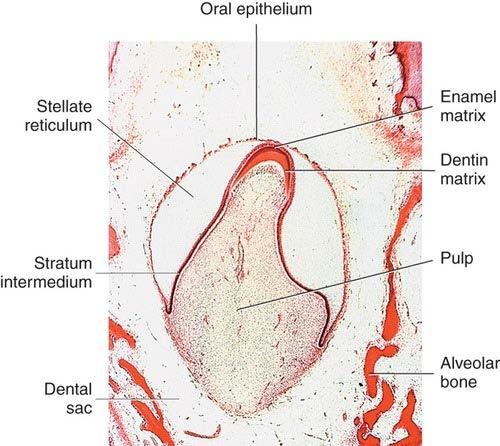
13 + Tooth development (Odontogenesis) Bell stage 11 th to 12 th weeks Proliferation, differentiation*, morphogenesis Enamel organ with four cell layers Dental papilla with two cell types Tooth germ enamel organ dental papilla dental follicle
14 ** Cell Layers of the Tooth during the Bell stage Enamel Organ 1 inner enamel epithelium, IEE Innermost tall, columnar cells Will differentiate into ameloblasts 2 stratum intermedium, SI More inner compressed layer of flat to cuboidal cells 3 stellate reticulum, SR More outer star-shaped cells in many layers, forming a network within the enamel organ 4 outer enamel epithelium, OEE Outer cuboidal cells
15 ** Cell Layers of the Tooth during the Bell stage Dental papilla 1 Outer cells of dental papilla peripheral layer of cells of the dental papilla nearest the inner enamel epithelium of the enamel organ will differentiate into odontoblast 2 Central cells of dental papilla inner cell mass of the dental papilla will differentiate into pulp tissue

16 ** Cell Layers of the Tooth during the Bell stage Dental follicle Increasing amount of collagen fibers forming around the enamel organ will differentiate into cementum, periodontal ligament, and alveolar bone

17 + Tooth development (Odontogenesis) Stages of apposition and maturation The final stage of tooth development Apposition stage (or secretory stage) Enamel, dentin, cementum are secreted in successive layers. Maturation stage Matrices of the hard dental tissue types subsequently fully mineralize Ameloblasts Odontoblasts Amelogenesis & Dentiogenesis Formation of preameloblasts Formation of odontoblasts and dentin matrix Formation of ameloblasts, dentinoenamel junction, and enamel matrix Enamel Predentin Dentin
18 + Formation of preameloblasts IEE cells grow even more columnar or elongate preameloblasts Repolarization : the nucleus in preameloblasts moves away from the center of the cell to the position farthest away from the basement membrane Preameloblasts will first induce dental papilla cells to differentiate into dentin-forming cells (odontoblasts) Preameloblasts will differentiate into enamel-forming cells (ameloblasts)

19
20 + Formation of odontoblasts and dentin matrix outer cells of the dental papilla are differentiated into odontoblasts. Repolarization Dentinogenesis : apposition of predentin (dentin matrix) by odontoblasts


21
22 + Formation of ameloblasts, dentinoenamel junction, and enamel matrix Disintegration of basement membrane between preameloblasts and odontobalsts Predentin induces the preameloblasts to differentiate into ameloblasts. Amelogenesis : Apposition of enamel matrix by ameloblasts Dentinoenamel junction (DEJ) formation : With enamel matrix in contact with predentin, mineralization of disintegrating basement membrane occurs. Odontoblasts will leave attached cellular extensions in the length of the predentin. : odontoblast process dentinal tubule Tomes process


23

24


25 ** Common dental developmental disturbances and involved stage 1. Initiation stage 2. Bud stage 3. Cap stage 4. Apposition and maturation stages <Anodontia> complete partial <Supernumerary teeth (Hyperdontia)>

26 ** Common dental developmental disturbances and involved stage 1. Initiation stage 2. Bud stage 3. Cap stage 4. Apposition and maturation stages <Microdontia/Macrodontia>


27 ** Common dental developmental disturbances and involved stage 1. Initiation stage 2. Bud stage 3. Cap stage 4. Apposition and maturation stages <Dens in dente> <Gemination> <Fusion> <Tubercle>


28 ** Common dental developmental disturbances and involved stage 1. Initiation stage 2. Bud stage 3. Cap stage 4. Apposition and maturation stages <Enamel dysplasia> <Concrescence> <Enamel pearl>
29 + Root development Cervical loop most cervical portion of enamel organ Grows deeper into the dental sac to become Hertwig s epithelial root sheath (HERS) Hertwig s epithelial root sheath (HERS) Bilayer rim consisting of ONLY inner and outer enamel epithelium Function of HERS is to shape the root(s). Also induces dentin formation in root area so that it is continuous with coronal dentin, as well as cementum on roots overlying the newly formed dentin. Epithelial rests of Malassez (ERM)


30


31 + Root development Root dentin formation Cervical loop IEE cells of HERS Outer cells of the dental papilla odontoblasts Begin to secrete predentin Root dentin formation Disintegration of basement membrane Disintegration of HERS Epithelial rest of Malassez
32 + Root development Cementum and pulp formation Cementogenesis Disintegration of HERS Dental follicle cells contact with root dentin Dental follicle cells cementoblasts Cementoid (cementum matrix) setretion Early : leave no cellular bodies in their secreted products Later: become entrapped by their products (cementocyte) mineralization or maturation cementum *** DCJ (dentinocemental junction)

33 + Root development Cementum and pulp formation Pulp formation Central cells of the dental papilla Periodontal ligament and alveolar bone development
34 + Periodontal ligament and alveolar bone development Periodontal ligament (PDL) formation After cementum formation The ectomesenchyme (from the dental sac) begins to form the PDL Collagen fiber formation These fibers insert into the cementum and alveolar bone Alveolar bone formation The ectomesenchyme from the dental follicle


35 + Development of multirooted teeth Root trunk A single root on the base of the crown Differential growth of HERS divides the root trunk into the correct number of root Cervical loop of multirooted teeth Long, tongue like horizontal epithelial extensions Extensions can be present on multirooted teeth, depending on the similar number of roots on the mature tooth.
36 + Primary tooth eruption and shedding Root growth Existence of a temporary ligament Vascular pressure Contractile collagen Hormonal signals
37 + Primary tooth eruption and shedding After enamel apposition ameloblasts place an acellular dental cuticle on new enamel surface the enamel organ is compressed, forming reduced enamel epithelium (REE) The REE fuses with oral epithelium lining the oral cavity Disintegration of the central part in the fused tissues epithelial tunnel Cervical part of the fused tissues attachs to the neck of the tooth Junctional epithelium


38


39 + Primary tooth shedding Is lost, exfoliated, or shed, as the succedaneous permanent tooth develops lingual to it Resorption of tooth Osteoclast: alveolar bone Odontoclast: primary s root dentin, cementum, small parts of enamel
 Tooth germ of nonsuccedaneous permanent molars Developing mandibular dental arch Developing primary teeth Tongue Developing")
40 + Permanent tooth eruption Erupts into the oral cavity in a position lingual to the roots of the shedding primary tooth Additional teeth Successional dental lamina of permanent teeth primordia Oral epithelium (cut to show tooth buds) Tooth germ of nonsuccedaneous permanent molars Developing mandibular dental arch Developing primary teeth Tongue Developing mandible


41 ** Developmental disturbances during eruption 1. Dentigerous cyst 2. Eruption cyst Nasmyth s membrane Eruption cyst
CAP STAGE. Ans 1 The following are the stages of tooth development :
 Ans 1 The following are the stages of tooth development : 1. Bud stage 2. Cap stage 3. Bell stage 4. Advanced bell stage 5. Formation of Hertwig s epithelial root sheath BUD STAGE 1. Around the eighth
Development of teeth. 5.DM - Pedo
 Development of teeth 5.DM - Pedo Tooth development process of continuous changes in predetermined order starts from dental lamina A band of ectodermal cells growing from the epithelium of the embryonic
Tooth eruption and movement
 Tooth eruption and movement Dr. Krisztián Nagy Diphydont dentition Deciduous dentition primary dentition Diphydont dentition Permanent dentition secondary dentition Mixed Dentition: Presence of both dentitions
Semester Credits: 3 Lecture Hours: 3. Prerequisites:
 Revised: Fall 2015 Semester Credits: 3 Lecture Hours: 3 21THistology DNH 115 Admission into dental hygiene program. Prerequisites: Course Description: Presents a study of the microscopic and macroscopic
DHYG 121 Winter, 2009 COURSE OUTLINE
 CAMOSUN COLLEGE School of Health & Human Services Dental Hygiene Department DHYG 121 Winter, 2009 COURSE OUTLINE The Approved Course Description is available on the web @ http://www.camosun.bc.ca/calendar/current/web/dhyg.html#dhyg121
Dentin Formation(Dentinogenesis)
") Lecture four Dr. Wajnaa Oral Histology Dentin Formation(Dentinogenesis) Dentinogenesis begins at the cusp tips after the odontoblasts have differentiated and begin collagen production. Dentinogenesis growth
Lecture four Dr. Wajnaa Oral Histology Dentin Formation(Dentinogenesis) Dentinogenesis begins at the cusp tips after the odontoblasts have differentiated and begin collagen production. Dentinogenesis growth
AMELOGENESIS. Prof. Shaleen Chandra
 AMELOGENESIS Epithelial Enamel Organ Outer Enamel Epithelium Stellate Reticulum Stratum Intermedium Inner Enamel Epithelium Cervical Loop Life Cycle of Ameloblasts Morphogenic stage Organizing Stage Formative
Oral Embryology and Histology
 Oral Embryology and Histology Chapter 8 Copyright 2018, Elsevier Inc. All Rights Reserved. 1 Learning Objectives Lesson 8.1: Oral Embryology 1. Pronounce, define, and spell the key terms. 2. Define embryology
DHYG 221 Oral Sciences 1 Winter 2014 COURSE OUTLINE
 CAMOSUN COLLEGE School of Health & Human Services Dental Hygiene Department DHYG 221 Oral Sciences 1 Winter 2014 COURSE OUTLINE The Approved Course Description is available on the web @ http://camosun.ca/learn/calendar/current/web/dhyg.html
and Non-Human MODULE No.17: Structural Variation in Teeth- Human and Non-Human
 SUBJECT Paper No. and Title Module No. and Title Module Tag MODULE No.17: Structural Variation in Teeth- Human and FSC_P11_M17 TABLE OF CONTENTS 1. Learning Outcomes 2. Introduction 3. Structure of Human
ANATOMY OF THE PERIODONTIUM. Dr. Fatin Awartani
 ANATOMY OF THE PERIODONTIUM Part II Cementum and Alveolar bone Associate Professor Periodontal division King Saud university Cementum Calcified mesenchymal tissue that forms the outer covering of the anatomic
DENTIN-PULP COMPLEX. Erlina Sih Mahanani. School of Dental sciences Universiti Sains Malaysia. Erlina Sih Mahanani
 DENTIN-PULP COMPLEX School of Dental sciences Universiti Sains Malaysia Introduction Overview anatomy & histology of dentin and pulp. Development of dentin and pulp Structure of dentin and pulp Dentin
Periodontal ligament
 Periodontal ligament The periodontium The periodontium includes: The gingiva Cementum Periodontal ligament Alveolar bone Def: The periodontal ligament is the dense fibrous connective tissue that occupies
Eruption and Shedding of Teeth
 Eruption and Shedding of Teeth Mixed Dentition: Presence of both dentitions Figure from Ten Cate s Oral Histology, Ed., Antonio Nanci, 6 th edition Tooth eruption is the process by which developing teeth
Applied Equine Dental Development
 Published in IVIS with the permission of the AAEP Close this window to return to IVIS Applied Equine Dental Development Kirstie Dacre, BVMS, MSc, Cert EM (Int Med), PhD Author s address: Veterinary Teaching
Lec. 11 & 12 Dr. Ali H. Murad Dental pulp 1- Coronal pulp
 Lec. 11 & 12 Dr. Ali H. Murad Dental pulp Is the soft connective tissue located in the central portion of each tooth. All pulps have similar morphologic characteristic, such as a soft, gelatinous consistency
Chapter 5. Developmental Disorders. Copyright 2014, 2009, 2004, 2000, 1996, 1992 by Saunders, an imprint of Elsevier Inc 1
 Chapter 5 Developmental Disorders Copyright 2014, 2009, 2004, 2000, 1996, 1992 by Saunders, an imprint of Elsevier Inc 1 Outline Ø Embryonic Development of the Face, Oral Cavity, and Teeth Ø Developmental
P. J. Slootweg Dental Pathology
 P. J. Slootweg Dental Pathology Pieter J. Slootweg Dental Pathology A Practical Introduction With 197 Figures 123 Pieter J. Slootweg, MD, DMD, PhD Radboud University Medical Center Nijmegen Department
TOOTH dens, dentis odus, odonotos
 TOOTH dens, dentis odus, odonotos Teeth (Dentes) arcus dentalis superior (maxillaris) ellipse arcus dentalis inferior (mandibularis) parabola permanent teeth (dentes permanentes) 32 deciduous teeth (dentes
Using Dental Stem Cells to Regenerate Tooth Tissue and Whole Tooth Replacement Peretz Rapoport
 and Whole Tooth Replacement Peretz Rapoport Peretz Rapoport graduated January 2018 with a BS in Biology and is starting Touro School of Dental Medicine in fall 2018 Abstract Irreversible dental problems
Fundamental & Preventive Curvatures of Teeth and Tooth Development. Lecture Three Chapter 15 Continued; Chapter 6 (parts) Dr. Margaret L.
 Dr. Margaret L.") Fundamental & Preventive Curvatures of Teeth and Tooth Development Lecture Three Chapter 15 Continued; Chapter 6 (parts) Dr. Margaret L. Dennis Proximal contact areas Contact areas are on the mesial and
Fundamental & Preventive Curvatures of Teeth and Tooth Development Lecture Three Chapter 15 Continued; Chapter 6 (parts) Dr. Margaret L. Dennis Proximal contact areas Contact areas are on the mesial and
Oral cavity Lab exercises
 Oral cavity Lab exercises Slide #190 (GT-1-32). Oral cavity, goat. large conical buccal papillae stratified squamous epithelium keratinized or non-keratinized no muscularis mucosae connective tissue represents
IMMUNOHISTOCHEMICAL PROFILE OF ODONTOGENIC EPITHELIUM OF DEVELOPING DOG TEETH (CANIS FAMILIARIS)
") IMMUNOHISTOCHEMICAL PROFILE OF ODONTOGENIC EPITHELIUM OF DEVELOPING DOG TEETH (CANIS FAMILIARIS) By SULETTE NEL Submitted in partial fulfilment of the requirements for the degree of Master of Science (Odontology)
IMMUNOHISTOCHEMICAL PROFILE OF ODONTOGENIC EPITHELIUM OF DEVELOPING DOG TEETH (CANIS FAMILIARIS) By SULETTE NEL Submitted in partial fulfilment of the requirements for the degree of Master of Science (Odontology)
DENT-1311: DENTAL ANATOMY, HISTOLOGY & EMBRYOLOGY
 DENT-1311: Dental Anatomy, Histology & Embryology 1 DENT-1311: DENTAL ANATOMY, HISTOLOGY & EMBRYOLOGY Cuyahoga Community College Viewing:DENT-1311 : Dental Anatomy, Histology & Embryology Board of Trustees:
TOX 2017 Magyar Toxikológusok Társasága Tudományos konferencia Bükfürdő, Október
 TOX 2017 Magyar Toxikológusok Társasága Tudományos konferencia Bükfürdő, Október 11-13. Histopathological evaluation of the teeth in regulatory studies: what are the needs and what can we learn from it
ODONTOGENESIS- A HIGHLY COMPLEX CELL-CELL INTERACTION PROCESS
 ODONTOGENESIS- A HIGHLY COMPLEX CELL-CELL INTERACTION PROCESS AMBRISH KAUSHAL, MALA KAMBOJ Department of Oral and Maxillofacial Pathology Career Post Graduate Institute of Dental Sciences and Hospital
Tooth Variations. Suruedee Chinthakanan
 Tooth Variations Suruedee Chinthakanan Tooth variations Dental anomalies Cause : hereditary factor Developmental disturbances of teeth www.ectodermaldysplsia.org Tooth variations Enamel is formed from
Postnatal Tooth Development in Cattle
 Postnatal Tooth Development in Cattle W. A. BARRY BROWN, L.D.S.; PAUL V. CHRISTOFFERSON, D.V.M.; MAURY MASSLER, D.D.S., M.S.;MARVIN B. WEISS, D.D.S. Chicago, Illinois INTEREST in the teeth of cattle dates
ORAL ANATOMY AND PHYSIOLOGY
 CHAPTER 7 ORAL ANATOMY AND PHYSIOLOGY INTRODUCTION This chapter covers the oral anatomy and physiology of the teeth, the histology of the tissues and supporting structures, and concentrates on the external
Chapter 2 Tooth Development
 Chapter 2 Tooth Development Experimental research on tooth development or odontogenesis is based very largely on the teeth of murine rodents (Butler 1967 ). Pioneering work by Shirley Glasstone on rat
Proceedings of the 36th World Small Animal Veterinary Congress WSAVA
 www.ivis.org Proceedings of the 36th World Small Animal Veterinary Congress WSAVA Oct. 14-17, 2011 Jeju, Korea Next Congress: http://www.ivis.org October 14(Fri) ~ 17(Mon) 2011 ICC Jeju, Korea 2011 WSAVA
DENTIN It a hard vital tissue, surrounds the pulp & underlies the enamel on the crown & the cementum on the roots of the teeth.
 Lec. 7 Dr. Ali H.Murad DENTIN It a hard vital tissue, surrounds the pulp & underlies the enamel on the crown & the cementum on the roots of the teeth. Physical properties: 1-Dentin is pale yellow in color,
BIOCHEMISTRY / PHYSIOLOGY. Bld. The fluid phase of coagulated blood is known as: Blood hematocrit. Blood cells. Blood serum.
 BIOCHEMISTRY / PHYSIOLOGY Bld The fluid phase of coagulated blood is known as: Blood hematocrit Blood cells Blood serum Blood plasma Copyright 2006-2007 DENTAL DECKS Blood serum Serum = plasma fibrinogen
Dental Morphology and Vocabulary
 Dental Morphology and Vocabulary Palate Palate Palate 1 2 Hard Palate Rugae Hard Palate Palate Palate Soft Palate Palate Palate Soft Palate 4 Palate Hard Palate Soft Palate Maxillary Arch (Maxilla) (Uppers)
SPACE MAINTAINER. Multimedia Health Education. Disclaimer
 Disclaimer This movie is an educational resource only and should not be used to manage your health. All decisions about the management of premature loss of primary teeth and use of space maintainers must
CHAPTER 4 ORAL ANATOMY
 CHAPTER 4 ORAL ANATOMY This chapter covers the oral anatomy and physiology of the teeth, the histology of their tissues and supporting structures, and concentrates on the external features of the teeth.
PREMATURE PRIMARY TOOTH LOSS
 Disclaimer This movie is an educational resource only and should not be used to manage your dental health. All decisions about the management of premature primary tooth loss must be made in conjunction
Sponsored document from Trends in Cell Biology. Stem cell-based biological tooth repair and regeneration
 Sponsored document from Trends in Cell Biology Stem cell-based biological tooth repair and regeneration Ana Angelova Volponi 1, Yvonne Pang 1,2, and Paul T. Sharpe 1 Paul T. Sharpe: paul.sharpe@kcl.ac.uk
The eternal tooth germ is formed at the apical end of continuously growing teeth*
 The eternal tooth germ is formed at the apical end of continuously growing teeth* Hayato Ohshima 1, Naohiro Nakasone 1, 2, Emi Hashimoto 1, Hideo Sakai 1, Kuniko Nakakura-Ohshima 3 and Hidemitsu Harada
II. Disturbances in Size.
 I. Introduction. Variation of teeth has been an enduring interest to the clinical practitioner and the laboratory scientist. No two teeth are alike. The day-to-day variation of teeth that we see is the
Development of the dentition
 4 Development of the dentition 85 Humans have two dentitions, the deciduous (primary) and permanent (secondary). Each dentition is heterodont, meaning that it consists of teeth with different shapes and
We are IntechOpen, the world s leading publisher of Open Access books Built by scientists, for scientists. International authors and editors
 We are IntechOpen, the world s leading publisher of Open Access books Built by scientists, for scientists 4,000 116,000 120M Open access books available International authors and editors Downloads Our
Digestive System. Presented by: Dr M. Arianmanesh PhD in Reproductive and Developmental Biology Dept. of Anatomical Sciences
 Digestive System Presented by: Dr M. Arianmanesh PhD in Reproductive and Developmental Biology Dept. of Anatomical Sciences Today we will discuss: Histological layers of alimentary canal Oral cavity Lip
Stem cell-based biological tooth repair and regeneration
 Review Special issue CellBio-X Stem cell-based biological tooth repair and regeneration Ana Angelova Volponi 1, Yvonne Pang 1,2 and Paul T. Sharpe 1 1 Department of Craniofacial Development and MRC Centre
HISTOLOGY VIRTUAL LABORATORY GASTROINTESTINAL SYSTEM
 HISTOLOGY VIRTUAL LABORATORY GASTROINTESTINAL SYSTEM LIP (Slides GI 1, 2) Identify the outer portion lined by stratified squamous (keratinized) epithelium. Note the hair follicles and sebaceous glands
Objectives. Discuss the physiology of tooth eruption. Identify the causes of anomalies associated with
 Eruption and eruption disorders Objectives Discuss the physiology of tooth eruption. Identify the causes of anomalies associated with Identify the causes of anomalies associated with tooth eruption. Introduction
Odontomes and Odontogenic tumours
 Odontomes and Odontogenic tumours Odontomes Developmental hamartoma Hamartoma: normal tissue in abnormal location Any cells to be neoplastic it must be able to replicate, which is not seen in hamartoma
Division of Health Professions/Dental Hygiene Department. Oral Embryology and Histology Course Syllabus
 Bergen Community College Division of Health Professions/Dental Hygiene Department Oral Embryology and Histology Course Syllabus Course Title: Oral Embryology and Histology DHY 109 Term: Fall 2014 Hours/Credits:
Medical NBDE-II. Dental Board Exams Part I.
 Medical NBDE-II Dental Board Exams Part I http://killexams.com/exam-detail/nbde-ii Question: 149 Anatomically, the term "clinical root" can be defined as which of the following: A. The space in the tooth
2.79J/3.96J/BE.441/HST522J DENTAL TISSUE REPLACEMENT AND REGENERATION
 Massachusetts Institute of Technology Harvard Medical School Brigham and Women s/massachusetts General Hosp. VA Boston Healthcare System 2.79J/3.96J/BE.441/HST522J DENTAL TISSUE REPLACEMENT AND REGENERATION
Lecture 2 Maxillary central incisor
 Lecture 2 Maxillary central incisor Generally The deciduous tooth appears in the mouth at 3 18 months of age, with 6 months being the average and is replaced by the permanent tooth around 7 8 years of
Only 40% of the Story
 X-RAY, X-RAY, READ ALL ABOUT IT! The Use and Utility of Dental Radiographs in Practice Lisa Fink, DVM, DAVDC Dentistry & Oral Surgery Service October 4, 2015 Only 40% of the Story Radiographs of teeth
Institution : College of Dentistry Academic Department : Maxillofacial surgery and Diagnostic sciences
 Institution : College of Dentistry Academic Department : Maxillofacial surgery and Diagnostic sciences [MDS] Program : Bachelor of Dentistry [ BDS ] Course : Oral Biology Course Coordinator : Saleem Shaikh
Developmental Biology of Cementum
 Int. J. Dev. Biol. 45: 695-706 (2001) Review Developmental Biology of Cementum THOMAS G.H. DIEKWISCH* Allan G. Brodie Laboratory for Craniofacial Genetics, University of Illinois at Chicago, USA CONTENTS
1. What is the highest and sharpest cusp on the lower first deciduous molar? 2. Which of the following is NOT the correct location of an embrasure?
 1 1. What is the highest and sharpest cusp on the lower first deciduous molar? a. mesiobuccal b. distobuccal c. distolingual d.mesiolingual 2. Which of the following is NOT the correct location of an embrasure?
Chapter 20: Pathology
 : Pathology There are many pathologic conditions, which occur in and around the mouth. Some of them have already been discussed in Chapters 7, 13, 15, 16, 17, 18 and 19. This chapter will cover a few more
Dental Anatomy and Physiology for Clinical Dental Technicians. with Marnie Hayward
 Dental Anatomy and Physiology for Clinical Dental Technicians with Marnie Hayward Salivary glands Parotid Submandibular Sublingual Salivary glands position Parotid glands Lie below ear and behind angle
COPYRIGHTED MATERIAL. Oral embryology, histology and anatomy. Sheila Phillips. Summary. Introduction. Oral embryology
 1 Oral embryology, histology and anatomy Sheila Phillips Summary This chapter covers: Oral embryology Early tooth development Development of the dental tissues Histology of oral tissues Histology of dental
Enamel: Composition, Formation & Structure PROF. DR. KARTHIKEYAN RAMALINGAM
 Enamel: Composition, Formation & Structure PROF. DR. KARTHIKEYAN RAMALINGAM ENAMEL It is the hardest calcified matrix in the body. Ameloblasts are the cells responsible for enamel formation. These cells
EXPRESSION OF SOX 11 DURING TOOTH FORMATION. Reem Atout. Dr. Mary MacDougall, CHAIR Dr. Amjad Javed Dr. Firoz Rahemtulla Dr. Michael Reddy A THESIS
 EXPRESSION OF SOX 11 DURING TOOTH FORMATION by Reem Atout Dr. Mary MacDougall, CHAIR Dr. Amjad Javed Dr. Firoz Rahemtulla Dr. Michael Reddy A THESIS Submitted to the graduate faculty of The University
6610 NE 181st Street, Suite #1, Kenmore, WA
 660 NE 8st Street, Suite #, Kenmore, WA 9808 www.northshoredentalacademy.com.08.900 READ CHAPTER The Professional Dental Assistant (p.-9) No Key Terms Recall Questions:,,,, and 6 CLASS SYLLABUS DAY READ
Oral Histology. Alveolar bone or process: Functions of alveolar bone: Chemical composition: Development of the alveolar process: Dr.
 Oral Histology Lec.12 Alveolar bone or process: Dr. Nada Al-Ghaban Alveolar bone is a specialized part of the mandibular and maxillary bones that forms the primary support structure for teeth. Although
WHO Histological typing of odontogenic tumors, A. Epithelial Odontogenic Tumors
 Cheng-Chung Lin, Prof. in Oral Pathology College of Dental Medicine, KMU 2007 Classification: The following classification is based upon the inductive effect of one dental tissue upon another. In normal
Teeth and supporting tissues
 Teeth and supporting tissues 1. The permanent teeth shape, relief and histological structure 2. Periodontium 3. Abnormalities of teeth 4. Phylogenetic and ontogenetic tooth development 5. The deciduous
04 Development of the Face and Neck. Development of the Face Development of the neck
 04 Development of the Face and Neck Development of the Face Development of the neck Development of the face Overview of facial development The fourth week ~ the twelfth week of prenatal development Between
Jaws: Cysts and Odontogenic Neoplasms
 Topic 10: Jaw Cysts General Features of Jaw Cysts Sources of Epithelium in Cysts Radiographic Features of Jaw Cysts Microscopic Features of Jaw Cysts Treatment and Prognosis of Jaw Cysts Classification
DEVELOPMENTAL DISTURBANCES AFFECTING TEETH
 DEVELOPMENTAL DISTURBANCES AFFECTING TEETH A) DISTURBANCES DURING INTIATION OF TOOTH GERMS Abnormalities in the number A Reduced number of teeth (ANODONTIA) I Total anodontia It is a very rare condition
Effects of diet and fluoride on early phases of odontogenesis in rats
 Izabela Maciejewska et al., Conditions of odontogenesis ORIGINAL ARTICLE Folia Morphol. Vol. 59, No. 1, pp. 37 42 Copyright 2000 Via Medica ISSN 0015 5659 Effects of diet and fluoride on early phases of
Origin of Odontogenic Cysts & Tumors
 Origin of Odontogenic Cysts & Tumors Odontogenic Apparatus Origin of Odontogenic Cysts & Tumors Odontogenic Apparatus Remnants of dental lamina Reduced enamel epithelium Odontogenic rests Basal cell layer
Advanced Probing Techniques
 Module 21 Advanced Probing Techniques MODULE OVERVIEW The clinical periodontal assessment is one of the most important functions performed by dental hygienists. This module begins with a review of the
Primary Teeth Chapter 18. Dental Anatomy 2016
 Primary Teeth Chapter 18 Dental Anatomy 2016 Primary Teeth - Introduction Synonyms deciduous teeth, baby teeth, temporary teeth, milk teeth. There are 20 primary teeth, designated as A thru T in the Universal
7. Development of digestive system I. Yolk sac. Primitive gut. Stomodeum. Teeth. Branchial clefts, pouches and arches. Tongue.
 7. Development of digestive system I. Yolk sac. Primitive gut. Stomodeum. Teeth. Branchial clefts, pouches and arches. Tongue. Gut tube the roof of the yolk sac is cephalocaudally and laterally folded
An Overview of Dental Anatomy
 Continuing Education Brought to you by An Overview of Dental Anatomy Course Author(s): Vickie Parrish Foster, RDH, MEd CE Credits: 1 hour Intended Audience: Dental Hygienists, Dental Assistants, Dental
BCL11B Regulates Epithelial Proliferation and Asymmetric Development of the Mouse Mandibular Incisor
 BCL11B Regulates Epithelial Proliferation and Asymmetric Development of the Mouse Mandibular Incisor Kateryna Kyrylkova 1, Sergiy Kyryachenko 1, Brian Biehs 2 *, Ophir Klein 2, Chrissa Kioussi 1 *, Mark
An Overview of Dental Anatomy
 An Overview of Dental Anatomy Vickie P. Overman, RDH, MEd Continuing Education Units: 1 hour Online Course: www.dentalcare.com/en-us/professional-education/ce-courses/ce500 Disclaimer: Participants must
Digestive system 1. Oral cavity: Lips Tongue Palate soft - hard Tooth
 Digestive system 1 Oral cavity: Lips Tongue Palate soft - hard Tooth Common structure of the wall of GIT tube The mucosa epithelial linning lamina propria /loose connect. tissue/ the muscularis mucosae
NEW YORK CITY COLLEGE OF TECHNOLOGY CUNY Dental Hygiene Department WINTER SESSION 2016
 Instructor: Maria-Elena Bilello, RDH, MSPH Office: Pearl 201E Office# (718) 260-4927 email: mbilello@citytech.cuny.edu NEW YORK CITY COLLEGE OF TECHNOLOGY CUNY Dental Hygiene Department WINTER SESSION
Dental Assisting COURSE ONE
 Dental Assisting COURSE ONE 1 History of Dentistry False teeth used to be made of wood or ivory Often painful dental treatments were done by "guzzling some vodka" Wilhelmina Conrad Roentgen discovered
The Epithelial-Mesenchymal Interaction Plays a Role in the Maintenance of the Stem Cell Niche of Mouse Incisors via Fgf10 and Fgf9 Signaling
 The Open Biotechnology Journal, 2008, 2, 111-115 111 The Epithelial-Mesenchymal Interaction Plays a Role in the Maintenance of the Stem Cell Niche of Mouse Incisors via Fgf10 and Fgf9 Signaling Tamaki
Anatomy Sheet: Oral cavity Done by: rasha Rakan edited by: khansaa Mahmoud
 Anatomy Sheet: Oral cavity Done by: rasha Rakan edited by: khansaa Mahmoud The oral cavity has 2 parts: 1. Oral vestibule: outer part that consists of outside the teeth, between the teeth, the cheeks and
FRACTURES AND LUXATIONS OF PERMANENT TEETH
 FRACTURES AND LUXATIONS OF PERMANENT TEETH 1. Treatment guidelines and alveolar bone Followup Procedures INFRACTION Clinical findings Radiographic findings Treatment Follow-Up Favorable Outcome Unfavorable
Applications in Dermatology, Dentistry and LASIK Eye Surgery using LASERs
 Applications in Dermatology, Dentistry and LASIK Eye Surgery using LASERs http://www.medispainstitute.com/menu_laser_tattoo.html http://www.life123.com/bm.pix/bigstockphoto_close_up_of_eye_surgery_catar_2264267.s600x600.jpg
The clinical appearance and diagnosis of odontogenic cysts. SE Arc-Állcsont-Szájsebészeti és Fogászati Klinika BUDAPEST
 The clinical appearance and diagnosis of odontogenic cysts SE Arc-Állcsont-Szájsebészeti és Fogászati Klinika BUDAPEST DEFINITION A cyst is a sac with walls of connective tissue, lined by epithelium, containing
Development of the Pharyngeal Arches
 Development of the Pharyngeal Arches Thomas A. Marino, Ph.D. Temple University School of Medicine Competencies: Upon completion of this section of the course, the student must be able to: 1. Recall the
Restorative Dentistry and it s related to Pulp health. Dr.Ahmed Al-Jobory
 Restorative Dentistry and it s related to Pulp health Dr.Ahmed Al-Jobory Pulp Is a viscous connective tissue of collagen fibers and ground substance supporting the vital cellular, vascular, and nerve structures
Australian Dental Journal
 Australian Dental Journal The official journal of the Australian Dental Association Australian Dental Journal 2014; 59:(1 Suppl): 55 80 doi: 10.1111/adj.12130 Three-dimensional analysis of the early development
Changes in marginal ridge alignment from early childhood to late adulthood in an untreated Caucasian population using the Iowa growth study sample
 University of Iowa Iowa Research Online Theses and Dissertations Spring 2017 Changes in marginal ridge alignment from early childhood to late adulthood in an untreated Caucasian population using the Iowa
Dental Anatomy and Occlusion
 CHAPTER 53 Dental Anatomy and Occlusion Ma Lou C. Sabino DDS, and Emily G. Smythe, DDS What numerical system is used most commonly in the United States for designating the adult dentition? Pediatric dentition?
The Histology of Dentin
 The Histology of Dentin Pauline Hayes Garrett, D.D.S. Department of Endodontics, Prosthodontics, and Operative Dentistry University of Maryland, Baltimore This material was taken from: Essentials of Oral
Connective tissue The Digestive System
 Connective tissue The Digestive System Part 1 Structure of digestive system Functions Basic Structure of the Alimentary Canal Wall Tube is made up of four layers: 1. Mucosa 2. Submucosa 3. Muscularis externa
T O O T H A T L A S C O U R S E G U I D E A S S I S T A N T E D I T I O N
 T O O T H A T L A S C O U R S E G U I D E A S S I S T A N T E D I T I O N The information in this guide was prepared by ehuman with contributions from: Cara Miyasaki, RDHEF, MS, Foothill College Kay Murphy,
Morphology of an Anatomic Crown. By: Assistant Professor Dr. Baydaa Ali Al - Rawi
 Morphology of an Anatomic Crown By: Assistant Professor Dr. Baydaa Ali Al - Rawi October 4, 2009 Elevated landmarks Depressed landmarks A) Elevated landmarks : 1. Dental lobe : is one of the primary centers
To the root of the stem cell problem. The evolutionary importance of the epithelial stem cell niche during tooth development
 To the root of the stem cell problem The evolutionary importance of the epithelial stem cell niche during tooth development Mark Tummers Developmental Biology Programme Institute of Biotechnology University
Hertwig s epithelial root sheath: A panaromic view
 2017; 3(4): 21-25 ISSN Print: 2394-7489 ISSN Online: 2394-7497 IJADS 2017; 3(4): 21-25 2017 IJADS www.oraljournal.com Received: 05-08-2017 Accepted: 06-09-2017 Dr. Suchetha A Head of the department, Dr.
Lab activity manual Histology of the digestive system
 Lab activity manual Histology of the digestive system Jeanne Adiwinata Pawitan Prerequisite: Histology of the 4 basic tissues In this module we learn about the histology of the digestive system, from the
INTERPRETATION RADIOGRAPHIC INTERPRETATION. Law of Symmetry. We will be reviewing: 7/30/16
 RADIOGRAPHIC INTERPRETATION Pam Wood, CDA, RDH, M.Ed, CAGS Community College of Rhode Island pwood@ccri.edu INTERPRETATION the ability to read what is revealed on a dental radiograph any dental professional
Teeth, orofacial development and
 Teeth, orofacial development and cleft anomalies Miroslav Peterka Variability of jaws in vertebrates. (A) cartilaginous fish shark; (B) an example of a bone fish; (C ) amphibian frog; (D) reptile - turtle;
NEW YORK CITY COLLEGE OF TECHNOLOGY CUNY Dental Hygiene Department STUDENT COURSE DOCUMENT Fall 2016
 NEW YORK CITY COLLEGE OF TECHNOLOGY CUNY Dental Hygiene Department STUDENT COURSE DOCUMENT Fall 2016 COURSE CODE: Den 1114 COURSE TITLE: Histology & Embryology INSTRUCTOR: Maria-Elena Bilello, RDH, MSPH
DENTAL TRAUMA IN DECIDUOUS TEETH
 Disclaimer This movie is an educational resource only and should not be used to manage your health. All decisions about the management of Dental Trauma in Deciduous Teeth must be made in conjunction with