Oral cavity Lab exercises
|
|
|
- Meghan Sullivan
- 6 years ago
- Views:
Transcription

1 Oral cavity Lab exercises


2 Slide #190 (GT-1-32). Oral cavity, goat. large conical buccal papillae stratified squamous epithelium keratinized or non-keratinized no muscularis mucosae connective tissue represents both the lamina propria and submucosa.


3 Slide #83 (SP-1-79) - Lip, sheep. oral cavity side that has a nonkeratinized stratified squamous epithelium and only glands in the lamina propria/submucosa. keratinized stratified squamous epithelium and adnexa in the CCT Mixed glands non-keratinized stratified squamous epithelium

4 Slide #83 (SP-1-79) - Lip, sheep Plant material being walled off with CCT
5

6
.")


7 Slide #12 (1101). Tongue, rabbit. Filiform papillae Skeletal muscle Foliate papillae that possess Taste buds Serous glands
8
 -")


9 DEMO SLIDE BOX 157 (852) - Tongue, cat large filiform papillae that are heavily keratinized Mixed glands in lamina propria/ submucosa fungiform papillae with taste buds skeletal muscle unilocular adipose tissue present that is common for this species


10 DEMO SLIDE BOX Tongue, monkey. taste buds circumvallate papilla Glands that wash out one taste for the next keratinized stratified squamous epithelium. Note the numerous taste buds

11

12

13

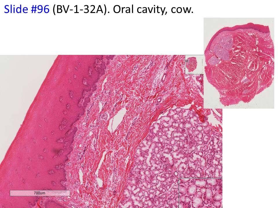
14 Slide #22 (BV3-113a1) Tongue, bovine. Top of tongue skeletal muscle Bottom epithelium

15

16


17 DEMO SLIDE BOX Oral cavity, dental pad, sheep. (SP-1-179) lamina propria/submucosa, and underlying bone and hyaline cartilage stratified squamous epithelium (both keratinized and non-keratinized),
 20x dentin odontoblasts periodontal")
18 DEMO SLIDE BOX 47 - Commercial preparation. Tooth. dental pulp (pulp cavity) 20x dentin odontoblasts periodontal membrane alveolar bone


19 DEMO SLIDE BOX 46 - Teeth, dog. dental pulp odontoblasts dentin and predentin cementum with cementocytes periodontal membrane alveolar bone


20 DEMO SLIDE BOX 45 - Demo slide # 45b. Tooth. non-erupted tooth; therefore the ameloblasts predentin odontoblasts enamel alveolar bone The cementum and periodontal ligament are not yet developed.

21

22
.")


23 Demo Slide #186 (SP-1-108). Parotid salivary gland, sheep lobulated organ striated ducts interlobular ducts Nerve bundles simple or stratified columnar or cuboidal epithelium

24
.")


25 Demo Slide #186(SP-1-108). Parotid salivary gland, she Striated ducts intercalated ducts secretory cells are serous type cells. Secretory asini

26


27 Ds 186 Striated ducts striations Striations are formed by lining up of mitochondria by enfoldings of the plasma membrane to increase surface area for salt removal from secretions to produce hypotonic saliva

28
.")


29 Demo Slide #221 (SP-1-85). Mandibular salivary gland and thymus, sheep. mixed type of salivary gland


30 Demo Slide #221 (SP-1-85). Mandibular salivary gland and thymus, sheep. white fat (unilocular adipose tissue) Thymus cortex and medulla large vein muscular artery large duct autonomic ganglion nerve bundles THYMIC CORPUSCLES. THEY ARE FORMED FROM EPITHELIORETICULAR CELLS (ER)
 - Salivary glands, dog.")


 intercalated ducts")
31 DEMO SLIDE BOX 162 (C003-H-113B; C007-H-88) - Salivary glands, dog. X mandibular salivary gland parotid salivary gland is on the left, the mandibular salivary gland is in the middle, and the polystomatic portion of the sublingual salivary gland (no typical ducts) intercalated ducts Striated duct
- Mandibular")


32 DEMO SLIDE BOX 183 (BV-1-85A)- Mandibular salivary gland, cow. White fat Striated duct intercalated duct Serous demilume interlobular ducts


33 DEMO SLIDE BOX 48 - Esophagus, dog. Non-keratinized stratified squamous epithelium lamina propria submucosa submucosal esophageal glands MUCOUS BUT SOME SEROUS ACINI AND DEMILUNES mucous membrane Muscularis mucosae tunica muscularis SKELETAL and two layers adventitia


34 Slide #71 (Pf5-73/205). Esophagus and trachea, pig. muscularis mucosae non-keratinized stratified squamous epithelium adventitia mucous membrane submucosa tunica muscularis


35 DEMO SLIDE BOX 177 (831)- Esophagus, proventriculus, and gizzard, chicken. Thick muscularis mucosae esophageal mucous glands are located in the lamina propria submucosa is very thin tunica muscularis is smooth muscle


36 Slide #29 (CK1-75/98/115A). Esophagus, proventriculus, and gizzard, chicken. esophageal mucous glands tunica muscularis muscularis mucosae tunica muscularis Smooth muscle
GI Histology Lab 1. Prepared by: Zeina Kalaji
 GI Histology Lab 1 Prepared by: Zeina Kalaji Lip ORAL MUCOSA -Arrow shows labial salivary glands in the submucosa. VERMILLION transitional zone. SKIN Stratified Squamous epithelium, keratinized -Arrow
HISTOLOGY VIRTUAL LABORATORY GASTROINTESTINAL SYSTEM
 HISTOLOGY VIRTUAL LABORATORY GASTROINTESTINAL SYSTEM LIP (Slides GI 1, 2) Identify the outer portion lined by stratified squamous (keratinized) epithelium. Note the hair follicles and sebaceous glands
Laboratory exercises for abdominal organs
 Laboratory exercises for abdominal organs Slide #77 (C007- H- 107A). Pancreas, dog. pancreatic islets CENTROACINAR CELLS ARE THE BEGINNING CELLS OF THE INTERCALATED DUCTS THAT DRAIN THE SECRETORY ACINI
Tongue In the buccal cavity of the digestive system
 Tongue In the buccal cavity of the digestive system same layers as those of tubular organs Mucosa, submucosa, and muscularis muscularis = the muscularis externa no muscularis mucosa 1 Tongue ling = tongue
Connective tissue The Digestive System
 Connective tissue The Digestive System Part 1 Structure of digestive system Functions Basic Structure of the Alimentary Canal Wall Tube is made up of four layers: 1. Mucosa 2. Submucosa 3. Muscularis externa
Objectives. Describe the cells of the GI tract and their function. Differentiate between different parts of the GI tract
 GI Histology 1 Objectives Describe the cells of the GI tract and their function Describe the histological features of each part of the GI tract. Differentiate between different parts of the GI tract Appreciate
Connective tissue The Digestive System
 Connective tissue The Digestive System Part 1 Structure of digestive system Functions Basic Structure of the Alimentary Canal Wall Tube is made up of four layers: 1. Mucosa 2. Submucosa 3. Muscularis externa
BIOL& 253 Lab Manual for Practical #2 Page 1 Rausch. For all slides, know a function for structures marked with a single asterisk (*).
.") BIOL& 253 Lab Manual for Practical #2 Page 1 Rausch Lab equipment: slides, models SLIDES For all slides, know a function for structures marked with a single asterisk (*). DIGESTIVE SYSTEM Layers of the
BIOL& 253 Lab Manual for Practical #2 Page 1 Rausch Lab equipment: slides, models SLIDES For all slides, know a function for structures marked with a single asterisk (*). DIGESTIVE SYSTEM Layers of the
Digestive system. Dr. Sami Zaqout. IUG
 Digestive system Digestive system Digestive tract Associated glands Oral cavity Salivary glands Esophagus Liver Stomach Pancreas Small and large intestines Rectum and anus General Structure of the Digestive
Lab activity manual Histology of the digestive system
 Lab activity manual Histology of the digestive system Jeanne Adiwinata Pawitan Prerequisite: Histology of the 4 basic tissues In this module we learn about the histology of the digestive system, from the
Dorsum of the tongue. Oral Part exhibit lingual papillae of the 4 types. Oral Part of Tongue divided into Left & right halves by shallow median groove
 Histology of TONGUE Figure 22.13 Dorsum of the tongue Oral Part of Tongue divided into Left & right halves by shallow median groove Oral Part exhibit lingual papillae of the 4 types a. filiform papillae,
DIGESTIVE. CHAPTER 17 Lecture: Part 1 Part 2 BIO 212: ANATOMY & PHYSIOLOGY II
 BIO 212: ANATOMY & PHYSIOLOGY II 1 CHAPTER 17 Lecture: DIGESTIVE Part 1 Part 2 Dr. Lawrence G. Altman www.lawrencegaltman.com Some illustrations are courtesy of McGraw-Hill. Processes of DIGESTION Mechanical
LABORATORY EXERCISES FOR THE URINARY SYSTEM
 LABORATORY EXERCISES FOR THE URINARY SYSTEM cortex Medulla DEMO SLIDE BOX 172 (450-E001-H-76). Kidney, horse. the inner medulla medullary rays, Uriniferous tubules expand both the cortex and medulla corticomedullary
Histology Lab. looking at microscopic pictures of tissues, for more information use Junqueira book and you can use BlueHistolgy website
 Done By: Aseel Twaijer & Laith Sorour Histology Lab *These notes help in differentiating tissues and you must read them while looking at microscopic pictures of tissues, for more information use Junqueira
MICROSTRUCTURES LIPS TOOTH TONGUE OESOPHAGUS STOMACH, CARDIAC, PYLORIC FUNDIC GLANDS
 MICROSTRUCTURES LIPS TOOTH TONGUE OESOPHAGUS STOMACH, CARDIAC, PYLORIC FUNDIC GLANDS HUMAN ANATOMY: MICROSTRUCTURES CLASSIFICATION: LOCATION AND BOUNDARIES, FORM, FUNCTION, MICROSCOPIC STRUCTURE: A hollow
A deep groove encircles the body of the circumvallate papilla. Serous (von Ebner s) glands (serous) drain into the base of this groove.
 glands (serous) drain into the base of this groove.") By Dr. Raja Ali A deep groove encircles the body of the circumvallate papilla. Serous (von Ebner s) glands (serous) drain into the base of this groove. The flow of fluid from these glands serves to wash
By Dr. Raja Ali A deep groove encircles the body of the circumvallate papilla. Serous (von Ebner s) glands (serous) drain into the base of this groove. The flow of fluid from these glands serves to wash
Small Intestine, Large Intestine and anal cannel
 Small Intestine, Large Intestine and anal cannel 32409 Small intestine Large intestine Small intestine General Structure of the Digestive Tract rat 32409 Epithelium with goblet cells and absorptive cells
Digestive System. Presented by: Dr M. Arianmanesh PhD in Reproductive and Developmental Biology Dept. of Anatomical Sciences
 Digestive System Presented by: Dr M. Arianmanesh PhD in Reproductive and Developmental Biology Dept. of Anatomical Sciences Today we will discuss: Histological layers of alimentary canal Oral cavity Lip
Dental Anatomy and Physiology for Clinical Dental Technicians. with Marnie Hayward
 Dental Anatomy and Physiology for Clinical Dental Technicians with Marnie Hayward Salivary glands Parotid Submandibular Sublingual Salivary glands position Parotid glands Lie below ear and behind angle
MH1001 Larynx, Monkey, H&E
 MH1001 Larynx, Monkey, H&E - False vocal fold : ciliated pseuodostratified columnar epithelium - True vocal fold : nonkeratinized stratified squamous epithelium vocalis muscle, vocal ligament - Thyroid
DIGESTIVE SYSTEM II ACCESSORY DIGESTIVE ORGANS
 DIGESTIVE SYSTEM II ACCESSORY DIGESTIVE ORGANS Dr. Larry Johnson Texas A& M University Objectives Distinguish between the parotid and submandibular salivary glands. Understand and identify the structural
14 Cardiovascular System
 14 Cardiovascular System The goal of this topic is to examine and understand the structure of the heart, blood vessels and the lymphatic vessels. You should aim to understand how the structure of blood
Digestive system 1. Oral cavity: Lips Tongue Palate soft - hard Tooth
 Digestive system 1 Oral cavity: Lips Tongue Palate soft - hard Tooth Common structure of the wall of GIT tube The mucosa epithelial linning lamina propria /loose connect. tissue/ the muscularis mucosae
The Digestive System. Chapter 16. Introduction. Overview of Digestive System. Histological Organization. Movement and Mixing of Digestive Materials
 The Digestive System Chapter 16 Introduction Structure of the digestive system A tube that extends from mouth to anus Accessory organs are attached Functions include Ingestion Movement Digestion Absorption
Common details: blood vessels, nerve trunks, ganglions, adipose tissue, lymphocytes
 Common details: blood vessels, nerve trunks, ganglions, adipose tissue, lymphocytes Outer lining of the organs: ADVENTITIA (connective tissue) or SEROSA (thin layer of connective tissue lined by simple
Digestive Anatomy Lab
 Digestive Anatomy Lab In-Lab Exercises I have included the word list in this document. Any descrepencies between this document and the wordlist, you should default to this document. There is a lot of repetition
Bio 322 Human Anatomy Objectives for the laboratory exercise Digestive System
 Bio 322 Human Anatomy Objectives for the laboratory exercise Digestive System Required reading before beginning this lab: Saladin, KS: Human Anatomy 5 th ed (2017) Chapter 24 For this lab you will use
c. Heparin sulfate proteoglycans d. Type IV collagen e. Type II collagen 3. The following statements concerning regular bronchioles are correct: a.
 This is just some examples of questions they could ask on the exam. For the multiple parts questions you would probably have 8-10 parts but they should tell you the exact pattern in one of the lectures.
Descriptive Histology
 Atlas of Descriptive Histology Michael H. Ross University of Florida College of Medicine Gainesville, Florida Wojciech Pawlina Mayo Medical School College of Medicine, Mayo Clinic Rochester, Minnesota
بسم هللا الرحمن الرحيم
 بسم هللا الرحمن الرحيم Today, we will leave all hormones and start with another topic which is GI system This lecture is talking about general histology of Gastrointestinal system.. The gastrointestinal
The Digestive System. Chapter 25
 The Digestive System Chapter 25 Introduction Structure of the digestive system A tube that extends from mouth to anus Accessory organs are attached Functions include Ingestion Movement Digestion Absorption
bolus. The bolus is passed to the pharynx which will convey it to the esophagus, the start of the digestive tube proper. This muscular tube will
 Chapter 13 Digestive System (Oral Cavity and the Alimentary Canal) 13.1. Basic Concepts The digestive system is involved with the intake, mechanical and chemical breakdown, and absorption of food. It also
PRACTICAL ROADMAP EPITHELIUM A. JOVANOVIĆ
 PRACTICAL ROADMAP EPITHELIUM A. JOVANOVIĆ Epithelia Simple epithelia Stratified epithelia Simple squamous Simple cuboidal Simple columnar Pseudostratified Stratified squamous - non keratinized - keratinized
Slide 154: Pancreas, H&E
 Slide 154: Pancreas, H&E the pancreas, located adjacent to the duodenum, is a mixed exocrine and endocrine gland; it is usually readily identifiable by the presence of the interspersed endocrine pancreatic
Cell and Tissue Types. Epithelial, Connective, Muscle, Nerve
 Cell and Tissue Types Epithelial, Connective, Muscle, Nerve Objectives Explain the major stages of the cell cycle and cellular division (mitosis). Describe specific events occurring in each of the phases
The Digestive System Laboratory
 The Digestive System Laboratory 1 The Digestive Tract The alimentary canal is a continuous tube stretching from the mouth to the anus. Liver Gallbladder Small intestine Anus Parotid, sublingual, and submaxillary
Semester Credits: 3 Lecture Hours: 3. Prerequisites:
 Revised: Fall 2015 Semester Credits: 3 Lecture Hours: 3 21THistology DNH 115 Admission into dental hygiene program. Prerequisites: Course Description: Presents a study of the microscopic and macroscopic
Gastrointestinal Tract
 CTO Lab #5 GI TRACT & GLANDS; ENDOCRINE SYSTEM Page 1 Gastrointestinal Tract Slide 126 This section through the esophagus shows the characteristic layers of the gastrointestinal tract. Examine the non-keratinized
The Digestive System and Body Metabolism Premedical Biology
 The Digestive System and Body Metabolism Premedical Biology Copyright 2003 Pearson Education, Inc. publishing as Benjamin Cummings The Digestive System and Body Digestion Metabolism Breakdown of ingested
Alimentary Canal (I) Salivatory Glands. (Esophagus and Stomach) Color index: Slides.. Important..Notes..Extra..
 Salivatory Glands. (Esophagus and Stomach) Color index: Slides.. Important..Notes..Extra..") Alimentary Canal (I) (Esophagus and Stomach) Salivatory Glands Color index: Slides.. Important..Notes..Extra.. Objectives: 1. By the end of this lecture, the student should be able to discuss the microscopic
Alimentary Canal (I) (Esophagus and Stomach) Salivatory Glands Color index: Slides.. Important..Notes..Extra.. Objectives: 1. By the end of this lecture, the student should be able to discuss the microscopic
Prelab #4 BLOOD; BONE MARROW; RESPIRATORY; INTEGUEMENT Page 1
 Prelab #4 BLOOD; BONE MARROW; RESPIRATORY; INTEGUEMENT Page 1 Blood Slide 101 This a classic slide of blood cells using a Wright stain. Inspect red blood cells and their appearance. Note the approximate
Organs Histology D. Sahar AL-Sharqi. Digestive System
 Digestive System The digestive system consists of the digestive tract oral cavity, esophagus, stomach, small and large intestines, and anus and its associated glands salivary glands, liver, and pancreas.
HISTOLOGY. Simple squamal lungs
 HISTOLOGY Lab Objectives: Students should be able to... 1. Visually identify each class of tissue and examples within each class 2. Indicate the location (in the human body and/or organ) and function of
The Tissue Level of Organization
 The Tissue Level of Organization Study of this lecture is to be accomplished in conjunction with the Histology Module on the Web!! 1. Introduction Cell Tissue Histology A. General Tissue Types i. Epithelial
HISTOLOGY OF THE RESPIRATORY SYSTEM I. Introduction A. The respiratory system provides for gas exchange between the environment and the blood. B.
 HISTOLOGY OF THE RESPIRATORY SYSTEM I. Introduction A. The respiratory system provides for gas exchange between the environment and the blood. B. The human respiratory system may be subdivided into two
Bio 104 Digestive System
 13 Lecture Outline: Digestive System Hole s HAP [Chapters 17 & 18] General Characteristics of the Alimentary Canal A. Functions 1. Ingestion 2. Mechanical digestion 3. Chemical digestion 4. Propulsion
The Digestive system
 The Digestive system The GI tract (gastrointestinal tract) Mouth Pharynx Esophagus Stomach Small intestine Large intestine Anus The accessory digestive organs Supply secretions contributing to the breakdown
Exercise 11 The Digestive System
 Exercise 11 The Digestive System Objectives Introduction After completing the laboratory exercise of the digestive system, you should be able to: Identify in the microscope sections of the oral cavity,
HUMAN ANATOMY II STUDY NOTES. At the end of this chapter the student should be able to answer the following questions:
 HUMAN ANATOMY II STUDY NOTES CHAPTER ONE The Special Senses Learning objectives At the end of this chapter the student should be able to answer the following questions: 1. What is the gross and histological
Classification of Tissues
 6 R e v i e w S h e e t Exercise Classification of Tissues NAME LAB TIME/DATE Tissue Structure and Function General Review 1. Define tissue. A group of cells similar to one another in structure that perform
Name: Test Date: Chapter 4- Tissues. Use the choices to identify the major tissue types found below:
 Name: Test Date: Chapter 4- Tissues Use the choices to identify the major tissue types found below: A. Connective B. Epithelium C. Muscle D. Nervous 1. B Lines body cavities and covers the body s external
Lesson 9A Tissues in Animals
 Lesson 9A Tissues in Animals Levels of Organization in the Human Body Similar types of cells Different types of tissues Different organs Many organ systems cell tissue organ organ system organism Levels
Basic Anatomy and Physiology of the Lips and Oral Cavity. Dr. Faghih
 Basic Anatomy and Physiology of the Lips and Oral Cavity Dr. Faghih It is divided into seven specific subsites : 1. Lips 2. dentoalveolar ridges 3. oral tongue 4. retromolar trigone 5. floor of mouth 6.
Epithelium. Four primary tissue types:
 Epithelium Four primary tissue types: Epithelial (covering) Connective (support) Nervous (control) Muscular (movement) Smooth muscle Cardiac muscle Skeletal muscle 1 Epithelial Tissue Features Epithelial
Epithelia will be discussed according to the following scheme: Type Number of layers Shape Line drawing. Squamous Cuboidal Columnar
 Epithelia Epithelia will be discussed according to the following scheme: Type Number of layers Shape Line drawing Simple Squamous Cuboidal Columnar Covering and Lining epithelium Pseudostratified Stratified
06 Tooth Development and Eruption
 + 06 Tooth Development and Eruption Tooth development Root development PDL and alveolar bone development Primary tooth eruption and shedding Permanent tooth eruption Q. Where and how tooth starts to form?
Use for reference if needed:
 A- 2.5 Describe how structure and function are related in terms of cell and tissue types. I can recognize different types of body tissue. I can explain how different tissue structures affect their functions.
Common details: blood vessels, nerve trunks, ganglions, adipose tissue, lymphocytes
 Common details: blood vessels, nerve trunks, ganglions, adipose tissue, lymphocytes Outer lining of the organs: ADVENTITIA (connective tissue) or SEROSA (thin layer of connective tissue lined by simple
Alimentary Canal (I)
") Alimentary Canal (I) Esophagus and Stomach (Objectives) By the end of this lecture, the student should be able to discuss the microscopic structure in correlation with the function of the following organs:
Alimentary Canal (I) Esophagus and Stomach (Objectives) By the end of this lecture, the student should be able to discuss the microscopic structure in correlation with the function of the following organs:
Urinary Anatomy. Lab 40. Kidneys. Nephrons. Renal Corpuscle
 Urinary Anatomy Lab 40. Urinary Anatomy and Kidney Dissection Kidneys: filters blood, produces urine Ureters: convey urine to bladder Bladder: holding tank Urethra: carries urine to the outside for elimination
Al s 202 study guide answers Answers Respiratory System 1 External nares (nostrils) 33 Carina 2 Vestibule 34 Left primary bronchus 3 Nasal cavity 35
 33 Carina 2 Vestibule 34 Left primary bronchus 3 Nasal cavity 35") Trachea & Respiratory Histology 1 Epiglottis 26 Capillary 2 Larynx 27 Alveolar sac 3 Thyroid cartilage 28 Alveoli/Alveolus 4 Cricoid cartilage 29 Basement membrane 5 Vocal folds (True vocal cords) 30 Cilia
Trachea & Respiratory Histology 1 Epiglottis 26 Capillary 2 Larynx 27 Alveolar sac 3 Thyroid cartilage 28 Alveoli/Alveolus 4 Cricoid cartilage 29 Basement membrane 5 Vocal folds (True vocal cords) 30 Cilia
Respiratory & Digestive Organs of the Head and Neck, Human;
 Name Date Lab Exercise 5: Lab Exercise 6: Lab Exercise 7: Lab Exercise 8: Respiratory & Digestive Organs of the Head and Neck, Human; Histology of the Respiratory System Digestive System Models, Human
LABORATORY EXERCISES FOR MALE REPRODUCTIVE SYSTEM
 LABORATORY EXERCISES FOR MALE REPRODUCTIVE SYSTEM Slide #101 (1096). Testis, rat. sustentacular ( Sertoli ) cells Nuclei of Sustentacular cells Leydig cells Spermatogonia Spermatocytes Spermatids pale
Tissues and Structures to Know for the Lab Practical
 Ch. 3 - Cells and Tissues Tissues and Structures to Know for the Lab Practical Miss School, Miss Out! Simple squamous epithelium line and cover; site of diffusion Simple squamous epithelium apical surface
Upper Respiratory Histology
 Upper Respiratory Histology - Today we ll discuss the histology of larynx, trachea, primary, secondary, and tertiary bronchus. *First: The Larynx: -The picture below represents a section in the larynx,
Hole s Human Anatomy and Physiology
 Hole s Human Anatomy and Physiology 1 Chapter 5 Tissues Four major tissue types 1. Epithelial 2. Connective 3. Muscle 4. Nervous 2 Epithelial Tissues General characteristics - cover organs and the body
TISSUES. Dr. Gary Mumaugh
 TISSUES Dr. Gary Mumaugh Tissues Tissues - Groups of cells similar in structure and function and perform a common function Histology The study of tissues The four types of tissues Epithelial Connective
Tissue: The Living Fabric: Part A
 PowerPoint Lecture Slides prepared by Janice Meeking, Mount Royal College C H A P T E R 4 Tissue: The Living Fabric: Part A Tissues Groups of cells similar in structure and function Types of tissues Epithelial
Chapter 5. Tissues. 4 Types of Body Tissues. Tissues
 Chapter 5 Tissues Tissues Tissues - groups of cells that are similar in structure & function RBC, WBC, & platelets are a group of cells working together to form BLOOD tissue Histology Pathohistology study
口腔生理學 ( 含顎咬合 ) Oral physiology (occlusion included) 學習目標. Oral moucosa 參考資料. Classification. Mucosa
 Oral physiology (occlusion included) 學習目標. Oral moucosa 參考資料. Classification. Mucosa") 口腔生理學 ( 含顎咬合 ) Oral physiology (occlusion included) Mucosa 臺北醫學大學牙醫學系張維仁老師 E-mail cweijen1@tmu.edu.tw 學習目標 1. let the student to understand the base knolwedge of oral physisology. 2.The student can firsther
口腔生理學 ( 含顎咬合 ) Oral physiology (occlusion included) Mucosa 臺北醫學大學牙醫學系張維仁老師 E-mail cweijen1@tmu.edu.tw 學習目標 1. let the student to understand the base knolwedge of oral physisology. 2.The student can firsther
Development of teeth. 5.DM - Pedo
 Development of teeth 5.DM - Pedo Tooth development process of continuous changes in predetermined order starts from dental lamina A band of ectodermal cells growing from the epithelium of the embryonic
Massachusetts Institute of Technology Harvard Medical School Brigham and Women s Hospital VA Boston Healthcare System 2.79J/3.96J/BE.
 Massachusetts Institute of Technology Harvard Medical School Brigham and Women s Hospital VA Boston Healthcare System 2.79J/3.96J/BE.441/HST522J TISSUE TYPES M. Spector, Ph.D. and I.V. Yannas, Ph.D. The
UNIVERSITY OF NAIROBI
 UNIVERSITY OF NAIROBI UNIVERSITY EXAMINATIONS 2013/2014 LEVEL I MID-SEMESTER II EXAMINATION FOR THE DEGREE OF BACHELOR OF SCIENCE IN NURSING (BScN) AND BACHELOR OF PHARMACY (B.PHARM) MARKING SCHEME HNS101/UPC106:
Urinary system. Urinary system
 Distal convoluted tubule (DCT) Highly coiled, ~ 5 mm in length Last part of the nephron. Wall; simple cuboidal epithelium Less metabolically active than the PCT no brush border light eosinophilic cytoplasm
A. cells that perform related functions and are similar in structure. B. extracellular material - made by cells and secreted into interstitial space
 I. tissue components A. cells that perform related functions and are similar in structure B. extracellular material - made by cells and secreted into interstitial space II. tissue types A. epithelium (e.)
The Tissue Level of Organization
 Tissue The Tissue Level of Organization Chapter 3 Definition an aggregation of cells in which each cooperates with all others in the performance of a given function Examples of general functions Movement
The Digestive System
 The Digestive System Identify the Structure and Function. Mesentery of the Large Intestine The mesentery functions to connect the visceral organs to the abdominal wall. Identify the Structure. Nasal Cavity
Tissues (Histology) Ch. 3 Human Anatomy lecture
 Ch. 3 Human Anatomy lecture") I. Histology the study of tissues A. 4 basic tissue types epithelial connective muscle nervous Tissues (Histology) Ch. 3 Human Anatomy lecture B. Usually found in combinations to form organs. C. As you
I. Histology the study of tissues A. 4 basic tissue types epithelial connective muscle nervous Tissues (Histology) Ch. 3 Human Anatomy lecture B. Usually found in combinations to form organs. C. As you
Slide 74: Pituitary (Masson s trichrome)
") histo074 Slide 74: Pituitary (Masson s trichrome) Infundibular stalk Pars tuberlaris Carcinoma Pars nervosa Pars intermedia Pars distalis Pars distalis Pars intermedia Pars nervosa Pars Pars intermedia
histo074 Slide 74: Pituitary (Masson s trichrome) Infundibular stalk Pars tuberlaris Carcinoma Pars nervosa Pars intermedia Pars distalis Pars distalis Pars intermedia Pars nervosa Pars Pars intermedia
Organs Associated with the Digestive Tract. Dr. Emad I H Shaqoura M.D, M.Sc. Anatomy Faculty of Medicine, IUG March, 2016
 Organs Associated with the Digestive Tract Dr. Emad I H Shaqoura M.D, M.Sc. Anatomy Faculty of Medicine, IUG March, 2016 2 Salivary Glands Salivary Glands Major 90% of saliva Minor 10% of saliva Parotid
-Ibrahim Al-Naser. -Dr Al- Muhtaseb. 1 P a g e
 -1 -Ibrahim Al-Naser - -Dr Al- Muhtaseb 1 P a g e The Digestive System The doctor started the lecture by talking about the class rules. The GI system is an organ system, it is divided into: The Alimentary
Dr. Abeer.c.Yousif. Histology -2 nd stage. What is histology?
 What is histology? Histology is the science of microscopic anatomy of cells and tissues, in Greek language Histo= tissue and logos = study and it's tightly bounded to molecular biology, physiology, immunology
General Human Histology. The Urinary System
 General Human Histology Lecture 8 Assist. Prof. Ahmed Anwar Albir The Urinary System Collecting Tubules & Ducts Urine passes from the distal convoluted tubules to collecting tubules that join each other
Indo Asian Journal of Multidisciplinary Research (IAJMR) ISSN:
 ISSN:") Available online at www.jpsscientificpublications.com Volume 3; Issue - 1; Year 2017; Page: 969 973 Indo Asian Journal of Multidisciplinary Research (IAJMR) ISSN: 2454-1370 MORPHOLOGICAL AND HISTOLOGICAL
Available online at www.jpsscientificpublications.com Volume 3; Issue - 1; Year 2017; Page: 969 973 Indo Asian Journal of Multidisciplinary Research (IAJMR) ISSN: 2454-1370 MORPHOLOGICAL AND HISTOLOGICAL
Topic and Aims. Contractile cells. Nervous system. Muscle tissue is one of the four basic tissue types.
 Topic and Aims Contractile cells Muscle tissue is one of the four basic tissue types. You should be able to: 1. Describe and identify the types of contractile cells, and summarise similarities and differences.
Chapter 05. Review. Copyright The McGraw-Hill Companies, Inc. Permission required for reproduction or display.
 Chapter 05 Review 5.1: Introduction Similar cells with a common function are called tissues. The study of tissues is called histology. There are four (4) primary or major tissue types: 1. Epithelial Tissue
Histology. Study of body tissues
 Histology Study of body tissues 2 Introduction to Body Tissues 1. Composed of specialized cells of similar structure and perform a common function 2. Four major types (4 Cs) a. Epithelial - Cover b. Connective
PRACTICAL ROADMAP. GLANDS AFFECTING LIFESTYLE WJ van der Spuy & T Tshabalala
 PRACTICAL ROADMAP GLANDS AFFECTING LIFESTYLE WJ van der Spuy & T Tshabalala GLANDS AFFECTING LIFESTYLE Submandibular gland (salivary gland) Liver Pancreas Hypophysis (pituitary gland) Thyroid Suprarenal
consists of: Muscular, hollow tube (= digestive tract ) + Various accessory organs
 + Various accessory organs") DIGESTIVE SYSTEM consists of: Muscular, hollow tube (= digestive tract ) + Various accessory organs FUNCTION Individual parts function in: ingestion mechanical digestion chemical and enzymatic digestion
DIGESTIVE SYSTEM consists of: Muscular, hollow tube (= digestive tract ) + Various accessory organs FUNCTION Individual parts function in: ingestion mechanical digestion chemical and enzymatic digestion
Epithelial Tissue. Functions include: 1. Protection 4. Absorption 2. Secretion 5. Filtration 3. Sensory reception
 Tissues There are 4 primary tissue types in the human body: 1. Epithelial (covering/lining) 2. Connective (support) 3. Muscle (movement) 4. Nervous (control) Epithelium Epithelial Tissue Covers the surface
TISSUES. Objectives. Tissues
 TISSUES Objectives Introduce the four major types of tissues Describe the general characteristics and functions of epithelial & connective tissue Name the major types of epithelial & connective tissues
Classification of Tissues
 M06_MARI0000_00_SE_CH06.qxd 3/28/11 4:37 PM Page 35 NAME LAB TIME/DATE R E V I E W S H E E T EXERCISE 6 Classification of Tissues Tissue Structure and Function General Review 1. Define tissue. A group
Anatomy Sheet: Oral cavity Done by: rasha Rakan edited by: khansaa Mahmoud
 Anatomy Sheet: Oral cavity Done by: rasha Rakan edited by: khansaa Mahmoud The oral cavity has 2 parts: 1. Oral vestibule: outer part that consists of outside the teeth, between the teeth, the cheeks and
Epithelial Tissue. By the end of this lecture, you should be able to: different types of epithelial membranes.
 Epithelial Tissue Objectives: By the end of this lecture, you should be able to: n Describe general characteristics of epithelial tissue. n Discuss microscopic structure and distribution of different types
DIGESTIVE TRACT ESOPHAGUS
 DIGESTIVE TRACT From the lower esophagus to the lower rectum four fundamental layers comprise the wall of the digestive tube: mucosa, submucosa, muscularis propria (externa), and adventitia or serosa (see
Animal Histology: Form and Function
 Animal Histology: Form and Function This presentation includes representatives of the major animal tissue types. It also includes a description of the form and function of each tissue or organ. The photographs
the serous membranes lining the peritoneal cavity continuously produce what?
 Basic A & P II Dr. L. Bacha Chapter Outline (Martini & Nath 2010) - two groups of organs form the digestive system (see Fig. 22-1): 1. digestive tract what is it also called? list the organs that make
ACTIVITY 11: RESPIRATORY AND DIGESTIVE SYSTEMS RESPIRATORY SYSTEM
 ACTIVITY 11: RESPIRATORY AND DIGESTIVE SYSTEMS OBJECTIVES: 1) How to get ready: Read Chapters 25 and 26, McKinley et al., Human Anatomy, 4e. All text references are for this textbook. 2) Identify structures
Chapter 9. The digestive system. Glossary. Louise McErlean
 Chapter 9 The digestive system Louise McErlean Glossary Absorption Process whereby the products of digestion move into the blood or lymph fluid. Acini glands Produce pancreatic juice. Amylase Carbohydrate