Rad Lab 4 Unknowns: Genitourinary!
|
|
|
- Merilyn West
- 5 years ago
- Views:
Transcription
1 Rad Lab 4 Unknowns: Genitourinary! Peter Clarke MD! Don Di Salvo, MD! Clerkship Directors for Radiology! Harvard Medical School! Brigham and Women s Hospital! Dana Farber Cancer Institute!
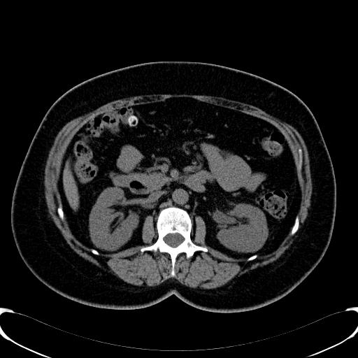
2 Case 1: 69 year old male with recurrent urinary tract infections! Name two ways we know that this study was performed with intravenous contrast?! Identify the abnormality.! How did this abnormality occur?! What are the low attenuation and the high attenuation areas in the kidneys?! CASE 1!
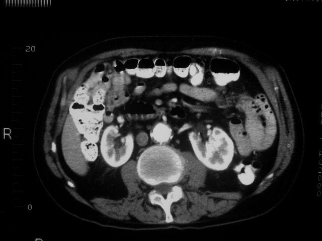
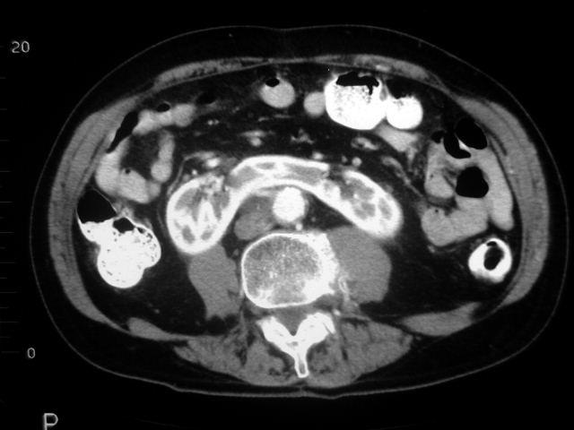
3 CASE 1!
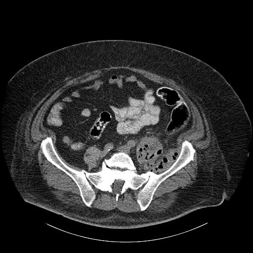
4 Case 2: 70 year old diabetic male with fever and flank pain! Would you use intravenous contrast for this patient and why? What was done in this case?! Do you see an air-fluid level? Where is it? Is there abnormal gas in other places?! Your diagnosis?! CASE 2!

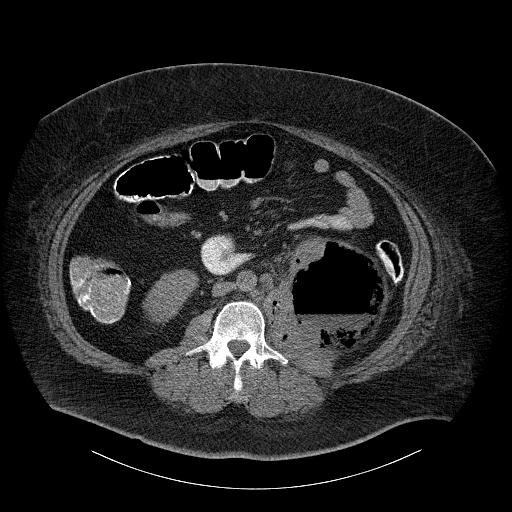
5 1! 2! 3! 4! CASE 2!

6 These images were obtained from the same study. What plane are we looking at?! Identify any abnormal gas. Point out any muscle with abnormal gas. CASE 2!
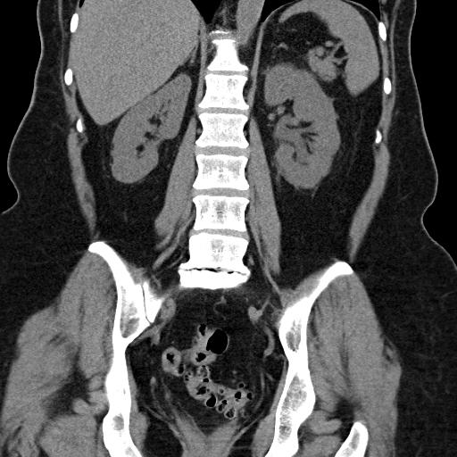
7 Case 3: 32 year old, otherwise healthy woman with fever and flank pain! Why do these kidneys enhance more uniformly than in Case 1?! Which flank hurt?! Identify any area of abnormal enhancement, and explain its appearance.! CASE 3!
8 1! 2! 3! 4! CASE 3!

9 coronal reconstruction! CASE 3!
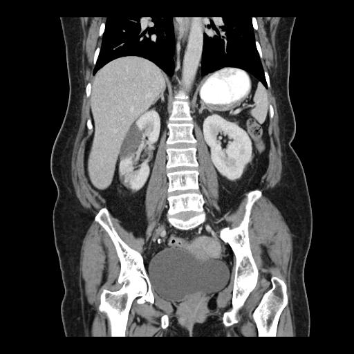
10 Case 4: 53 year old woman with progressive renal failure! Identify the kidneys. Are they normal in size or attenuation? Are there discrete areas of abnormality?! Does the appearance of the liver narrow your differential diagnosis?! What is the difference in appearance between a cyst and a solid lesion?! CASE 4!

11 CASE 4! Clue: compare lesion attenuation with that of muscle, spleen, or liver.!

12 coronal econstruction! CASE 4!
13 Case 5: 80 year old woman with hematuria and weight loss! Identify the abnormality. Name ways in which its appearance is different from that of a simple cyst.! Your diagnosis?! What does that area of low attenuation in the center of the lesion indicate pathologically and physiologically? How is this area different from a simple cyst?! CASE 5!

14 1! 2! 3! 4! CASE 5!


15 2 different patients with renal masses, one benign, the other malignant! What ultrasound features distinguish these masses?!


16 How can color Doppler help distinguish benign from malignant renal masses?!
17 Case 6: Young man with sudden onset of severe flank pain and hematuria! You suspect an obstructing kidney stone. Would you perform the study with or without IV contrast, and why?! Identify the collecting systems of both kidneys. How are they different? Which flank hurt?! Identify the cause of the obstruction.!
18 CASE 6!


19 These renal sonograms are from a 25 yo woman with! identical complaints. You learn her LMP was 5 weeks ago! RK! LK! Compare carefully the renal pelvis and calyces from each kidney. Do you see any differences?! How would you proceed with management?!

20 This is an image obtained through the bladder trigone! slightly to the left of midline. The echogenic structure! between the cross hairs is a stone. Where is it located?! Why is the sonogram preferable in this instance?!
21 Case 7: 60 year old male with atrial fibrillation, flank pain, no fever! Identify the abnormality. Would it have been easily seen without intravenous contrast. Why or why not?! How does the shape of the areas of abnormal enhancement help in the differential diagnosis! What is the diagnosis? What could have caused this?! CASE 7!
22 CASE 7!

23 Identify the aorta and any evidence of vascular disease.! Any evidence for prior surgery?! CASE 7!
24 Case 8: 19 year old intoxicated female in a roll-over motor vehicle accident! Identify any free fluid. What type of fluid could this be, especially with this history?! Can you identify the bladder foley catheter? How does the fluid in the bladder compare with the free fluid?! Contrast material was introduced into the bladder, and the second set of images taken. What happened to the bladder?! CASE 8!
25 CASE 8! before contrast administration!

26 CASE 8! after a cystogram!
27 Case 9: 29 yr old woman with pelvic discomfort! What type of imaging would you first order?! Look at the normal pelvic organs from a different! patient, then the images from this patient! What other history would you like to obtain?! CASE 9!


28 Normal uterus right ovary! Normal left ovary! What type of exam is this and how is it done?!


29 Patient s right ovary! Patient s left ovary! Comparing these to the previous normal images,! which ovary do you suspect may be abnormal and why?! Hint: review the pelvic CT image, case 7, Radlab 3!! CASE 9!

30 Left ovary! CASE 9! What technique is being used here?! How might it be helpful?!
31 Case 10: 29 yr old woman with sudden pelvic pain! What could be going on?! What type of imaging would you order?!


32 transabdominal! transvaginal! Both of these images show a right ovarian cyst and a part of the uterus, but with different technique! What differences in detail do you see?! How does this help make a diagnosis?!
Find Medical Solutions to Your Problems HYDRONEPHROSIS. (Distension of Renal Calyces & Pelvis)
") HYDRONEPHROSIS (Distension of Renal Calyces & Pelvis) Hydronephrosis is the distension of the renal calyces and pelvis due to accumulation of the urine as a result of the obstruction to the outflow of
HYDRONEPHROSIS (Distension of Renal Calyces & Pelvis) Hydronephrosis is the distension of the renal calyces and pelvis due to accumulation of the urine as a result of the obstruction to the outflow of
Rad Lab 6 Unknowns: Musculoskeletal
 Rad Lab 6 Unknowns: Musculoskeletal Peter Clarke MD Associate Clerkship Director for Radiology Harvard Medical School Brigham and Women s Hospital Dana Farber Cancer Institute Here are two men, one 70,
CASE STUDY. Presented by: Jessica Pizzo. CFCC Sonography student Class of 2018
 CASE STUDY Presented by: Jessica Pizzo CFCC Sonography student Class of 2018 Case Presentation April 4, 2017 56 yr old woman presented to ED with lower abdominal pain & swelling, along with constipation.
Genitourinary. Common Clinical Scenarios Protocoling Module. Patty Ojeda & Mariam Shehata
 The following training module was developed as a quality improvement project to serve as an educational tool for junior radiology residents. The following diagnostic radiology protocoling modules were
PROFESSIONAL SKILLS 1 3RD YEAR SEMESTER 6 RADIOGRAPHY. THE URINARY SYSTEM Uz. Fatema shmus aldeen Tel
 PROFESSIONAL SKILLS 1 3RD YEAR SEMESTER 6 RADIOGRAPHY THE URINARY SYSTEM Uz. Fatema shmus aldeen Tel. 0925111552 Professional skills-2 THE URINARY SYSTEM The urinary system (review anatomy and physiology)
ISUOG Basic Training. Distinguishing between Normal & Abnormal Appearances of the Urinary Tract. Seshadri Suresh, India
 ISUOG Basic Training Distinguishing between Normal & Abnormal Appearances of the Urinary Tract Seshadri Suresh, India Learning objectives 13 & 14 At the end of the lecture you will be able to: describe
CLINICAL PRESENTATION AND RADIOLOGY QUIZ QUESTION
 Donald L. Renfrew, MD Radiology Associates of the Fox Valley, 333 N. Commercial Street, Suite 100, Neenah, WI 54956 2/12/2011 Radiology Quiz of the Week # 7 Page 1 CLINICAL PRESENTATION AND RADIOLOGY QUIZ
Imaging Ejaculatory Disorders and Hematospermia
 ATHENS 4-6 October 2018 European Society of Urogenital Radiology Imaging Ejaculatory Disorders and Hematospermia Parvati Ramchandani, MD Professor, Radiology and Surgery University of Pennsylvania Medical
Female Genital Tract Lab. Dr. Nisreen Abu Shahin Assistant Professor of Pathology University of Jordan
 Female Genital Tract Lab Dr. Nisreen Abu Shahin Assistant Professor of Pathology University of Jordan Ovarian Pathology A 20-year-old female presented with vague left pelvic pain. Pelvic exam revealed
Bladder Trauma Data Collection Sheet
 Bladder Trauma Data Collection Sheet If there was no traumatic injury with PENETRATION of the bladder DO NOT proceed Date of injury: / / Time of injury: Date of hospital arrival: / / Time of hospital arrival:
My Patient Has Abdominal Pain PoCUS of the Biliary Tract and the Urinary Tract
 My Patient Has Abdominal Pain PoCUS of the Biliary Tract and the Urinary Tract Objectives PoCUS for Biliary Disease PoCUS for Renal Colic PoCUS for Urinary Retention Biliary Disease A patient presents
Uroradiology For Medical Students
 Uroradiology For Medical Students Lesson 4: Cystography & Urethrography - Part 2 American Urological Association Review Cystography is useful in evaluating the bladder, the urethra and the competence of
Abdominal Ultrasonography
 Abdominal Ultrasonography David A. Masneri, DO, FACEP, FAAEM Assistant Professor of Emergency Medicine Assistant Director, Emergency Medicine Residency Medical Director, Operational Medicine Division Center
Leiomyoma of the Bladder in a 23-year-old Male: Case Report
 in a 23-year-old Male: Ahmet Camtosun, Huseyin Celik Inonu University, Turgut Ozal Medical Center, Department of Urology, Malatya, Turkey Abstract Leiomyoma is a rare mesenchymal tumor that can occur in
The Focused Assessment with Sonography for Trauma, (FAST) procedure.
 procedure.") The Focused Assessment with Sonography for Trauma, (FAST) procedure. ROBERT H. WRIGLEY Professor Veterinary Diagnostic Imaging University of Sydney Veterinary Teaching Hospital Professor Emeritus Colorado
The Focused Assessment with Sonography for Trauma, (FAST) procedure. ROBERT H. WRIGLEY Professor Veterinary Diagnostic Imaging University of Sydney Veterinary Teaching Hospital Professor Emeritus Colorado
Pelvic Pain. What you need to know. 139 Dumaresq Street Campbelltown Phone Fax
 Pelvic Pain What you need to know 139 Dumaresq Street Campbelltown Phone 4628 5292 Fax 4628 0349 www.nureva.com.au September 2015 PELVIC PAIN This is a common problem and most women experience some form
CLINICAL PRESENTATION AND RADIOLOGY QUIZ QUESTION
 Donald L. Renfrew, MD Radiology Associates of the Fox Valley, 333 N. Commercial Street, Suite 100, Neenah, WI 54956 1/22/2011 Radiology Quiz of the Week # 4 Page 1 CLINICAL PRESENTATION AND RADIOLOGY QUIZ
A Practical Approach to Adnexal Masses
 A Practical Approach to Adnexal Masses Darcy J. Wolfman, MD Section Chief of Genitourinary Imaging American Institute for Radiologic Pathology Clinical Associate Johns Hopkins Community Radiology Division
Obstructive Nephropathy
 Obstructive Nephropathy Liza A. Lucero RN, FNP-C, MSN Renal Medicine Associates Conflicts No conflict of interests Obstructive Nephropathy Objectives Definition of Obstructive Nephropathy Causes Clinical
Sara Schaenzer Grand Rounds January 24 th, 2018
 Sara Schaenzer Grand Rounds January 24 th, 2018 Bladder Anatomy Ureter Anatomy Areas of Injury Bladder: Posterior bladder wall above trigone Ureter Crosses beneath uterine vessels At pelvic brim when ligating
Ultrasound - Pelvis. What is Pelvic Ultrasound Imaging?
 Scan for mobile link. Ultrasound - Pelvis Ultrasound imaging of the pelvis uses sound waves to produce pictures of the structures and organs in the lower abdomen and pelvis. There are three types of pelvic
What is ovarian cancer?
 What is ovarian cancer? Ovarian cancer is a type of cancer that forms in tissues of the ovary. Most ovarian cancers are either ovarian epithelial cancers (cancer that begins in the cells on the surface
Outline. Introduction to imaging modalities of the urinary system. Case base learning of common diseases in urinary tract
 Outline Introduction to imaging modalities of the urinary system Case base learning of common diseases in urinary tract Outline Introduction to imaging modalities of the urinary system Case base learning
Outline. Introduction to imaging modalities of the urinary system. Case base learning of common diseases in urinary tract
 Outline Introduction to imaging modalities of the urinary system Case base learning of common diseases in urinary tract Diagnostic Investigations in Urinary System PLAIN KUB EXCRETORY UROGRAPHY RETROGRADE
American Journal of Oral Medicine and Radiology
 American Journal of Oral Medicine and Radiology e - ISSN - XXXX-XXXX ISSN - 2394-7721 Journal homepage: www.mcmed.us/journal/ajomr ULTRASONOGRAPHIC EVALUATION OF ADNEXAL MASSES Nageswar Rao* Professor,
Paediatric surgical emergencies. Mani Thyagarajan BWCH
 Paediatric surgical emergencies Mani Thyagarajan BWCH General points Always discuss Call consultant for help ASAP CT scan is a bad modality in paediatrics Ultrasound? Intussusception? Renal colic? UTI
MODULE 5: HEMATURIA LEARNING OBJECTIVES DEFINITION. KEY WORDS: Hematuria, Cystoscopy, Urine Cytology, UTI, bladder cancer
 MODULE 5: HEMATURIA KEY WORDS: Hematuria, Cystoscopy, Urine Cytology, UTI, bladder cancer LEARNING OBJECTIVES At the end of this clerkship, the learner will be able to: 1. Define microscopic hematuria.
Hydronephrosis. What is hydronephrosis?
 What is hydronephrosis? Hydronephrosis Hydronephrosis describes the situation where the urine collecting system of the kidney is dilated. This may be a normal variant or it may be due to an underlying
Ovarian Lesion Benign vs Malignant?
 Ovarian Lesion Benign vs Malignant? Michele Keenan 1,2 Bernice Dunne 2 Mary Moran 1 Therese Herlihy 1 1. Radiography and Diagnostic Imaging, School of Medicine, University College Dublin, Ireland 2. Midland
Renal Artery Stenosis With Severe Hypertension: A Case Report
 CASE REPORT Renal Artery Stenosis With Severe Hypertension: A Case Report Suwaid MA ABSTRACT Background: Renal artery stenosis (RAS) is found in 77% of hypertensive patients and is responsible for 1-2%
The many faces of Endometriosis
 The many faces of Endometriosis Beryl Benacerraf M.D Harvard Medical School What is Endometriosis? Endometriosis is defined as the presence of normal endometrial tissue occurring outside of the endometrial
Sonographic Detection of Cervical Carcinoma With Metastases
 634026JDMXXX10.1177/8756479316634026Journal of Diagnostic Medical SonographyChappell and Fisher research-article2016 Case Study Sonographic Detection of Cervical Carcinoma With Metastases Journal of Diagnostic
Pelvic Ultrasound.
 Pelvic Ultrasound Before Your Exam: Drink 32 oz. of water one hour before your examination time. Try to drink all the liquid within 30 minutes. Do not urinate before the exam. Arrive for your exam with
Chapter 6: Genitourinary and Gastrointestinal Systems 93
 Chapter 6: Genitourinary and Gastrointestinal Systems 93 Chapter 6 Genitourinary and Gastrointestinal Systems Embryology Three sets of excretory organs or kidneys develop in human embryos: Pronephros:
L. Alexandre Frigini MD; Aaron Thomas, MD; Veronica Lenge de Rosen, MD
 Computed Tomography Urography (CTU) for Evaluation of Asymptomatic microscopic hematuria. Is intravenous contrast administration warranted for all patients? A retrospective evaluation utilizing ACR s Appropriateness
UBC Department of Urologic Sciences Lecture Series. Urological Trauma
 UBC Department of Urologic Sciences Lecture Series Urological Trauma Disclaimer: This is a lot of information to cover and we are unlikely to cover it all today These slides are to be utilized for your
Do ovarian cysts cause back pain
 Toggle navigation Do ovarian cysts cause back pain 10-3-2018 Ovarian Cysts - Symptoms. What. Since March 2017 had back pain radiating to groin and leg associated with. Ovarian Cysts - Causes. What was
Zoltan Harkanyi M.D., Ph.D. Department of Radiology, Heim Pal Children s Hospital, Budapest, Hungary
 Zoltan Harkanyi M.D., Ph.D. Department of Radiology, Heim Pal Children s Hospital, Budapest, Hungary CEUS expereince 10 years Department of Radiology, Heim Pal Children s Hospital, Budapest US N o 1 study
Renal Pelvis Squamous Cell Carcinoma and Renal Cell Carcinoma in a Tuberculous Kidney
 Case Study TheScientificWorldJOURNAL (2004) 4, 965 968 ISSN 1537-744X; DOI 10.1100/tsw.2004.196 Renal Pelvis Squamous Cell Carcinoma and Renal Cell Carcinoma in a Tuberculous Kidney M. Al-Assiri 1, M.F.
Acute Pyelonephritis
 Acute Pyelonephritis Variant 1: Acute pyelonephritis. Uncomplicated patient (eg, no history of diabetes or immune compromise or history of stones or obstruction or prior renal surgery or lack of response
Appendix 5. EFSUMB Newsletter. Gastroenterological Ultrasound
 EFSUMB Newsletter 87 Examinations should encompass the full range of pathological conditions listed below A log book listing the types of examinations undertaken should be kept Training should usually
performed to help sway the clinician in what the appropriate diagnosis is, which can substantially alter the treatment of management.
 Hello, I am Maura Polansky at the University of Texas MD Anderson Cancer Center. I am a Physician Assistant in the Department of Gastrointestinal Medical Oncology and the Program Director for Physician
West Yorkshire Major Trauma Network Clinical Guidelines 2015
 WYMTN: Pelvic fracture with urogenital trauma KEY RECOMMENDATIONS 1. During the initial exploratory survey / secondary survey, a. The external urethral meatus and the transurethral bladder catheter (if
CLINICAL PRESENTATION AND RADIOLOGY QUIZ QUESTION
 Donald L. Renfrew, MD Radiology Associates of the Fox Valley, 333 N. Commercial Street, Suite 100, Neenah, WI 54956 8/20/2011 Radiology Quiz of the Week # 34 Page 1 CLINICAL PRESENTATION AND RADIOLOGY
X-Plain Ovarian Cancer Reference Summary
 X-Plain Ovarian Cancer Reference Summary Introduction Ovarian cancer is fairly rare. Ovarian cancer usually occurs in women who are over 50 years old and it may sometimes be hereditary. This reference
Step up to the iu22 The key reasons are now even more compelling
 Step up to the iu22 The key reasons are now even more compelling The reasons to step up to the iu22 are now more compelling than ever Reason #1 Reduce failed ultrasound exams on your technically difficult
Metastatic Colonic Adenocarcinoma Simulating Primary Ovarian Neoplasm in Transvaginal Doppler Sonography
 J Clin Ultrasound 22:121-125, February 1994 0 1994 by John Wiley & Sons, Inc. CCC 0091-2751/94/020121-05 Case Report Metastatic Colonic Adenocarcinoma Simulating Primary Ovarian Neoplasm in Transvaginal
Abdominal Ultrasound : Aorta, Kidneys, Bladder
 Abdominal Ultrasound : Aorta, Kidneys, Bladder Nilam J. Soni, MD, MSc Associate Professor of Medicine Divisions of Hospital Medicine and Pulmonary/Critical Care Medicine Department of Medicine University
BLADDER CANCER: PATIENT INFORMATION
 BLADDER CANCER: PATIENT INFORMATION The bladder is the balloon like organ located in the pelvis that stores and empties urine. Urine is produced by the kidneys, is conducted to the bladder by the ureters,
Gastrointestinal & Genitourinary Emergencies. Lesson Goal. Learning Objectives 9/10/2012
 Gastrointestinal & Genitourinary Emergencies Lesson Goal Recognize, assess & provide care to patients with abdominal cavity injuries Learning Objectives Discuss different causes of nontraumatic abdominal
Abdomen and Pelvis CT (1) By the end of the lecture students should be able to:
 By the end of the lecture students should be able to:") RAD 451 Abdomen and Pelvis CT (1) By the end of the lecture students should be able to: State the common indications for Abdomen and pelvis CT exams Identify possible contra indications for Abdomen and
RAD 451 Abdomen and Pelvis CT (1) By the end of the lecture students should be able to: State the common indications for Abdomen and pelvis CT exams Identify possible contra indications for Abdomen and
Index. mri.theclinics.com. Note: Page numbers of article titles are in boldface type.
 Index Note: Page numbers of article titles are in boldface type. A Angiogenesis, and cancer of prostate, 689 690 Angiography, MR. See MR angiography. Apoptosis, MR imaging of, 637 Apparent diffusion coefficient,
Abdomen and Retroperitoneum Ultrasound Protocols
 Abdomen and Retroperitoneum Ultrasound Protocols Reviewed By: Anna Ellermeier, MD Last Reviewed: March 2018 Contact: (866) 761-4200, Option 1 **NOTE for all examinations: 1. If documenting possible flow
CLINICAL PRESENTATION AND RADIOLOGY QUIZ QUESTION
 Donald L. Renfrew, MD Radiology Associates of the Fox Valley, 333 N. Commercial Street, Suite 100, Neenah, WI 54956 2/19/2011 Radiology Quiz of the Week # 8 Page 1 CLINICAL PRESENTATION AND RADIOLOGY QUIZ
ULTRASOUND NOMENCLATURE
 Chapter 1: Ultrasound Nomenclature, Image Orientation, and Basic Instrumentation CYNTHIA SIKOWSKI Ultrasound waves are sound waves that have a frequency exceeding 20,000 Hz. When sound waves are transmitted
DAll that you need to know
 DAll that you need to know ouble - J Stenting D.Dalela UroHealth Education Cell UroHealth Research Centre, Lucknow What is a Double-J Stent? Double J (D.J.) Stent is a fine tube made of silicone coated
Five Views of Transitional Cell Carcinoma: One Man s Journey
 September 2006 Five Views of Transitional Cell Carcinoma: One Man s Journey Amsalu Dabela, Harvard Medical School III Outline Overview: Renal Anatomy Our Patient s Story Diagnostic Imaging Studies Appearance
Pelvic Pain: Diagnosis and Management
 Pelvic Pain: Diagnosis and Management Mr N Pisal Consultant Gynaecologist Advanced Laparoscopic Surgeon www.london-gynaecology.com History LMP Dysmenorrhoea / Dyspareunia Cyclical pain related to menstrual
n Make tremendous difference in patients lives: n Diagnosing or excluding disease and injury n Evaluating response to therapy
 Imaging: Choosing the Appropriate Exam Rob Milman, MD Austin Radiological Association What is a Radiologist? A physician who specializes in diagnosing and treating disease and injury by using medical imaging
Acute renal colic Radiological investigation in patients with renal colic
 Acute renal colic Radiological investigation in patients with renal colic Mikael Hellström Professor Department of Radiology Sahlgrenska University Hospital Göteborg University 0.9-1.8/1.000 inhabitants
Genitourinary Radiology In-Training Test Questions for Diagnostic Radiology Residents
 Genitourinary Radiology In-Training Test Questions for Diagnostic Radiology Residents March, 2013 Sponsored by: Commission on Education Committee on Residency Training in Diagnostic Radiology 2013 by American
Bladder Cancer Early Detection, Diagnosis, and Staging
 Bladder Cancer Early Detection, Diagnosis, and Staging Detection and Diagnosis Catching cancer early often allows for more treatment options. Some early cancers may have signs and symptoms that can be
US in non-traumatic acute abdomen. Lalita, M.D. Radiologist Department of radiology Faculty of Medicine ChiangMai university
 US in non-traumatic acute abdomen Lalita, M.D. Radiologist Department of radiology Faculty of Medicine ChiangMai university Sagittal Orientation Transverse (Axial) Orientation Coronal Orientation Intercostal
Nonurographic evaluation of renal calculous disease 1
 Contributions Nonurographic evaluation of renal calculous disease 1 Gregory P. Borkowski, M.D. Craig R. George, M.D. Peter B. O'Donovan, M.D. While excretory urography has been useful in the evaluation
Lec-8 جراحة بولية د.نعمان
 4th stage Lec-8 جراحة بولية د.نعمان 11/10/2015 بسم هللا الرحمن الرحيم Ureteric, Vesical, & urethral stones Ureteric Calculus Epidemiology like renal stones Etiology like renal stones Risk factors like
Imaging the Urogenital System
 maging the Urogenital System Tony Pease, DVM, MS, DACVR Assistant Professor of Radiology North Carolina State University Reading Thrall Chapters 42-46 Prostate Gland Not visible radiographically in normal
ASSESSING THE PLAIN ABDOMINAL RADIOGRAPH M A A M E F O S U A A M P O F O
 ASSESSING THE PLAIN ABDOMINAL RADIOGRAPH M A A M E F O S U A A M P O F O Introduction The abdomen (less formally called the belly, stomach, is that part of the body between the thorax (chest) and pelvis,
RADIOLOGY ORDERING GUIDE
 DOWNLOAD OR READ : DIAGNOSTIC ULTRASOUND ABDOMEN AND PELVIS E BOOKDIAGNOSTIC ULTRASOUND PHYSICS BIOLOGY AND INSTRUMENTATIONDIAGNOSTIC ULTRASOUND PRINCIPLES AND INSTRUMENTS PDF EBOOK EPUB MOBI Page 1 Page
Understanding Women's Sexuality after Bladder Cancer webinar. Part I: The Physical Impact
 Understanding Women's Sexuality after Bladder Cancer webinar Tuesday, December 1, 2015 Part I: The Physical Impact Presented by LaShon Day received her Masters of Science as a Physician s Assistant at
Fetal Renal Malformations: The Role of Ultrasound in Diagnosis & Management
 Fetal Renal Malformations: The Role of Ultrasound in Diagnosis & Management 12 weeks Alfred Abuhamad, M.D. Eastern Virginia Medical School 13 weeks 2nd trimester Medullary pyramids Renal Sinus Cortex 2nd
Continuous Bladder Irrigation
 Continuous Bladder Irrigation Introduction Continuous bladder irrigation, or CBI, is the infusion of a sterile solution into the urinary bladder. The purpose of CBI is to prevent the formation of blood
Acute flank pain in children: Imaging considerations
 Acute flank pain in children: Imaging considerations Carlos J. Sivit MD Rainbow Babies and Children s Hospital Case Western Reserve School of Medicine Flank pain Results from distention of ureter or renal
Male groin pain icd 9
 Cari untuk: Cari Cari Male groin pain icd 9 Free, official information about 2013 (and also 2015) ICD-9-CM diagnosis code 789.0, including coding notes, detailed descriptions, index cross-references and
CLINICAL PRESENTATION AND RADIOLOGY QUIZ QUESTION
 Donald L. Renfrew, MD Radiology Associates of the Fox Valley, 333 N. Commercial Street, Suite 100, Neenah, WI 54956 2/5/2011 Radiology Quiz of the Week # 6 Page 1 CLINICAL PRESENTATION AND RADIOLOGY QUIZ
H(a)ematuria. FX Keeley Consultant Urologist Bristol Urological Institute
ematuria. FX Keeley Consultant Urologist Bristol Urological Institute") H(a)ematuria FX Keeley Consultant Urologist Bristol Urological Institute From Philadelphia to Bristol, England Southmead Hospital, 1916 Southmead Hospital, 2013 Southmead Hospital, 2014 H(a)ematuria Blood
H(a)ematuria FX Keeley Consultant Urologist Bristol Urological Institute From Philadelphia to Bristol, England Southmead Hospital, 1916 Southmead Hospital, 2013 Southmead Hospital, 2014 H(a)ematuria Blood
What a history of cancer may mean for you and your family and the steps you can take to reduce the risk
 What a history of cancer may mean for you and your family and the steps you can take to reduce the risk www.myriadtests.com 1-800-469-7423 Family Ties and Colon Cancer In every family, certain traits are
Bladder Cancer in Primary Care. Dr Penny Kehagioglou Consultant Clinical Oncologist
 Bladder Cancer in Primary Care Dr Penny Kehagioglou Consultant Clinical Oncologist Objectives Patient presentation in primary care Investigating bladder cancer Management of bladder cancer Differential
What is endometrial cancer?
 Uterine cancer What is endometrial cancer? Endometrial cancer is the growth of abnormal cells in the lining of the uterus. The lining is called the endometrium. Endometrial cancer usually occurs in women
8/14/2017. Kidney location & visualization. Brief Review with tips & Case Based Illustrations. Size = x L2. Size =
 Dr. Russell Tucker, DACVR Brief Review with tips & Case Based Illustrations Kidney location & visualization K9 Kidneys: Rt @ T13-L1 Lt @ L2-L4 Kidney visualization K9 Kidneys: Rt @ T13-L1 Lt @ L2-L4 Size
APPROACH TO ABDOMINAL MASS
 Thomas Hong APPROACH TO ABDOMINAL MASS General Presentation An abdominal mass in a neonate, young child, or adolescent patient is something that every pediatrician needs to be wary of as these masses can
Endometrioma With Calcification Simulating a Dermoid on Sonography
 Case Report Endometrioma With Calcification Simulating a Dermoid on Sonography Kiran A. Jain, MD Several investigators have explored the sonographic diagnostic criteria of endometriomas. Endometriomas
4 th Year Urology Core Objectives Keith Rourke (Revised June 1, 2007)
") 4 th Year Urology Core Objectives Keith Rourke (Revised June 1, 2007) I. Genitourinary Trauma: 1. Goal: The student will be able to demonstrate a basic clinical approach to the management & diagnosis of
4 th Year Urology Core Objectives Keith Rourke (Revised June 1, 2007) I. Genitourinary Trauma: 1. Goal: The student will be able to demonstrate a basic clinical approach to the management & diagnosis of
Pediatric Retroperitoneal Masses Radiologic-Pathologic Correlation
 Acta Radiológica Portuguesa, Vol.XVIII, nº 70, pág. 61-70, Abr.-Jun., 2006 Pediatric Retroperitoneal Masses Radiologic-Pathologic Correlation Marilyn J. Siegel Mallinckrodt Institute of Radiology, Washington
A Case Report Hydronephrosis and Hydrodureter due to Ureteral Deep Infiltrating Endometriosis mimic Ureteral Stricture Suryamanggala SI 1, Satria ML 2
 A Case Report Hydronephrosis and Hydrodureter due to Ureteral Deep Infiltrating Endometriosis mimic Ureteral Stricture Suryamanggala SI 1, Satria ML 2 1 Departement of Obstetric and Gynecology Faculty
objectives Pitfalls and Pearls in PET/CT imaging Kevin Robinson, DO Assistant Professor Department of Radiology Michigan State University
 objectives Pitfalls and Pearls in PET/CT imaging Kevin Robinson, DO Assistant Professor Department of Radiology Michigan State University To determine the regions of physiologic activity To understand
Is Structured Reporting More Accurate Than Conventional Reporting in CT Reporting of the Abdomen and Pelvis?
 Is Structured Reporting More Accurate Than Conventional Reporting in CT Reporting of the Abdomen and Pelvis? A M Almuslim, MBBS; J G Ryan, MD; A Murtaza, MD Purpose The purpose of this research is to determine
Lab Schedule for Rest of Semester
 Laboratory 9 Cat Dissection II Respiratory Urinary/Reproductive Systems Lab Schedule for Rest of Semester Cat dissection labs Dissection II (today) Respiratory (Ex. 57 in Hole) Human Reproductive Systems
PREAMBLE GENERAL DIAGNOSTIC RADIOLOGY
 PREAMBLE The General Diagnostic Radiology category is intended to cover the body of knowledge a practicing board certified Diagnostic Radiologist should know. Since the range of content relevant to the
Chemotherapy for Breast Cancer - General
 Chemotherapy for Breast Cancer - General Introduction Breast cancer is a common condition that affects one out of every 11 women. Your doctor has recommended chemotherapy for your breast cancer. Chemotherapy
FHS Appendicitis US Protocol
 FHS Appendicitis US Protocol Reviewed By: Shireen Khan, MD; Sarah Farley, MD; Anna Ellermeier, MD Last Reviewed: May 2018 Contact: (866) 761-4200 **NOTE for all examinations: 1. If documenting possible
2. Blunt abdominal Trauma
 Abdominal Trauma 1. Evaluation and management depends on: a. Mechanism (Blunt versus Penetrating) b. Injury complex in addition to abdomen c. Haemodynamic stability assessment: i. Classically patient s
January Details of the fee code revisions can be found highlighted in Schedule A, attached.
 Government of Newfoundland and Labrador Department of Health and Community Services January 2018 18-01 TO: RE: ALL FEE-FOR-SERVICE PHYSICIANS CHANGES TO DOPPLER ULTRASOUND FEE CODES The Department of Health
Gynaecology Cancer Red Flags. Dr Dina Bisson Consultant Obstetrician and Gynaecologist Southmead Hospital North Bristol NHS Trust 27 April 2017
 Gynaecology Cancer Red Flags Dr Dina Bisson Consultant Obstetrician and Gynaecologist Southmead Hospital North Bristol NHS Trust 27 April 2017 Gynaecological Cancers Endometrial Cancer Ovarian Cancer Cervical
Renal Trauma: Management Options
 Renal Trauma: Management Options Immediate surgical repair Nephrectomy Conservative management Alonso RC et al. Kidney in Danger: CT Findings of Blunt and Penetrating Renal Trauma. RadioGraphics 2009;
Basic Training Programme. 16 Februrary 2018, ROTTERDAM. Pre and Post-Course Test Answers
 Basic Training Programme 16 Februrary 2018, ROTTERDAM Pre and Post-Course Test Answers Your details: Name: Conference registration number/ BT delegate number: Email address: Are you already performing
Learning Objectives for Rotations in Vascular Surgery Year 3 Basic Clerkship
 Learning Objectives for Rotations in Vascular Surgery Year 3 Basic Clerkship CLINICAL PROBLEMS IN VASCULAR SURGERY 1. ABDOMINAL AORTIC ANEURYSM A 70 year old man presents in the emergency department with
US Applications. Case Based Wrap-Up 1. Case 1 E-FAST. Case presentations E-FAST Abdominal. Pearls for each indication
 Case Based Wrap-Up 1 Stephanie J. Doniger MD RDMS FAAP FACEP Associate Director, Pediatric Emergency Ultrasound Stanford University Medical Center US Applications Case presentations E-FAST Abdominal Aorta
Prostate Gland Disorders
 Prostate Gland Disorders THE PROSTATE GLAND A male s prostate gland is located in the floor of the pelvis surrounding the urethra between the bladder and the penis. The prostate is positioned immediately
Acute Kidney Injury. I. David Weiner, M.D. Division of Nephrology, Hypertension and Transplantation University of Florida and NF/SGVHS
 Acute Kidney Injury I. David Weiner, M.D. Division of Nephrology, Hypertension and Transplantation University of Florida and NF/SGVHS 374-6102 David.Weiner@medicine.ufl.edu www.renallectures.com Concentration
General history. Basic Data : Age :62y/o Date of admitted: Married status : Married
 General history Basic Data : Age :62y/o Date of admitted:940510 Married status : Married General history Chief Complain : bilateral ovarian cyst incidentally being found out during pap smear. Present Illness