Abdominal Ultrasonography
|
|
|
- Angel Lyons
- 5 years ago
- Views:
Transcription
1 Abdominal Ultrasonography David A. Masneri, DO, FACEP, FAAEM Assistant Professor of Emergency Medicine Assistant Director, Emergency Medicine Residency Medical Director, Operational Medicine Division Center for Applied Learning Wake Forest University School of Medicine

2
3 Disclosure None: I do not have any significant financial relationships to disclose.
4 Outline of Topics: Detection of intra-abdominal free fluid and Aorta Clinical Application Anatomy Technique Normal US Pathologic US Pitfalls/Take Home Points
5 Clinical Application Question: Why learn how to identify intra-abdominal free fluid? Answer: Clinically significant Trauma patient assessment Part of US evaluation of a hypotensive patient Component of RUSH Exam Ascites assessment Assist in US Guided Paracentesis
6 How good is US for detection of free fluid? Detection of Intraperitoneal fluid - Excellent Increased sensitivity with Increasing number of views Positioning (Trendelenburg) Serial examinations Important to visualize as much of the areas as possible and not just obtain one quick view Multiple windows may be required to fully evaluate for free fluid
7 US is POOR for assessing: Solid organ disruption without significant bleed Hollow viscus injury Retroperitoneal injury or hemorrhage


8 Appearance of Free Fluid Anechoic collections Free flowing blood Urine, bile, ascites, peritoneal dialysate Echogenic Clotted blood Bowel contents
9 Peritoneal Windows - Dependent Spaces LUQ: Perisplenic space liver 2 RUQ: Morison s pouch Pelvic: Retrovesical space Retrouterine space 7 7


10 RUQ (Hepatorenal) View Mid-axillary line in 10, 11 interspaces Must see junction of right kidney and liver Morison's pouch Visualize subdiaphragmatic space Visualize the pleural space 8

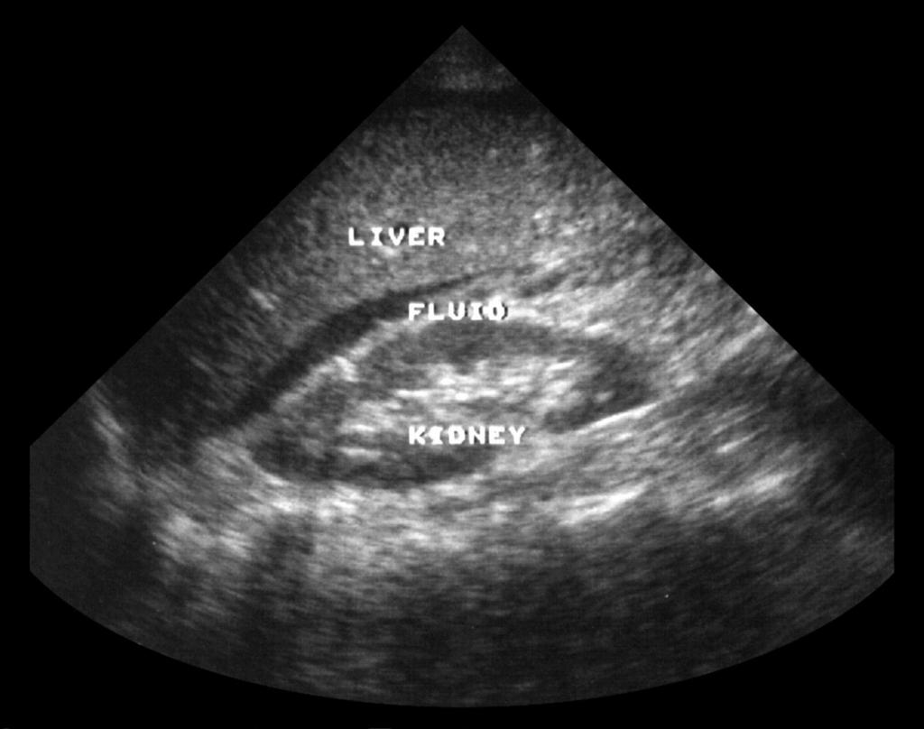
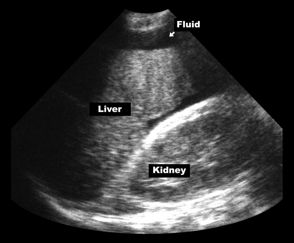
11 RUQ View Liver cephalad Kidney inferiorly Morison s Pouch * * * *


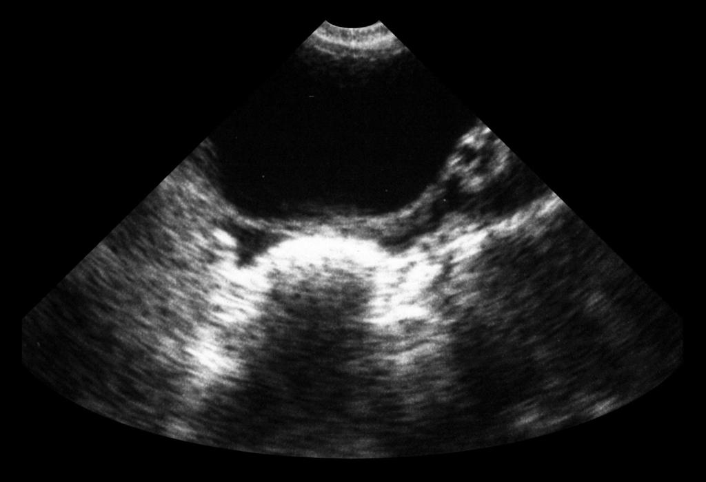
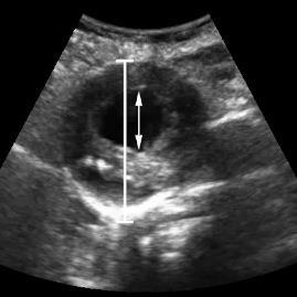
12 Free fluid in Morison s Pouch Normal Morison s Pouch

13 Positive RUQ
14
15
16

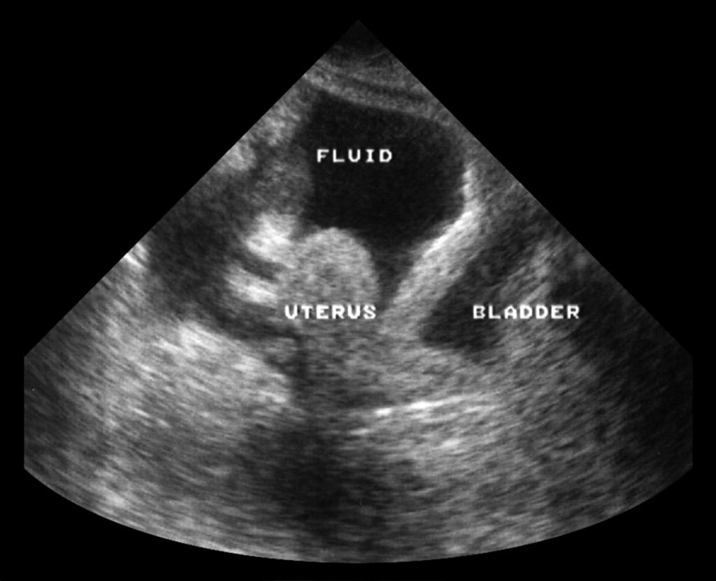
17 Pelvic (Suprapubic or Bladder) View Probe should be placed in the suprapubic position Full bladder is window Helpful to image before placement of a Foley Trick of trade: If bladder is empty or Foley already placed: IV bag on abdomen Scan through bag


18 Pelvic View Where to look? Behind bladder in men Retrovesicular space Behind uterus in women Retrouterine space Reduce far gain Sagittal and transverse 7

19 Sagittal Pelvic View

20 Positive Sagittal Pelvic View
21

22 Transverse Pelvic View

23 Positive Pelvis Transverse View
24
 Spleen is a smaller window Close attention to perisplenic area: Diaphragm spleen interface Splenorenal space not as")
25 LUQ (Perisplenic) View Probe at left posterior axillary line at Left 8 th or 9 th Intercostal space Angle probe obliquely (avoid ribs) Spleen is a smaller window Close attention to perisplenic area: Diaphragm spleen interface Splenorenal space not as important

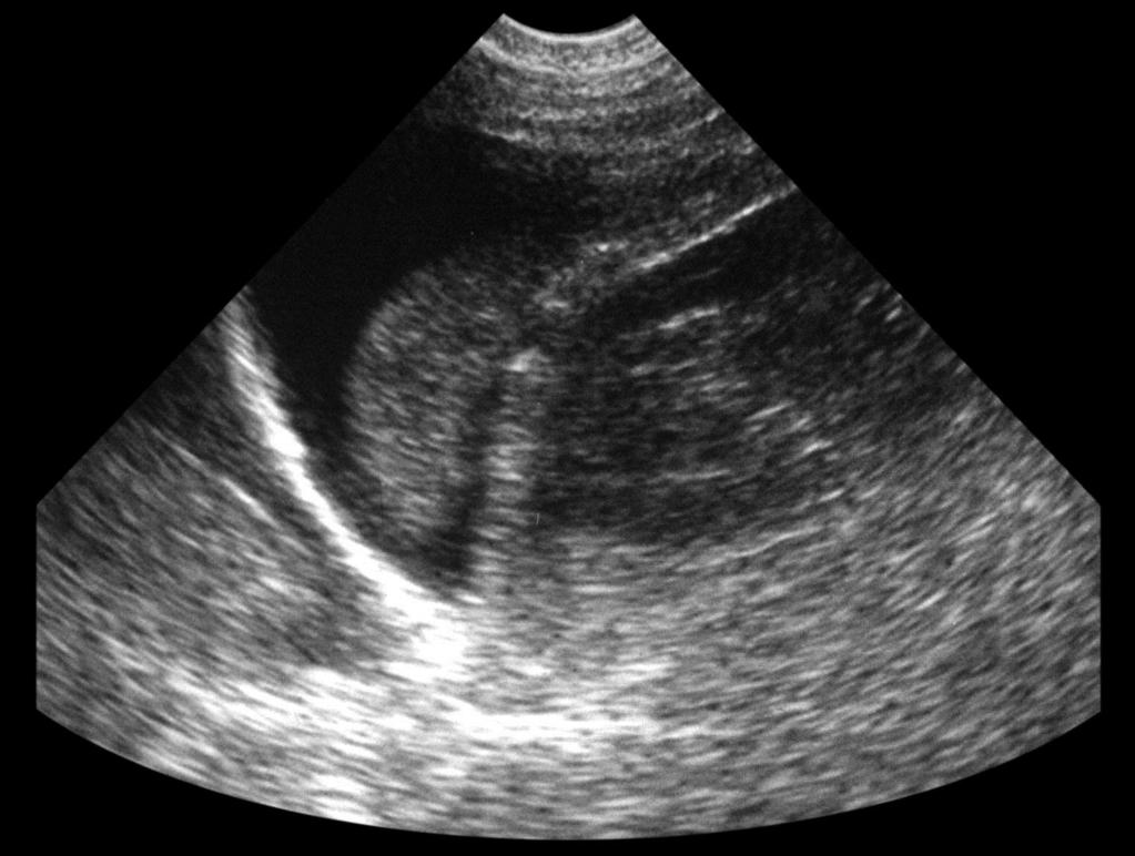
26 Normal LUQ diaphragm spleen kidney

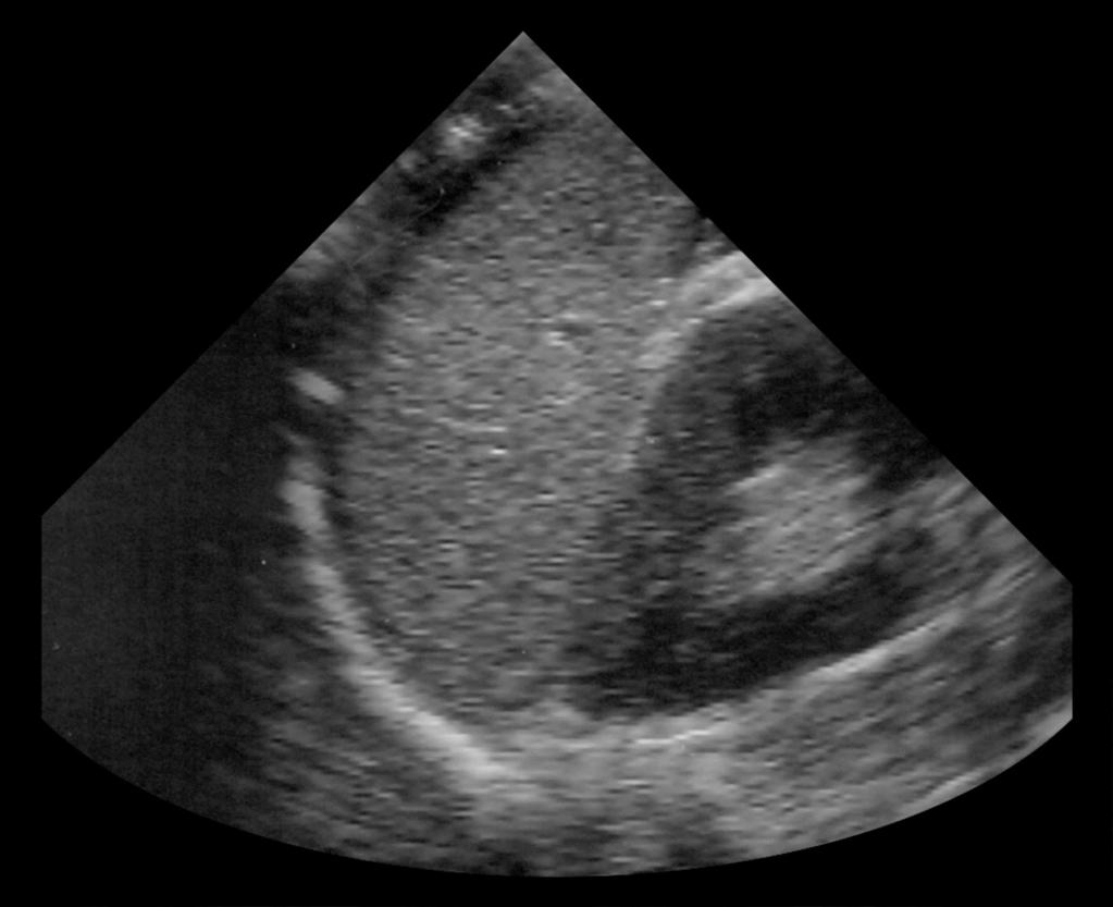
27 Positive LUQ View
28
29
30 Assessment for Intra-peritoneal Free Fluid Interpretation: Exam positive If any one abd view positive Exam negative All three abd views negative Indeterminate Cannot adequately visualize any one view Window not available Technically limited study Body habitus

31 OK the FAST is positive, but home much fluid is in the belly? 1cm stripe 1 L of fluid 0.5 cm stripe 500 ml Thin stripe 250 ml
32 Aorta Ultrasound Clinical Application Question: Why learn Aorta Ultrasound? Answer: Clinically significant in assessing or AAA Flank or back pain in the elderly patient Part of US evaluation of a hypotensive patient Component of RUSH Exam Good screening study
33 Aorta Ultrasound Excellent sensitivity for presence of AAA High degree of accuracy with brief training Improve time to diagnosis and OR vs. CT Poorly identifies retroperitoneal rupture
 Superior Mesenteric (SMA) Renal (RA) Inferior Mesenteric (IMA) From: Gray, H. Anatomy of the Human Body 20 th ed.")
34 Aorta - Anatomy Courses from Xyphoid to umbilicus Originates at diaphragm and extends to iliacs Main branches Celiac (CA) Superior Mesenteric (SMA) Renal (RA) Inferior Mesenteric (IMA) From: Gray, H. Anatomy of the Human Body 20 th ed. 2000
35 Aorta - Anatomy Maximal diameter Proximal: 2.1 cm for males, 1.8 cm for females > 55 yrs Distal: 1.5 cm Aneurysm considered when diameter 3 cm Measure diameter from outer surface of walls

36 Aorta Technique Curved or phased array probe Supine position preferred
37 Aorta Technique Transverse Aorta Anterior to vertebral body Abd wall Ao vs IVC: Aorta on pt s left IVC on pt s right Right Left IVC Aorta Back
38 Normal Appearance - Transverse Vertebral body posterior, Hyperechoic arch Aorta and IVC anechoic circles anterior to vertebral body Aorta divides at bifurcation Right IVC Abd wall Aorta Left Back
39 Bifurcation- Transverse
40 Aorta Technique Longitudinal images Abd Wall Rotate probe 90 Head Feet Rock and slide technique Back
41 Normal Appearance - Longitudinal Longitudinal Aorta Courses left to right Branches Celiac Superior Mesenteric Course anterior to Ao Head CA Abd Wall Ao SMA Feet Back
42 Technically Difficult Studies Bowel gas and obesity may obscure images Maneuvers: Gentle pressure with probe Left lateral decubitus position Coronal plane images from right flank

43 Coronal Right Head Feet Cava Aorta Left
44 IVC Aorta On patient s right Undulating motion Thin walled Compressible Collapses with negative intrathoracic pressure Sniff, Valsalva On patient s left Pulsatile motion Thick walled Non-compressible Non-collapsing

45 Color Doppler to Differentiate
46 AAA Best visualized in transverse view Many AAAs have greater transverse than AP diameters If you see the aorta VERY WELL it is probably a AAA Fusiform Symmetric, concentric dilatation More common Saccular Localized out-pouching 90 % occur at and below level of renals (region of SMA) MUST visualize Ao bifurcation for complete scan

47 AAA Transverse View Fusiform Abd wall Right Left Back

48 AAA - Longitudinal View Fusiform Abd Wall Head Feet Back

49 AAA Transverse View Saccular
50

51 AAA - Transverse View with Thrombus
52 Intro to RUSH Exam Rapid Ultrasound for Shock and Hypotension Assess the Pipes, Pump, Tank HI MAP Heart IVC Morison s Pouch (Abdominal FAST views) Aorta Pneumothorax

53 Thank You
Focused Assessment Sonography of Trauma (FAST) Scanning Protocol
 Scanning Protocol") Focused Assessment Sonography of Trauma (FAST) Scanning Protocol Romolo Gaspari CHAPTER 3 GOAL OF THE FAST EXAM Demonstrate free fluid in abdomen, pleural space, or pericardial space. EMERGENCY ULTRASOUND
Focused Assessment Sonography of Trauma (FAST) Scanning Protocol Romolo Gaspari CHAPTER 3 GOAL OF THE FAST EXAM Demonstrate free fluid in abdomen, pleural space, or pericardial space. EMERGENCY ULTRASOUND
Focused Assessment with Sonography in Trauma (FAST) UC Irvine School of Medicine
 UC Irvine School of Medicine") Focused Assessment with Sonography in Trauma (FAST) UC Irvine School of Medicine Purpose of FAST exam Quickly evaluate patient s status in emergency situations Blunt or penetrating trauma Visualize fluid
Focused Assessment with Sonography in Trauma (FAST) UC Irvine School of Medicine Purpose of FAST exam Quickly evaluate patient s status in emergency situations Blunt or penetrating trauma Visualize fluid
Background & Indications
 Teresa S. Wu, MD, FACEP Director, EM Ultrasound Program & Fellowship Co-Director, Simulation Based Training Program & Fellowship Maricopa Medical Center Simulation Curriculum Director Associate Professor,
Point-of-Care Ultrasound Guide for Landmarks, Recording, and Report Content. TJUH/MHD EM Ultrasound Division 2012
 Point-of-Care Ultrasound Guide for Landmarks, Recording, and Report Content TJUH/MHD EM Ultrasound Division 2012 Table of Contents 1 - Objectives 2 - Procedural 3 - AAA 4 - Abdominal OB 5 - Transvaginal
Archiving in Qpath Defining Adequate
 General Archiving Information for QPath Users As you become familiar with Qpath and how to archive your clips you will want to be sure you are capturing good quality clips for review. The properly captured,
Point of Care Ultrasound (PoCUS)
") Point of Care Ultrasound (PoCUS) Competency Assessment Forms AORTA Competency A Focussed Assessment of the Aorta (AAA) Guidance Please follow this guidance as closely as possible to ensure consistency
Point of Care Ultrasound (PoCUS) Competency Assessment Forms AORTA Competency A Focussed Assessment of the Aorta (AAA) Guidance Please follow this guidance as closely as possible to ensure consistency
Abdominal Ultrasound : Aorta, Kidneys, Bladder
 Abdominal Ultrasound : Aorta, Kidneys, Bladder Nilam J. Soni, MD, MSc Associate Professor of Medicine Divisions of Hospital Medicine and Pulmonary/Critical Care Medicine Department of Medicine University
The FAST Exam! Dr. David Easton MD FRCPC Critical Care and Emergency Medicine University of Manitoba Canada
 The FAST Exam! Dr. David Easton MD FRCPC Critical Care and Emergency Medicine University of Manitoba Canada Dr. David Easton MD FRCPC Assistant Professor Section of Critical Care and Emergency Medicine
Certificate in Clinician Performed Ultrasound (CCPU) Syllabus. Extended Focussed Abdominal Scan for Trauma (E-FAST)
 Syllabus. Extended Focussed Abdominal Scan for Trauma (E-FAST)") Certificate in Clinician Performed Ultrasound (CCPU) Syllabus Extended Focussed Abdominal Scan for Trauma (E-FAST) Page 1 of 6 01/17 ACN 001 679 161 ABN 64 001 679 Extended Focussed Abdominal Scan for
Certificate in Clinician Performed Ultrasound (CCPU) Syllabus Extended Focussed Abdominal Scan for Trauma (E-FAST) Page 1 of 6 01/17 ACN 001 679 161 ABN 64 001 679 Extended Focussed Abdominal Scan for
FAST Focused Assessment with Sonography in Trauma
 FAST Focused Assessment with Sonography in Trauma Wilma Rodriguez Mojica,MD,FACR Professor of Radiology UPR School of Medicine Ultrasound Section - Radiological Sciences Department OBJECTIVES Understand
Advanced Imaging Practice CSB068
 Advanced Imaging Practice CSB068 Week 1 Peer Review - Evaluation of work by one or more people of similar competence to the producer - A form of self-regulation about improving quality and upholding standards
My Patient Has Abdominal Pain PoCUS of the Biliary Tract and the Urinary Tract
 My Patient Has Abdominal Pain PoCUS of the Biliary Tract and the Urinary Tract Objectives PoCUS for Biliary Disease PoCUS for Renal Colic PoCUS for Urinary Retention Biliary Disease A patient presents
L o o k L i s t e n F e e l S c a n. Your Pocus Cards For Your Every Day Scanning.
 L o o k L i s t e n F e e l S c a n Your Pocus Cards For Your Every Day Scanning E-FAST Extended Focused Assessment by Sonography in Trauma Subcostal Heart View Pleural Sliding on M-mode (Sea-shore sign)
Background & Indications Probe Selection
 Teresa S. Wu, MD, FACEP Director, EM Ultrasound Program & Fellowship Co-Director, Simulation Based Training Program & Fellowship Associate Program Director, EM Residency Program Maricopa Medical Center
Basic of Ultrasound Physics E FAST & Renal Examination. Dr Muhammad Umer Ihsan MBBS,MD, DCH CCPU,DDU1,FACEM
 Basic of Ultrasound Physics E FAST & Renal Examination Dr Muhammad Umer Ihsan MBBS,MD, DCH CCPU,DDU1,FACEM What is Sound? Sound is Mechanical pressure waves What is Ultrasound? Ultrasounds are sound waves
Abdominal ultrasound:
 Abdominal ultrasound: Non-traumatic acute abdomen Wittanee Na-ChiangMai, MD Department of Radiology ChiangMai University 26/04/2017 Contents Technique of examination Normal anatomy Emergency conditions
Abdomen and Retroperitoneum Ultrasound Protocols
 Abdomen and Retroperitoneum Ultrasound Protocols Reviewed By: Anna Ellermeier, MD Last Reviewed: March 2018 Contact: (866) 761-4200, Option 1 **NOTE for all examinations: 1. If documenting possible flow
Guidelines, Policies and Statements D5 Statement on Abdominal Scanning
 Guidelines, Policies and Statements D5 Statement on Abdominal Scanning Disclaimer and Copyright The ASUM Standards of Practice Board have made every effort to ensure that this Guideline/Policy/Statement
Scanning Mesenteric and Hypogastric Artery Aneurysms
 Scanning Mesenteric and Hypogastric Artery Aneurysms Marsha M. Neumyer, BS, RVT, FSVU, FSDMS, FAIUM International Director Vascular Diagnostic Education Services Vascular Resource Associates Harrisburg,
Extended FAST Exam. Goal of Trauma Care. Golden Hour of Trauma
 Extended FAST Exam Goal of Trauma Care Golden Hour of Trauma Best INITIAL screening modality in trauma efast 2014 LLSA Article (ACEP Policy Statement) Level B Recommendation: In hemodynamically unstable
Objectives. The Extended FAST Exam. Focused Assessment e With Sonography In. Trauma (FAST)
") Northern California Emergency Ultrasound Course Objectives The Extended FAST Exam Rimon Bengiamin, MD, RDMS UC SF Discuss the components of the EFAST exam Evaluate the utility of the EFAST Review how to
Northern California Emergency Ultrasound Course Objectives The Extended FAST Exam Rimon Bengiamin, MD, RDMS UC SF Discuss the components of the EFAST exam Evaluate the utility of the EFAST Review how to
Abdominal Ultrasound
 Abdominal Ultrasound Imaging Control Buttons Depth The organ imaged should take up 3/4 of the screen Frequency = Penetration Use high frequencies (harmonics) for fluid filled and superficial structures
Abdominal Aortic Aneurysm (AAA)
") Abdominal Aortic Aneurysm (AAA) Vascular Workshop: Objectives Anatomy Keith VanHaltren Indications Technique Cases Abdominal Aorta: Normal Size Abdominal aortic aneurysm: Definition Normal diameter of
Abdominal Aortic Aneurysm (AAA) Vascular Workshop: Objectives Anatomy Keith VanHaltren Indications Technique Cases Abdominal Aorta: Normal Size Abdominal aortic aneurysm: Definition Normal diameter of
Basic Abdominal Sonography
 24S Basic Abdominal Sonography Procedural Overview JOHN FATCHETT II, RDMS is provided. Patient preparation (i.e., fasting) scanning techniques, spleen, transducer. evaluation of abdominal anatomy in the
AAENP US WORKSHOP 2/25/17
 Know the components of the Rapid Ultrasound for Shock & Hypotension & Extended Focused Assessment Sonography in Trauma & how they can help quickly determine diagnosis. Be comfortable obtaining and interpreting
FHS Appendicitis US Protocol
 FHS Appendicitis US Protocol Reviewed By: Shireen Khan, MD; Sarah Farley, MD; Anna Ellermeier, MD Last Reviewed: May 2018 Contact: (866) 761-4200 **NOTE for all examinations: 1. If documenting possible
ASSESSING THE PLAIN ABDOMINAL RADIOGRAPH M A A M E F O S U A A M P O F O
 ASSESSING THE PLAIN ABDOMINAL RADIOGRAPH M A A M E F O S U A A M P O F O Introduction The abdomen (less formally called the belly, stomach, is that part of the body between the thorax (chest) and pelvis,
PAH EMERGENCY Department Aortic Scanning module
 PAH EMERGENCY Department Aortic Scanning module Abdominal Aorta Scan is a goal directed ultrasound examination to detect the presence of an Abdominal Aortic Aneurysm (AAA). The Abdominal Aorta scan is
Ultrasound. FAST Focused Assessment with Sonography in Trauma
 Ultrasound FAST Focused Assessment with Sonography in Trauma Rohit Patel, MD University of Florida Health Director, Critical Care Ultrasound Surgical ICU Center for Intensive Care Gainesville, Florida
Deb Coghlan AMS (Vascular and General ) Brisbane, Australia
 Brisbane, Australia") Deb Coghlan AMS (Vascular and General ) Brisbane, Australia ANEURYSMAL DIISEASE The infrarenal aorta enlarges with age, and is the commonest site for arterial aneurysms. An aneurysm is a permanent focal
Deb Coghlan AMS (Vascular and General ) Brisbane, Australia ANEURYSMAL DIISEASE The infrarenal aorta enlarges with age, and is the commonest site for arterial aneurysms. An aneurysm is a permanent focal
Intro Case. Outline What We ll Cover. What we won t cover. Cardiac Ultrasound and The RUSH Exam: Bedside Ultrasound in Resuscitation and Shock
 Cardiac Ultrasound and The RUSH Exam: Bedside Ultrasound in Resuscitation and Shock Justin Davis, MD, MPH, RDMS Associate Physician Subchief for Emergency Ultrasound Services Kaiser Oakland Medical Center
Abdominal Ultrasound. Diane Hallinen, MD. Bloodroot
 Abdominal Ultrasound Diane Hallinen, MD Bloodroot Abdominal Ultrasound Vasculature Hepatobiliary Spleen Kidney Bladder Bowel Where to put the probe? Vasculature We are going to talk about Celiac Trunk
ASSOC. PROF. DR. SADIK GİRİŞGİN Necmettin Erbakan Uni. Meram Medicine School
 ASSOC. PROF. DR. SADIK GİRİŞGİN Necmettin Erbakan Uni. Meram Medicine School Portable Probe variety Real time imaging Cheap No radiation Repeatable Cost effective 1988 1971 1990 2001 2008 FAST First case
The Role of the FAST exam in the EDRU
 The Role of the FAST exam in the EDRU A. Robb McLean, MD, MHCM Vice Chair of Clinical Operations, Department of Emergency Medicine Joint Trauma Conference June 20, 2017 Disclosures Goals Describe the performance,
Objectives. Hepatobiliary Ultrasound: Anatomy, Technique, Pathology. RUQ: Normal Anatomy. Emergency Ultrasound: Gallbladder Location
 Hepatobiliary Ultrasound: Anatomy, Technique, Pathology Laleh Gharahbaghian, MD FAAEM Associate Director, EM Ultrasound Co-Director, EM Ultrasound Fellowship Stanford University Medical Center Seric Cusick,
The Human Body: An Overview of Anatomy. Anatomy. Physiology. Anatomy - Study of internal and external body structures
 C H A P T E R 1 The Human Body: An Orientation An Overview of Anatomy Anatomy The study of the structure of the human body Physiology The study of body function Anatomy - Study of internal and external
1=presence Of Abdominal Aorta Aneurysm, 0=no Presence Of Abdominal Aorta Aneurysm
 Derived Variable Dataset (AAA_DERV) ARIC V5 / NCS Code Book III, S.2 Page 1 of 9 Derived Variable Dataset: FORM CODE=AAA_DERV VERSION=1.0 Instructions: Derived variables for AAA study. AAA 1=presence Of
Visceral Vascular Ultrasound. Joel Thompson, MD, MPH Borg & Ide Imaging
 Visceral Vascular Ultrasound Joel Thompson, MD, MPH Borg & Ide Imaging Objectives: Review major abdominal vascular structures Identify normal peak systolic velocity (PSV) for major abdominal arteries.
Vascular CT Protocols
 Vascular CT Protocols V 1D: Chest and abdominal CT angiogram (aortic dissection protocol) V 1T: Chest CT angiogram (aortic trauma protocol) V 2: Abdominal and pelvis CT angiogram (aortic aneurysm protocol)
Session 2: Ultrasonography for Primary Care Clinicians Learning Objectives
 Session 2: Ultrasonography for Primary Care Clinicians Learning Objectives 1. Assess the main components and functions of a portable ultrasound unit. 2. Identify three clinical applications of portable
The Focused Assessment with Sonography for Trauma, (FAST) procedure.
 procedure.") The Focused Assessment with Sonography for Trauma, (FAST) procedure. ROBERT H. WRIGLEY Professor Veterinary Diagnostic Imaging University of Sydney Veterinary Teaching Hospital Professor Emeritus Colorado
The Focused Assessment with Sonography for Trauma, (FAST) procedure. ROBERT H. WRIGLEY Professor Veterinary Diagnostic Imaging University of Sydney Veterinary Teaching Hospital Professor Emeritus Colorado
GASTRIC ULTRASOUND. A Point-of-care tool for aspiration risk assessment.
 GASTRIC ULTRASOUND A Point-of-care tool for aspiration risk assessment edu@gastricultrasound.org Indications Any clinical situation where aspiration risk is uncertain. For example: Lack of adherence to
Hepatobiliary Ultrasound Rimon Bengiamin, MD, RDMS Assistant Clinical Professor Director of Emergency Ultrasound UCSF Fresno. Objectives. Why?
 Hepatobiliary Ultrasound Rimon Bengiamin, MD, RDMS Assistant Clinical Professor Director of Emergency Ultrasound UCSF Fresno Objectives Discuss the goals of point-of-care biliary ultrasound Review the
ISUOG Basic Training. Examining Fetal Anatomy from Longitudinal Sections Titia Cohen-Overbeek, The Netherlands
 ISUOG Basic Training Examining Fetal Anatomy from Longitudinal Sections Titia Cohen-Overbeek, The Netherlands Learning objectives 2 & 3 At the end of the lecture you will be able to: describe how to obtain
Evolution of Clinical Anatomy with Ultrasonography Past Present and Future
 College of Medicine Evolution of Clinical Anatomy with Ultrasonography Past Present and Future Andrew F. Payer, Ph.D. Human Body Structure and Function Module Director Christine Bellew, M.D., Caridad Hernandez,
Lecture 56 Kidney and Urinary System
 Lecture 56 Kidney and Urinary System The adrenal glands are located on the superomedial aspect of the kidney The right diagram shows a picture of the kidney with the abdominal walls and organs removed
EFAST. Extended Focussed Assessment with Sonography for Trauma. Ultrasound Logbook. Name
 EFAST Extended Focussed Assessment with Sonography for Trauma Ultrasound Logbook ame Contents EFAST Accreditation Requirements 25 Abdominal Aorta Report Forms 3 Formative Assessments 1 Summative Assessment
US Applications. Case Based Wrap-Up 1. Case 1 E-FAST. Case presentations E-FAST Abdominal. Pearls for each indication
 Case Based Wrap-Up 1 Stephanie J. Doniger MD RDMS FAAP FACEP Associate Director, Pediatric Emergency Ultrasound Stanford University Medical Center US Applications Case presentations E-FAST Abdominal Aorta
**Confirm accuracy of above with your instructor.** Revised 8/22/2017 1
 AP1 Lab 1 Cavities, Organs, Serous Membranes, Quadrants, Regions, and Directional Terms, Planes & Sections Project 1 Directional Terminology Step 1: Define/Describe what is known as the "ANATOMICAL POSITION."
Emergency Ultrasound Standard Reporting Guidelines
 Emergency Ultrasound Standard Reporting Guidelines October 2011 *heterogenous corrected to heterogeneous on pages 9 & 11, January 2016 Emergency Ultrasound Standard Reporting Guidelines: Introduction and
Lab Monitor Images Dissection of the Abdominal Vasculature + Lower Digestive System
 Lab Monitor Images Dissection of the Abdominal Vasculature + Lower Digestive System Stomach & Duodenum Frontal (AP) View Nasogastric tube 2 1 3 4 Stomach Pylorus Duodenum 1 Duodenum 2 Duodenum 3 Duodenum
Background Focused Assessment with Sonography in Trauma. Johann Baptist Dormagen, MD, PhD
 Focused Assessment with Sonography in Trauma Johann Baptist Dormagen, MD, PhD Unit of Abdominal and Oncologic Radiology Department of Radiology and Nuclear Medicine Oslo University Hospital, Norway 8 th
The faculty will include physicians with international reputations as outstanding ultrasound educators.
 Ultrasound Courses Course Description Whether you re a beginner or a seasoned sonographer, this year s AAEM pre-conference ultrasound course will be worth your time. We will be offering a half day course
A Frame of Reference for Anatomical Study. Anatomy and Physiology Mr. Knowles Chapter 1 Liberty Senior High School
 A Frame of Reference for Anatomical Study Anatomy and Physiology Mr. Knowles Chapter 1 Liberty Senior High School Anatomical Terms of Direction and Position Created for communicating the direction and
Policies, Standards, and Guidelines. Guidelines for Abdominal Ultrasound Examination
 Policies, Standards, and Guidelines Guidelines for Abdominal Ultrasound Examination Approved by Council Feb 2018 Disclaimer and Copyright The ASUM Standards of Practice Board have made every effort to
Abdominal Doppler Mastering the next level of vascular anatomy in the belly. Cindy A. Owen, RDMS, RVT
 Abdominal Doppler Mastering the next level of vascular anatomy in the belly Cindy A. Owen, RDMS, RVT Introduction Abdominal Doppler is a tough exam Success is dependent on: Patient body habitus Patient
Abdominal Aortic Aneurysm. Ultrasound Logbook. Name
 Abdominal Aortic Aneurysm Ultrasound Logbook ame Contents AAA Accreditation Requirements 15 Abdominal Aorta Report Records 3 Formative Assessments 1 Summative Assessment AAA Accreditation Abdominal Aortic
Which Blunt Trauma Patients Should Be Studied by Abdominal CT?
 MDCT of Bowel and Mesenteric Injury: How Findings Influence Management 4 th Nordic Trauma Radiology Course 2006 4 th Nordic Trauma Radiology Course 2006 Stuart E. Mirvis, M.D., FACR Department of Radiology
Guide to Small Animal Vascular Imaging using the Vevo 770 Micro-Ultrasound System
 Guide to Small Animal Vascular Imaging using the Vevo 770 Micro-Ultrasound System January 2007 Objectives: After completion of this module, the participant will be able to accomplish the following: Understand
The abdominal Esophagus, Stomach and the Duodenum. Prof. Oluwadiya KS
 The abdominal Esophagus, Stomach and the Duodenum Prof. Oluwadiya KS www.oluwadiya.com Viscera of the abdomen Abdominal esophagus: Terminal part of the esophagus The stomach Intestines: Small and Large
US in non-traumatic acute abdomen. Lalita, M.D. Radiologist Department of radiology Faculty of Medicine ChiangMai university
 US in non-traumatic acute abdomen Lalita, M.D. Radiologist Department of radiology Faculty of Medicine ChiangMai university Sagittal Orientation Transverse (Axial) Orientation Coronal Orientation Intercostal
Introduction to Anatomical Terms. Packet #3
 Introduction to Anatomical Terms Packet #3 Directional Terms Directional terms describe the positions of structures relative to other structures or locations in the body. Introduction Superior vs. Inferior
SPECIAL DIAGNOSTIC STUDIES IN BLUNT TRAUMA OLEH : Prof.DR.Dr Abdul Rasyid SpRad (K),Ph.D Dr.Evo Elidar Sp.Rad
,Ph.D Dr.Evo Elidar Sp.Rad") SPECIAL DIAGNOSTIC STUDIES IN BLUNT TRAUMA OLEH : Prof.DR.Dr Abdul Rasyid SpRad (K),Ph.D Dr.Evo Elidar Sp.Rad Trauma Emergency Room layout Ideally the trauma emergency room is centrally located to provide
SPECIAL DIAGNOSTIC STUDIES IN BLUNT TRAUMA OLEH : Prof.DR.Dr Abdul Rasyid SpRad (K),Ph.D Dr.Evo Elidar Sp.Rad Trauma Emergency Room layout Ideally the trauma emergency room is centrally located to provide
POLICY STATEMENT. Emergency Ultrasound Imaging Criteria Compendium. Approved by the ACEP Board of Directors October 2014
 POLICY Approved by the ACEP Board of Directors October 2014 Emergency Ultrasound Imaging Criteria Compendium Developed by the following members of ACEP's Emergency Ultrasound Section: John L. Kendall,
Introduction to The Human Body
 1 Introduction to The Human Body FOCUS: The human organism is often examined at seven structural levels: chemical, organelle, cell, tissue, organ, organ system, and the organism. Anatomy examines the structure
Case Report 1. CTA head. (c) Tele3D Advantage, LLC
 Tele3D Advantage, LLC") Case Report 1 CTA head 1 History 82 YEAR OLD woman with signs and symptoms of increased intra cranial pressure in setting of SAH. CT Brain was performed followed by CT Angiography of head. 2 CT brain Extensive
Case Report 1 CTA head 1 History 82 YEAR OLD woman with signs and symptoms of increased intra cranial pressure in setting of SAH. CT Brain was performed followed by CT Angiography of head. 2 CT brain Extensive
Biology Human Anatomy Abdominal and Pelvic Cavities
 Biology 351 - Human Anatomy Abdominal and Pelvic Cavities Please place your name and I.D. number on the back of the last page of this exam. You must answer all questions on this exam. Because statistics
MDCT signs differentiating retroperitoneal and intraperitoneal lesions- diagnostic pearls
 MDCT signs differentiating retroperitoneal and intraperitoneal lesions- diagnostic pearls Poster No.: C-0987 Congress: ECR 2015 Type: Educational Exhibit Authors: D. V. Bhargavi, R. Avantsa, P. Kala; Bangalore/IN
Anatomical Terminology
 Anatomical Terminology Dr. A. Ebneshahidi Anatomy Anatomy : is the study of structures or body parts and their relationships to on another. Anatomy : Gross anatomy - macroscopic. Histology - microscopic.
Realistic simulation in Point of Care Ultrasound
 Realistic simulation in Point of Care Ultrasound 2 Preparing for PoCUS... BodyWorks Eve is an ultra-realistic female patient simulator designed for interactive and immersive Point of Care Ultrasound (PoCUS)
Normal Sonographic Anatomy
 hapter 2:The Liver DUNSTAN ABRAHAM Normal Sonographic Anatomy Homogeneous, echogenic texture (Figure 2-1) Measures approximately 15 cm in length and 10 12.5 cm anterior to posterior; measurement taken
Perioperative Ultrasonography Ehab Farag, MD, FRCA Hesham Elsharkawy David G. Anthony, M.D.
 Perioperative Ultrasonography Ehab Farag, MD, FRCA Hesham Elsharkawy David G. Anthony, M.D. Cleveland Clinic, Cleveland OH 1 Complications during central venous catheterization (CVC) occur 2% -15% of the
Disclosures. Cardiac Ultrasound. Introductory Case. 80 y/o male Syncope at home Emesis x 3 in ambulance Looks sick. No pain.
 Disclosures Cardiac Ultrasound Justin A Davis, MD MPH RDMS Subchief for Emergency Ultrasound Kaiser Permanente East Bay Medical Center I have nothing to disclose. Introductory Case HR 118 BP 65/43 RR 27
Abdomen and Pelvis CT (1) By the end of the lecture students should be able to:
 By the end of the lecture students should be able to:") RAD 451 Abdomen and Pelvis CT (1) By the end of the lecture students should be able to: State the common indications for Abdomen and pelvis CT exams Identify possible contra indications for Abdomen and
RAD 451 Abdomen and Pelvis CT (1) By the end of the lecture students should be able to: State the common indications for Abdomen and pelvis CT exams Identify possible contra indications for Abdomen and
Ultrasound (2 weeks,) v PGY 1 & 3
 v PGY 1 & 3") Ultrasound (2 weeks,) v 11.14.10 PGY 1 & 3 PLEASE READ THESE GUIDELI ES CAREFULLY. YOU ARE RESPO SIBLE FOR FAMILIARITY WITH THIS CO TE T A D FOR ACHIEVI G THE GOALS OF THE ROTATIO. PLEASE CO TACT A ULTRASOU
Ultrasound (2 weeks,) v 11.14.10 PGY 1 & 3 PLEASE READ THESE GUIDELI ES CAREFULLY. YOU ARE RESPO SIBLE FOR FAMILIARITY WITH THIS CO TE T A D FOR ACHIEVI G THE GOALS OF THE ROTATIO. PLEASE CO TACT A ULTRASOU
Bedside RUQ Ultrasound. Replace Formal ULS? Why Bedside ULS RUQ? RUQ Ultrasound. Bedside ULS is Limited, Goal-Directed
 Bedside RUQ Ultrasound RUQ Ultrasound Why do it How to do it Elizabeth Kwan UCSF Emergency Ultrasound Fellow Why Bedside ULS RUQ? Dx or Rule Out Acute Cholecystitis Cholelithiasis, Choledocolithiasis Earlier
Advanced Bedside Ultrasound Course for Primary Care Clinicians MUSE 2.0
 M U S E McGill UltraSound Evaluation Program Advanced Bedside Ultrasound Course for Primary Care Clinicians MUSE 2.0 Table of Contents Course description... 2 Introduction... 2 Accreditation... 2 Course
Peritoneum: Def. : It is a thin serous membrane that lines the walls of the abdominal and pelvic cavities and clothes the viscera.
 Peritoneum: Def. : It is a thin serous membrane that lines the walls of the abdominal and pelvic cavities and clothes the viscera. Layers of the peritoneum: 1. Outer Layer ( Parietal Peritoneum) : lines
Transducer Selection. Renal Artery Duplex Exam. Renal Scan. Renal Scan Echogenicity. How to Perform an Optimal Renal Artery Doppler Examination
 How to Perform an Optimal Renal Artery Doppler Examination Director of Ultrasound Education & Quality Assurance Baylor College of Medicine Division of Maternal-Fetal Medicine Maternal Fetal Center Imaging
ISUOG Basic Training. Distinguishing between Normal & Abnormal Appearances of the Urinary Tract. Seshadri Suresh, India
 ISUOG Basic Training Distinguishing between Normal & Abnormal Appearances of the Urinary Tract Seshadri Suresh, India Learning objectives 13 & 14 At the end of the lecture you will be able to: describe
CT abdomen and pelvis
 CT abdomen and pelvis General indications: Assessment of vague abdominal symptoms (pain, colics,distenstion,...) Varifecation of a lesion discovered by other diagnostic modalities as US, barium,ivp, Staging
Diseases of the Aorta
 Diseases of the Aorta ASE Review 2018 Susan E Wiegers, MD, FASE, FACC Professor of Medicine My great friend Dr. Roberto Lang Disclosure None related to this presentation 1 Objectives Aneurysm Dissection
Introduction & Physics of ED Ultrasound. Objectives. What? - Limited Studies. Who? - ED Docs
 Introduction & Physics of ED Ultrasound Martine Sargent, MD Ultrasound Director, Assistant Professor UCSF Department of Emergency Medicine San Francisco General Hospital & Trauma Center Objectives Who?
Ultrasound basics Part 1
 Ultrasound basics Part 1 'Ultrasound enhanced critical care medicine' Rohit Patel, MD University of Florida Health Director, Critical Care Ultrasound Surgical ICU Center for Intensive Care Gainesville,
#5 Cardiovascular II Blood Vessels
 Page1 #5 Cardiovascular II Blood Vessels Objectives: Observe slide of artery and vein cross-section Identify a list of human arteries and veins using a virtual human dissection Dissect and identify a list
Abdominal Examination Benchmarks
 Abdominal Examination Benchmarks Preparation and Positioning: Stand on the right side of the patient. The patient should be supine and double draped so only the abdomen is exposed o To relax the abdominal
Certificate in Clinician Performed Ultrasound (CCPU) Syllabus
 Syllabus") Certificate in Clinician Performed Ultrasound (CCPU) Syllabus Abdominal Aortic Aneurysm (AAA) Page 1 of 6 12/18 Abdominal Aortic Aneurysm (AAA) Syllabus Purpose: This unit is designed to cover the theoretical
Certificate in Clinician Performed Ultrasound (CCPU) Syllabus Abdominal Aortic Aneurysm (AAA) Page 1 of 6 12/18 Abdominal Aortic Aneurysm (AAA) Syllabus Purpose: This unit is designed to cover the theoretical
#1 - Chapter 1 - Anatomy. General Anatomical Terms The Anatomical Position
 #1 - Chapter 1 - Anatomy General Anatomical Terms The Anatomical Position The anatomical position is a stance in which a person stands erect with the feet flat on the floor and close together, arms at
Caudal Edge of the Liver in the Right Upper Quadrant (RUQ) View Is the Most Sensitive Area for Free Fluid on the FAST Exam
 View Is the Most Sensitive Area for Free Fluid on the FAST Exam") Original Research Caudal Edge of the Liver in the Right Upper Quadrant (RUQ) View Is the Most Sensitive Area for Free Fluid on the FAST Exam Viveta Lobo, MD* Michelle Hunter-Behrend, MD* Erin Cullnan,
Original Research Caudal Edge of the Liver in the Right Upper Quadrant (RUQ) View Is the Most Sensitive Area for Free Fluid on the FAST Exam Viveta Lobo, MD* Michelle Hunter-Behrend, MD* Erin Cullnan,
Introduction to A & P Medical Terminology
 Human Anatomy & Physiology PHA322.10 D. Matesic, Spring, 2012 Class Notes Introduction to A & P Medical Terminology Levels of Structural Organization Anatomy the study of the structure of body parts and
Mesenteric/Splanchnic Artery Duplex Imaging
 VASCULAR TECHNOLOGY PROFESSIONAL PERFORMANCE GUIDELINES Mesenteric/Splanchnic Artery Duplex Imaging This Guideline was prepared by members of the Society for Vascular Ultrasound (SVU) as a template to
Human Anatomy Key Points Unit 1/ Study Guide
 Human Anatomy Key Points Unit 1/ Study Guide I. Anatomy and Physiology a. Anatomy 1. Means cutting apart (dissection) 2. Study of the body and the relationships of its parts to each other. 3. Dissection
Pelvic Injuries. Chapter 21
 Chapter 21 Introduction Injuries of the pelvis are an uncommon, but potentially lethal, battlefield injury. Blunt injuries may be associated with major hemorrhage and early mortality. Death within the
Basic Training. ISUOG Basic Training The 20 Planes Approach to the Routine Mid Trimester Scan
 ISUOG The 20 Planes Approach to the Routine Mid Trimester Scan Learning objective At the end of the lecture you will be able to: Explain how to perform a structured routine examination, including measurements,
Duodenum retroperitoneal
 Duodenum retroperitoneal C shaped Initial region out of stomach into small intestine RETROperitoneal viscus Superior 1 st part duodenal cap ; moves upwards and backwards to lie on the R crura medial to
MEDICAL TERMINOLOGY. Complete! Second Edition CHAPTER. The Human Body in Health and Disease Content Review Slides
 MEDICAL TERMINOLOGY Complete! Second Edition CHAPTER 4 The Human Body in Health and Disease Content Review Slides Learning Objectives Define and spell the word parts used to create terms for the human
Duplex ultrasound is first-line imaging for all
 Our Protocol for Transabdominal Pelvic Vein Duplex Ultrasound A summary of s protocol for pelvic vein duplex ultrasonography, including equipment, patient positioning, ultrasound settings, and technique.
Chapter 3 General Anatomy and Radiographic Positioning Terminology General Anatomy
 Chapter 3 General Anatomy and Radiographic Positioning Terminology General Anatomy Definition of Terms Anatomy- term applied to the science of the structure of the body Physiology- study of the function
Artery 1 Head and Thoracic Arteries. Arrange the parts in the order blood flows through them.
 Artery 1 Head and Thoracic Arteries 1. Given the following parts of the aorta: 1. abdominal aorta 2. aortic arch 3. ascending aorta 4. thoracic aorta Arrange the parts in the order blood flows through
Muscle spasm Diminished bowel sounds Nausea/vomiting
 3 4 5 6 7 8 9 0 Chapter 8: Abdomen and Genitalia Injuries Abdominal Injuries Abdomen is major body cavity extending from to pelvis. Contains organs that make up digestive, urinary, and genitourinary systems.