Effects of Plant Stress on Facultative Apomixis in Boechera (Brassicaceae)
|
|
|
- Stephen Banks
- 5 years ago
- Views:
Transcription


1 Utah State University All Graduate Theses and Dissertations Graduate Studies Effects of Plant Stress on Facultative Apomixis in Boechera (Brassicaceae) Mayelyn Mateo de Arias Utah State University Follow this and additional works at: Part of the Plant Sciences Commons Recommended Citation Mateo de Arias, Mayelyn, "Effects of Plant Stress on Facultative Apomixis in Boechera (Brassicaceae)" (2015). All Graduate Theses and Dissertations This Dissertation is brought to you for free and open access by the Graduate Studies at It has been accepted for inclusion in All Graduate Theses and Dissertations by an authorized administrator of For more information, please contact
2 EFFECTS OF PLANT STRESS ON FACULTATIVE APOMIXIS IN BOECHERA (BRASSICACEAE) by Mayelyn Mateo de Arias A dissertation submitted in partial fulfillment of the requirements for the degree of DOCTOR OF PHILOSOPHY in Plant Science Approved: Dr. John G. Carman Major Professor Dr. Paul G. Wolf Committee Member Dr. Paul G. Johnson Committee Member Dr. B. Shaun Bushman Committee Member Dr. David J. Hole Committee Member Dr. Mark R. McLellan Vice President for Research and Dean of the School of Graduate Studies UTAH STATE UNIVERSITY Logan, Utah 2015
3 i ABSTRACT Effects of Plant Stress on Facultative Apomixis in Boechera (Brassicaceae) By Mayelyn Mateo de Arias, Doctor of Philosophy Utah State University, 2015 Major Professor: Dr. John G. Carman Department: Plants, Soils and Climate In angiosperms, apomixis is asexual reproduction by seed. In gametophytic apomixis, every nucleus in the female gametophyte is unreduced, and the embryo forms parthenogenetically. Different types of gametophytic apomixis occur depending on the origin of the embryo. In this research we documented that both aposporous and diplosporous apomixis occur in the genus Boechera (Brassicaceae). Our results from cytological analysis show that in B. gunnisoniana, B. lignifera, B. retrofracta x exilis, and B. retrofracta x stricta the type of apomixis is diplospory; and in B. microphylla it is apospory. Sexual reproduction was observed in B. stricta. By using quantitative real time PCR (qrt-pcr), we obtained the same pattern of expression of four stress-related genes and one ribosomal gene that had been previously documented in a project carried out with microarray analysis in our lab. In this part of the project, we could appreciate differences in gene expression in pistils and ovules of sexual and apomictic Boechera species. A major objective of this study was to evaluate the effects of stress on megasporogenesis, seed set, and gene expression in sexual and apomictic species of the
4 ii genus Boechera. Cytology of ovule development, flow cytometry of single seeds and qrt-pcr of pistil samples were conducted on the five apomictic Boechera species and the one sexual species mentioned above. Sets of plants from each species were grown under ideal control conditions and other sets were exposed to drought stress or drought plus heat stress. The cytological evaluation of ovules showed an increase in the frequency of sexual ovule development (meiosis instead of apomeiosis) under stress in some species. However, no alteration was detected in frequencies of sexual and apomictic seed set regardless as to whether the plants were grown with or without stress. Drought stress triggered changes in gene expression analyzed by qrt-pcr. While these changes may be related to the switch from apomeiotic to meiotic megasporogenesis, these changes did not cause an increase in sexual seed formation. Low frequency sexual seed formation (relative sexual sterility) may be the result of the interspecific or triploid nature of the apomictic plants being tested. (128 pages)
5 iii PUBLIC ABSTRACT Effects of Plant Stress on Facultative Apomixis in Boechera (Brassicaceae) Mayelyn Mateo de Arias In flowering plants, apomixis is asexual reproduction by seeds. Apomixis allows the production of offspring with the same genetic characteristics as the mother plant. Fertilization is not required. Apomixis could become a tool for naturally cloning highyielding crop hybrids through their own seed. However, apomixis does not occur in major crop plants, except for citrus. In the present study, genes that might cause apomixis in naturally occurring apomictic plants were investigated. Sexual and apomictic species of the genus Boechera were exposed to stressed and non-stressed conditions. Effects of these treatments on the expression of apomixis was then measured. Stress triggered an increase in the frequency of sexual development in apomictic plants, but continuation of sexual development to form sexual seeds did not occur. Stress also triggered alterations in the expression of stress-related genes.
6 iv ACKNOWLEDGMENTS I would like to thank my husband, Amaury Arias, for his great support, encouragement and comprehension during the development of this program. In the same way, I thank my parents, Rafael and Milagros Mateo, and the rest of my family for helping me when I needed anything from them, and for believing in me. A very special thanks to my adviser, Dr. John Carman, for his important guidance during these years of academic experience. Also, thanks to Dr. Manoj Srivastava for his great support in this research. In the same way, I would like to thank my committee members for their contribution, Dr. Paul Wolf, Dr. Shawn Bushman, Dr. David Hole and Dr. Paul Johnson. I thank Dr. Krishna Dwivedi and my lab partners, Lei Gao and David Sherwood, for their contributions in the development of this project. Finally, my most special thanks to my heavenly Father for all my achievements, since He is the reason of my existence and the one who guides me in this life. Mayelyn Mateo de Arias
7 v CONTENTS ABSTRACT....ii PUBLIC ABSTRACT.. iv ACKNOWLEDGMENTS..v CONTENTS...vi LIST OF TABLES..viii LIST OF FIGURES...ix CHAPTER Page 1. INTRODUCTION LITERATURE CITED DIPLOSPOROUS AND APOSPOROUS APOMIXIS IN BOECHERA (BRASSICACEAE)...34 ABSTRACT INTRODUCTION.35 MATERIALS AND METHODS Plant Materials...37 Embryological analyses...38 Single Seed Flow Cytometry...39 RESULTS AND DISCUSSION 39 LITERATURE CITED DIFFERENTIALLY-EXPRESSED GENES IN PISTILS AND OVULES OF SEXUAL AND APOMICTIC BOECHERA (BRASSICACEAE).. 53 ABSTRACT...53 INTRODUCTION. 54 MATERIALS AND METHODS...56 Plant Materials...56 Embryological staging of pistils for sample collection.. 56 Quantitative Real Time PCR (qpcr)...57 RESULTS AND DISCUSSION....58
8 vi Gene selection qpcr analyses LITERATURE CITED EFFECTS OF STRESS ON MEGASPOROGENESIS, SEED SET, AND GENE EXPRESSION IN SEXUAL AND APOMICTIC BOECHERA (BRASSICACEAE)...74 ABSTRACT...74 INTRODUCTION...75 MATERIALS AND METHODS...77 Plant materials and treatments Embryological analyses.79 Single Seed Flow Cytometry.80 qrt-pcr RESULTS AND DISCUSSION 81 LITERATURE CITED SUMMARY LITERATURE CITED APPENDIXES.106 APPENDIX I APPENDIX II..111 APPENDIX III
9 vii LIST OF TABLES Table Page 1.1 Terminology important to my study Accession numbers and collection information for Boechera species and species hybrids evaluated embryologically and by single seed flow cytometry Frequencies of sexual tetrad and sexual or diplosporous dyad formation and frequencies of aposporous (AS) embryo sac formation by taxon as determined by DIC microscopy of ovules in the meiocyte (M) to early embryo sac (ES) formation stages Reproductive pathways by taxon as determined by single-seed embryo:endosperm C values (2C:3C sexual, others apomictic) Primer pairs used in the present study Genes differentially-expressed in ovule and pistil microarray experiments and chosen for qpcr analyses Analysis of variance statistics by gene Reproductive pathways by taxon as determined by single-seed embryo:endosperm C values (2C:3C sexual, others apomictic) of both treatment and control Analysis of variance statistics by gene
10 viii LIST OF FIGURES Figure Page 1.1 Polygonum type embryo sac development (sexual) Allium type embryo sac development (sexual, bispory) Forms of apomeiosis that directly involve the megaspore mother cell (MMC) Meiosis in the genetically doubled MMC Apomeiosis that involves the formation of an unreduced embryo sac from nucellar cells or rarely from integument cells (Apospory) Panicum type embryo sac development (apomictic, apospory) Sexual polygonum type embryo sac Sexual megasporogenesis of the Polygonum type in B. stricta. a-c: tetrads of genetically-reduced megaspores Diplosporous megasporogenesis of the Taraxacum type in apomictic B. lignifera Aposporous embryo sac formation of the Hieracium type in apomictic B. microphylla Representative flow-cytometry histograms derived from single seeds of various Boechera species Sexual diploid progenitors of the apomictic interspecific Boechera hybrids evaluated herein Differential gene expression as determined by qpcr Stress treatment effects on tetrad and dyad formation frequencies in sexual B. stricta, diplosporous B. gunnisoniana and diplosporous B. retrofracta x exilis and B. retrofracta x stricta (B. retrofracta x) and on frequencies of sexual and aposporous embryo sac formation in aposporous B. microphylla Megaspores derived by meiotic processes in sexual and apomictic Boechera Unreduced apomeiotic dyads of megaspores from non-stressed apomictic B. retrofracta x exilis (a), drought and heat-stressed B. retrofracta x stricta (b), drought and heat-stressed B. lignifera (c), non-stressed apomictic triploid B.
11 ix gunnisoniana (d), drought-stressed apomictic triploid B. gunnisoniana (e) and drought-stressed apomictic B. lignifera (f) Histogram of diploid sexual B. stricta with peaks 2C, 3C, for embryo and endosperm; and 4C and 6C for endoreplication of embryo and endosperm, correspondingly Histogram of diploid apomictic B. retro x stricta, showing peaks 2C and 6C, for embryo and fertilized endosperm, correspondingly Histogram of diploid apomictic B. retrofracta, showing peaks 2C, 4 C and 6C, for embryo, endoreplication of embryo and fertilized endosperm, correspondingly Histogram of diploid apomictic B. lignifera, showing peaks 2C, 4 C and 6C, for embryo, endoreplication of embryo and fertilized endosperm, correspondingly Histogram of diploid apomictic B. microphylla with peaks 2C and 5C, for embryo and fertilized endosperm, correspondingly Differential gene expression as determined by qpcr. Data were adjusted so that expression levels are relative to those observed in the control plants of sexual B. stricta...91
12 CHAPTER 1 INTRODUCTION Apomixis in flowering plants (angiosperms) is asexual reproduction by seed. Two major types of apomixis occur in plants, gametophytic and sporophytic. In gametophytic apomixis, every nucleus in the female gametophyte (embryo sac, ES), including the egg cell and the polar nuclei, is unreduced, and the embryo forms parthenogenetically. The unreduced ES originates in one of two ways, from a nucellar cell of the ovule wall (apospory) or directly from the megaspore mother cell (MMC) either before or during the early stages of meiosis (diplospory). Hence, all nuclei of the entire ES are unreduced, and the unreduced egg develops into an embryo without fertilization, i.e. parthenogenetically (Nogler, 1984). The endosperm in gametophytic apomicts forms pseudogamously or autonomously, i.e. it may or may not require fertilization of the ES central cell depending on the species (Ozias-Akins and van Dijk, 2007; Tucker and Koltunow, 2009; Yamada-Akiyama et al., 2009). In sporophytic apomixis, an abnormal unreduced parthenogenetically active cell (capable of embryogenesis without fertilization) of the ovule wall forms and undergoes embryo formation directly in a process referred to as adventitious or nucellar embryony. This clonal embryo uses the endosperm of the sexually derived ES for nutrition. Hence, sporophytic apomixis involves both gametophytic and sporophytic processes. The term apomixis was first used by Winkler (1908) to describe absence of mixis (absence of meiotic recombination) followed by parthenogenesis. Some authors have expanded the definition to include all types of vegetative reproduction, e.g. the production of rhizomes, tillers and vegetative buds, etc., but this modification of
13 2 Winkler s original intension has fallen out of favor (Asker and Jerling, 1992; Carman, 1997; Nogler, 1984; Tucker and Koltunow, 2009). Today, apomixis refers to a two steps process: i) formation of an unreduced cell, gamete or gametophyte from cells or related tissues normally involved in reduced gamete or gametophyte formation (apomeiosis), and ii) parthenogenesis. Based on this definition, the term apomixis has been appropriately used to describe apomeiosis followed by parthenogenesis in all kingdoms of eukaryotes, i.e. it occurs in Protozoa (Bilinski et al., 1989; Asker and Jerling, 1992); in Chromista (Bilinski et al., 1989); in Animalia (Maniatsi et al., 2010; Dufresne et al., 2011; Robinson et al., 2011), including many insects (Rozek et al., 2009); as well as in Plantae and Fungi (Suomalainen et al., 1987; Asker and Jerling, 1992; Donahoo and Lamour, 2008; Carman et al., 2011; Hörandl and Hojsgaard, 2012). Therefore, apomixis occurs in every kingdom. Because reproduction by apomixis produces clones of the mother plant, it may someday be a useful trait for crop improvement where it will cause progeny plants of a crop to express desired characteristics of the mother plant. This could be a great advantage with regard to hybrid seed formation particularly for cereals and legumes, and it includes inbred crops where hybridity could be very important but is not currently easy to achieve such as in rice, wheat, soybean and cotton (De Meeûs et al., 2007; Ozias- Akins and van Dijk, 2007; Tucker and Koltunow, 2009). It is first necessary to understand the genetic basis of apomixis in order to convert sexual crops to apomixis. Because my research involved gametophytic apomixis, I review herein types of diplospory and apospory, which are differentiated by i) the type of apomeiosis involved and ii) the number and organization of nuclei in the subsequent ES. Before discussing
14 3 these forms, I first review sexual meiosis and ES formation. In sexual angiosperms, meiosis usually results in a linear or T-shaped tetrad of four spores with a single reduced nucleus (1n) in each. During pre-meiotic interphase, a single DNA replication (S phase) occurs such that each chromosome consists of two replicate sister chromatids. Meiotic chromosome pairing and crossing over (genetic recombination of chromatids from homologous chromosomes) occurs during prophase I. This is followed by metaphase I and anaphase I. The division that occurs (meiosis I) is unique to meiosis in that it is reductional, i.e., homologous chromosomes separate from each other. This produces two cells with only half the number of chromosomes (1n) in each. However, each chromosome still contains two chromatids (which possess recombined regions distil to crossover events) that are attached at their centromeres. The intervening interphase is unique to meiosis in that it does not include an S-phase (DNA replication). During anaphase II, the chromatids of each chromosome separate from each other. This mitoticlike division produces four spores with half the chromosome number (1n). Except in Oenothera, the three micropylar-most spores degenerate. The surviving megaspore at the chalazal-most end, or at the micropylar end in Oenothera (Johri et al., 1992), expands and undergoes three mitotic divisions to produce an 8-nucleate ES with an egg and two synergids at the micropylar end, two polar bodies (not to be confused with meiotic polar bodies, e.g. those formed during animal oogenesis) that fuse to form the 2n 1-nucleate central cell, and three antipodals at the chalazal end. This is the most common type of meiosis and ES formation observed in angiosperms and is referred to as the Polygonum type.
15 4 Megasporocyte Meiosis 1 Meiosis 2 Degeneration of 3 spores After 3 mitoses 2n n n n Antipodals Central cell (fusion of polar bodies) Synergids 1 n egg Figure 1.1 Polygonum type embryo sac development (sexual) In some sexual species, the mature ES contains only four reduced nuclei or 16 to as many as 32 nuclei (Johri et al., 1992; Carman, 1997). In other sexual species, both karyokineses of meiosis occur, but the second cytokinesis does not. Hence, the product of meiosis is a dyad with each dyad member containing two genetically reduced nuclei. Because each spore contains two nuclei, this sexual mechanism is referred to as bispory. The micropylar member of the dyad degenerates, and the two reduced nuclei in the surviving dyad member undergo two (instead of three) mitotic divisions to produce an eight-nucleate sexual ES (Fig. 1.2), which otherwise resembles a Polygonum type ES. This bisporic type of meiosis and ES formation is referred to as the Allium type (Johri et al., 1992; Carman, 1997; Crane, 2001). Megasporocyte Meiosis 1 Meiosis 2 Degeneration of 2 spores and functional megaspore After 2 mitoses 2n n n n Antipodals Synergids 1 n egg Figure 1.2 Allium type embryo sac development (sexual, bispory) Additionally, in some sexual species of angiosperms, neither cytokinesis occurs such that the immediate product of meiosis is a single spore with four reduced nuclei.
16 5 This is referred to as tetraspory, and the four nuclei may i) immediately form a mature 4- nucleate sexual ES, ii) divide once to form a mature 8-nucleate sexual ES, iii) divide twice to form a mature 16-nucleate sexual ES, or iv) divide three times to form a mature 32-nucleate sexual ES (Johri et al., 1992; Carman, 1997). Allium type embryo sac development (apomictic, diplospory) Megasporocyte Meiosis 1 Meiosis 2 Degeneration of 2 spores and functional megaspore After 2 mitoses Antipodals 2n 2 n 2 n 2 n Synergids 2 n egg Central nuclei Antenaria type embryo sac development (apomictic, diplospory) Megasporocyte Enlargement and vacuolation After 3 mitoses 2n 2n 2n Eragrostis type embryo sac development (apomictic, diplospory) Megasporocyte After 2 mitoses 2n 2n Taraxacum type embryo sac development (apomictic, diplospory) Megasporocyte Meiosis 1 fails to complete Creation of 2 cells through meiosis II Degeneration of one of the two dyad After 3 mitoses 2n 2n 2n 2n 2n Figure 1.3. Forms of apomeiosis that directly involve the megaspore mother cell (MMC)
17 6 In most cases, the various types of gametophytic apomixis are conveniently described by comparing them to the more common Polygonum and Allium types of sexual ES formation. As mentioned above, forms of apomeiosis that directly involve the MMC are referred to as diplospory. These include the Allium odorum type, the Antennaria type, the Eragrostis type and the Taraxacum type (Fig. 1.3). The Elymus types are modifications of the Antennaria and Taraxacum types (Crane, 2001). In Allium odorum type diplospory, two pre-meiotic S phases occur. This produces four chromatids per chromosome (instead of the normal two chromatids per chromosome), all four of which are joined at their centromeres (Fig. 1.4). A modified meiosis then occurs. Chromosome pairing is replaced by pseudo-chromosome pairing wherein all recombination is restricted to the four, genetically-identical sister chromatids of each chromosome. This intra-chromosome recombination (among sister chromatids) does not have genetic recombination implications because it involves sister chromatids that are identical replicates of each other. No recombination occurs between chromatids of homologous chromosomes, as would occur during normal meiosis. Metaphase I also differs from a normal meiosis in that all non-paired chromosomes align on the metaphase plate individually (Fig. 1.4). Thus, there are twice as many chromosomes on the metaphase plate during metaphase I (2n) as would occur during a normal meiosis I (n). During anaphase I, each chromosome (four interconnected chromatids) separates such that two interconnected chromatids move to each pole. The resulting cells are 2n. Like in a normal meiosis, no S phase occurs. During anaphase II, each chromosome divides again such that the two remaining chromatids of each chromosome migrate to their respective
18 7 poles. However, the second cytokinesis fails. This produces two nuclei that are genetically identical to the nuclei of undoubled somatic tissues of the sporophyte. Hence the products of Allium odorum type diplospory are a 2n 2-nucleate surviving megaspore at the chalazal end of the dyad and a 2n 2-nucleate spore that degenerates at the micropylar end (Fig. 1.4). The nuclei of the surviving spore divide twice to form an 8- nucleate genetically unreduced ES similar in design to a Polygonum type ES. Second premeiotic endoreplication 8 diploid spores Premeiotic endoreplication Recombination within the doubled sister chromatids Figure 1.4. Meiosis in the genetically doubled MMC Pre-meiotic chromosome doubling is rare among plants, occurring in Allium and possibly a few other genera (Carman, 1997), but it is a relatively common form of apomeiosis in animals where i) an unreduced egg forms and ii) the three unreduced polar bodies of oogenesis (analogous to the three degenerating megaspores of a sexual plant tetrad) degenerate (Suomalainen et al., 1987). In Antennaria type diplospory, meiosis is completely eliminated, and the MMC forms an unreduced ES directly. Various meiosis-like processes occur in the other types of diplospory (Fig. 3). In all cases, the resulting ES is unreduced. Note that the mature ES is only 4-nucleate in the Eragrostis type (Fig. 1.3). Here, the central cell is not the product of polar body fusion but consists of a single polar body.
19 8 Types of apomeiosis that involve the formation of an unreduced ES from nucellar cells (rarely integument cells) are referred to as apospory (Fig. 1.5). These include i) the Hieracium type, which results in an unreduced 8-nucleate ES that is cytologically similar to a Polygonum type sexual ES, and ii) the Panicum type (fig. 1.6), which results in an unreduced 4-nucleate ES that is similar to a 4-nucleate Eragrostis type apomictic ES or a 4-nucleate Oenothera type sexual ES. Aposporous ES replace the female meiocyte or immature sexual ES in what appears cytologically to be a competitive process (Nogler, 1984). This is like the apomeiosis phase of sporophytic apomixis wherein nucellar cells produce embryos, but in apospory, a nucellar cell first produces an unreduced ES, and then parthenogenesis occurs from the unreduced egg in the aposporous ES. p ( p p p y) Nucellar cell Nucellar cell After 2 mitoses 3 mitoses Antipodals 2n Zygote Polar nuclei, Unreduced megagametocyte (2n) Egg cell (2n) Synergids 2n Figure 1.6. Panicum type embryo sac development (apomictic, apospory) Endosperm Embryo Figure 1.5. Apomeiosis that involves the formation of an unreduced embryo sac from nucellar cells or rarely from integument cells (Apospory)
20 9 In the common sexual Polygonum type ES, the two central-most genetically reduced nuclei fuse to form the central cell, and this cell is fertilized by a single reduced nucleus (sperm) to produce an endosperm with a 2:1 maternal:paternal genome ratio (Fig. 1.7). By contrast, the maternal:paternal genome ratio is 1:1 for embryos. Modifications made to the 2:1 endosperm ratio, e.g., by crossing plants of different ploidy, disrupt endosperm formation usually leading to embryo and seed abortion (Tucker and Koltunow, 2009). In the Eragrostis and Panicum types of apomixis, the central cell of the mature ES contains only one unreduced maternal genome, which is usually fertilized by a genetically reduced sperm. This maintains the acceptable 2:1 endosperm balance ratio, and this may provide an advantage in the evolution of these types of apomixis. However, in most angiospermous apomicts, the ES central cell is a fusion product of two unreduced maternal nuclei, and it is usually fertilized by a reduced sperm to yield a 4:1 maternal:paternal genome ratio for the endosperm. Hence, the endosperm of apomicts tends to have an ability to tolerate unusual maternal:paternal genome ratios and to proceed in making functional seeds. 2 Polar nuclei (1n+1n=2n) Sperm (n) Endosperm 2:1 maternal:paternal genome ratio Sperm (n) Embryo 1:1 maternal:paternal genome ratio Figure 1.7. Sexual polygonum type embryo sac
21 10 In angiosperms, apomixis is a strictly female phenomenon. Paternal apomixis, where an apomeiotic (unreduced) and parthenogenetically competent pollen grain enters the female megagametophyte and undergoes embryogenesis (without syngamy), has been reported in the gymnosperm Cupressus dupreziana (Pichot et al., 2000; Nava et al., 2010). According to Pichot et al. (2000), this species is the only known member of the plant kingdom whose regular mode of reproduction is male apomixis. Cupressus dupreziana may provide opportunities for researching various aspects of apomixis. Apomixis occurs in 78 angiospermous families, with adventitious embryony, apospory and diplospory occurring in 148, 110 and 68 genera, respectively (Hojsgaard et al., 2014). Apomixis is wide-spread in angiosperms, but it does not occur in major crop plants except for citrus (Ozias-Akins, 2006; van Dijk and Bakx-Schotman, 2004) and possibly mango (Koltunow et al., 2001). Apomixis is observed in many genera of the Asteraceae, Poaceae and Rosaceae; and it is almost exclusively restricted to perennial plants (Richards, 2003). Examples of well-known genera that contain apomictic species include Crataegus (hawthorns), Amelanchier (shadbush), Sorbus (whitebeams), Rubus (blackberries), Hieracium (hawkweeds), Poa (including Kentucky bluegrass), and Taraxacum (dandelions). Apomixis may be obligate, cyclical or facultative. Obligate apomixis is when apomixis is the only mode of reproduction in a specific species. Cyclical apomixis occurs when the mode of reproduction in an individual organism alternates from asexual to sexual due to seasonal signals or other environmental changes. Facultative apomixis occurs when both sexual and apomictic reproduction occur in the same organism. Changes in facultativeness, i.e., the percentage of seeds formed apomictically vs.
22 11 sexually, have been observed to occur in response to specific environmental stressors. In general, stress increases the frequency of sexual reproduction in apomictic plants (Carman et al., 2011). Therefore, apomixis occurs in different ways, and sometimes it depends on the environment. The origin of apomixis is unknown. According to Tucker and Koltunow (2009), apomixis has evolved many times during evolution, although the evolutionary origin of apomixis is not well understood. Lovell et al. (2013) assumed that gametophytic apomixis in Boechera (Brassicaceae) has some connection with hybridization and polyploidy, but these may not be the apomixis inducing factors (Voigt-Zielinski et al., 2012). Contrary to that assumption, Hörandl and Hojsgaard (2012) suggest that polyploidization and/or hybridization could activate the switch from sex to apomixis. Hybridization is a very important component that contributed to the evolution of species diversity in Boechera (Dobes et al., 2004; Koch et al., 2003; Sharbel et al., 2009). Carman (1997) suggested that the recurring processes of polyploidization and paleopolyploidization may be a key factor for the evolution of apomixis. According to Gustafsson (1947), apomixis might be the product of genetic interaction within polyploid taxa such that it would not be related to evolutionary change (by mutations, etc.) but to polyploidy itself. However, Sharbel et al. (2010) suggest that because most hybrids and polyploids are sexual, other genetic processes must be the key factors for the switch from sexual to asexual reproduction. In the same sense as Sharbel et al. (2010), and contrary to Gustafsson (1947), Nogler (1984) supposed that apomixis evolved by the evolution of certain genes. Once apomixis is established in a lineage, it may occasionally serve as a reproductively-stable springboard in the evolution of new groups of sexual plants,
23 12 possibly including new genera (Carman, 1997; Hörandl and Hojsgaard, 2012; Hojsgaard et al., 2014). Beck et al. (2011) show that some apomictic diploids of Boechera came from hybridization. But this analysis was done just in a small number of diploid species. For an angiospermous plant to be apomictic means that the embryo develops parthenogenetically from an unreduced somatic cell (sporophytic apomixis) or from an unreduced egg (gametophytic apomixis). Sharbel et al. (2009) suggested that each of these factors has its own genetic regulation. Hence, the odds for the evolution of these genetic controls occurring by random mutation from a sexual ancestor at the same time should be very rare. In this respect, Carman proposed that the basic apomictic mechanism (apomeiosis followed by parthenogenesis) is an ancient epigenetically-regulated program (conserved pathways hypothesis) that was inherited from angiospermous progenitors with innovations in apomictic types evolving thereafter (Carman et al., 2011; Hojsgaard et al., 2014). This is an important field of study with lots of work remaining to be done. It has been proposed that various factors intervene in plants to induce apomixis. Gene expression profiling studies recently conducted in John Carman s lab have identified several biological processes that are strongly correlated with the onset of apomixis. These processes are regulated by specific genes that include environmental signal receptor and transduction genes and genes associated with retrograde signaling and circadian clock genes. These environmental signal reception and transduction genes could influence plants to be apomictic due to their function. Plants capture signals from the environment and use the information to control developmental responses epigenetically, including dormancy-breaking and germination (Penfield and Hall, 2009). Also, the shutdown of genes responsible for meiosis may be a factor that intervenes
24 13 during apomixis thus allowing apomeiosis to occur. According to Changbin et al. (2010), 68 genes function during meiosis in Arabidopsis, and some genes that were found greatly up-regulated in meiocytes of the same species are controlled by transposable elements. These results suggest that these genes might be involved in apomixis. Genes responsible for retrograde and anterograde signaling might also affect apomixis. Retrograde genes are responsible for controlling the emission of signals that regulate the expression of nuclear genes (Eckardt, 2007); and anterograde genes are responsible for controlling organelle gene expression (Sota and Kinya, 2008). Both retrograde and anterograde regulations are associated with cytoplasmic male sterility. They affect fertilization by pollen and make pollen non-functional due to genome barriers between the nucleus and foreign mitochondria (Sota and Kinya, 2008). Retrograde signaling genes are also understood to regulate MADS box genes during cytoplasmic male sterility (Yang et al., 2008). In our expression profiling studies, some MADS box genes were differentially regulated between apomicts and sexual during early ovule development, including PI, STK, AP3 and SHP1. On another hand, circadian clock genes have been understood to control different processes in Arabidopsis thaliana. They coordinate metabolism and physiology with daily changes in the environment (McWatters et al., 2007). The circadian clock synchronizes physiological and developmental processes such as flowering time, with seasonal and diurnal times in most organisms (Más and Yanovsky, 2009). The circadian clock is also in charge of regulating several factors of development and growth, promoting plant fitness, and affecting signaling pathways necessary for plants to respond appropriately to the environment (Harmer, 2009). Circadian Clock Associated 1 (CCA1)
25 14 and Late Elongated Hypocotyl (LHY) together with Timing Of Cab Expression 1 (TOC1) constitute the central oscillator in plants (Green and Tobin, 2002; Zhaojun et al., 2007), and therefore, they control seasonal flowering and photomorphogenesis (Zhaojun et al., 2007). The circadian clock via histone methylation controls floral repressors to avoid premature flowering (Fekih et al., 2009). Another important gene in the circadian clock of Arabidopsis is the Early Flowering 4 (ELF4) locus. Due to its function, it has been found to be the central gene in the circadian clock (Kolmos and Seth, 2007). The function of this gene is entrainment to an environmental cycle and rhythm under constant conditions (McWatters et al., 2007). This gene is essential for oscillatory properties of the circadian clock. Due to the functions of all these genes, we think that they or some of them might participate in regulating apomixis. Some other assumptions exist about what causes apomixis. Some researchers state that apomeiosis might occur because of the mutation of a single gene in a sexual plant. According to Olmedo-Monfil et al. (2010), female gamete formation in Arabidopsis is controlled by the mutation of the Arabidopsis protein ARGONAUTE 9 (AGO9). In the same sense, alteration of the Arabidopsis gene DYAD/SWITCH1 (SWI1) induces unreduced gamete formation (Ravi et al., 2008). On another hand, several Mendelian genetics factors make it difficult to understand the genetic basis of apomixis. These include epistatic gene interactions, or agents that modify gene expression, aneuploidy, polyploidy, segregation alteration, and repressed recombination, among others (Ozias- Akins and van Dijk, 2007). So, the situation is complicated, and much additional research is needed to understand what causes this phenomenon.
26 15 In the profiling studies carried out in Carman s lab, ovules and whole pistils from apomictic and sexual species of Boechera were analyzed and differences were found in the expression of genes whose functions are related to stress. Some of these genes were up-regulated either in apomictic species or in sexual species. In general, stress response genes and genes affiliated with the transduction of environmental stresses were upregulated in ovules during sexual meiosis, in sexual Boechera, when compared to ovules in which apomeiosis was occurring in apomictic Boechera. A hypothesis tested by my research is that the ability to perceive environmental stresses during early ovule development is impaired in apomictic plants, and this absence of stress signals, which normally induces meiosis, is responsible for the occurrence of apomeiosis (Carman et al., 2011). To test this, I applied severe drought and temperature stresses to apomictic Boechera in an attempt to convert apomeiosis to sexual meiosis and apomictic seed set to sexual seed set. I also tested if shifts from apomeiosis and apomictic seed set to meiosis and sexual seed set are correlated with the expression of stress response genes as determined by quantitative real time PCR (qpcr). Some of the genes discussed above were differentially expressed in the expression profiling studies conducted in our lab (manuscripts in preparation). My studies, which monitored effects of stress on reproduction-related gene networks, were conducted to find biological characteristics that differentiate these two forms of reproduction and to discover genes that may be involved. Specifically, my study determined effects of severe drought and high temperature stress on facultativeness of apomictic expression and on the expression of genes previously shown in our lab to be correlated with mode of reproduction in Boechera.
27 16 Several studies have been done to analyze the response of abiotic stress in plants due to different environmental agents. Some of these studies are based on alkaline stress, resulting in the change of gene expression due to changes in the expression of transcription factors. Metabolism genes, transcription regulators, genes involved in cell structure, energy, and protein synthesis as well as abscisic acid-mediated signaling factors and stress response genes were altered in plants of Glycine soja after being exposed to alkaline stress (Ge et al., 2011). Other stress factors that change gene expression in plants are temperature and drought. Drought triggers the up regulation of certain stress-response genes in Arabidopsis and Boechera holboellii species. According to Knight et al. (2006), some responsive proteins for desiccation, ABA and dehydration, as well as some transcription factors, the pentatricopeptide (PPR) genes and genes whose functions are unknown were up regulated when these species were submitted to water stress. On the same sense, the development of chickpea (Cicer arietinum L.) plants cultivated under chilling stress improved when the plants were treated with proline (Kaura et al., 2011). In Arabidopsis, the size and morphology of plants exposed to high temperature were altered (Liu et al., 2011). In addition, heavy metals can also cause changes in organisms. In Arabidopsis, it was demonstrated that uranium can alter morphology, physiology, type of nutrition and cellular redox equilibrium (Vanhoudt et al., 2008, 2011). Therefore, it is clearly understood that environmental factors trigger changes that can affect morphology, physiology and metabolic functions. As a consequence, such changes could alter the normal reproduction pathway in some plants, making sexual plants to reproduce asexually or vice versa.
28 17 Stress triggers changes in the function of genes and transcription factors in animals as well. In the apparent absence of stress, the expression of heat shock transcription factor 1 is nonetheless required for normal meiosis to occur in mice (Metchat et al., 2009). Stress is the direct regulator of mode of reproduction, i.e. sexual versus apomictic, in cyclically apomictic animals (Suomalainen et al., 1987). Hence, stress related genes should be investigated when investigating possible causes of apomixis. How stress affects an organism might also depend on the stage and time of development of an organism as well as on the genotype. In Vicia villosa drought stress triggered changes in morphological and phonological characteristics as well as in productivity, but these changes were affected by time and stage of development in which the stress was applied (Andersen et al., 2002; Sun et al., 2004; Petraitytė et al., 2007; Aragón et al., 2008). In wheat, the effect of drought was shown to be dependent on genotype (Rampino et al., 2006) and on the stage of development the plant was in when drought was imposed (Zhu et al., 2005). C3 plants are particularly responsive to the level of drought stress (Araus et al., 2002). In rice it was shown that the effect also depends on the organ stressed (Zhou et al., 2007). Environmental stresses trigger changes in plant development because they affect mechanisms at the cellular, physiological and molecular levels (Barnabás et al., 2008). In some eukaryotes, apomixis is cyclical or facultative, i.e., both sex and apomixis occur. In facultative apomicts, sex and apomixis occur simultaneously as is found among the numerous seeds of an inflorescence. In cyclical apomixis, the organism is apomictic in one season, generally under favorable conditions, and sexual generally during stressful
29 18 conditions (Suomalainen et al., 1987). Several plants tend to behave like cyclical apomixis in that the frequency of sexual seed set increases under stressful conditions. Stress also influences cell apoptosis and the cell cycle, although the mechanisms are not well understood (Nedelcu and Michod, 2010). Since apomixis is facultative in some species and dependent on stress, I imposed different types of stress in my study to advance our understanding of the responsible mechanisms. Drought, high temperatures, salt and unusual photoperiods influence reproductive organs and related functions. Drought increases the senescence of reproductive organs (Sun et al., 2004). High temperature stress of Brassica napus during flowering reduced micro and megagametophyte fertility, caused fruit abortion and inhibited the further development of seeds (Young et al., 2004). In the same sense, salt stress inhibited microsporogenesis and stamen filament elongation and caused ovule abortion in Arabidopsis (Sun et al., 2004). Also, according to Nan et al. (2002), photoperiod influences the levels of hormones present in tissues of wheat. Submitting plants to more than one type of abiotic stress simultaneously can enhance the stress response. Hence, I used both heat and drought stress treatments in my study. Stress response genes may cause changes in cascades of different genes. In Arabidopsis, mitogen-activated protein kinases (MAPKs) trigger changes in the cascades of different stress-response genes whose function is to defend the plant from different environmental stressors. Most environmental stress-response pathways converge into the H2O2 signaling pathway (Kovtun et al., 2000). Closely linked genes on chromosomes are often co-regulated in that they share transcription regulators and are co-expressed. Proteins associated with the same function,
30 19 e.g., development of a particular tissue, are the products of co-expressed genes (Wuchty et al., 2006) and the expression of these interacting proteins is altered in a coordinated manner across species (Fraser et al., 2004). It is possible that with the change of one or more genes due to environmental stress, other genes from the same genome neighborhood may change. If such genes are linked to reproduction, stress might change the entire mechanism of reproduction. On another hand, according to Hörandl and Hojsgaard (2012), epigenetic modifications might cause a sexual plant to become apomictic after events such as polyploidization or hybridization. This suggestion is supported by Grimanelli (2012) who suggests that epigenetic modifications could produce parthenogenesis in apomictic plants. According to Kota and Feil (2010) epigenetic reprogramming of histone modifications and DNA methylation provide important functions during the production of germ cells in sexual plants. Hence, epigenetic reprogramming might trigger or influence the switch in mode of reproduction, especially when facultative apomictic plants are subjected to stress. A reversion from apomeiosis to meiosis in anthers, as a result of stress, with some evidence of the same phenomenon occurring in ovules, was observed in plants of Boechera by Böcher (1951). However, because the apomictic Boechera were interspecific hybrids or triploids, meiosis was highly disturbed and did not produce viable pollen grains. Shifts from apomixis to mixis in response to stress have also been reported in other apomictic plants and animals (Suomalainen et al., 1987; Carman et al., 2011). Therefore, I exposed apomictic plants to different drought and high temperature stresses and evaluated the effects of these treatments on frequencies of sexual meiosis and seed set vs. apomeiosis and apomictic seed set.
31 20 Since genes causing apomixis are not known, my aim was to conduct plant stress experiments in facultatively apomictic Boechera, to evaluate the effect of stress on frequencies of sexual meiosis and seed set vs. apomictic meiosis (apomeiosis) and seed set and to correlate any changes in mode of reproduction with changes in the expression of certain genes that we have identified as candidates for the regulation of mode of reproduction. Sexually versus apomictically-produced seeds were differentiated by analyzing the ploidy of individual seeds, i.e., the embryo and endosperm components, using flow cytometry. To carry out the flow cytometry analyses, we analyzed the ploidy level of dried seed collected from each treatment. Seeds with a 2C embryo and 3C endosperm were considered sexual. Seeds with a 2C embryo and a 5-6C endosperm were considered apomictic. The methods used were those of Matzk et al. (2000). Additionally, effects of stress on megasporogenesis (meiotic versus apomeiotic) were quantified by embryology as in Carman et al. (2011). Gene expression profiling of plants (excised immature pistils) subjected to stress versus a control was conducted using qpcr. This method uses the polymerase chain reaction to amplify and quantify a specific cdna region of interest with a fluorescent dye spiked into the PCR cocktail. The fluorescence is measured after every PCR cycle, i.e., in real time. Results of this research will be used in the future to define functional analyses of specific candidate genes by up and down regulating these genes transgenically and evaluating the effects on apomixis. If we can identify these genes, they could be used to convert sexual crops to apomictic crops thus fixing hybrid vigor and other desired characteristics.
32 21 Table 1.1 Terminology important to my study. Term Sexual reproduction Apomixis Apomeiosis Parthenogenesis Facultative Apomixis Obligate Apomixis Cyclical Apomixis Definition The production of gametes through meiosis followed by the formation of embryos through syngamy (fertilization). In animals, reproduction without mixis of gametes (Suomalainen et al. 1987). Apomixis in angiosperms involves three processes: (1) formation of an unreduced, embryogenically-competent cell, either in an unreduced ES by apospory or diplospory wherein the egg assumes the embryogenic fate (gametophytic apomixis) or from a nucellar cell, which assumes the embryogenic fate (sporophytic apomixis); (2) embryo formation without fertilization (parthenogenesis) from the embryogenically-competent egg or nucellar cell; (3) endosperm formation in an ES with or without fertilization of the central cell, i.e., pseudogamously or autonomously. Without meiosis: it is usually defined as formation of an unreduced egg. In Dr. Carman`s lab, it is defined as the process of producing a parthenogenetically-competent unreduced egg in the ES, as a part of gametophytic apomixis, or producing a parthenogenetically-competent nucellar cell, as a part of sporophytic apomixis. The resulting cells are genetically identical to the parent plant. Embryogenesis from a reduced or unreduced egg cell or from a nucellar cell without fertilization. The occurrence of both sexual and apomictic reproduction within the same organism, e.g., where frequencies of sexual and apomictic seeds produced in an apomict may vary depending on environmental conditions. Obligate apomixis is when apomixis is the only mode of reproduction in a specific species under any environmental condition or season. Cyclical apomixis occurs when the mode of reproduction in an individual organism is alternated from asexual to sexual or vice versa due to seasonal or other environmental changes. Terms important to my study are defined in Table 1.1. I conducted plant stress experiments in facultatively apomictic and completely sexual Boechera and evaluated the effects of these treatments on (1) frequencies of apomeiotic versus meiotic megasporogenesis, (2) frequencies of sexual seed set vs. apomictic seed set, and (3) the
33 22 expression of a particular set of genes previously shown to be differentially expressed at different stages of ovule development between sexual and apomictic Boechera. Specifically, I determined effects of environmental stresses (drought and temperature) on: frequency of sexual vs. apomictic seed set as determined by flow cytometry frequency of sexual vs. apomictic megasporogenesis as determined cytologically expression of stress-related apomixis candidate genes as determined by qrt-pcr My experiments were conducted using five species of facultatively apomictic Boechera and one sexual species. My null hypothesis was that alterations in environmental stress will have no effect on frequencies of meiotic vs. apomeiotic megasporogenesis, frequencies of sexual vs. apomictic seed set, or on expression levels of stress related candidate genes in pistils. Based on the literature reviewed above, I expected to observe an increase in the frequency of sexual seed set of apomictic Boechera as a response to stress and to correlate this with changes in candidate gene expression. I observed that the frequency of sexual (meiotic) megasporogenesis in facultatively apomictic plants was greatly increased by stress. However, ovules in which sexual meiosis occurred did not produce seed. In contrast, ovules that did produce seed were those in which megasporogenesis had occurred apomeiotically. This result opens the door for future research to identify genes responsible for the stress-induced switch from apomeiosis to meiosis. Once we know the genes and gene networks responsible for apomixis, future research will include introducing appropriate modifications in genes and gene networks in crops that will permit apomixis to be harnessed for agricultural benefits. This could greatly advance
34 23 agriculture by reducing investments in hybrid seed production and by the conversion of currently inbred crops to superior-yielding hybrid crops. LITERATURE CITED ANDERSEN, M.N., F. ASCH, Y. WU, C. R. JENSEN, H. NAESTED, V. O. MOGANSEN, AND K. E. KOCH Soluble invertase expression in an early target of drought stress during the critical, abortion-sensitive phase of young ovary development in maize. Plant Physiology 130: ARAGÓN, C. F., A. ESCUDERO, AND F. VALLADARES Stress-induced dynamic adjustments of reproduction differentially affect fitness components of a semi-arid plant. Journal of Ecology 96: ARAUS, J. L., G. A. SLAFER, M. P. REYNOLDS, AND C. ROYO Plant breeding and drought in C3 cereals: what should we breed for? Annals of Botany 89: ASKER, S. E., AND L. JERLING Apomixis in Plants. CRC Press, Boca Raton, Florida, USA. BARNABÁS, B., K. JAGER, AND A. FEHÉR The effect of drought and heat stress on reproductive processes in cereals. Plant, Cell and Environment 31: BECK, J. B., P. J. ALEXANDER, L. ALLPHIN, I. A. AL-SHEHBAZ, C. RUSHWORTH, C. D. BAILEY, AND M. D. WINDHAM Does Hybridization drive the transition to asexuality in diploid Boechera? Evolution 66:
35 24 BILINSKI, C.A., N. MARMIROLI, AND J. J. MILLER Apomixis in Saccharomyces cerevisiae and other eukaryotic micro-organisms. Adv Microbial Physiology APL 30: BÖCHER, T.W Cytological and embryological studies in the amphi-apomictic Arabis holboellii-complex. Biologiske Skrifter 6: CARMAN, J.G Asynchronous expression of duplicate genes in angiosperms may cause apomixis, bispory, tetraspory, and polyembryony. Biological Journal of the Linnean Society 61: CARMAN, J.G., M. JAMISON, E. ELLIOTT, K. K. DWIVEDI, AND T. N. NAUMOVA Apospory appears to accelerate onset of meiosis and sexual embryo sac formation in sorghum ovules. BMC Plant Biology 11:9, CHANGBIN, C., A. D. FARMER, R. J. LANGLEY, J. MUDGE, J. A. CROW, G. D. MAY, J. HUNTLEY, A. G. SMITH, AND E. F. RETZEL Meiosis-specific gene discovery in plants: RNA-Seq applied to isolated Arabidopsis male meiocytes. BMC Plant Biology, CRANE, C. F Classification of apomixis mechanisms. In Y. Savidan, T. Dresselhaus and JG Carman Eds. The Flowering of Apomixis: From Mechanisms to Genetic Engineering, Mexico D.F.: CIMM YT, IRD, European Commission DG VI (FAIR) DE MEEÛS, T. T., F. F. PRUGNOLLE, AND P. P. AGNEW Asexual reproduction: Genetics and evolutionary aspects. Cellular & Molecular Life Sciences, 64(11):
36 25 DOBES, C.H., T. MITCHELL-OLDS, AND M. A. KOCH Extensive chloroplast haplotype variation indicates Pleistocene hybridization and radiation of North American Arabis drummondii, A. x divaricarpa, and A. holboellii (Brassicaceae). Molecular Ecology 13: DONAHOO, R. S., AND K. H. LAMOUR Interspecific hybridization and apomixis between Phytophthora capsici, and Phytophthora tropicalis. Mycologia, 100(6): DUFRESNE, F., S. MARKOVÁ, R. VERGILINO, M. VENTURA, AND P. KOTLÍK Diversity in the Reproductive Modes of European Daphnia pulicaria Deviates from the Geographical Parthenogenesis. Plos ONE 6(5): ECKARDT, N. A Retrograde Signaling from Chloroplast to Nucleus. The Plant Cell 19:1722. FEKIH, R., R. NEFISSI, K. MIYATA, H. EZURA, AND T. MIZOGUCHI Roles of Circadian Clock and Histone Methylation in the Control of Floral Repressors. Advances in Botanical Research FRASER, H. B., A. E. HIRSH, D. P. WALL, AND M. B. EISEN Coevolution of gene expression among interacting proteins. Proceedings Of The National Academy Of Sciences Of The United States Of America 101(24): GE Y., Y. LI : D.-K. LV : X. BAI : W. JI : H. CAI : A.-X. WANG, Y.-M. ZHU Alkaline-stress response in Glycine soja leaf identifies specific transcription factors and ABA-mediated signaling factors. Functional Integrative Genomics 11:
37 26 GREEN, R. M., AND E. M. TOBIN The Role of CCA1 and LHY in the Plant Circadian Clock. Developmental Cell 2: GRIMANELLI, D Epigenetic regulation of reproductive development and the emergence of apomixis in angiosperms. Current Opinion In Plant Biology 15(1): GUSTAFSSON, A Mutations in agricultural plants. - Hereditas XXXIII: HARMER, S The Circadian System in Higher Plants. Annual Review of Plant Biology 60(1): HÖRANDL, E. E., AND D. D. HOJSGAARD The evolution of apomixis in angiosperms: A reappraisal. Plant Biosystems 146(3): HOJSGAARD D., S. KLATT, R. BAIER, J. G. CARMAN, AND E. HÖRANDL Taxonomy and biogeography of apomixis in Angiosperms and associated biodiversity characteristics. Critical Reviews in Plant Sciences 33: JOHRI, B. M., K. B. AMBEGAOKAR, AND P. S. SRIVASTRA Comparative embryology of angiosperms. Vol. 1 and 2 Springer-Verlag, New York, New York, USA. KAURA G., S. KUMARA, P. THAKURA, J. A. MALIKA, K. BHANDHARIA, K. D. SHARMAB, AND H. NAYYAR Involvement of proline in response of chickpea (Cicer arietinum L.) to chilling stress at reproductive stage. Scientia Horticulturae 128: KNIGHT C.A., H. VOGEL., J. KROYMANN, A. SHUMATE, H. WITSENBOER, AND T. MITCHELL-OLDS Expression profiling and local adaptation of
38 27 Boechera holboellii populations for water use efficiency across a naturally occurring water stress gradient. Molecular Ecology 15: KOCH, M.A., C. DOBES., AND T. MITCHELL-OLDS Multiple hybrid formation in natural populations: concerted evolution of the internal transcribed spacer of nuclear ribosomal DNA (ITS) in North American Arabis divaricarpa (Brassicaceae). Society for Molecular Biology and Evolution 20: KOLMOS, E., AND J. D. SETH ELF4 as a Central Gene in the Circadian Clock. Plant Signaling and Behavior. 2(5): KOLTUNOW, A. M., N. S. SCOTT, AND A. M. CHAUDHURY The use of apomixis in cloning horticultural plants: current applications and molecular prospects. Acta Hortic 560: KOTA, S. K., AND R. FEIL Epigenetic Transitions in Germ Cell Development and Meiosis. Developmental Cell, 19(5): KOVTUN, Y., W. L. CHIU, G. TENA, AND J. SHEEN Functional analysis of oxidative stress-activated mitogen-activated protein kinase cascade in plants. Proceedings of the National Academy of Sciences of the United States of America 97(6): LIU H-C, H. T. LIAO, AND Y. Y. CHARNG The role of class A1 heat shock factors (HSFA1s) in response to heat and other stresses in Arabidopsis. Plant, Cell and Environment 34: LOVELL, J. T., O. M. ALIYU, M. MAU, M. SCHRANZ, M. KOCH, C. KIEFER, B. H. SONG, T. MITCHELL-OLDS, AND T. F. SHARBEL On the origin and evolution of apomixis in Boechera. Plant Reproduction 26(4):
39 28 MANIATSI, S., K. BOURTZIS, AND T. J. ABATZOPOULOS May parthenogenesis in Artemia be attributed to Wolbachia? Hydrobiologia, 651(1): MÁS, P., AND M. YANOVSKY Time for circadian rhythms: plants get synchronized. Current Opinion in Plant Biology 12(5): MATZK, F., A. MEISTER, AND I. SCHUBERT An efficient screen for reproductive pathways using mature seeds of monocots and dicots. Plant Journal 21(1): McWATTERS, H., E. KOLMOS, A. HALL, M. DOYLE, R. AMASINO, AND P. GYULA ELF4 Is Required for Oscillatory Properties of the Circadian Clock. Plant Physiology 144(1): METCHAT A., M. ÅKERFELT, C. BIERKAMP, V. DELSINNE, L. SISTONEN, H. ALEXANDRE, AND E. S. CHRISTIANS Mammalian Heat Shock Factor 1 Is Essential for Oocyte Meiosis and Directly Regulates Hsp90_ Expression. The Journal of Biological Chemistry 284(14): NAN R., J. G. CARMAN, AND F. B. SALISBURY Water stress, CO2 and photoperiod influence hormone levels in wheat. Journal of Plant Physiology. 159: NAVA, J., A. BUONAMICI, G. VENDRAMIN, AND C. PICHOT Molecular evidence for the natural production of homozygous Cupressus sempervirens L. lines by Cupressus dupreziana seed trees. Heredity 104(2):
40 29 NEDELCU, A. M. AND R. E. MICHOD Antioxidants on sexual induction in a facultatively sexual Sex as a response to oxidative stress: the effect of lineage. Royal Society London 270: S136-S139. NOGLER, G. A Gametophytic apomixis. In Johri BM (ed) Embryology of Angiosperms Springer-Verlag, Berlin, Germany. OLMEDO-MONFIL, V., N. DURÁN-FIGUEROA, M. ARTEAGA-VÁZQUEZ, E. DEMESA-ARÉVALO, D. AUTRAN, AND D. GRIMANELLI Control of female gamete formation by a small RNA pathway in Arabidopsis. Nature 464(7288): OZIAS-AKINS, P Apomixis: Developmental Characteristics and Genetics. Critical Reviews in Plant Sciences 25(2): OZIAS-AKINS, P. AND P. VAN DIJK, PETER Mendelian Genetics of Apomixis in Plants. Annual Review of Genetics 41(1): PENFIELD, S., AND A. HALL A Role for Multiple Circadian Clock Genes in the Response to Signals That Break Seed Dormancy in Arabidopsis. Plant Cell 21(6): PETRAITYTĖ, N., A. SLIESARAVIČIUS, AND A. DASTIKAITĖ Potential reproduction and real seed productivity of Vicia villosa L. Biologija 53(2): PICHOT C., B. FADY, AND I. HOCHU Lack of mother tree alleles in zymograms of Cupressus dupreziana A. Camus embryos. Annals of Forest Science, Springer Verlag, Germany 57 (1):
41 30 RAMPINO P., S. PATALEO, C. GERERDI, G. MITA, AND C. PERROTTA Drought response in wheat: physiological and molecular analysis of resistant and sensitive genotypes. Plant, Cell and Environment 29: RAVI, M., M. MARIMUTHU, AND I. SIDDIQI Gamete formation without meiosis in Arabidopsis. Nature 451(7182): RICHARDS, A. J. (2003). Apomixis in flowering plants: an overview. Philosophical Transactions of the Royal Society of London B: Biological Sciences 358(1434): ROBINSON, D. P., W. W. BAVERSTOCK, A. A. AL-JARU, K. K. HYLAND, AND K. A. KHAZANEHDARI Annually recurring parthenogenesis in a zebra shark Stegostoma fasciatum. Journal of Fish Biology 79(5): ROZEK M, D. LACHOWSKA, M. HOLECOVA, AND L. KAJTOCH Karyology of parthenogenetic weevils (Coleoptera, Curculionidae): Do meiotic prophase stages occur? Micron 40(2009): SHARBEL, T. F., M. VOIGT, J. M. CORRAL, G. GALLA, J. KUMLEHN, C. KLUKAS, F. SCHREIBER, H. VOGEL, AND B. ROTTER Apomictic and Sexual Ovules of Boechera Display Heterochronic Global Gene Expression Patterns. Plant Cell 22(3): SHARBEL, T. F., M. VOIGT, J. CORRAL, T. THIEL, A. VARSHNEY, J. KUMLEHN, H. VOGEL, AND B. ROTTER Molecular signatures of apomictic and sexual ovules in the Boechera holboellii complex. Plant Journal 58(5):
42 31 SOTA, F., AND T. KINYA Genome Barriers between Nuclei and Mitochondria Exemplified by Cytoplasmic Male Sterility. Plant and Cell Physiology 49(10): SUN, K., K. HUNT, AND B. A. HAUSER. (2004). Ovule abortion in Arabidopsis triggered by stress. Plant Physiology, 135, SUOMALAINEN, E., A. SAURA, AND J. LOKKI Cytology and evolution in parthenogenesis. CRC Press, Boca Raton, Florida, USA. TUCKER, M. R., AND A. G. KOLTUNOW Sexual and asexual (apomictic) seed development in flowering plants: molecular, morphological and evolutionary relationships. Functional Plant Biology 36(6): VAN DIJK, P. J., AND J. BAKX-SCHOTMAN Formation of Unreduced Megaspores (Diplospory) in Apomictic Dandelions (Taraxacum officinale, s.l.) Is Controlled by a Sex-Specific Dominant Locus. Genetics 166(1): VANHOUDT N., H. VANDENHOVE, N. HOREMANS, T. REMANS, K. OPDENAKKER, K. SMEETS, B. D. MARTINEZ, J. WANNIJN, M. VAN HEES, J. VANGRONSVELD, AND A. CUYPERS Unraveling uranium induced oxidative stress related responses in Arabidopsis thaliana seedlings. Part I: responses in the roots. Journal of Environmental Radioactivity 102(2011): 630e637. VANHOUDT N., H. VANDENHOVE, K. SMEETS, T. REMANS, M. VAN HEES, J. WANNIJN, J. VANGRONSVELD, AND A. CUYPERS A Effects of uranium and phosphate concentrations on oxidative stress related responses
43 32 induced in Arabidopsis thaliana. Plant Physiology and Biochemistry 46(2008): VOIGT-ZIELINSKI, M., M. PIWCZYŃSKI, AND T. SHARBEL Differential effects of polyploidy and diploidy on fitness of apomictic Boechera. Sexual Plant Reproduction 25(2): WINKLER, H Über parthenogenesis und apogamie im pflanzenreich. Progressus rei Botanicae 2: WUCHTY, S., A. L. BARABÁSI, AND M. T. FERDIG Stable evolutionary signal in a Yeast protein interaction network. BMC Evolutionary Biology. YAMADA-AKIYAMA, H., Y. AKIYAMA, M. EBINA, Q. XU, S. TSURUTA, J. YAZAKI, N. KISHIMOTO, S. KIKUCHI, M. TAKAHARA, T. TAKAMIZO, S. I. SUGITA, AND H. NAKAGAWA Analysis of expressed sequence tags in apomictic guineagrass (Panicum maximum). Journal of Plant Physiology 166(7): YANG, J. H., X. H. QI, M. F. ZHANG, AND J. Q. YU MADS-box genes are associated with cytoplasmic homeosis in cytoplasmic male-sterile stem mustard as partially mimicked by specifically inhibiting mtetc. Plant Growth Regululation 56: YOUNG, L.W., R. W. WILEN, AND P. C. BONHAM-SMITH High temperature stress of Brassica napus during flowering reduces micro- and megagametophyte fertility, induces fruit abortion, and disrupt seed production. Journal of Experimental Botany 55:
44 33 ZHAOJUN, D., R. D. MARK, M. A. RICHARD, AND J. D. SETH A Complex Genetic Interaction between Arabidopsis thaliana TOC1 and CCA1/LHY in Driving the Circadian Clock and in Output Regulation. Genetics 176(3): ZHOU J., X. WANG, AND Y. JIAO Global genome expression analysis of rice in response to drought and high-salinity stresses in shoot, flag leaf, and panicle. Plant Molecular Biology 63: ZHU X., H. GONG, G. CHEN, S. WANG, AND C. ZHANG Different solute levels in two spring wheat cultivars induced by progressive field water stress at different developmental stages. Journal of Arid Environments 62: 1 14.
45 34 CHAPTER 2 DIPLOSPOROUS AND APOSPOROUS APOMIXIS IN BOECHERA (BRASSICACEAE) ABSTRACT Boechera encompasses approximately 70 sexual diploid species and hundreds of apomictic diploid and polyploid hybrids. Methods other than cytological investigations of the female developmental pathway have generally been used to identify apomixis in Boechera. While these methods are definitive, they do not identify the type of apomixis expressed. The embryology of only three apomictic species of Boechera has been reported, B. holboellii, B. gunnisoniana and B. retrofracta. These undergo Taraxacumtype diplospory, in which the first division of the megaspore mother cell fails and is followed by the second meiotic division and the formation of two unreduced megaspores. Herein, we document, using differential interference contrast microscopy, Taraxacumtype diplospory in diploid accessions of B. exilis x retrofracta, B. lignifera, and B. retrofracta x stricta. We also document Hieracium-type apospory as the major form of reproduction in several accessions of B. microphylla. This is the first formal report of apospory being a major mechanism of reproduction in Boechera and in the entire Brassicaceae. Apospory involves the formation of an unreduced embryo sac from a nucellar cell adjacent to the degenerating megaspore mother cell or its meiotic products. Sexual reproduction, involving 8-nucleate Polygonum-type embryo sac formation, was also documented for B. stricta.
46 35 INTRODUCTION Boechera has become an important model genus for the study of apomixis (Aliyu et al., 2010, 2013; Sharbel et al., 2010; Carman et al., 2015). It belongs to the family Brassicaceae, which contains 340 genera (Schranz et al., 2005). Boechera contains 110 species, which are primarily distributed in Greenland, in the Russian Far East, and in North America (Al-Shehbaz and Windham, 2010). Its greatest diversity occurs in the Western United States (Mitchell-Olds, 2001) where many species are widely distributed in deserts, rocky scree and moist alpine meadows (Rushworth et al., 2015). Among the species found in North America, about 70 are sexual (Alexander et al., 2013), and 38 are apomictic (Al-Shehbaz and Windham, 2006, 2007a, 2007b). Boechera is the only genus in the Brassicaceae that contains verified apomictic species (Schranz et al., 2005, 2006). Taxonomically, it was separated from Arabis in 1975 by Love and Love, mainly due to differences in chromosome numbers (Dorn, 2003). Much work remains to be done in this genus to obtain a final classification, not only taxonomically, but also in terms of understanding the origins of apomixis in the genus and the species involved. Boechera (2n = 2x = 14) is closely related to Arabidopsis (2n = 2x = 10). According to Koch and Haubold (2001) and Schranz et al. (2007), the ancestors of these two genera diverged about 20 million years ago, and high levels of conservation in gene synteny and gene sequence homology exist. This allows researchers to investigate Boechera using the highly advanced genomic tools and information developed for Arabidopsis (Mitchell-Olds, 2001; Schmidt et al., 2014; and Carman et al., 2015). Triploid (2n = 21) and tetraploid (2n = 28) apomictic Boechera are also common, as are diploid interspecific hybrid apomicts and apomictic aneuploids. A common aneuploid
47 36 number for near-diploid apomicts is 15 (2n = 2x + 1 = 15) (Schranz et al., 2006; Kantama et al., 2007). Four papers have reported the type of apomixis expressed in Boechera. Taraxacum-type diplospory was reported in each. Here, the megaspore mother cell (MMC) undergoes an apomeiotic MI restitution and a normal MII that produces a dyad of unreduced megaspores. The micropylar megaspore degenerates, and the chalazal megaspore develops into a genetically-unreduced 8-nucleate Polygonum-type embryo sac. Unreduced eggs in diplosporous ovules of Boechera develop into embryos parthenogenically, but fertilization is required for endosperm formation, which is required for embryo development and healthy seed formation (pseudogamy). Taraxacumtype diplospory has previously been documented in diploid and triploid accessions of B. holboellii (Böcher, 1951; Naumova et al., 2001), diploid accessions of B. retrofracta (Sharbel et al., 2010) and triploid hybrids involving B. gunnisoniana (Taskin et al., 2004). The occurrence of apospory in Boechera microphylla was reported in a book chapter (Carman, 2007), but details were not provided. Apospory involves degeneration of the sexual MMC or its meiotic products coupled with unreduced embryo sac formation from a somatic cell of the ovule. Herein I provide details of apospory in multiple accessions of B. microphylla and of diplospory in accessions of B. gunnisoniana, B. lignifera, B. retrofracta x exilis, and B. retrofracta x stricta. I also document sexual reproduction in B. stricta.
48 37 MATERIALS AND METHODS Plant materials --Genotypes studied in this investigation were originally collected by John Carman and his students as live specimens (Table 2.1). Seeds harvested from these plants were placed on moist filter paper in petri dishes, stratified at 4 C for 3 weeks, and planted in cone-shaped pots (68 mm diameter x 255 mm tall, 600 ml volume) or square pots (85 mm wide x 95 mm tall, 350 ml volume), which were filled with Sunshine Mix #1 potting soil (Sun Gro Horticulture Canada Ltd, Vancouver, BC). Thirty to 40 plants of each genotype were established. After 6 weeks, seedlings (8-12 leaves) were vernalized for weeks in a cold room (4-6 C) with minimal lighting (8/16 day/night photoperiod) from soft-white fluorescent bulbs. The vernalized plants were then transferred to a controlled-environment greenhouse where a 16/8 h day/night photoperiod was maintained using supplemental light provided by 1000 W high-pressure sodium-vapor lamps. These provided a minimum photosynthetic photon flux of 600 µmol m -2 sec -1 at the tops of the canopies. Day/night temperatures were maintained at 22/16 C, and plants were watered regularly with a 15:20:20 (250 mg L -1 ) nutrient solution.
49 38 Table 2.1. Accession numbers and collection information for Boechera species and species hybrids evaluated embryologically and by single seed flow cytometry Taxon Accession Location GPS B. gunnisoniana hybrid CO11005 CO, Gunnison Co., hillside East of Highway 50 B. lignifera WY05001 WY, Sweetwater Co., bluffs South of Green River B. microphylla UT10003 UT, Millard Co., shaded N-facing slopes, Oak Creek Canyon B. retrofracta x exilis B. retrofracta x stricta UT11004 CO11010 UT, Utah Co., Right Fork, Hobble Creek Canyon CO, Rio Blanco Co., drainage near Cow Creek Access Road B. stricta UT10007 UT, Duchesne Co., North Fork of Duchesne River (west side) 38 o 31' 38.6" N 106 o 48' 51.7" W 41 o 33' 06" N 109 o 31' 31" W 39 o 21' 00" N 112 o 15' 50" W 40 o 00' 46" N 109 o 13' 19" W 39 o 42' 10.4" N 107 o 59' 54.8" W 40 o 33' 21.4" N 110 o 53' 30.3" W Embryological analyses -- For studies of megasporogenesis, clusters of floral buds at the late pre-anthesis stage and younger were fixed in formalin acetic acid alcohol (FAA) for 48 h and then transferred to 70 % ethanol for storage at room temperature. In preparation for analysis, buds were cleared in a series of solutions ending with a 2:1 concentration of benzyl benzoate and dibutyl phthalate (Crane and Carman, 1987). Thirty to forty pistils were then excised for each genotype, and those ranging from mm in length were mounted such that pistils of similar length occurred in separate columns on the slides. The pistils were mounted in a small volume of clearing solution, up to 16 per slide, and covered with a coverslip. Ovaries inside the cleared pistils were studied using a BX53 microscope (Olympus, Center Valley, PA, USA) equipped with differential interference contrast (DIC) optics. Details of ovule development were photographed using a MicroFire camera (Olympus).
50 39 Single Seed Flow Cytometry -- Nuclei were isolated from individual mature seeds of each genotype using a mortar, pestle and a few drops of Partec (Partec North America, Inc., Swedesboro, NJ) buffer, which contained DAPI (4,6-diamidino-2- phenylindole). Pestles were used only to crack the seeds open. The seed fragments were not ground. The seed fragments were exposed to the Partec buffer solution for several minutes. The nuclei-containing solutions were then filtered through 30 µm nylon filters into 1.2 ml tubes. Nuclear fluorescence values for each sample were determined using a Partec I flow cytometer per the manufacturer s instructions. Relative fluorescence intensities of nuclei were analyzed using Partec software. Embryo and endosperm ploidy levels were determined from 50 individually-analyzed seeds per genotype. Sexual seeds were identified by a 2:3 C embryo to endosperm ratio. Apomictic seeds were identified by a 2:5, 2:6, 2:7 or 3:9 C embryo to endosperm ratio (Matzk et al., 2000). RESULTS AND DISCUSSION Apomixis has been documented in Boechera based on the presence of unreduced pollen (Mulligan, 1995; Sharbel and Mitchell-Olds, 2001; Koch et al., 2003; Schranz et al., 2005; Taskin et al., 2009), progeny tests (Roy, 1995), and embryo to endosperm genome ratios (Aliyu et al., 2010). However, these methods, do not reveal the type of apomixis expressed. Herein, types of apomixis expressed for several species and species hybrids are reported. Also reported is extent of facultativeness for female apomeiosis (determined cytologically, Table 2.2), and extent of facultativeness for apomictic seed set (determined by single-seed flow cytometry, Table 2.3). Sexual reproduction was
51 40 observed in B. stricta, Hieracium-type apospory was observed in B. microphylla, and Taraxacum-type diplospory was observed in the remaining taxa (Fig ). Table 2.2. Frequencies of sexual tetrad and sexual or diplosporous dyad formation and frequencies of aposporous (AS) embryo sac formation by taxon as determined by DIC microscopy of ovules in the meiocyte (M) to early embryo sac (ES) formation stages. Taxon M to early ES Tetrad Dyad AS No % B. gunnisoniana hybrid B. lignifera B. microphylla B. retrofracta x exilis B. retrofracta x stricta B. stricta Table 2.3. Reproductive pathways by taxon as determined by singleseed embryo:endosperm C values (2C:3C sexual, others apomictic) Embryo Endosperm Taxon 2C 3C 3C 5C 6C 7C 9C Not detected B. gunnisoniana hybrid B. lignifera B. microphylla B. retrofracta x exilis B. retrofracta x stricta B. stricta


52 41 a b p c Figure 2.1. Sexual megasporogenesis of the Polygonum type in B. stricta. a-c: tetrads of genetically-reduced megaspores; white arrows, surviving megaspores; black arrows, degenerating megaspores; p, parietal cells. a d p b p v c p Figure 2.2 Diplosporous megasporogenesis of the Taraxacum type in apomictic B. lignifera. a-c: dyads of genetically-unreduced megaspores; d: rapidly-growing 1-nucleate unreduced embryo sac and degenerating megaspore; white arrows, surviving megaspores or embryo sacs; black arrows, degenerating megaspores; p, parietal cells; v, vacuoles.


53 42 a d b e c f Figure 2.3. Aposporous embryo sac formation of the Hieracium type in apomictic B. microphylla. a-f: white arrows point to aposporous initials or 1-2 nucleate geneticallyunreduced aposporous embryo sacs; b-c, e: black arrows point to degenerating spores from meiotically-produced megaspores; f: black arrows point to chalazal and micropylarmost ends of degenerating meiotically-produced tetrads; a, d: meiotic products completely degenerated Determining the mode of reproduction by flow cytometry of single seeds depends on an ample supply of nuclei from both embryo and endosperm cells within single seeds. In the taxa studied, plenty of embryo nuclei were available in individual seeds to
54 43 determine the ploidy level of the embryo. However, Boechera seeds at maturity, like Arabidopsis seeds, contain only a single layer of endosperm cells, which are compressed against the ovule wall by the nutrient-storage-containing cotyledons of the mature embryo. The results of the present study suggest that nuclei in these endosperm cells degenerate rapidly upon seed maturation. In this respect, ploidy levels of endosperm were difficult to obtain by single seed flow cytometry using old seeds. Even in freshlyharvested mature seeds, some species yielded favorable endosperm ploidy level results while others did not (Table 2.3). a 2C b 2C 3C 6C c 2C d 2C 5C 7C e 3C 9C Figure 2.4. Representative flow-cytometry histograms derived from single seeds of various Boechera species. a: B. stricta, 2C:3C; b: B. lignifera, 2C:6C; c-d: B. microphylla, 2C:5C and 2C:7C, respectively; e: B. gunnisoniana, 3C:9C. Seeds of B. lignifera and B. microphylla yielded favorable endosperm ploidy results, from which frequencies of sexually versus apomictically-produced seeds could be estimated (Fig. 2.4). Interestingly, the tabulated results (Table 2.3) were not consistent with those based on embryology (Table 2.2). In the case of B. lignifera, 16 % of ovules
55 44 were undergoing sexual megasporogenesis, but only 4 % of seeds were produced sexually. High-frequency abortion of seeds forming sexually presumably accounted for this discrepancy. Likewise, 35 % of B. microphylla ovules, in the meiocyte to early embryo-sac-formation stages, were observed to contain what appeared to be viable tetrads without obvious aposporous embryo sacs or aposporous initials (Table 2.2). Again, only about 4 % of flow-cytometry-tested B. microphylla seeds were being produced sexually (Table 2.3). In both cases, the data suggest these taxa are highly sterile sexually but that apomixis has provided an escape from sexual sterility. Sexual sterility is understandable because both taxa are considered to be interspecific hybrids, B. lignifera being derived from B. kelseyana and B. thompsonii, and B. microphylla being derived from B. yellowstonensis, B. imnahaensis and B. thompsonii (Fig. 2.5). KEY Sexual diploids Apomictic diploids (or 3x) B. yellowstonensis B. retrofracta B. retrofracta x exilis B. retrofracta x stricta B. stricta B. exilis B. oxylobula B. microphylla B. thompsonii B. gunnisoniana (3x) B. imnahaensis B. lignifera B. kelseyana Figure 2.5. Sexual diploid progenitors of the apomictic interspecific Boechera hybrids evaluated herein. These are tentative phylogenies based on unpublished molecular marker data (personal communication to John G. Carman from Michael D. Windham, 2015). The relationship between B. lignifera and B. kelseyana is according to Windham et al. (submitted).
56 45 The high frequency of presumably-functional sexual tetrads recorded for B. microphylla (Table 2.2) may be misleading. Such tetrads might have been completely replaced by late-forming aposporous embryo sacs had the pistils continued to develop, i.e., had not been killed and fixed for cytology. Additionally, as discussed above for B. lignifera, many sexual tetrads that formed in B. microphylla ovules (if not replaced by late-forming aposporous embryo sacs) were likely sterile due to genetic imbalances coincident with meiosis occurring in this interspecific hybrid. In many plants, including Arabidopsis (Kradolfer et al., 2013), a close relative of Boechera, endosperm formation fails or is greatly weakened when the maternal to paternal genome ratio differs from the normal 2:1, i.e., two maternal genomes (central cell) to one paternal genome (sperm nucleus). This endosperm balance number requirement is maintained in some apomicts by developmental modifications during embryo sac formation such that the mature embryo sac contains only four instead of eight nuclei, e.g., the 4-nucleate Panicum-type embryo sac wherein the central cell is derived from a single polar nucleus, or by modifications of the fertilization system. Examples of the latter include i) reduced sperm nuclei fertilizing unfused and unreduced polar nuclei and ii) unreduced central cells (fusion products of unreduced polar nuclei) being fertilized by unreduced pollen. In other apomicts, the 2:1 genome balance number requirement is completely relaxed such that maternal to paternal genome ratios, in endosperm cells, are highly divergent from the normal requirement of 2:1. Such ratios include 4:1 and 8:0 (Spielman et al., 2003). A relaxation of the endosperm balance number requirement in apomicts is also observed in Boechera (Aliyu et al., 2010), which is confirmed in the present study. The
57 46 normal 2:1 ratio was observed in diploid sexual B. stricta, where it produced a 3C endosperm (Fig. 2.4a), and in diploid apomictic B. lignifera, where it produced a 6C endosperm (Fig. 2.4b). In the latter case, apomeiosis occurs on the male side to produce unreduced pollen (Windham et al. submitted). Thus, central cells in ovules of this species are 4C, which are fertilized by 2C sperm to produce 6C endosperm (maintaining the 2:1 maternal to paternal genome ratio). The 2:1 ratio was also observed for the triploid B. gunnisoniana hybrid, in which the genome contributions were 6C from the central cell and 3C from unreduced pollen (Fig. 2.4e). Deviations from the 2:1 endosperm balance number rule were observed in B. microphylla. A 4:1 ratio was observed in most seeds, 4C from the central cell and 1C from the sperm, but a presumably 4:3 ratio (7C) was observed in one seed, and this 4:3 ratio was also observed in one seed of B. lignifera (Table 2.3). Both species were grown adjacent to each other and to the triploid B. gunnisoniana hybrid, which produces unreduced (3C) pollen. Hence, the likely origin of the endosperm in the 2C:7C seeds (Table 2.3) is a 4C central cell being fertilized by a 3C B. gunnisoniana sperm. When correctly identified, B. gunnisoniana is a diploid interspecific hybrid between sexual B. thompsonii and sexual B. oxylobula (Fig. 2.5). However, our B. gunnisoniana, which was collected at the type location for this species (Table 2.1), is triploid (Fig. 2.4e). It may be a tri-specific hybrid involving diploid B. gunnisoniana and another unknown Boechera species. As a triploid, the formation of viable male and female gametes by sexual meiosis is not expected. Here again, apomixis, on both the male and female side, appears to have provided an escape from the sterility that accompanies unbalanced male and female gametes in interspecific hybrids. Embryo and
58 47 endosperm ploidy levels were determined for only seven seeds of B. gunnisoniana. All of these had been produced by apomixis in which unreduced central cells (6C) were fertilized by unreduced sperm (3C) to produce a 3C to 9C embryo to endosperm ratio (Fig. 2.4e). This 3C:9C ratio was also consistently observed by Schmidt et al. (2014) for a different accession of triploid B. gunnisoniana. The B. retrofracta x exilis and B. retrofracta x stricta apomicts were only weakly apomictic embryologically (Table 2.2), but no sexually-produced seeds were detected (Table 2.3). This again indicates sexual sterility due to interspecific hybridity. The high degree of facultativeness at the cytological level (Table 2.2) might reflect a recent hybridization-induced induction of apomixis in these taxa. In this respect, older apomicts are expected to be more nearly obligate apomicts than youthful apomicts due to acquisition in the older apomicts of a genetic background, over time, by fortuitous mutations and recombination that support the high seed fertility (fecundity) offered by near-obligate apomixis (Hair, 1956; Carman, 2007). This report of apospory in B. microphylla is the first formal documentation of apospory as the primary mode of reproduction in the genus as well as in the entire Brassicaceae. It brings the number of angiospermous genera that contain aposporous species to 111 and the number of genera that contain both aposporous and diplosporous species to 18, 110 and 17, respectively, were previously documented (Hojsgaard et al., 2014). Aposporous embryo sac formation also occurs in high frequencies in certain Antennaria, which is generally diplosporous (Carman, 2007), and occasionally in Tripsacum (Carman, personal observations). This suggests that differences in the regulation of apospory and diplospory may be more subtle than previously thought. That
59 48 apospory and diplospory occur at the diploid level reinforces the conclusion that Boechera is highly unique among angiosperms. Boechera is an important genus for studying apomixis, and a vast amount of research is still needed to understand it at the evolutionary, phylogenetic and molecular levels. Such studies are aided by the fact that Boechera is closely related to Arabidopsis. Boechera are easy to grow, and of course its double mode of reproduction, apomixis and sex, make it appealing for molecular studies. In the present investigation we found that Boechera has the capability to reproduce aposporously and diplosporously, depending on the species. These findings will be important for future investigations, especially as they relate to types of apomixis. LITERATURE CITED ALEXANDER P. J., M. D. WINDHAM, J. B. BECK, I. A. AL-SHEHBAZ, L. ALLPHIN, AND C. D. BAILEY Molecular Phylogenetics and Taxonomy of the Genus Boechera and Related Genera (Brassicaceae: Boechereae). Systematic Botany 38(1): ALIYU O. M., M. SEIFERT, J. M. CORRAL, J. FUCHS, AND T. F. SHARBEL Copy number variation in transcriptionally active regions of sexual and apomictic Boechera demonstrates independently derived apomictic lineages. The Plant Cell 25: ALIYU O. M., M. E. SCHRANZ, AND T. F. SHARBEL Quantitative Variation for Apomictic Reproduction in the genus Boechera (Brassicaceae). American Journal of Botany (2010):
60 49 AL-SHEHBAZ, I. A., AND M. D. WINDHAM New and noteworthy species of Boechera (Brassicaceae) I: sexual diploids. Harvard Papers in Botany 11(1): AL-SHEHBAZ I. A., AND M. D. WINDHAM. 2007a. New and noteworthy species of Boechera (Brassicaceae) II: apomictic hybrids. Harvard Papers in Botany 1(2): AL-SHEHBAZ IA, WINDHAM MD. 2007b. New and noteworthy species of Boechera (Brassicaceae) III: additional sexual diploids and apomictic hybrids. Harvard Papers in Botany 12(1): AL-SHEHBAZ, I. A. AND M. D. WINDHAM Boechera in Flora of North America north of Mexico Vol 7., eds. Flora of North America Committee. New York and Oxford: Oxford University Press. BÖCHER T. W Cytological and embryological studies in the amphi-apomictic Arabis holboellii complex. Kongelige Danske Videnskabernes Selskab Biologiske Skrifter VI 7:1 57. CARMAN J. G Do duplicate genes cause apomixis? In Apomixis: Evolution, Mechanisms and Perspectives. ARG Gantner Verlag KG, Lichtenstein, CARMAN J. G., M. MATEO DE ARIAS, S. M. NELSON, X. ZHAO, L. GAO, M. SRIVASTAVA, D. SHERWOOD, AND M. D. WINDHAM Hot on the trail of the sex-apomixis switch in Boechera (Brassicaceae). Plant and Animal Genome XXIII Annual Conference, January 10-15, 2015, San Diego, CA
61 50 CRANE C. F., AND J. G. CARMAN Mechanisms of apomixis in Elymus rectisetus from Eastern Australia and New Zealand. American Journal Botany 74: DORN R. D A New Species of Boechera (Brassicaceae) from Utah and Colorado. Brittonia, 55(1): 1-3. HAIR J. B Subsexual reproduction in Agropyron. Heredity 10: HOJSGAARD D., S. KLATT, R. BAIER, J. G. CARMAN, AND E. HÖRANDL Taxonomy and biogeography of apomixis in Angiosperms and associated biodiversity characteristics. Critical Reviews in Plant Sciences 33: KANTAMA, L., T. F. SHARBEL, M. E. SCHRANZ, T. MITCHELL-OLDS, S. DE VRIES, AND H. DE JONG H Diploid apomicts of the Boechera holboellii complex display large-scale chromosome substitutions and aberrant chromosomes. Proceedings of the National Academy of Sciences 104: KOCH, M., AND B. HAUBOLD Molecular systematics of the Brassicaceae: Evidence from coding plastidic MATK and nuclear CHS sequences. American Journal of Botany 88(3), 534. KOCH M. A., C. DOBEŠ, AND T. MITCHELL-OLDS Multiple hybrid formation in natural populations: concerted evolution of the internal transcribed spacer of nuclear ribosomal DNA (its) in North American Arabis divaricarpa (Brassicaceae). Molecular Biology and Evolution 20: KRADOLFER, D., P. WOLFF, H. JIANG, A. SIRETSKIY, AND C. KÖHLER C An imprinted gene underlies postzygotic reproductive isolation in Arabidopsis thaliana. Developmental Cell 26:
62 51 MATZK, F., A. MEISTER, AND I. SCHUBERT An efficient screen for reproductive pathways using mature seeds of monocots and dicots. The Plant Journal 21: MITCHELL-OLDS T Arabidopsis thaliana and its wild relatives: a model system for ecology and evolution. Trends in Ecology and Evolution 16: MULLIGAN G. A Synopsis of the genus Arabis (Brassicaceae) in Canada, Alaska and Greenland. Rhodora 97: NAUMOVA T. N., J. VAN DER LAAK, J. OSADTCHIY, F. MATZK, A. KRAVTCHENKO, J. BERGERVOET, D. S. RAMULU, AND K. BOUTILIER Reproductive development in apomictic populations of Arabis holboellii (Brassicaceae). Sexual Plant Reproduction 14: ROY B. A The breeding systems of six species of Arabis (Brassicaceae). American Journal of Botany 82: RUSHWORTH, C. A., B. SONG, C. LEE, AND T. MITCHELL-OLDS Boechera, a model system for ecological genomics. Molecular Ecology 20(23): SCHRANZ M. E., C. DOBEŠ, M. A. KOCH, AND T. MITCHELL-OLDS Sexual reproduction, hybridization, apomixis, and polyploidization in the genus Boechera (Brassicaceae). American Journal of Botany 92: SCHRANZ, M. E., L. KANTAMA, H. DE JONG, T. AND MITCHELL-OLDS Asexual reproduction in a close relative of Arabidopsis: a genetic investigation of apomixis in Boechera (Brassicaceae). New Phytologist 171(3),
63 52 SCHRANZ, M. E., A. J. WINDSOR, S. BAO-HUA, A. LAWTON-RAUH, AND T. MITCHELL-OLDS Comparative Genetic Mapping in Boechera stricta, a Close Relative of Arabidopsis. Plant Physiology 144(1), SHARBEL T. F., AND T. MITCHELL-OLDS Recurrent polyploid origins and chloroplast phylogeography in the Arabis holboellii complex (Brassicaceae). Heredity 87: SHARBEL T. F., M. L. VOIGT, J. M. CORRAL, G. GALLA, J. KUMLEHN, C. KLUKAS, F. SCHREIBER, H. VOGEL, AND B. ROTTER Apomictic and sexual ovules of Boechera display heterochronic global gene expression patterns. The Plant Cell 22: SPIELMAN M., R. VINKENOOG, AND R. J. SCOTT Genetic mechanisms of apomixis. Philosophical Translations of the Royal Society of London B 358: SCHMIDT, A., M. W. SCHMID, U. C. KLOSTERMEIER, W. QI, D. GUTHÖRL, C SAILER, M. WALLER, P. ROSENSTIEL, AND U. GROSSNIKLAUS Apomictic and Sexual Germline Development Differ with Respect to Cell Cycle, Transcriptional, Hormonal and Epigenetic Regulation. PLoS Genetics 10(7): e TASKIN K. M., K., K. TURGUT, AND R. J. SCOTT Apomictic development in Arabis GUNNISONIANA. Israel Journal of Plant Sciences 52: TASKIN K. M., K. TURGUT, AND R. J. SCOTT Apomeiotic pollen mother cell development in the apomictic Boechera species. Biologia Plantarum 53:
64 53 CHAPTER 3 DIFFERENTIALLY-EXPRESSED GENES IN PISTILS AND OVULES OF SEXUAL AND APOMICTIC BOECHERA (BRASSICACEAE) ABSTRACT Previous microarray studies in our laboratory using mrna from sexual and apomictic Boechera have provided evidence that cells in pistils and ovules of apomictic Boechera are insensitive to stress. Herein, we chose four stress response genes that were differentially expressed in the previous studies and one ribosome gene. Our objective was to verify the microarray findings by quantitative real time PCR (qpcr) using an additional apomictic Boechera species, B. retrofracta x exilis, and two formerly-used species, sexual B. stricta and apomictic B. lignifera. Three sets of pistil samples were collected from each species, at the megaspore mother cell to early embryo sac formation stages, and these were analyzed by qpcr. For three of the five genes, expression differences followed those observed by the previous microarray studies. Expression valued for the two remaining genes were in the same direction as observed in the microarray studies, but the differences were not statistically significant. Further studies are required to determine whether other sexual and apomictic Boechera, which may be more phylogenetically divergent than those studied here, will also demonstrate the same differences in gene expression. Additional studies are also required to determine if these differences are at all involved in regulating mode of reproduction.
65 54 INTRODUCTION Being rooted in soil, plants are unable to escape environmental stresses. Thus, plants tolerate stress by homeostasis adjustments. During these adjustments, developmental processes may be altered including the alteration of normal physiological schedules, and these alterations often depend on stress intensity. Cells subjected to high levels of stress (intensity or duration) experience greater changes in developmental and physiological programs (Khandelwal et al., 2008). How stress affects an organism might also depend on the stage and time of development of the organism as well as on the genotype. In angiosperms, the time interval from the vegetative to the reproductive stage is an especially vulnerable period, with the environment contributing negatively or positively to flowering (Jiang et al., 2009). In the presence of certain stresses, chromatin may suffer changes, allowing differences in the transcription of genes (Wang et al., 2008; Jiang et al., 2009). In Vicia villosa, drought stress triggered changes in morphological characteristics as well as in productivity, but these changes were affected by time and stage of development in which the stress was applied (Andersen et al., 2002; Sun et al., 2004; Petraitytė et al., 2007; Aragón et al., 2008). In wheat, the effects of drought are highly dependent on genotype (Rampino et al., 2006) and on the stage of development in which the plant is exposed to drought (Zhu et al., 2005). C3 plants are particularly responsive to drought stress (Araus et al., 2002). In rice, it was shown that the effect of drought also depends on the organ stressed (Zhou et al., 2007). Environmental stresses trigger changes in plant development because they affect mechanisms at the cellular, physiological and molecular levels (Barnabás et al., 2008).
66 55 Abiotic and biotic stresses often affect the expression of the same stress response genes. The expression of stress response and defense genes were modified in Arabidopsis by exposure to RNA viruses (Whitham et al., 2003) and to the fungus Botrytis cinerea (AbuQamar et al., 2006). According to Wullschleger et al. (2009), drought stress triggered a deep modification in the transcriptome in Populus. Drought increases the senescence of reproductive organs (Sun et al., 2004). High temperature stress of Brassica napus during flowering caused fruit abortion and inhibited the further development of seeds (Young et al., 2004). In the same way, salt stress caused ovule abortion in Arabidopsis (Sun et al., 2004). In sexual Arabidopsis and in apomictic Boechera holboellii, drought triggers the up regulation of certain stress-response genes. Liu et al. (2011) reported that in Arabidopsis, the size and morphology of plants were changed when exposed to high temperature. Thus, submitting plants to one or more abiotic or biotic stressors often causes morphological and biological alterations. Microarray studies conducted in our laboratory using mrna from sexual and apomictic Boechera have provided strong evidence that cells in pistils and ovules of apomictic Boechera are insensitive to stress. Stress response genes were up-regulated in pistils and ovules of sexual Boechera compared to apomictic Boechera. The objective of the present study was to verify the microarray findings by qpcr using additional sexual and apomictic Boechera. To do this, four stress-related genes and one ribosomal gene were selected and their expression in pistils of two facultatively-apomictic Boechera species and one sexual species were analyzed.
67 56 MATERIALS AND METHODS Plant materials -- Two diploid apomictic taxa were studied, B. retrofracta x exilis and B. lignifera, and one sexual species, B. stricta (Chapter 2). Seeds of these plants were placed on moist filter paper in petri dishes, stratified at 4 C for 3 weeks, and planted in cone-shaped pots (68 mm diameter x 255 mm tall, 600 ml volume) or square pots (85 mm wide x 95 mm tall, 350 ml volume), which were filled with Sunshine Mix #1 potting soil (Sun Gro Horticulture Canada Ltd, Vancouver, BC). Thirty to 40 plants of each genotype were established. After 6 weeks, seedlings (8-12 leaves) were vernalized for weeks in a cold room (4-6 C) with minimal lighting (8/16 day/night photoperiod) from soft-white fluorescent bulbs. The vernalized plants were then transferred to a controlled-environment greenhouse where a 16/8 h day/night photoperiod was maintained using supplemental light provided by 1000 W high-pressure sodiumvapor lamps. These provided a minimum photosynthetic photon flux of 600 µmol m -2 sec -1 at the tops of the canopies. Day/night temperatures were maintained at 22/16 C, and plants were watered regularly with a 15:20:20 (250 mg L -1 ) nutrient solution. Embryological staging of pistils for sample collection -- Pistils and ovules analyzed by the previous microarray studies were collected from the megaspore mother cell stage through the mid embryo sac formation stage. The intention of the present study was to obtain pistil samples at the same stage of development. To accomplish this, clusters of floral buds at the late pre-anthesis stage and younger were fixed in formalin acetic acid alcohol (FAA) for 48 h and then transferred to 70 % ethanol for storage at room temperature. The buds were then cleared in a series of solutions ending with a 2:1 concentration of benzyl benzoate and dibutyl phthalate (Crane and Carman, 1987). Thirty
68 57 to 40 pistils were then excised for each genotype, and those ranging from mm in length were mounted such that pistils of similar length occurred in separate columns on the slides. The pistils were mounted in a small volume of clearing solution, up to 16 per slide, and covered with a coverslip. Ovaries inside the cleared pistils were studied using a BX53 microscope (Olympus, Center Valley, PA, USA) equipped with differential interference contrast (DIC) optics. Quantitative Real Time PCR (qpcr) -- Appropriately-staged pistils ( per sample, three samples per species) were excised, placed immediately in RNALater (Qiagen), and stored at -80 C. For qpcr process we used three biological replicates and two technical replicates from all the species and treatments. We extracted total RNA from pistils at different stage development using Qiagen RNeasy Plant Mini Kit, and then the RNA was treated with DNaseI before the conversion to cdna. 1.8 µg of RNA from sample was converted to cdna using the Qiagen Easy First Strand kit in a 14-µL reaction. Primers were designed using Primer3 ( and the size of the amplicons used was between 163 and 230 bases, and an average melting temperature of 60 degrees. Additionally, we included a housekeeping gene for normalization, BoechACT2/Actin 2, which had previously been used in qpcr studies of Boechera mrna (Pellino et al., 2011). Primers were tested for amplification of the expected cdna region using Boechera gdna. All samples concentrations were analyzed using Nanodrop 2000 UV visible spectrophotometer (Thermo Scientific, USA).The qpcr reaction was carried out in in 20-µL reaction volumes having QuantiTect SYBR Green PCR kit (Qiagen), 250 nm of both forward and reverse primers, and 3 µl of a 1:20 dilution of cdna. The reaction cycles used were as:
69 58 50 ºC, 2 min; 95 ºC, min; 40 cycles of 94 ºC, 15sec; 58 ºC, 30 sec; 72 ºC, 30 sec. qpcr were performed using a DNA Engine, Opticon 2, Continuous Fluorescence Detection System (Bio-Rad), in MicroAmp Optical 96-well 12 reaction plates with optical covers ANOVA was used to analyze the expression of each gene in the different species. Table 3.1. Primer pairs used in the present study p p y Gene name Primer Tm Product size AT2G40140 AT2G40140_Forward:CAGATGTGTCGTGGGTGAAC AT2G40140_Reverse:TATGCCACAATCTGCTGCTC AT4G02730 AT4G02730_Forward:TCGCATTACACTTGCTCTGC AT4G02730_Reverse:ATCGTCTCATCGAACGATCC AT3G52940 AT3G52940_Forward:TTCAGGCTATTGGGGAATTG AT3G52940_Reverse:TCGAACCTCGTCTCTTCGTT AT3G13580 AT3G13580_Forward: TCATCTGCGTTGAGGATCTG AT3G13580_Reverse: GATGAAGTTCTCGCGGTTTC AT2G48020 AT2G48020_Forward:AGTTGACAGAGCCGGAAGAA AT2G48020_Reverse:CCATTCCTGCTGAAAACGAT RESULTS AND DISCUSSION Gene selection -- The present study was designed to test whether relationships previously observed between specific gene networks and modes of reproduction, i.e., sexual or apomictic, based on microarray analyses of mrna in Boechera pistils and ovules (in preparation), are also observed in additional Boechera taxa. The previous microarray studies involved two apomictic species, diplosporous B. lignifera and aposporous B. microphylla, and two sexual species, B. formosa and B. stricta. Over 5000 genes were identified in these microarray studies as being differentially expressed in ovules or pistils between sexual and apomictic species, and these genes were responsible
70 59 for hundreds of gene ontology (GO) categories being enriched (significantly more differentially expressed genes within a GO category than expected by chance). Many of the same enriched GO categories were observed for both the pistil-only and ovule-only analyses. These included many stress-response-related and ribosome-related GO categories. Consequently, for further analysis with additional Boechera taxa, we chose stress-response genes and ribosome-related genes that were differentially expressed in both pistils and ovules based on microarray analyses. Criteria used in narrowing down differentially-expressed genes selected for the present qpcr study included their responsiveness to stress. Based on stress responsiveness, as observed in other species, seven genes were selected: AT3G03190 (glutathione S-transferase F11), AT2G40140 (zinc finger (CCCH-type) family protein), AT4G02730 (transducing/wd40 repeat-like superfamily protein), AT3G52940 (Ergosterol biosynthesis ERG4/ERG24 family), AT3G51300 (RHO-related protein from plants), AT3G21175 (ZIM-like 1), and AT2G48020 ((major facilitator superfamily protein). We also considered genes that encode ribosome components that differentially methylate mrnas thus post-transcriptionally regulating the expression of other genes. These genes were AT3G13580 (ribosomal protein L30/L7 family protein) and AT2G44120 (ribosomal protein L30/L7 family protein). Because the PCR primers used were designed from the Arabidopsis sequence, some failed to amplify genomic DNA of the Boechera species tested. Five of the nine originally-selected genes provided favorable amplifications: AT2G40140, AT4G02730, AT3G52940, AT3G13580 and AT2G48020, and these were used for qpcr in the present experiment. In previous experiments, our success rate in designing primers for Boechera
71 60 based on Arabidopsis sequence information has been about 80%, which is similar to what we observed here. The degree of differential expression for the chosen genes as observed in the previous microarray experiments is shown in Table 3.2. Table 3.2. Genes differentially-expressed in ovule and pistil microarray experiments and chosen for qpcr analyses. q y Fold change Gene Tissue Up in sexuals AT2G48020 Ovules 9.5 Pistils 2.6 AT2G40140 Ovules 9.9 Pistils 2.0 Up in apomicts AT4G02730 Ovules 3.7 Pistils 2.1 AT3G52940 Ovules 3.2 Pistils 2.0 AT3G13580 Ovules 94.6 Pistils 60.4 qpcr analyses -- The directions of differential expression for genes analyzed by qpcr appeared identical to those observed by microarray analyses (Table 3.2, Fig. 3.1). However, differential expression levels determined by qpcr were not significant for two of the genes, AT2G40140 and AT4G0273 (Fig. 3.1, Table 3.3). Note also that the sample variable was significant for all but one of the analyses (Table 3.3). Samples of pistils were taken from the same sets of plants, and some taxa produced enough pistils to complete all three sets of samples within a few days. Collection of samples from other taxa, because of growth differences, occurred over a period of several weeks. This likely introduced variability in the data, and it may be the major reason why stronger significant
72 61 differences were not observed. Also, the levels of significance for differentially expressed genes were much high in ovules than in pistils (Table 3.2). This suggests that the gene expression differences are more highly restricted to the germline and its associated tissues in ovules, with pistil tissue having a dilutive effect in the present analysis a AT2G a b a b AT2G40140 a Relative expression AT4G02730 a AT53G52940 a a b a c AT3G13580 a b c B. stricta B. retrofracta x exilis Taxon B. lignifera Figure 3.1. Differential gene expression as determined by qpcr. Data were adjusted so that expression levels are relative to those observed in sexual B. stricta.
73 62 Table 3.3. Analysis of variance statistics by gene y y g Sum of squares Mean squares Significance level Source of variation DF F-ratio AT2G48020 Taxa (T) Samples(T) (S(T)) AT2G40140 T S(T) AT4G02730 T S(T) AT3G52940 T S(T) AT3G13580 T S(T) The most significantly different gene in terms of expression was AT3G This gene encodes a ribosomal protein of 80 S and 60 S ribosomes in Arabidopsis (Giavalisco et al., 2005; Carroll et al., 2008; Abbasi et al., 2010). It is a L30/L7 family protein that functions in regulating transcription and influencing translation. It is expressed during several growth stages, including flowering and maturation of embryos; and it is expressed in flowers, seeds, embryos, among other organs and tissues (Tair.org.). Ribosomal proteins are influenced by stress, which changes their expression. In maize, the expression of ribosomal proteins at the transcriptional and post-transcriptional levels was affected by the length of stress, with anaerobic proteins being produced under hypoxic stress (Bailey-Serres and Freeling, 1990). Hence, this gene likely influences protein synthesis.
74 63 The next most significantly different gene in terms of expression was AT2G This gene encodes a sugar transporter (Weber et al., 2005, Coupe et al., 2006, and Carter et al., 2004) known as ZIF2, Zinc-Induced Facilitator 2 and ERD6 (Early Response to Dehydration)-like 7 (tair.org; Remy et al., 2014). It is expressed during flowering, particularly during the globular and mature embryo stages. It is also expressed in roots and most other tissues of Arabidopsis (tair.org, Remy et al., 2014). Its participation in embryo and seed development make it important for the present study. The next most significant gene in terms of expression was AT3G It is known as ELL1, EXTRA-LONG-LIFESPAN 1, FACKEL, FK and HYD2 and is linked to brassinosteroids biosynthesis, embryo and seed development, among others. It is expressed in flowers, embryos, seeds, pollen and other organs (Tair.org). In animals, this gene encodes proteins responsible for sterol production and transcriptional and posttranscriptional regulation processes that affect meiosis, apoptosis and other cellular activities (Edwards and Ericsson, 1999; He et al., 2003). It is also involved in regulating sterol production in plants, including brassinosteroids, sitosterol and stigmasterol. In Arabidopsis, sterols influence embryonic and post-embryonic development, and FACKEL mutants produce irregular embryos in Arabidopsis, possibly because of poor brassinosteroid synthesis (Jang et al., 2000; Clouse, 2000; Schrick et al., 2000, Souter et al., 2002; Lindsey et al., 2003; Schaller, 2003; Pullen et al., 2010; Clouse, 2011; Qian et al., 2013). For brassinosteroids to work correctly and contribute to normal organ formation, it is important to maintain a normal cellular homeostasis (Tanaka et al., 2005). Therefore, an apparent insensitivity to stress, as putatively observed in Boechera apomicts, may cause considerable changes in the expression of this gene (Clouse, 2011).
75 64 Because of its importance on embryo development, its relation to stress, and its differential expression observed herein between pistils of sexual and apomictic plants, it is a good gene for further study. AT2G40140 is a stress response gene, a CCCH-type zinc finger transcription factor and an ankyrin-repeat protein (Jae-Heung et al., 2004; Kaplan et al., 2006; tair.org). It is also known as SZF1 and SZF2 (Dong Hwan et al., 2008), AtSZF2 (Sun et al., 2007) or CZF1 (Vergnolle et al., 2005). It is regulated by other zinc-finger transcription factors that regulate multiple genes associated with stress. It also regulates various morphogenesis and signal transduction processes (Sun et al., 2007; Lin et al., 2011; Guohua et al., 2012), and its action involves hormone responses linked to development (Bogamuwa and Jang, 2013). It has motifs characterized by cysteines and histidines that are involved in multiple functions (Guohua et al., 2012), including growth, regulation of genes associated with abscisic acid (ABA) and gibberellic acid (GA) production, seed development, maturation and germination, embryo development (Li and Thomas, 1998; Lin et al., 2011; Bogamuwa and Jang, 2013), and light and oxidative stresses (Davletova et al., 2005). In Arabidopsis, AT2G40140 was down-regulated under high light stress (Khandelwal et al., 2008), and there was an alteration of its expression due to stress by virus infection (Whitham et al., 2003), as well as an alteration due to fungi (AbuQamar et al., 2006). In the same way, the expression of this gene showed an alteration when exposed to low temperature (Vogel et al., 2005; tair.org). Different expressions of this gene were found for orthologues of the Arabidopsis gene (Dong et al., 2008), including Populus trichocarpa (Guohua et al., 2012). In Populus, the response of this gene to drought was different between two different genotypes, and it yielded
76 65 different responses depending on the time of day (Wilkins et al., 2009). In the same way, Hamanishi et al. (2010) found in a study of six Populus species that the closer two genotypes were phylogenetically the more similar their responses to stress were, confirming that there are differences in how this gene responds to drought stress among genotypes. Sun et al. (2007) also reported that this gene is a salt stress response regulator in Arabidopsis. Therefore, this gene shows to be very important for plant development, and biotic and abiotic stresses are very important since they may influence plant metabolism and plant reproduction. AT4G02730 encodes a protein expressed during the mature embryo stage, globular embryo stage, the flowering stage and others (tair.org). It is named ATWDR5B in humans. In Arabidopsis, WDR5 (WD40 REPEAT) Homolog B (WDR5B) upregulates Flowering Locus C (FLC) (Jiang et al., 2009). Hence, it is an interesting choice with regard to the expression of apomixis. The present study provides evidence that the differential expression of genes observed previously by microarray analyses of pistils and ovules among specific sexual and apomictic species of Boechera may be extended to the apomictic hybrid B. retrofracta x exilis. The data provide evidence for the hypothesis that sexual and apomictic pistils perceive their environment differently. Whether this perception is at all responsible for apomixis will require further study. LITERATURE CITED ABBASI N., H. B. KIM, N. I. PARK, H. S. KIM, Y. K. KIM, Y. I. PARK, AND S. B. CHOI APUM23, a nucleolar Puf domain protein, is involved in pre-
77 66 ribosomal RNA processing and normal growth patterning in Arabidopsis. The Plant Journal 64: ABUQAMAR, S., X. CHEN, R. DHAWAN, B. BLUHM, J. SALMERON, S. LAM, AND T. MENGISTE Expression profiling and mutant analysis reveals complex regulatory networks involved in Arabidopsis response to Botrytis infection. Plant Journal 48(1): ANDERSEN, M. N., F. ASCH, Y. WU, C. R. JENSEN, H. NAESTED, V. O. MOGANSEN, AND K. E. KOCH Soluble invertase expression in an early target of drought stress during the critical, abortion-sensitive phase of young ovary development in maize. Plant Physiology 130: ARAUS J.L., G. A. SLAFER, M. P. REYNOLDS, AND C. ROYO Plant breeding and drought in C3 cereals: what should we breed for? Annals of Botany 89: ARAGÓN C. F., A. ESCUDERO, AND F. VALLADARES Stress-induced dynamic adjustments of reproduction differentially affect fitness components of a semi-arid plant. Journal of Ecology 96: BAILEY-SERRES J. AND M. FREELING Hypoxic Stress-induced Changes in Ribosomes of Maize Seedling Roots. Plant Physiology 94: BARNABÁS B., K. JAGER, AND A. FEHÉR A The effect of drought and heat stress on reproductive processes in cereals. Plant, Cell and Environment 31: BOGAMUWA S. AND J. C. JANG The Arabidopsis tandem CCCH zinc finger proteins AtTZF4, 5 and 6 are involved in light-, abscisic acid- and gibberellic acid-
78 67 mediated regulation of seed germination. Plant, Cell and Environment 36: CARROLL A. J., J. L. HEAZLEWOOD, J. ITO, AND A. H. MILLAR Analysis of the Arabidopsis Cytosolic Ribosome Proteome Provides Detailed Insights into Its Components and Their Post-translational Modification. Molecular and Cellular Proteomics 7: CARTER, C., S. PAN, J. ZOUHAR, E. L. AVILA, T. GIRKE, AND N. V. RAIKHEL The Vegetative Vacuole Proteome of Arabidopsis thaliana Reveals Predicted and Unexpected Proteins. Plant Cell 16(12): CLOUSE S. D Plant development: A role for sterols in embryogenesis. Current Biology Vol 10(16): R601-R604. CLOUSE S. D Brassinosteroids. The Arabidopsis Book. doi: /tab COUPE, S. A., B. G. PALMER, J. A. LAKE, S. A. OVERY, K. OXBOROUGH, F. I. WOODWARD, AND W. P. QUICK Systemic signalling of environmental cues in Arabidopsis leaves. Journal of Experimental Botany 57(2): CRANE C. F., AND J. G. CARMAN Mechanisms of apomixis in Elymus rectisetus from Eastern Australia and New Zealand. American Journal Botany 74: DAVLETOVA S., K. SCHLAUCH, J. COUTU, AND R. MITTLER The Zinc- Finger Protein Zat12 Plays a Central Role in Reactive Oxygen and Abiotic Stress Signaling in Arabidopsis. Plant Physiology 139: DONG HWAN, K., S. YAMAGUCHI, L. SOOHWAN, O. EUNKYOO, J. PARK, A. HANADA, AND C. GILTSU SOMNUS, a CCCH-Type Zinc Finger Protein
79 68 in Arabidopsis, Negatively Regulates Light-Dependent Seed Germination Downstream of PIL5. Plant Cell 20(5): DONG, W., G. YINGHUI, W. CHANGAI, Y. GUODONG, L. YINGYING, AND Z. CHENGCHAO Genome-wide analysis of CCCH zinc finger family in Arabidopsis and rice. BMC Genomics, EDWARDS, P. A., AND J. ERICSSON STEROLS AND ISOPRENOIDS: Signaling Molecules Derived from the Cholesterol Biosynthetic Pathway. Annual Review of Biochemistry 68(1): 157. GIAVALISCO P., D. WILSON, T. KREITLER, H. LEHRACH, J. KLOSE, J. GOBOM, AND P. FUCINI High heterogeneity within the ribosomal proteins of the Arabidopsis thaliana 80S ribosome. Plant Molecular Biology 57: GUOHUA, C., H. RUIBO, Z. DONGYUAN, Q. GUANG, Z. RAN, C. YINGPING, AND Z. GONGKE Comprehensive analysis of CCCH zinc finger family in poplar (Populus trichocarpa). BMC Genomics 13(1): HAMANISHI E., S. RAJ, O. WILKINS, B. R. THOMAS, S. D. MANSFIELD, A. L. PLANT, AND M. M. CAMPBELL Intraspecific variation in the Populus balsamifera drought transcriptome. Plant Cell Environ 33: HE J. X., S. FUJIOKA, T. C. LI, S. G. KANG, H. SETO, S. TAKATSUTO, S. YOSHIDA, AND J. C. JANG Sterols Regulate Development and Gene Expression in Arabidopsis. Plant Physiology 131(3): JAE-HEUNG, K., H. KYUNG-HWAN, P. SUNCHUNG, AND Y. JAEMO Plant Body Weight-Induced Secondary Growth in Arabidopsis and Its Transcription
80 69 Phenotype Revealed by Whole-Transcriptome Profiling. Plant Physiology 135(2): JANG J-C, S. FUJIOKA, M. TASAKA, H. SETO, S. TAKATSUTO, A. ISHII, M. AIDA, S. YOSHIDA, AND J. SHEEN A critical role of sterols in embryonic patterning and meristem programming revealed by the fackel mutants of Arabidopsis thaliana. Genes and Development 14: JIANG D., X. GU, AND Y. HEAB Establishment of the Winter-Annual Growth Habit via FRIGIDA-Mediated Histone Methylation at FLOWERING LOCUS C in Arabidopsis. Plant Cell 21(6): KAPLAN, B., O. DAVYDOV, H. KNIGHT, Y. GALON, M. R. KNIGHT, R. FLUHR, AND H. FROMM Rapid Transcriptome Changes Induced by Cytosolic Ca2+ Transients Reveal ABRE-Related Sequences as Ca2+-Responsive cis Elements in Arabidopsis. Plant Cell 18(10): KHANDELWAL A., T. ELVITIGALA, B. GHOSH, AND R. S. QUATRANO Arabidopsis Transcriptome Reveals Control Circuits Regulating Redox Homeostasis and the Role of an AP2 Transcription Factor. Plant Physiology 148(4): LI Z., AND T. L. THOMAS PEI1, an embryo-specific zinc finger protein gene required for heart-stage embryo formation in Arabidopsis. The Plant Cell 10: LIN P.C., M. C. POMERANZ, Y. JIKUMARU, S. G. KANG, C. HAH, S. FUJIOKA, Y. KAMIYA, AND J. C. JANG The Arabidopsis tandem zinc finger protein
81 70 AtTZF1 affects ABA- and GA-mediated growth, stress and gene expression responses. The Plant Journal 65: LINDSEY, K., M. L. PULLEN, AND J. F. TOPPING Importance of plant sterols in pattern formation and hormone signaling. Trends in Plant Science, 8(11), LIU H. C., H. T. LIAO, AND Y. Y. CHARNG The role of class A1 heat shock factors (HSFA1s) in response to heat and other stresses in Arabidopsis. Plant, Cell and Environment 34: PELLINO, M., T. F. SHARBEL, M. MAU, S. AMITEYE, AND J. M. CORRAL Selection of reference genes for quantitative real time PCR expression studies of microdissected reproductive tissues in apomictic and sexual Boechera. BMC Research Notes 4(1), PETRAITYTĖ, N., A. SLIESARAVIČIUS, AND A. DASTIKAITĖ Potential reproduction and real seed productivity of Vicia villosa L. Biologija 53(2): PULLEN M, N. CLARK, F. ZARINKAMAR, J. TOPPING, AND K. LINDSEY Analysis of Vascular Development in the hydra Sterol Biosynthetic Mutants of Arabidopsis. PLoS ONE 5(8): e QIAN, P., B. HAN, E. FORESTIER, Z. HU, N. GAO, W. LU, AND S. HOU Sterols are required for cell-fate commitment and maintenance of the stomatal lineage in Arabidopsis. Plant Journal 74(6): RAMPINO P., S. PATALEO, C. GERERDI, G. MITA, AND C. PERROTTA Drought response in wheat: physiological and molecular analysis of resistant and sensitive genotypes. Plant, Cell and Environment 29:
82 71 REMY, E., T. R. CABRITO, R. A. BATISTA, M. M. HUSSEIN, M. C. TEIXEIRA, A. ATHANASIADIS, P. ANDDUQUE Intron Retention in the 5 UTR of the Novel ZIF2 Transporter Enhances Translation to Promote Zinc Tolerance in Arabidopsis. PLoS Genetics 10(5), SCHALLER H The role of sterols in plant growth and development. Progress in Lipid Research 42: SCHRICK K., U. MAYER, A. HORRICHS, C. KUHNT, C. BELLINI, J. DANGL, J. SCHMIDT, AND G. GERD FACKEL is a sterol C-14 reductase required for organized cell division and expansion in Arabidopsis embryogenesis. Genes and Development 14: SOUTER M., J. TOPPING, M. PULLEN, J. FRIML, K. PALME, R. HACKETT, D. GRIERSON, AND K. LINDSEY Hydra Mutants of Arabidopsis Are Defective in Sterol Profiles and Auxin and Ethylene Signaling. The Plant Cell 14: SUN, K., K. HUNT, AND B. A. HAUSER Ovule abortion in Arabidopsis triggered by stress. Plant Physiology 135, SUN, J., H. JIANG, Y. XU, H. LI, W. WU, Q. XIE, AND C. LI The CCCH-type zinc finger proteins AtSZF1 and AtSZF2 regulate salt stress responses in Arabidopsis. Plant Cell Physiology 48: SYSTAT SYSTAT Software, Inc. TANAKA K., T. ASAMI, S. YOSHIDA, Y. NAKAMURA, T. MATSUO, AND S. OKAMOTO Brassinosteroid Homeostasis in Arabidopsis Is Ensured by
83 72 Feedback Expressions of Multiple Genes Involved in Its Metabolism. Plant Physiology 138; VERGNOLLE C., M. N. VAULTIER, L. TACONNAT, J. P. RENOU, J. C. KADER, A. ZACHOWSKI, AND E. RUELLAND The Cold-Induced Early Activation of Phospholipase C and D Pathways Determines the Response of Two Distinct Clusters of Genes in Arabidopsis Cell Suspensions. Plant Physiology 139: VOGEL, J. T., D. G. ZARKA, H. A. VAN BUSKIRK, S. G. FOWLER, AND M. F. THOMASHOW Roles of the CBF2 and ZAT12 transcription factors in configuring the low temperature transcriptome of Arabidopsis. Plant Journal 41(2): WANG Y., W. Z. ZHANG, L. F. SONG, J. J. ZOU, Z. SU, AND W. H. WU Transcriptome Analyses Show Changes in Gene Expression to Accompany Pollen Germination and Tube Growth in Arabidopsis. Plant Physiology 148(3): WEBER, A. P., R. SCHWACKE, AND U. FLÖGGE Solute Transporters of the Plastid Envelope Membrane. Annual Review of Plant Biology 56(1): WILKINS O., L. WALDRON, H. NAHAL, J. H. PROVART, AND M. M. CAMPBELL Genotype and time of day shape the Populus drought response. The Plant Journal 60: WHITHAM, S. A., S. QUAN, H. CHANG, B. COOPER, B. ESTES, T. ZHU, AND Y. HOU Diverse RNA viruses elicit the expression of common sets of genes in susceptible Arabidopsis thaliana plants. Plant Journal 33(2):
84 73 WULLSCHLEGER S. D., D. J. WESTON, AND J. M. DAVIS Populus Responses to Edaphic and Climatic Cues: Emerging Evidence from Systems Biology Research. Critical Reviews in Plant Science 28: YOUNG, L.W., R. W. WILEN, AND P. C. BONHAM-SMITH High temperature stress of Brassica napus during flowering reduces micro- and megagametophyte fertility, induces fruit abortion, and disrupt seed production. Journal of Experimental Botany 55: ZHOU J., X. WANG, AND Y. JIAO, Y. QIN, X. LIU, K. HE, C. CHEN, L. MA, J. WANG, L. XIONG, Q. ZHANG, L. FAN, AND X. WANG Global genome expression analysis of rice in response to drought and high-salinity stresses in shoot, flag leaf, and panicle. Plant Molecular Biology 63: ZHU X., H. GONG, G. CHEN, S. WANG, AND C. ZHANG Different solute levels in two spring wheat cultivars induced by progressive field water stress at different developmental stages. Journal of Arid Environments 62: 1 14.
85 74 CHAPTER 4 EFFECTS OF STRESS ON MEGASPOROGENESIS, SEED SET, AND GENE EXPRESSION IN SEXUAL AND APOMICTIC BOECHERA (BRASSICACEAE) ABSTRACT Boechera is an important genus for the study of apomixis. This importance is due to its proximity to Arabidopsis, easiness to reproduce and its advantage of having facultative apomictic species within the genus. Drought stress and drought plus heat stress were used to evaluate the effects of the stresses on five apomictic Boechera species and one sexual. Cytology, flow cytometry and qrt-pcr were used to analyze the samples. The cytological evaluation of ovules showed changes in the frequency of apomictic and sexual ovules, increasing the frequency of sexual ovules under stress in some species. Not changed was found in the frequencies of sexual and apomictic seeds grown with or without stress. Finally, gene expression profiling with qrt-pcr showed changes in some species and genes due to stress. However, these changes are not shown to be related to mode of reproduction.
86 75 INTRODUCTION Facultative apomixis has been observed to occur in response to specific environmental stressors. Stress increases the frequency of sexual reproduction in apomictic plants (Böcher, 1951; Carman et al., 2011). Facultative apomixis occurs in response to specific environmental stressors. The frequency of sexual reproduction of seeds in facultative apomictic species increases when the plants are grown under stress conditions (Böcher, 1951; Carman et al., 2011); and this phenomenon has also been reported in animals (Suomalainen et al., 1987). Therefore, to study gene expression profiling on facultative apomictic species developed under different environment is a great tool to understand causes of apomixis on plants. Biological characteristics and expression of stress response genes may be affected by several biotic and abiotic stressors some of them being drought and heat. Drought and heat stresses trigger alteration in the metabolism of different species. Drought stress was reported to affect wheat, depending on the genotype and stage of development (Zhu et al., 2005; Rampino et al., 2006). Due to drought, morphological characteristics and productivity were modified in Vicia villosa, rice, C3 plants and Populus (Andersen et al., 2002; Araus et al., 2002; Sun et al., 2004; Petraitytė, 2007; Zhou et al., 2007; Aragón et al., 2008; Wullschleger et. al., 2009). It has been reported that the combination of drought and heat to stress plants trigger different alteration on plants that when plants are subjected under either heat or drought. This phenomenon occurs due to the intervention of two multigene defense pathways (Rizhsky et al., 2004). In Tobacco, a combination of drought + heat stress triggered the suppression of photosynthesis and closure of stomata; and it triggered the induction of some transcripts and the suppression of others, that
87 76 would not be suppressed or induced when subjected to these stresses individually, and not combined (Rizhsky et al., 2002). Therefore, combination of these stresses is also good to analyze in Boechera, and see if it has a significant effect on stress response genes, and influences the mode of reproduction in facultative apomictic Boechera species. Several methodologies have been used to identify apomixis in Boechera. Some of the methodologies used are cytology, flow cytometry, genomic in situ hybridization, microarray and RNA-Seq Analyses (Schmidt et al., 2014). These methods, besides karyotype analysis, were used to analyze apomixis in the diploid apomictic B. divaricarpa and the diploid sexual B. stricta (Schranz et al., 2006). Therefore, different methodologies work for identification of apomixis. In this experiment we used flow cytometry, cytology and gene expression profiling with qpcr to analyze the effect of drought and drought plus heat stress in apomictic species of Boechera. For this research, we selected four stress-related responses genes and one ribosomal gene: AT2G40140, AT4G02730, AT3G52940, AT3G13580 and AT2G AT2G40140 is a protein coding and stress response gene, reported as a CCCH-type zinc finger protein and an ankyrin-repeat protein (Jae-Heung et al., 2004; Kaplan et al., 2006; tair.org). AT4G02730 is a protein-coding and stress response gene. AT3G52940 is also protein-coding and stress response gene, also known as ELL1, EXTRA-LONG-LIFESPAN 1, FACKEL, FK, and HYD2, which is linked to brassinosteroids and sterol biosynthetic process, embryo and seed development, among other processes. AT3G13580 is a coding-protein gene and a Ribosomal protein L30/L7 family protein. Finally, AT2G48020 is another protein-coding and stress response gene. It is also known as ZIF2, ZINC-INDUCED FACILITATOR 2 and ERD6 (Early
88 77 Response to Dehydration)-like 7 (tair.org; Remy et al., 2014). The reason for the selection of these genes is that they were differentially expressed in ovules and pistils of one apomictic and one sexual species in a previous microarray analysis carried out at Carman`s lab. In addition, with exception of AT3G13580, these genes are stress response related, and they are expressed during stages such as embryo, seed and flower development. On another hand, AT3G13580 is a ribosomal protein L30/L7 family protein that encodes ribosome components and influences expression of other genes. The hypothesis for this research is that apomictic plants do not have the ability or capability to perceive environmental stresses during ovule development, and this absence of stress signals, which normally triggers meiosis, is responsible for the occurrence of apomeiosis (Carman et al., 2011). We applied drought and temperature stresses to facultative apomictic Boechera species seeking to convert apomictic to sexual seeds set. My study determined the effects of drought and high temperature on facultativeness of apomictic expression and on the expression of the genes chosen. MATERIALS AND METHODS Plant materials and treatments -- Five apomictic Boechera were investigated in the present study, three are diplosporous diploids, B. lignifera, B. retrofracta x exilis and B. retrofracta x stricta, one is a diplosporous triploid, B. gunnisoniana, and one is an aposporous diploid, B. microphylla. A diploid sexual species was also studied, B. stricta. Details concerning collection locations and embryology are reported in Chapter 2. Seeds of the six taxa were placed on moist filter paper in petri dishes, stratified at 4 C for 3 weeks, and planted in cone-shaped pots (68 mm diameter x 255 mm tall, 600
89 78 ml volume) or square pots (85 mm wide x 95 mm tall, 350 ml volume), which were filled with Sunshine Mix #1 potting soil (Sun Gro Horticulture Canada Ltd, Vancouver, BC). Thirty to 40 plants of each genotype were established. After 6 weeks, seedlings (8-12 leaves) were vernalized for weeks in a cold room (4-6 C) with minimal lighting (8/16 day/night photoperiod) from soft-white fluorescent bulbs. Sets of vernalized control plants, drought-treated plants, and heat and drought-treated plants, 8-12 replicates of each were then selected at random for each species. We had previously observed that vernalized 8-12-leafed seedlings would begin to flower within several days of removal from vernalization. Hence, immediately upon removal from the vernalization chamber, control plants and drought-stressed plants were placed in a controlled-environment greenhouse, and drought and heat-stressed plants were placed in a controlled-environment growth chamber. The control and droughtstressed plants in the greenhouse were exposed to a 16/8 h day/night photoperiod that was maintained using supplemental light provided by 1000 W high-pressure sodium-vapor lamps. These provided a minimum photosynthetic photon flux of 600 µmol m -2 sec -1 at the tops of the canopies. Day/night temperatures were maintained at 22/16 C. The functioning of the greenhouse was continuously monitored using a Campbell Scientific (Logan, UT) CR1000 data logger that was connected to thermistors in the greenhouse. Drought treatments in the greenhouse were imposed by growing the plants at a transpiration rate that was maintained at 50% of the control plants. Transpiration rate was not controlled, but it was replaced by weighing the pots. This was accomplished by i) adding a layer of perlite to the surface of all pots, to inhibit evaporation directly from the soilless potting mix, ii) obtaining a field capacity weight for each pot (pot weight 2 h
90 79 after drenching), iii) adding enough water to the controls, measured by pot weight, each day to bring them back to field capacity, iv) and by adding a certain amount of water to the drought-stressed plants each day to equal 50% of the transpiration rate of the control plants. From our preliminary tests, Boechera plants grown with less than 50% of control water failed to flower and eventually died. The heat and drought-stressed plants in the growth chamber were exposed to a continuous 32 C environment and a photosynthetic photon flux of µmol m -2 sec -1 (16/8 day/night photoperiod). Drought treatments in the growth chamber were imposed as for those in the greenhouse. Embryological analyses -- Megasporogenesis in control and treated plants was investigated to evaluate the effects of stress on the mode of reproduction. Clusters of floral buds at the late pre-anthesis stage and younger, of control and treated plants, were fixed in formalin acetic acid alcohol (FAA) for 48 h and then transferred to 70 % ethanol for storage at room temperature. The buds were then cleared in a series of solutions ending with a 2:1 concentration of benzyl benzoate and dibutyl phthalate (Crane and Carman, 1987). Thirty to 40 pistils were excised for each genotype, and those ranging from mm in length were mounted such that pistils of similar length occurred in separate columns on the slides. The pistils were mounted in a small volume of clearing solution, up to 16 per slide, and covered with a coverslip. Ovaries inside the cleared pistils were studied using a BX53 microscope (Olympus, Center Valley, PA, USA) equipped with differential interference contrast (DIC) optics. Details of ovule development were photographed using a MicroFire camera (Olympus). Frequencies of sexual and apomictic ovule development were evaluated by restricting analyses to ovules in a diagnostic stage. For plants exhibiting sexual or
91 80 diplosporous megasporogenesis, numbers of dyad and tetrads were recorded. The dyad to tetrad ratio in sexual plants is low, because the sexual dyad stage in sexual plants is brief. In contrast, the dyad to tetrad ratio in diplosporous plants is high, because the diplosporous dyad stage is long. For plants exhibiting aposporous apomixis, the presence of aposporous initials (AI) or aposporous embryo sacs (AES) were recorded for ovules in the meiocyte to early embryo sac formation stages. By the 2-nucleate embryo sac stage, it is difficult to tell with certainty whether the embryo sac is sexual or aposporous. Hence, for quantification purposes, earlier-staged ovules were used. Single seed flow cytometry -- Nuclei were isolated from individual mature seeds produced from control and treated plants using a mortar, pestle and a few drops of Partec (Partec North America, Inc., Swedesboro, NJ) buffer, which contained DAPI (4,6- diamidino-2-phenylindole). Pestles were used only to crack the seeds open. The seed fragments were not ground. The seed fragments were exposed to the Partec buffer solution for several minutes. The nuclei-containing solutions were then filtered through 30 µm nylon filters into 1.2 ml tubes. Nuclear fluorescence values for each sample were determined using a Partec I flow cytometer per the manufacturer s instructions. Relative fluorescence intensities of nuclei were analyzed using Partec software. Embryo and endosperm ploidy levels were determined from 50 individually-analyzed seeds per genotype. Sexual seeds were identified by a 2:3 C embryo to endosperm ratio. Apomictic seeds were identified by a 2:5, 2:6, 2:7 or 3:9 C embryo to endosperm ratio (Matzk et al., 2000). qrt-pcr -- For qpcr, appropriately staged pistils (ovules in the MMC to early embryo sac formation stages) were excised from control and drought-stressed sexual B.
92 81 stricta and diplosporously-apomictic B. lignifera and B. retrofracta x exilis. Three samples of pistils each were collected for each set of control plants and for each set of drought-stressed plants. The pistils were immediately placed in RNALater (Qiagen), and stored at -80 C. RNA extraction, PCR primers (for five genes) and qpcr were as previously reported (Chapter 3). Briefly, RNA was extracted and treated with DNaseI, cdna was synthesized, and qpcr reactions were performed using a DNA Engine, Opticon 2, Continuous Fluorescence Detection System (Bio-Rad). For each gene, two technical replicates of each sample were analyzed (three samples per treatment by taxa combination). This provided six qpcr expression values per treatment by taxa combination for each gene. Each gene by taxa by treatment combination also included six technical replicates of the BoechACT2/Actin 2 housekeeping gene (Pellino et al., 2011), which were used to normalize the results. qpcr expression values were calculated for each gene as the difference between PCR cycle values and the average of the technical replicates of the housekeeping gene (delta Ct method). Delta Ct values were then normalized against the mean value for the sexual B. stricta control (delta delta Ct method). The data were subjected to analyses of variance with samples nested within treatments and taxa (SYSTAT II, 2004). The P 0.05 level was chosen to represent significance. Relative expression values were determined by raising 2 to the power of the negative delta-delta Ct mean values for each treatment-by-taxon combination. RESULTS AND DISCUSSION Stress decreased the frequency of occurrence of diplosporous megasporogenesis (dyad formation leading to unreduced embryo sac formation) in diplosporous B.
93 82 gunnisoniana and B. lignifera, and it reduced the occurrence of unreduced aposporous embryo sac formation in B. microphylla. For B. gunnisoniana and B. lignifera, drought plus heat caused a greater shift from apomeiotic to meiotic megasporogenesis than drought alone (Fig ; Appendices II, III). These observations are consistent with those of Böcher (1951), who reported a stress-induced shift during microsporogenesis, in apomictic B. holboellii, from apomeiotic to meiotic. Böcher (1951) provided only minimal evidence that this stress-induced shift from apomeiotic to meiotic development occurred on the female side as well. Herein, we document extreme shifts from apomeiotic to meiotic development on the female side as well. The side-by-side existence of sex and apomixis in facultative apomicts with sex being induced by stress indicates the existence of a sex apomixis switch that responds to environmental conditions. If sensitivity to the environment has been lost, the switch remains on or off (apomictic or sexual). If some sensitivity remains, then the plant may respond accordingly, as appears to be the case with the Boechera species observed herein. Percentage apomictic seed set in such plants is correlated with optimal growth/flowering conditions. As growth conditions deteriorate, percentage sexual development, often including sexual seed set, increases. In the case of Boechera, reversions from apomeiotic to meiotic development in young ovules do not guarantee that seeds will develop. Böcher (1951) noted that most meiotic microspores were sterile because the Boechera taxa were interspecific hybrids or triploids. The same appears to be the case with our apomictic Boechera species, which are of interspecific hybrid origin or are triploids (Chapter 2). As detailed below, sexual seed set in our apomictic plants remained low regardless of the increase in meiotic development induced by stress. Other
94 83 plants in which development reverts from apomeiotic to meiotic in response to stress include Calamagrostis purpurea (Nygren, 1951), Ageratina riparia (Sparvioli, 1960), Limonium transwallianum (Hjelmqvist and Grazi, 1964), Dichanthium aristatum (Knox and Heslop-Harrison 1963; Knox, 1967), Themeda australis (Evans and Knox, 1969), Dichanthium intermedium (Saran and de Wet, 1976), Paspalum cromyorrhizon (Quarin, 1986), and Brachiaria brizantha (Lutts et al., 1994). An important point to make is that B. gunnisoniana is a triploid obligate apomictic species. Therefore, although it cannot produce sexual seeds, yet the process to produce sexual seeds may start in the ovules developed under stress conditions. It is good to clarify that sample sizes were small for the B. retrofracta hybrids, so these were combined for Figure 4.1. Even with combining them, stress did not shift the mode of reproduction. Interestingly, these plants, and presumably the apomixis that they express, appear to be of very recent origin. Thus, it is possible that their high facultativeness (nearly 50:50 apomeiotic to meiotic in the controls) is the reason they do not shift in response to stress. Using single seed flow cytometry, we determined that percentage sexual seed set did not increase with stress (Table 4.1) even though the frequency of ovules undergoing meiosis had increased (Fig. 4.1). The data we obtained confirmed that the five apomictic taxa used in this study are strongly apomictic in terms of seed set. In the same sense, we confirm sexuality for B. stricta. Some species did not survive to the drought + heat stress, and therefore, we were not able to collect seeds for analysis. In addition, the concentration of the endosperm was very low in most of the samples, and this was a disadvantage to measuring ploidy level. However, following the pattern of the ones that
95 84 could be detected, we may conclude that mode of seed formation generally remained apomictic even though many ovules had reverted to meiosis. 100 B. stricta Tetrads Dyads B. retrofracta x Tetrads Dyads B. microphylla Sexual Aposporous Percentage C T B. gunnisoniana Tetrads Dyads C T C T B. lignifera Tetrads Dyads C T1 T2 C T1 T2 Treatment Figure 4.1. Stress treatment effects on tetrad and dyad formation frequencies in sexual B. stricta, diplosporous B. gunnisoniana and diplosporous B. retrofracta x exilis and B. retrofracta x stricta (B. retrofracta x) and on frequencies of sexual and aposporous embryo sac formation in aposporous B. microphylla. Numbers by pairs of bars represent ovules observed that were in a diagnostic stage. C, control; T, drought and drought and heat treatments combined; T1, drought treatment; T2, drought and heat treatment. For B. microphylla, the switch from aposporous apomeiosis to only sexual apomeiosis was not as pronounced as in the well-established diplosporous species. Again, this might reflect the high level of facultativeness observed in the control plants. For this species, as well as for B. stricta, an evaluation of ovules from treatment 2 was not carried out because the plants did not survive both drought and heat stress. As a conclusion, we demonstrated that growing facultative apomictic species of Boechera under stress increases the frequency of sexual meiosis in ovules. Meiotic ovules in the apomicts apparently aborted, thus reducing seed set per silique. Abortion of meiotic ovules in these


 sexual B.")


,")
, drought")
, drought and")

.")

96 85 apomicts is understandable because of interspecific hybridity and triploidy. Thus, apomixis in these species is truly an escape from sexual sterility. a b c d e f g h i j k l m n o p Figure 4.2. Megaspores derived by meiotic processes in sexual and apomictic Boechera. Meiotically-reduced tetrads of megaspores from non-stressed (a) and drought-stressed (b) sexual B. stricta; tetrads and pentads of genetically-reduced but potentially-unbalanced megaspores of drought and heat-stressed apomictic diploid B. retrofracta x exilis (c), nonstressed apomictic triploid B. gunnisoniana (d), drought stressed apomictic triploid B. gunnisoniana (e-h), drought and heat-stressed apomictic triploid B. gunnisoniana (i, j), drought-stressed apomictic diploid B. lignifera (k-o), and drought-stressed apomictic diploid B. microphylla (p). Black arrows indicate megaspores. White arrows indicate the occasional occurrence of subepidermal cells between the nucellar epidermis and the micropylar-most megaspores.
, drought and heat-stressed B. retrofracta x stricta (b), drought and heat-stressed B.")
 and drought-stressed apomictic B. lignifera (f). Black arrows indicate megaspores.")

 were the two 3C polar nuclei fuse to")
. In their study, seeds had 6C endosperm but one had a 5C endosperm.")
97 86 a b c d e f Figure 4.3. Unreduced apomeiotic dyads of megaspores from non-stressed apomictic B. retrofracta x exilis (a), drought and heat-stressed B. retrofracta x stricta (b), drought and heat-stressed B. lignifera (c), non-stressed apomictic triploid B. gunnisoniana (d), drought-stressed apomictic triploid B. gunnisoniana (e) and drought-stressed apomictic B. lignifera (f). Black arrows indicate megaspores. White arrow indicates a sub-epidermal layer between the nucellar epidermis and the dyad. In triploid B. gunnisoniana, an obligate apomict, the embryos were 3C, as expected, and endosperm 9 C (Table 4.1), in both control and treatment 1, with exception of one seed found to have 5C endosperm. This shows that in this species endosperm formation is pseudogamous (requiring fertilization of the central cell) were the two 3C polar nuclei fuse to produce a 6C central cell that is fertilized by a 3C sperm (unreduced sperm) to form the 9C endosperm. The results obtained with B. gunnisoniana in this research agree with what was obtained by Taskin et al. (2004). In their study, seeds had 6C endosperm but one had a 5C endosperm. There plant was apparently diploid, because it had 2C embryos. Taskin et al., 2004 reported that this 2C embryo correspond to an asexual 2C embryo developed from an unreduced egg cell; and the 5C endosperm formed from 2 unreduced polar nuclei fertilized by a reduced sperm. As we obtained 3C for all embryos, we conclude that the 5C peak might be the result of contamination.
98 87 In B. stricta, embryos were 2C and endosperms 3C (Fig. 4.4, Table 4.1), as was expected, thus verifying that B. stricta is sexual. In B. retrofracta x stricta and B. retrofracta x exilis, all samples detected were 2C for the embryos and 6C for the endosperms (Table 4.1), thus showing pseudogamous (fertilized) endosperm formation (Fig. 4.5, 4.6). In B. lignifera, embryos were 2C and most endosperms 6C (Fig. 4.7), again showing pseudogamous endosperm formation. It is good to point out that in the control of B. lignifera, two seeds of 50 produced a 3C endosperm, which is strong evidence for infrequent sexual reproduction in this species. Finally, in B. microphylla, embryos were 2C and most of the endosperms were 5C (Fig. 4.8). This establishes that endosperm formation is pseudogamous, were the two 2C central cells (unreduced polar nuclei) fused with a 1C reduced sperm to form 5C endosperm. This species was originally considered sexual because it produces reduced pollen (Windham, personal communication). Our embryological studies confirm that it is apomictic, and that it is an aposporous apomictic. Two seeds of B. microphylla also had 3C endosperms, showing that sexuality may occasionally occur in this species as well. Therefore, sexuality happens in facultative apomictic Boechera grown under normal conditions. Other peaks in the histograms corresponded to endoreduplication of genetic material from embryos or endosperm.
99 88 Table 4.1. Reproductive pathways by taxon as determined by single-seed embryo:endosperm C values (2C:3C sexual, others apomictic) of both treatment and control. Embryo Endosperm Taxon 2C 3C 3C 5C 6C 7C 9C Not detected B. gunnisoniana hybrid control B. gunnisoniana hybrid treatment B. lignifera control B. lignifera treatment B. microphylla control B. microphylla treatment B. retrofracta x exilis control B. retrofracta x exilis treatment B. retrofracta x exilis treatment B. retrofracta x stricta control B. retrofracta x stricta treatment B. retrofracta x stricta treatment B. stricta control B. stricta treatment Figure 4.4. Histogram of diploid sexual B. stricta with peaks 2C, 3C, for embryo and endosperm; and 4C and 6C for endoreplication of embryo and endosperm, correspondingly. Figure 4.5. Histogram of diploid apomictic B. retro x stricta, showing peaks 2C and 6C, for embryo and fertilized endosperm, correspondingly.
100 89 Figure 4.6. Histogram of diploid apomictic B. retrofracta, showing peaks 2C, 4 C and 6C, for embryo, endoreplication of embryo and fertilized endosperm, correspondingly. Figure 4.7. Histogram of diploid apomictic B. lignifera, showing peaks 2C, 4 C and 6C, for embryo, endoreplication of embryo and fertilized endosperm, correspondingly. Figure 4.8. Histogram of diploid apomictic B. microphylla with peaks 2C and 5C, for embryo and fertilized endosperm, correspondingly. The taxa we used for gene expression profiling were sexual B. stricta and diplosporous B. retrofracta x exilis and B. lignifera. The genes studied, to determine if stress affects their expression, were AT2G40140, AT4G02730, AT3G52940, AT3G13580 and AT2G For AT2G48020, the effect of stress treatments was not significant at P 0.05, but the taxa effect was highly significant (Table 4.2). Specifically,
101 90 gene expression was higher in the sexual B. stricta regardless of treatment (Fig. 4.9). These results match with those of the microarray performed in our lab (Chapter 3). Treatments and taxa significantly influenced the results with AT2G40140 (Table 4.2). Again, gene expression was lower in the apomicts, and this expression in both the sexual and the apomicts was reduced by stress (Fig. 4.9). For sure, there was an effect on the gene because of the stress. While this correlation is consistent with a shift from apomeiosis to meiosis, it does not mean that this change in gene expression triggered an approach to sexual meiosis. It may just be a coincidence. We assume that if a gene triggers a switch in mode of reproduction as part of the stress signaling pathway, its expression would be closer in what we observe in the sexual species. In the present case, the shift in expression does not appear to be moving in that direction. We would expect a proximity among them if the mode of reproduction of B. lignifera or B. retrofracta x B. stricta (apomictic) would have become closer to B. stricta which is sexual, since the frequency of facultative apomixis is expected to increase sexual reproduction under stress conditions. Thus, while there was a change on the regulation of this gene due to stress, being down-regulated in the treatment of apomictic species, the change probably is not related to the observed shift to meiosis. Gene expression differences due to stress were not detected for AT4G02730, but stress in B. lignifera enhanced the expression of AT3G52940 and AT3G13580 (Fig. 4.9); significant main effects of taxa and treatments (Table 4.2). Each of these genes were upregulated in apomicts in previous microarray studies (Chapter 3). AT3G13580 showed
102 91 a much higher expression in the apomictic species. This suggests that although an alteration is shown to occur due to stress in the apomictic species, the pattern is still the same of microarray previous analysis, where this gene was upregulated on apomictic species. The alteration seems not to be related to mode of reproduction. Only AT3G52940 showed an approach to what we might expect of a gene involved in the switch to apomeiosis, i.e., depression in gene expression was more severe in the apomicts than in the sexual, but this interaction was not significant (Table 4.2). Nevertheless, this gene, FACKEL, in animals encodes proteins responsible for sterol production and transcriptional and post-transcriptional regulation processes that affect meiosis (Edwards and Ericsson, 1999; He et al., 2003). Hence, it is of interest to the present study. In plants, it is involved in regulating sterol production, including brassinosteroids, and these influence embryonic and post-embryonic development a a AT2G48020 Control Stress b b b b 1.0 a a AT2G40140 Relative expression AT4G02730 a a AT53G52940 bc c ab b a ab a bc ab a a ab a ab AT3G13580 b b B. stricta a a B. retrofracta x exilis Taxon a a B. lignifera Figure 4.9. Differential gene expression as determined by qpcr. Data were adjusted so that expression levels are relative to those observed in the control plants of sexual B. stricta.
103 92 Table 4.2. Analysis of variance statistics by gene. Sum of squares Mean squares Significance level Source of variation DF F-ratio AT2G48020 Treatment (T) Lines (L) T*L Samples(T*L) (S(T*L)) AT2G40140 T L T*L S(T*L) AT4G02730 T L T*L S(T*L) AT3G52940 T L T*L S(T*L) AT3G13580 T L T*L S(T*L) Our hypothesis was that the ability to perceive environmental stresses during ovule development is impaired in apomictic plants. Thus, an absence of stress signals, which can normally induce meiosis, causes apomeiosis. The strong stress imposed by our experiments confirm that the stressing of Boechera apomicts shifts apomeiosis to meiosis. However, apomeiosis is only part of apomixis. For complete apomixis to occur, the surviving megaspore must be fertile. In the present experiments, interspecific hybridity and triploidy likely prevented fertility of sexually-derived ovules. Our results
104 93 are fully consistent with those of Böcher (1951), who observed stress-induced shifts from male apomeiosis to male meiosis, but as in our experiments, the male gametes were sterile because of the interspecific nature of the Boechera germplasm he used. Our results indicate that a switch to meiosis is not sufficient to produce viable seed. The meiotic products must also be viable, which was not the case for the Boechera taxa we used. More studies using other genotypes, and maybe other stresses, are required in order to find genes able to switch the mode of reproduction in facultative apomictic species. In our experiment, important results were obtained confirming the effect of stress on gene expression. The above results showed that stresses produce changes on stress related genes. Those changes depend on the type of stress, the magnitude of stress and also on the species or genotype in consideration (Andersen et al., 2002; Sun et al., 2004; Rampino et al., 2006; Petraitytė, 2007; Aragón et al., 2008; Khandelwal et al., 2008). The samples of the species under study were submitted to the same stress, and the methodologies to do the analyses were the same. However, not all the species had the same response to the stress. The alteration of gene expression observed does not show to be related to mode of reproduction with the possible exception of FACKEL. In another hand, making a comparison with previous microarray analyses carried out at Carman`s lab, the expressions of these genes largely confirm previous results. Both types of stress, drought and heat, had significant effects on the mode of reproduction as manifested by frequencies of sexual and apomictic ovule development, which were determined cytologically. In the triploid B. gunnisoniana and the diploid B. lignifera, the frequency of apomictic ovule formation was reduced from an average of 90 % for control plants to an average of 70 % for the drought-stress-treated plants, and finally to an average of 23
105 94 % for the drought and heat-stressed plants. For both species, B. gunnisoniana and B. lignifera, drought plus heat caused a greater effect than drought alone. In B. stricta, the frequency of observed dyads decreased under drought stress. As B. stricta is a repeatedly confirmed sexual species, this observation may be a result of a more rapid meiosis during stress such that the dyad stage is shorter in duration and thus less likely to be observed. The dyads observed in B. stricta were likely sexual in control plants and drought-treated plants. Our results with facultatively-apomictic Boechera are consistent with previous findings with male apomeiosis in Boechera (Böcher, 1951) and with findings of others that facultatively apomictic species may switch their mode of reproduction to sexual when submitted to stress conditions (Carman et al., 2011). Not all the species showed the same behavior with the stresses. Therefore, this result is showing that the effect may be depended on the genotype, with highly facultative taxa possibly being less sensitive to stress. As a conclusion, we demonstrated that growing facultative apomictic species of Boechera under stress may increase the frequency of sexual ovule development. As far as frequencies of sexual and apomictic seeds are concerned, when growing these Boechera species under stress conditions, several points need to be discussed. In this research, we confirmed the sexuality or asexuality of the species under study; and we also conclude that the frequencies of sexual and apomictic seed set did not change with the stress. These findings demonstrate that although the frequency of sexual ovules increased when the plants were submitted to stress, being greater the stronger the stress was, those ovules that changed their mode of reproduction did not develop into seeds. Therefore, we assume that seeds that started to form sexually, as verified by cytology,
106 95 subsequently aborted, and that is why they did not show up in the flow cytometry analysis. The results of qpcr indicate that drought and heat cause changes in the expression of some of the stress related genes studied. Those changes depended on the type of stress, the magnitude of stress and also on the species or genotype in consideration (Andersen et al., 2002; Sun et al., 2004; Rampino et al., 2006; Petraitytė et al., 2007; Aragón et al., 2008; Khandelwal et al., 2008). Some genes showed alterations due to stress (AT2G40140 and AT3G13580), but the others did not. The alteration in gene expression may or may not be related to mode of reproduction. LITERATURE CITED ANDERSEN, M.N., F. ASCH, Y. WU, C. R. JENSEN, H. NAESTED, V. O. MOGANSEN, AND K. E. KOCH Soluble invertase expression in an early target of drought stress during the critical, abortion-sensitive phase of young ovary development in maize. Plant Physiology 130: ARAGÓN C. F., A. ESCUDERO, AND F. VALLADARES Stress-induced dynamic adjustments of reproduction differentially affect fitness components of a semi-arid plant. Journal of Ecology 96: ARAUS J.L., G. A. SLAFER, M. P. REYNOLDS, AND C. ROYO Plant breeding and drought in C3 cereals: what should we breed for? Annals of Botany 89: BÖCHER, T.W Cytological and embryological studies in the amphi-apomictic Arabis holboellii-complex. Biology Skrifter 6(7): 1-59.
107 96 CARMAN, J. G., M. JAMISON, E. ELLIOTT, K. K. DWIVEDI, AND T. N. NAUMOVA Apospory appears to accelerate onset of meiosis and sexual embryo sac formation in sorghum ovules. BMC Plant Biology 11:9. EDWARDS, P. A., AND J. ERICSSON STEROLS AND ISOPRENOIDS: Signaling Molecules Derived from the Cholesterol Biosynthetic Pathway. Annual Review of Biochemistry 68(1): 157. EVANS L. T., AND R. B. KNOX Environmental control of reproduction in Themeda australis. Australian Journal of Botany 17(3): HE J. X., S. FUJIOKA, T. C. LI, S. G. KANG, H. SETO, S. TAKATSUTO, S. YOSHIDA, AND J. C. JANG Sterols Regulate Development and Gene Expression in Arabidopsis. Plant Physiology 131(3): HJELMQVIST H, AND F. GRAZI Studies on variation in embryo sac development. Botaniska Notiser 117: JAE-HEUNG, K., H. KYUNG-HWAN, P. SUNCHUNG, AND Y. JAEMO Plant Body Weight-Induced Secondary Growth in Arabidopsis and Its Transcription Phenotype Revealed by Whole-Transcriptome Profiling. Plant Physiology 135(2): KAPLAN, B., O. DAVYDOV, H. KNIGHT, Y. GALON, M. R. KNIGHT, R. FLUHR, AND H. FROMM Rapid Transcriptome Changes Induced by Cytosolic Ca2+ Transients Reveal ABRE-Related Sequences as Ca2+-Responsive cis Elements in Arabidopsis. Plant Cell 18(10): KHANDELWAL A., T. ELVITIGALA, B. GHOSH, AND R. S. QUATRANO Arabidopsis Transcriptome Reveals Control Circuits Regulating Redox
108 97 Homeostasis and the Role of an AP2 Transcription Factor. Plant Physiology (2008): KNOX R. B Apomixis: seasonal and population differences in a grass. Science 157: KNOX R. B., J. HESLOP-HARRISON Experimental control of aposporous apomixis in a grass of the Andropogoneae. Botaniska Notiser 116: LUTTS S, J. NDIKUMANA, B. P. LOUANT Male and female sporogenesis and gametogenesis in apomictic Brachiaria brizantha, Brachiaria decumbens and F1 hybrids with sexual colchicine induced tetraploid Brachiaria ruziziensis. Euphytica 78: MATZK, F., A. MEISTER, AND I. SCHUBERT An efficient screen for reproductive pathways using mature seeds of monocots and dicots. Plant Journal 21: NYGREN, A Form and biotype formation in Calamagrostis purpurea. Hereditas 37: PELLINO, M., T. F. SHARBEL, M. MAU, S. AMITEYE, AND J. M. CORRAL. (2011). Selection of reference genes for quantitative real time PCR expression studies of microdissected reproductive tissues in apomictic and sexual Boechera. BMC Research Notes 4(1): PETRAITYTĖ, N., A. SLIESARAVIČIUS, AND A. DASTIKAITĖ. (2007) Potential reproduction and real seed productivity of Vicia villosa L. Biologija 53(2): QUARIN C. L Seasonal changes in the incidence of apomixis of diploid, triploid, and tetraploid plants of Paspalum cromyorrhizon. Euphytica 35:
109 98 RAMPINO P., S. PATALEO, C. GERERDI, G. MITA, AND C. PERROTTA. (2006). Drought response in wheat: physiological and molecular analysis of resistant and sensitive genotypes. Plant, Cell and Environment 29: REMY, E., T. R. CABRITO, R. A. BATISTA, M. M. HUSSEIN, M. C. TEIXEIRA, A. ATHANASIADIS, AND P. DUQUE. (2014). Intron Retention in the 5 UTR of the Novel ZIF2 Transporter Enhances Translation to Promote Zinc Tolerance in Arabidopsis. PLoS Genetics 10(5): RIZHSKY L., H. LIANG, J. SHUMAN, V. SHULAEV, S. DAVLETOVA, AND R. MITTLER. (2004). When Defense Pathways Collide. The Response of Arabidopsis to a Combination of Drought and Heat Stress. Plant physiology (2004): RIZHSKY L., H. LIANG, AND R. MITTLER. (2002). The Combined Effect of Drought Stress and Heat Shock on Gene Expression in Tobacco. Plant Physiology 130(3): SARAN S., AND J. M. J. DEWET Environmental control of reproduction in Dichanthium intermedium. Journal of Cytology and Genetics 11: SCHMIDT, A., M. W. SCHMID, U. C. KLOSTERMEIER, W. QI, D. GUTHÖRL, C SAILER, M. WALLER, P. ROSENSTIEL, AND U. GROSSNIKLAUS Apomictic and Sexual Germline Development Differ with Respect to Cell Cycle, Transcriptional, Hormonal and Epigenetic Regulation. PLoS Genetics 10(7): e
110 99 SCHRANZ, M. E., L. KANTAMA, H. DE JONG, AND T. MITCHELL-OLDS. (2006). Asexual reproduction in a close relative of Arabidopsis: a genetic investigation of apomixis in Boechera (Brassicaceae). New Phytologist 171(3): SPARVIOLI E Osservazioni cito-embryologiche in Eupatorium riparium Reg. II. Megasporogenesi e sviluppo del gametofito femminile. Annali di Botanica 26: SUN, K., K. HUNT, AND B. A. HAUSER. (2004) Ovule abortion in Arabidopsis triggered by stress. Plant Physiology 135: SUOMALAINEN, E., A. SAURA, AND J. LOKKI Cytology and evolution in parthenogenesis. CRC Press, Boca Raton, Florida, USA. TASKIN K. M., TURGUT K., AND SCOTT R. J Apomictic development in Arabis Gunnisoniana, Israel Journal of Plant Sciences 52(2): WULLSCHLEGER S. D., D. J. WESTON, AND J. M. DAVIS Populus Responses to Edaphic and Climatic Cues: Emerging Evidence from Systems Biology Research. Critical Reviews in Plant Science 28: ZHOU J., X. WANG, AND Y. JIAO, Y. QIN, X. LIU, K. HE, C. CHEN, L. MA, J. WANG, L. XIONG, Q. ZHANG, L. FAN, AND X. WANG Global genome expression analysis of rice in response to drought and high-salinity stresses in shoot, flag leaf, and panicle. Plant Molecular Biology 63: ZHU X., H. GONG, G. CHEN, S. WANG, AND C. ZHANG Different solute levels in two spring wheat cultivars induced by progressive field water stress at different developmental stages. Journal of Arid Environments 62: 1 14.
111 100 CHAPTER 5 SUMMARY Boechera is an important genus for studying apomixis, a topic that needs more investigation in order to understand factors that intervene in the apomictic reproduction pathway. This genus has several advantages for the study of apomixis, which include its relationship with Arabidopsis, easiness to produce, and the capability to reproduce sexually and asexually. In this investigation we confirmed different types of apomixis (apospory and diplospory); and we also investigated the effects of drought stress on gene expression, emphasizing the effects of stress on mode of reproduction. Concerning apospory and diplospory, our results through cytology demonstrated that diplospory is present in the triploid species B. gunnisoniana, and in the diploid species B. lignifera, B. retrofracta x stricta, and B. retrofracta x exilis; but in B. microphylla the type of apomixis present is apospory. This finding in B. microphylla confirms Carman s (2007) report. We also conclude that apomixis occurs at the diploid level in Boechera, and that apospory as well as diplospory are present in this genus. To evaluate the effect of stress on mode of reproduction in Boechera, drought and drought plus heat were imposed. Both stresses significantly affected mode of reproduction as shown in the frequencies of sexual and apomictic ovules produced. Not all species showed the same behavior. In B. lignifera the frequency of apomictic ovules were reduced from approximately 84 % in the control to 61 % in drought stress, and 25 % in the drought plus heat stress; showing that in this species the effect of drought plus heat was greater than the effect of drought only. Similar responses occurred in B. stricta in treatment 1, but here the decrease in frequency of sexual dyads observed may have been
112 101 caused by meiosis, and hence the dyad stage, occurring more rapidly during stress. These results agree with papers reviewed by Carman et al. (2011) that reported the frequency of sexual seed formation increases when facultative apomictic plants are grown under certain stress conditions. In B. gunnisoniana, a triploid, and therefore an obligate apomict, although it cannot produce sexual seeds, yet the process to produce sexual seeds may start in the ovules developed under stress conditions. In the control of this species, 96 % of ovules produced dyads (apomictic reproduction); and this frequency was reduced to 77 % and 23 % in treatments 1 and 2, respectively. In B. microphylla, the switch in mode of reproduction was less pronounced; and in B. retrofracta x stricta combined with B. retrofracta x exilis the frequency of apomictic ovules was reduced in treatment 1 but increased slightly in treatment 2, although it is good to point out that for these two taxa, only small ovules were analyzed, and this may have biased the result toward dyad formation. Nevertheless, the data indicate that genotype is a very important factor affecting facultativeness in Boechera and the effect that stress has on facultativeness. Similar conclusions were reached by Rampino et al. (2006). As a conclusion for this part of the experiment, we demonstrated that growing facultative apomictic species of Boechera under drought or under drought plus heat stress increases the frequency of sexual ovule development. Another methodology used to evaluate the effect of stress on reproductive processes was flow cytometry, with which we analyzed the stress response on the frequency of sexual and apomictic seed set. In this study we confirmed the sexuality or asexuality of the species under study; however, we found that the frequency of sexual and apomictic seed set did not change with the stresses. In triploid B. gunnisoniana, embryos were 3C
113 102 and most of the endosperms were 9C (endosperm peaks shown not by peaks detected by the machine, but by the agglomeration of many black dots at the 9C location in the histograms). No sexual seeds were detected, demonstrating that although the frequency of sexual ovules increased when the plants were submitted to stress, those ovules, that had changed their mode of reproduction, did not developed into seeds. If stress causes a reversion to sexual megasporogenesis, there will be an increase of the frequency of aborted seeds. The seeds that reach a complete formation are formed from apomictic ovules only; and the ones that started to form from sexual ovules are aborted. As a result, the frequency of aborted seeds (per silique) increases when apomicts are stressed, and this increase in aborted seeds may largely be due to the sterility of sexually-derived embryo sacs, because of genetically-unbalanced gamete formation in interspecific hybrids and triploids. Seeds that started to form from sexual ovules, in this investigation, aborted. That is the reason why although the frequency of sexual ovules of stressed samples increased (shown in the cytology analysis), sexual seeds did not appear in the flow cytometry analysis. It is good to point out that in the control of B. lignifera and B. microphylla, 2 seeds out of 50 were sexual. Therefore, sexuality happens within facultative apomictic Boechera species grown under normal conditions; however stress produced sexual ovules, but it did not produce sexual seeds. The results of qpcr show that drought and heat stresses changed the expression of some genes. One of these genes was a ribosomal protein gene, and the other one was a stress-response gene. Alteration happened according to the type of stress, the magnitude of stress and the species or genotype in consideration (Andersen et al., 2002; Sun et al., 2004; Rampino et al., 2006; Petraitytė, 2007; Aragón et al., 2008; Khandelwal et al.,
114 ). The expression of genes AT2G40140 and AT3G13580 was altered due to the stress. But, stress did not trigger any alteration on genes AT4G02730, AT3G52940 and AT2G Nevertheless, the alteration of gene expression on the species that were affected by the stress may not be related to mode of reproduction. In another hand, the results are basically in agreement with those of previous microarray analyses carried out at Carman`s lab. We conclude that drought stress triggered alterations in the expression of some of the genes, but it has not been shown that these genes affect mode of reproduction. Our hypothesis was that the ability to perceive environmental stresses during ovule development is impaired in apomictic plants, and this absence of stress signals, which may often induce reproduction, including meiosis, is responsible for the occurrence of apomeiosis. We have clearly seen that stress triggered an alteration in the frequency of sexual and apomictic ovule formation, increasing the frequency of sexual ovules coming from plants developed under stress conditions. As a result, we conclude that the stresses triggered changes in mode of reproduction. Stress also significantly affected the expression of the chosen genes. The main effect of stress was significant for all genes and the P 0.10 level and was significant for three of the five genes at the P 0.05 level. The results suggest that it is very important to continue evaluating additional stress-response genes related to mode of reproduction in future investigations. There is no contradiction between the results obtained by the different methodologies, cytology, flow cytometry and qpcr. The switch detected in mode of reproduction by cytology was not seen by flow cytometry, probably because sexual seeds aborted. As a suggestion, it is important to carry out more studies using probably other genotypes, stresses or genes, in order to
115 104 find specific genes able to switch the mode of reproduction of facultative apomictic species with the final aim of finding genes responsible for apomixis. LITERATURE CITED ANDERSEN, M.N., F. ASCH, Y. WU, C. R. JENSEN, H. NAESTED, V. O. MOGANSEN, AND K. E. KOCH Soluble invertase expression in an early target of drought stress during the critical, abortion-sensitive phase of young ovary development in maize. Plant Physiology 130: ARAGÓN C. F., A. ESCUDERO, AND F. VALLADARES Stress-induced dynamic adjustments of reproduction differentially affect fitness components of a semi-arid plant. Journal of Ecology 96: BÖCHER, T.W Cytological and embryological studies in the amphi-apomictic Arabis holboellii-complex. Biology Skrifter 6(7): CARMAN J. G Do duplicate genes cause apomixis? In Apomixis: Evolution, Mechanisms and Perspectives. ARG Gantner Verlag KG, Lichtenstein, CARMAN, J.G., M. JAMISON, E. ELLIOTT, K. K. DWIVEDI, AND T. N. NAUMOVA Apospory appears to accelerate onset of meiosis and sexual embryo sac formation in sorghum ovules. BMC Plant Biology 11:9. KHANDELWAL A., T. ELVITIGALA, B. GHOSH, AND R. S. QUATRANO Arabidopsis Transcriptome Reveals Control Circuits Regulating Redox Homeostasis and the Role of an AP2 Transcription Factor. Plant Physiology 148(4): 2058.
116 105 PETRAITYTĖ, N., A. SLIESARAVIČIUS, AND A. DASTIKAITĖ Potential reproduction and real seed productivity of Vicia villosa L. Biologija 53(2): RAMPINO P., S. PATALEO, C. GERERDI, G. MITA, AND C. PERROTTA Drought response in wheat: physiological and molecular analysis of resistant and sensitive genotypes. Plant, Cell and Environment 29: SUN, K., K. HUNT, AND B. A. HAUSER Ovule abortion in Arabidopsis triggered by stress. Plant Physiology 135:
117 APPENDIXES 106
118 107 APPENDIX I Gene sequences with left and right primers identified for qrt-pcr 1. AT2G40140: >AT2G Symbols: CZF1, ZFAR1, SZF2, ATSZF2 zinc finger (CCCH-type) family protein chr2: FORWARD LENGTH=2310 >GACGAAAGAAAGAGCGTACCTTCCTCTTTCCTTCTTCGTAAAAACCAAGTTCCTTTTAAAAGGAGCCTCT CCTTTCTCATTTGATCCTTCTTCAAAAACCCCAACCACTTCTTCTCCCCAAAAACCTCCAAAGTTTCAATC TTTACTTCTCTCTTTTTCTCCAAGTTATCTTCTTTTCTAGGAAGAGATATGTGCGGTGCAAAGAGCAACCT TTGCTCATCTAAAACCCTAACAGAAGTCGAATTCATGAGGCAGAAATCAGAAGACGGAGCTTCCGCCACGT GTCTCCTCGAATTCGCCGCCTGTGATGATCTTTCATCGTTTAAGAGAGAGATCGAAGAGAATCCATCGGTG GAGATTGATGAGTCAGGGTTTTGGTATTGCAGACGGGTCGGGTCTAAGAAGATGGGTTTTGAAGAAAGAAC ACCACTTATGGTTGCTGCTATGTATGGAAGCATGGAAGTGTTGAATTACATAATTGCCACAGGAAGATCCG ATGTGAACAGAGTTTGCAGTGACGAGAAAGTCACTGCTCTTCACTGTGCAGTTTCTGGCTGTTCTGTTTCT ATCGTTGAGATCATCAAGATCTTGCTTGATGCTTCTGCTTCACCTAATTGTGTTGACGCTAATGGGAACAA ACCGGTTGATTTGTTGGCTAAAGATTCTCGGTTTGTTCCTAACCAGAGTAGAAAGGCGGTTGAGGTTTTAC TGACCGGGATTCATGGTTCGGTTATGGAAGAAGAGGAGGAGGAACTGAAGAGTGTTGTGACTAAGTATCCA GCTGATGCATCACTTCCTGATATTAACGAAGGTGTTTATGGAACTGATGATTTTAGGATGTTTAGCTTTAA GGTTAAGCCATGTTCTAGGGCTTATTCACATGATTGGACTGAATGTCCTTTTGTTCATCCTGGTGAGAATG CAAGGAGGAGAGATCCTAGGAAGTATCCTTACACTTGTGTGCCTTGTCCCGAGTTTCGTAAAGGGTCTTGT CCTAAAGGAGATTCGTGTGAGTACGCGCACGGTGTTTTCGAGTCTTGGCTTCACCCGGCGCAGTATAGGAC ACGGCTTTGCAAAGATGAGACTGGTTGTGCTAGGAGAGTTTGTTTCTTTGCTCATAGACGGGATGAGTTAA GACCGGTTAATGCTTCTACTGGTTCTGCAATGGTTTCACCAAGGTCGTCTAATCAGTCTCCTGAGATGTCT GTTATGTCTCCTTTGACGCTGGGATCATCGCCAATGAACTCTCCTATGGCTAATGGTGTTCCTTTGTCTCC AAGAAATGGTGGTTTATGGCAGAACAGAGTTAATAGCCTTACACCACCACCGTTGCAGCTTAATGGTAGCA GATTGAAGTCGACTTTGAGTGCTAGAGATATGGATATGGAGATGGAACTTAGGTTTCGCGGTTTGGATAAC CGGAGACTTGGTGATCTCAAGCCATCCAACCTCGAAGAGACTTTCGGATCATATGACTCAGCTTCTGTGAT GCAACTTCAATCACCAAGCAGGCATTCTCAGATGAACCACTATCCGTCTTCACCTGTGAGGCAGCCTCCTC CTCATGGATTCGAATCTTCAGCAGCCATGGCAGCTGCAGTGATGAATGCAAGATCCTCAGCGTTTGCGAAA CGCAGCTTGAGTTTCAAACCAGCTCCAGTAGCTTCTAATGTCTCCGATTGGGGATCACCAAATGGGAAGCT TGAGTGGGGAATGCAAAGAGATGAGCTGAACAAGTTGAGGAGAAGTGCCTCCTTCGGCATTCATGGAAACA ACAACAACAGTGTGTCACGCCCTGCTAGAGACTACAGTGACGAGCCAGATGTGTCGTGGGTGAACTCACTG GTGAAAGAGAATGCACCAGAGAGAGTGAATGAGAGGGTTGGGAATACGGTGAATGGTGCAGCGAGTAGAGA CAAGTTTAAGCTGCCGTCGTGGGCAGAGCAAATGTATATAGACCATGAGCAGCAGATTGTGGCATAAGAAG CAGAAAGAAAGATGTGGGATTTATATTGCTTTTGTCTTCTGGGCCTCTCTACACAGAATCTAACAAATCTG GCAATAATTCTTTGATTTGTGTTTGACCCATAGTTTGGTTACTAGTATATGTTTTTTTATGTTCTTTTTTT CTTTGTCATTCTCTTGTCCTTCGTGACACTATGTAATGATTAAAAGCAAATAATTGATGCATGAGTTCAAA TGTTCTTTGAAGGATCCATCTTATTAGCTTTGTAATTGTTGTGATATCTTAATCTTATTGGTTACGTATTT CAAGTGCTTTAGAAAAAATGGGCCTAAGAGATTTTGGGG OLIGO start len tm gc% any 3' seq LEFT PRIMER CAGATGTGTCGTGGGTGAAC RIGHT PRIMER TATGCCACAATCTGCTGCTC SEQUENCE SIZE: 2310 INCLUDED REGION SIZE: 2310 PRODUCT SIZE: 163, PAIR ANY COMPL: 6.00, PAIR 3' COMPL: AT4G02730: >AT4G Symbols: Transducing/WD40 repeat-like superfamily protein chr4: FORWARD LENGTH=1257
119 108 >ATCTCCACTCTGCTAAATCCGACGAGAGACGACGATGCCAAGCGGTGGTAACGGAACAAGTAACGGAGTC GCTAACGCGAATTCCACCGGTAACGCAGGAACAAGCGGAAACGTTCCGATATATAAGCCATACCGTCACCT GAAAACCCTAGAAGGTCACACGGCGGCGATCTCTTGCGTCAAATTCTCCAACGACGGCAATCTTCTCGCTT CCGCTTCCGTAGACAAAACCATGATCCTCTGGTCAGCAACAAACTACTCTCTCATTCATCGTTACGAAGGT CACTCTAGTGGTATCTCCGATCTAGCTTGGTCTTCTGATTCGCATTACACTTGCTCTGCTTCTGATGATTG TACGCTTCGAATCTGGGACGCTAGATCTCCTTATGAATGTCTTAAGGTTCTTAGAGGTCATACGAATTTCG TCTTCTGTGTTAATTTCAATCCTCCTTCGAATTTGATTGTGTCTGGATCGTTCGATGAGACGATTCGTATT TGGGAGGTTAAGACTGGGAAGTGTGTTAGGATGATTAAAGCTCATTCGATGCCGATTTCGAGTGTGCATTT TAATAGAGATGGATCGTTGATTGTTTCGGCAAGTCATGATGGTTCTTGTAAGATTTGGGATGCTAAAGAAG GAACTTGCTTGAAGACTCTTATTGATGATAAATCTCCGGCTGTTTCTTTCGCTAAGTTCTCGCCCAATGGC AAGTTTATACTCGTTGCTACTCTCGATAGTACTCTCAAGCTGTCGAACTATGCGACGGGGAAGTTTTTGAA AGTGTATACAGGACATACTAACAAAGTGTTCTGTATCACATCGGCGTTTTCTGTAACGAATGGGAAGTACA TTGTGAGTGGATCAGAGGATAATTGTGTGTATCTGTGGGATCTTCAAGCGAGAAATATACTGCAGAGACTA GAAGGCCACACAGACGCAGTGATCTCAGTGAGTTGCCATCCGGTTCAGAACGAGATATCTTCATCGGGTAA TCATTTGGATAAAACTATCAGGATTTGGAAACAGGATGCTTGAAAAACATGAATATATGTGTATGGAATTA ATGGTAACAACAATAGTTGAGATTGTGAATCTGTTTGATCTGAACACACTATTTTGATGTTTCGGAATGGG ATGAAATCTCTTGCAAGTTGGTGTTAATATTTATGACAGAGATATCATCTATGTTCTCTCCCCAATATGTG GATGATACTTACTTTAAAACTAAAGCTTTTTAATAAGAGCTACTAAACTAA OLIGO start len tm gc% any 3' seq LEFT PRIMER TCGCATTACACTTGCTCTGC RIGHT PRIMER ATCGTCTCATCGAACGATCC SEQUENCE SIZE: 1257 INCLUDED REGION SIZE: 1257 PRODUCT SIZE: 167, PAIR ANY COMPL: 4.00, PAIR 3' COMPL: AT3G52940: >AT3G Symbols: FK, HYD2, ELL1 Ergosterol biosynthesis ERG4/ERG24 family chr3: REVERSE LENGTH=1465 >AAGGGTTCTTTTTGGTAATTTTCCCGAATCACACAAGTGAGCTAGCTCATCAGAGTCCACGAGCTTCCCA CTAAAAAATTGAAAATTGTTGCTTCTGTCATCTGAAATTAAACAAAGCGAGAAAAGGCGATACAAACGATT TCGAATGCTTCATCTTCTCCTTTGAAAATCCTTCTTCTGCTTAATGCTGCTAGATATGGATCTCGGTGTTC TTCTTCCATCATTGCAATCTGTTTATGTGCTGGTGTTTTACTTCGTTTACTTGGCCGTTGCCGGAGAAATT CTCCCCGGGAAAGTTATTCGCGGCGTCCTTTTATCAGATGGCTCTCAACTTCGTTACCGATGCAATGGTCT ATTGGCACTAATATTGTTGGTAGCTATTTTGGGAATCTGTGCAAAACTTGGCATTGTATCACCTCTTGTGG TTGCGGATAGAGGACTTGAGTTACTCTCAGCTACTTTTATTTTCTGTGTTTTGGTGACATTAGCATTGTAT GTTACTGGGCGAAGTTCCTCGAATAAGGGTTCTTCCCTAAAGCCTCATGTCTCAGGAAATCTTGTACATGA CTGGTGGTTTGGAATACAGCTGAATCCTCAGTTTATGAGCATTGATCTCAAGTTTTTCTTTGTCAGAGCCG GGATGATGGGATGGCTGCTTATCAATCTCTCTATTCTGGCAAAAAGTGTGCAGGATGGTTCCTTGAGTCAG TCGATGATTCTTTACCAGATCTTCTGTGCGTTATATATATTGGACTACTTTGTTCATGAAGAATACATGAC CTCTACGTGGGACATAATTGCAGAGAGACTAGGCTTCATGCTAGTGTTTGGAGATCTCCTGTGGATTCCTT TCACTTTTAGCATTCAGGGCTGGTGGCTTTTGCACAACAAAGTAGAACTAACAGTTCCTGCGATTGTAGTC AATTGCCTTGTCTTCTTGATAGGGTACATGGTTTTTCGAGGAGCTAACAAACAAAAACATATCTTTAAGAA GAACCCAAAAACACCAATATGGGGCAAGCCTCCAGTGGTAGTTGGTGGAAAGTTACTGGTTTCAGGCTATT GGGGAATTGCAAGGCACTGTAATTACCTTGGCGACTTGATGCTTGCTCTGTCCTTCAGTTTGCCATGTGGA ATAAGTTCTCCGGTTCCATATTTCTACCCGATATACCTTCTGATACTATTGATATGGAGAGAACGAAGAGA CGAGGTTCGATGTGCAGAGAAGTACAAGGAGATATGGGCAGAGTATCTTAGACTTGTCCCCTGGAGAATAC TTCCTTATGTTTATTAGATGTGCCAAGAGCCAAGTCATGAATCCTTTCAGATTCATCCTCTTGTGTCTTAT TTTTTCATAATCTTGTTTTATTTTAGCAATGCTCGAGTGAAACTTTGTAGTACACGTTTGAGAATAACTTC AGTCCTTATTATTATTTTAGCATTGATATCAGCATTTTCGGATTTT OLIGO start len tm gc% any 3' seq LEFT PRIMER TTCAGGCTATTGGGGAATTG
120 109 RIGHT PRIMER TCGAACCTCGTCTCTTCGTT SEQUENCE SIZE: 1465 INCLUDED REGION SIZE: 1465 PRODUCT SIZE: 163, PAIR ANY COMPL: 3.00, PAIR 3' COMPL: AT3G13580: >AT3G Symbols: Ribosomal protein L30/L7 family protein chr3: FORWARD LENGTH=1569 >CGCCGGTTAGTCTTCTTCTTTCTCTTCGCCCTCTCAACATACATCGTTGCAGAAATCAATCGTGTTTCGA GCTGCGCGATTGCATTGGTCAGGTTTAGGAAAGCTATGTGTGTTTTGGTAGTGGTTCGGCTTTATTTGGCC TAAACAAATCTAGCTATTGGTTCAGAAGAAGGTGGAATCCTGAGAAAGCTCTCTAAACAACGCATGTGATC TAGCAATTGGATTACTTTACAAAAATTAAGAGTCTTTAAGGTGCATAACCTGTGTGATAGAATGTAATTGG ATCTATGACTTGTGGACCATTTCCTAGGTTTTTTAAAAGATTTTTTCTGATATGTATGACAACAGACACGA GGATTATGCTAGTAATTGCCTGAAAGTATTGATTTTTGGAGCCTAAAACCTTTTGTATCATTTTGGTGAAA GGTTATGTTTCACTCGTGTAACACAATTCCATTTCACTTGTTACATTTTTATATATGTTTTCACTTGCATT ATCTAAGAAGCTGGAATTCTTGCTTTGATTTTATTAGCAGTAGCATCTAATTTTGTTTGTTTTTCAGTTTC TCTCTTCAGGAAAAGTGTAGTTAAACGATTGAGTATTCCAATTTTGTTCTTTGGCTATGTAATTTACAGAA ATGACTGAAGCAGAGTCCAAGACTGTTGTTCCTGAGTCAGTGTTGAAGAAGAGAAAGAGGGAGGAAGAATG GGCACTTGCCAAGAAACAGGAGCTTGAGGCTGCCAAAAAGCAGAATGCTGAGAAGAGGAAACTCATATTTA ACCGGGCTAAACAGTACTCCAAGGAGTACCAGGAGAAAGAAAGGGAATTAATCCAGCTGAAGCGTGAGGCA AAATTGAAAGGAGGCTTTTATGTTGACCCAGAAGCTAAACTGCTTTTCATTATCCGTATCCGTGGTATCAA TGCCATTGACCCAAAGACAAAGAAGATTTTGCAACTTTTGCGTTTAAGACAGATTTTCAATGGTGTGTTCT TGAAGGTCAACAAGGCAACCATTAACATGCTTCGCCGTGTTGAACCCTATGTAACCTATGGATACCCGAAC TTAAAAAGTGTGAAGGAATTGATTTACAAACGAGGTTTTGGAAAGCTTAACCACCAGAGGACTGCCTTAAC AGACAATTCTATTGTAGATCAGGGGCTAGGAAAGCATGGCATCATCTGCGTTGAGGATCTGATCCATGAGA TCATGACGGTTGGGCCACATTTCAAGGAAGCCAATAACTTTTTGTGGCCATTCCAGTTGAAGGCTCCATTG GGAGGGATGAAGAAGAAGAGGAACCATTACGTGGAAGGAGGAGATGCTGGAAACCGCGAGAACTTCATCAA CGAGCTCGTTAGGAGAATGAACTGAAGCGTAAGCGTTATTGCTCTGAAACTCCCTAGGAAACGTTTTGCTA TAGGTGGAAAACTTCTGTTCGCTTGCTTGTGTTGCCATTGAGGCGAAGTAAACATTTACGGTGAAAGACTT TGATATTTTATAAGTTTGAAATTGTAAGAACACATCATTTATTTCCTTCCACATTACATCGTCACTTGCAT TGCATTTT OLIGO start len tm gc% any 3' seq LEFT PRIMER TCATCTGCGTTGAGGATCTG RIGHT PRIMER GATGAAGTTCTCGCGGTTTC SEQUENCE SIZE: 1569 INCLUDED REGION SIZE: 1569 PRODUCT SIZE: 170, PAIR ANY COMPL: 5.00, PAIR 3' COMPL: AT2G48020: >AT2G Symbols: Major facilitator superfamily protein chr2: FORWARD LENGTH=1751 >GGATTTCTTGCCTAAAGGGTCACTCTTACCCATAAAAAGTTTTCACCTTTAATTCTTGTCCTCCGCTTTT CCATTTTACTCTTCTTGTATGTTTCTGTCTTCTTACTCAACTTTTGCTTCAGGCTTCGTCTCTTTTGCAAT TTTTGAGTTAGCCAAGTGCTCAAGGGAGATACATAATACCTGAAGATGTCCAAGGCAAGTGATGCCGTCAG AGAACCATTGGTGGACAAGAATATGGCTGGATCAAAACCAGACCAGCCGTGGATGGTTTATCTCAGCACAT TCGTTGCGGTCTGTGGTTCTTTTGCGTTCGGTTCTTGTGCGGGATACTCTTCACCTGCTCAGGCTGCAATT AGGAATGACCTTTCATTGACTATAGCTGAGTTTTCACTCTTTGGTTCTTTACTAACTTTTGGCGCAATGAT CGGTGCTATAACAAGCGGGCCTATAGCTGATTTAGTTGGAAGAAAGGGGGCGATGAGAGTTTCCTCTGCAT TTTGTGTAGTCGGGTGGCTAGCAATCATCTTTGCCAAGGGAGTAGTGGCTCTGGACCTTGGAAGACTGGCA ACGGGATATGGAATGGGAGCATTTTCCTATGTGGTGCCAATCTTTATAGCAGAAATTGCACCTAAAACTTT CAGAGGGGCTCTAACCACACTGAACCAGATTCTGATCTGCACTGGAGTGTCGGTTTCCTTCATCATAGGCA
121 110 CACTAGTGACGTGGAGAGTCTTGGCATTAATAGGAATCATCCCATGCGCTGCCTCCTTCCTTGGCCTCTTT TTTATCCCTGAGTCTCCAAGATGGCTGGCAAAAGTGGGGCGTGATACGGAGTTTGAAGCTGCACTGAGGAA GCTCCGTGGGAAGAAGGCTGATATTTCGGAGGAGGCAGCAGAGATCCAGGATTATATCGAAACTCTGGAAA GGCTACCGAAAGCCAAGATGCTGGATTTGTTTCAGAGGAGATACATACGCTCTGTTCTTATAGCTTTCGGG TTGATGGTGTTTCAGCAGTTTGGAGGAATCAACGGAATATGTTTCTACACAAGCTCGATATTTGAGCAAGC AGGTTTTCCCACAAGACTTGGGATGATAATATATGCTGTTCTTCAGGTGGTAATCACTGCGCTTAATGCAC CGATAGTTGACAGAGCCGGAAGAAAACCATTGCTACTGGTTTCTGCAACAGGGTTAGTGATAGGCTGTTTG ATAGCAGCGGTTTCTTTCTATCTCAAGGTTCACGACATGGCGCACGAAGCAGTCCCAGTCCTGGCTGTTGT TGGTATAATGGTGTACATAGGATCGTTTTCAGCAGGAATGGGAGCAATGCCGTGGGTGGTCATGTCTGAGA TATTTCCCATAAACATAAAAGGAGTAGCAGGAGGCATGGCGACGCTGGTGAACTGGTTTGGAGCGTGGGCT GTTTCTTACACTTTCAACTTCCTCATGTCCTGGAGCTCTTACGGAACTTTCCTCATTTACGCTGCCATCAA CGCGCTGGCCATTGTCTTCGTCATTGCCATCGTGCCTGAGACAAAAGGGAAGACATTGGAGCAGATCCAAG CTATAGTCAATCCATAGCTCAAACACTTCCTAGATGTTCGTTTCTATTAGCCAGCTGGGACATAAATTGCA ATCTTGGATCTCTTTGCTTTTATGATTCTTTACTGAGTTCAATTGTTATCACTCTCTCTATTTCTAATCTC TTCTTAATGAACTTTGGACTTCAATTTGATGTTTTTCCATAGTTCAGC OLIGO start len tm gc% any 3' seq LEFT PRIMER AGTTGACAGAGCCGGAAGAA RIGHT PRIMER CCATTCCTGCTGAAAACGAT SEQUENCE SIZE: 1751 INCLUDED REGION SIZE: 1751 PRODUCT SIZE: 179, PAIR ANY COMPL: 6.00, PAIR 3' COMPL: 1.00


122 111 APPENDIX II Additional Photomicrographs of Tetrads in Sexual and Apomictic Boechera Drought-stressed sexual B. stricta


123 112 Non-stressed apomictic triploid B. gunnisoniana Drought-stressed apomictic triploid B. gunnisoniana


124 113 Drought and heat-stressed apomictic triploid B. gunnisoniana Non-stressed apomictic diploid B. lignifera


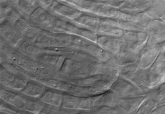


125 114 Drought-stressed apomictic diploid B. lignifera Drought-stressed apomictic diploid B. microphylla


126 115 APPENDIX III Additional Photomicrographs of Apomeiotic Dyads in Apomictic Boechera Drought-stressed diploid apomictic B. retrofracta x exilis Drought and heat-stressed diploid apomictic B. retrofracta x exilis


127 Non-stressed triploid apomictic B. gunnisoniana 116


128 117 Drought-stressed triploid apomictic B. gunnisoniana Drought and heat-stressed triploid apomictic B. gunnisoniana


129 Drought-stressed diploid apomictic B. lignifera 118
Sexual Reproduction in Flowering Plants
 Sexual Reproduction in Flowering Plants Question 1: Name the parts of an angiosperm flower in which development of male and female gametophyte take place. Answer :- The male gametophyte or the pollen grain
03. Apomixis classification and significance in plant breeding
 03. Apomixis classification and significance in plant breeding Apomixis, derived from two Greek word "APO" (away from) and "mixis" (act of mixing or mingling). It refers to the occurrence of an sexual
Introduction 1. INTRODUCTION
 1. INTRODUCTION Early in their evolution, plants have acquired a life cycle that alternates between a multicellular haploid organism, the gametophyte and a multicellular diploid organism, the sporophyte.
NCERT Solutions for Class 12 Biology Chapter 2
 NCERT Solutions for Class 12 Biology Chapter 2 Sexual Reproduction in Flowering Plants Class 12 Chapter 2 Sexual Reproduction in Flowering Plants Exercise Solutions Exercise : Solutions of Questions on
Fixing complex genotypes in plants: progress with apomixis in the Brassicaceae
 Fixing complex genotypes in plants: progress with apomixis in the Brassicaceae Global Institute for Food Security (GIFS) IPK Gatersleben Apomixis scientists everywhere thank you to everyone Apomixis in
SEXUAL REPRODUCTION IN FLOWERING PLANTS
 SEXUAL REPRODUCTION IN FLOWERING PLANTS 9 CHAPTER 2 SEXUAL REPRODUCTION IN FLOWERING PLANTS MULTIPLE-CHOICE QUESTIONS 1. Among the terms listed below, those that of are not technically correct names for
Embryological Evidence of Apomixis in Eulaliopsis binata
 Acta Botanica Sinica 2004, 46 (1): 86 92 http://www.chineseplantscience.com Embryological Evidence of Apomixis in Eulaliopsis binata YAO Jia-Ling *, YANG Ping-Fang, HU Chun-Gen, ZHANG You-De, LUO Bin-Shan
Introduction. Copyright 2002 Pearson Education, Inc., publishing as Benjamin Cummings
 Introduction It has been said that an oak is an acorn s way of making more acorns. In a Darwinian view of life, the fitness of an organism is measured only by its ability to replace itself with healthy,
Apomixis in cassava: advances and challenges
 Apomixis in cassava: advances and challenges D.Y.H. Freitas 1 and N.M.A. Nassar 2 1 Programa de Pós-Graduação em Botânica, Universidade de Brasília, Brasília, DF, Brasil 2 Departamento de Genética e Morfologia,
Kingdom Plantae, Part II - Gymnosperms and Angiosperms
 Kingdom Plantae, Part II - Gymnosperms and Angiosperms I. Introduction Reproduction in the seed plants (Gymnosperms and Angiosperms) has been greatly influenced by the requirements of a terrestrial existence.
plant reproduction Alternation of Generations chapter 38
 Alternation of Generations Haploid (n) plant reproduction chapter 38 Diploid (2n) Sporangium Spore dispersal Spore (n) Young Mature (n) ARCHEGONIUM ANTHERIDIUM Sperm Mature Sorus Sporangium sporophyte
Introduction. Copyright 2002 Pearson Education, Inc., publishing as Benjamin Cummings
 Introduction It has been said that an oak is an acorn s way of making more acorns. In a Darwinian view of life, the fitness of an organism is measured only by its ability to replace itself with healthy,
Biology 3201 Unit 2A Notes
 Biology 3201 Unit 2A Notes Mitosis Somatic cell: refers to a body cell; anything not a germ (sex) cell Cell cycle: a continuous sequence of cell growth and division The cell cycle consists of two main
Mastery. Sexual Reproduction and Meiosis. Chapter Content CHAPTER 3 LESSON 1. Directions: Study the diagram. Then answer the following questions.
 Chapter Content Mastery Sexual Reproduction and Meiosis Directions: Study the diagram. Then answer the following questions. LESSON 1 Interphase Prophase I Metaphase I Anaphase I Telophase I Meiosis II
BIOLOGY 3201 REPRODUCTION
 BIOLOGY 3201 REPRODUCTION Asexual vs. Sexual Reproduction MODES OF REPRODUCTION (1) Asexual one parent cell divides into two by mitosis to produce 2 identical cells which are clones of the parent (2) Sexual
Downloaded from CHAPTER 2 SEXUAL REPRODUCTION IN FLOWERING PLANTS POINTS TO REMEMBER
 CHAPTER 2 SEXUAL REPRODUCTION IN FLOWERING PLANTS POINTS TO REMEMBER Autogamy : When pollen grains of a flower are transferred from anther to stigma of the same flower. Coleorhiza : A protective sheath
Chapter 31: Plant Reproduction
 Chapter 31: Plant Reproduction Plants and Pollinators Pollen had evolved by 390 million years ago Sperm packed inside a nutritious package Transferred first by wind currents Later transferred by insects
plant reproduction chapter 40 Alternation of Generations
 Alternation of Generations plant reproduction chapter 40 Haploid (n) Diploid (2n) Sporangium Spore dispersal Spore (n) Young Mature (n) Archegonium Antheridium Sperm Sporangium Mature sporophyte (2n) New
Chapter 38 Angiosperm Reproduction and Biotechnology
 Chapter 38 Angiosperm Reproduction and Biotechnology Concept 38.1 Pollination enables gametes to come together within a flower Diploid (2n) sporophytes produce spores by meiosis; these grow into haploid
Chapter 38: Angiosperm Reproduction and Biotechnology
 Name: Chapter 38: Angiosperm Reproduction and Biotechnology 38.1 Flowers, double fertilization, and fruits are unique features of the angiosperm life cycle This may be a good time for you to go back and
Organisms that reproduce Sexually are made up of two different types of cells.
 MEIOSIS Organisms that reproduce Sexually are made up of two different types of cells. 1. Somatic Cells are body cells and contain the normal number of chromosomes.called the Diploid number (the symbol
Chapter 38: Angiosperm Reproduction and Biotechnology: To Seed or Not to Seed
 Chapter 38: Angiosperm Reproduction and Biotechnology: To Seed or Not to Seed The parasitic plant Rafflesia arnoldi produces huge flowers that produce up to 4 million seeds Many angiosperms reproduce sexually
Reproduction 19/02/2016. Asexual Reproduction. Budding: Types of asexual reproduction: SEXUAL VS. ASEXUAL
 Asexual Reproduction Reproduction SEXUAL VS. ASEXUAL One parent cell divides by mitosis to produce 2 daughter cells which are clones of the parent Types of asexual reproduction: Budding: 1. Budding 2.
The form of cell division by which gametes, with half the number of chromosomes, are produced. Chromosomes
 & Karyotypes The form of cell division by which gametes, with half the number of chromosomes, are produced. Homologous Chromosomes Pair of chromosomes (maternal and paternal) that are similar in shape,
Ontwikkeling; bevruchting
 Ontwikkeling; bevruchting http://www.lima.ohiostate.edu/biology/archive/flowers. html Young Lily anther x40. Four pollen sacs and a cross section of the fillament are visible. Diploid (2N chromosomes)
Pharmacologically Induced Meiosis Apomeiosis Interconversions in Boechera, Arabidopsis and Vigna
 Utah State University DigitalCommons@USU All Graduate Theses and Dissertations Graduate Studies 8-2018 Pharmacologically Induced Meiosis Apomeiosis Interconversions in Boechera, Arabidopsis and Vigna Lei
Angiosperm Reproduction
 Name Angiosperm Reproduction Today you will examine closely the reproductive aspects of the Anthophyta (aka Magnoliophyta aka Angiosperms) to finish your phylogenetic study of reproduction and evolution
Megasporogenesis and Megagametogenesis in Autotetraploid Indica/ Japonica Rice Hybrid
 Rice Science, 2010, 17(4): 296 302 Copyright 2010, China National Rice Research Institute. Published by Elsevier BV. All rights reserved DOI: 10.1016/S1672-6308(09)60030-5 Megasporogenesis and Megagametogenesis
CHAPTER 2 Reproduction of Flowering Plants. Bui Tan Anh College of Natural Sciences
 CHAPTER 2 Reproduction of Flowering Plants Bui Tan Anh College of Natural Sciences Rafflesiaarnoldii in Indonesia Asexual Reproduction Sexual Reproduction Seeds and Fruits Flower Plant Reproduction Many
Apomixis and cassava. Genet. Mol. Res. 1 (2): (2002) Received June 4, 2002 Published June 29, Apomixis and cassava 147
: (2002) Received June 4, 2002 Published June 29, Apomixis and cassava 147") Apomixis and cassava 147 Apomixis and cassava Nagib M.A. Nassar Departamento de Genética e Morfologia, Universidade de Brasília, Brasília, DF, Brazil Corresponding author: N.M.A. Nassar E-mail: nagnassa@rudah.com.br
Apomixis and cassava 147 Apomixis and cassava Nagib M.A. Nassar Departamento de Genética e Morfologia, Universidade de Brasília, Brasília, DF, Brazil Corresponding author: N.M.A. Nassar E-mail: nagnassa@rudah.com.br
Modes of reproduction Types of cultivar
 Modes of reproduction Types of cultivar 2012 What is a Cultivar? Distinctness:.. Shall be clearly distinguishable, by one or more important characteristic, from any other plant variety Uniformity:.. Similar
meiosis asexual reproduction CHAPTER 9 & 10 The Cell Cycle, Meiosis & Sexual Life Cycles Sexual reproduction mitosis
 meiosis asexual reproduction CHAPTER 9 & 10 The Cell Cycle, Meiosis & Sexual Sexual reproduction Life Cycles mitosis Chromosomes Consists of a long DNA molecule (represents thousands of genes) Also consists
Chapter 8: Cellular Reproduction
 Chapter 8: Cellular Reproduction 1. The Cell Cycle 2. Mitosis 3. Meiosis 2 Types of Cell Division 2n 1n Mitosis: occurs in somatic cells (almost all cells of the body) generates cells identical to original
Class XII Chapter 1 Reproduction in Organisms Biology
 Question 1: Why is reproduction essential for organisms? Reproduction is a fundamental feature of all living organisms. It is a biological process through which living organisms produce offspring s similar
THE ENDOSPERM OF ANGIOSPERMS
 THE ENDOSPERM OF ANGIOSPERMS CONTRIBUTIONS FROM THE HULL BOTANICAL LABORATORY I50 JOHN M. COULTER In a recent analysis of all the available testimony in reference to the morphological nature of the endosperm
BIOLOGY CLASS: VIII TOPIC: Life Processes: Growth, Reproduction & Development (plants) Difference between self-pollination & cross pollination
 Difference between self-pollination & cross pollination") BIOLOGY CLASS: VIII TOPIC: Life Processes: Growth, Reproduction & Development (plants) Difference between self-pollination & cross pollination Self -Pollination 1. It is transfer of pollen grains from
BIOLOGY CLASS: VIII TOPIC: Life Processes: Growth, Reproduction & Development (plants) Difference between self-pollination & cross pollination Self -Pollination 1. It is transfer of pollen grains from
Unit E: Plant Propagation. Lesson 1: Understanding Sexual Reproduction
 Unit E: Plant Propagation Lesson 1: Understanding Sexual Reproduction 1 Vocabulary Cross-pollination Diploid Endosperm Fertilization Gametes Genes Haploid Hybrids Pollination Seed Self-pollination Sexual
Modes of Reproduction
 Modes of Reproduction Very Short answer Questions 1. What is the dominant phase in the life cycle of an angiosperm? A: Sporophyte phase (diploid phase). 2. What is meant by heterospory? Mention the two
Biology Class 12 th NCERT Solutions
 Chapter.2 Sexual Reproduction in Flowering Plants Class XII Subject Biology 1. Name the parts of an angiosperm flower in which development of male and female gametophyte take place. Answer 1. Pollen grains
Plant Science 1203L Laboratory 5 - Sexual Reproduction (Pollination and Double Fertilization)
") Plant Science 1203L Laboratory 5 - Sexual Reproduction (Pollination and Double Fertilization) Today s lab is about sexual reproduction in plants. As with stem or root structure there are numerous definitions
Plant Science 1203L Laboratory 5 - Sexual Reproduction (Pollination and Double Fertilization) Today s lab is about sexual reproduction in plants. As with stem or root structure there are numerous definitions
Cellular Reproduction, Part 2: Meiosis Lecture 10 Fall 2008
 Mitosis & 1 Cellular Reproduction, Part 2: Lecture 10 Fall 2008 Mitosis Form of cell division that leads to identical daughter cells with the full complement of DNA Occurs in somatic cells Cells of body
4. Which of the following cell parts can be found in human cells, but not plant cells? A. mitochondria B. chloroplast C. centrioles D.
 http://chss.sd57.bc.ca/~spearce/science_9/practice%20tests/reproductionpracticetest.htm Sc.9 Biology Practise Test 1. Single-celled organisms use cell division for growth. 2. Chromosomes are made of two
Sexual Reproduction and Genetics. Section 1. Meiosis
 Chromosomes and Chromosome Number! Human body cells have 46 chromosomes! Each parent contributes 23 chromosomes! Homologous chromosomes one of two paired chromosomes, one from each parent Chromosomes and
Sexual Reproduction in Flowering Plants
 Sexual Reproduction in Flowering Plants Pre Fertilisation Events Several hormonal and structural changes result in the development of a flower. Inflorescences bear the flower buds, and then the flowers.
Cell Division and Inheritance
 Cell Division and Inheritance Continuing life relies on reproduction Individual organism replacing dead or damaged cells Species making more of same species Reproduction Cells divide, grow, divide again
Chapter 38. Plant Reproduction. AP Biology
 Chapter 38. Plant Reproduction 1 Animal vs. Plant life cycle Animal multicellular 2n Plant multicellular sporophyte 2n gametes 1n spores 1n unicellular gametes 1n multicellular gametophyte 1n 2 Alternation
Meiosis. Oh, and a little bit of mitosis
 Meiosis Oh, and a little bit of mitosis Haploid Cells- The sex cells (egg and sperm) only contain half of the genetic diversity that diploid cells do. For humans this would mean 23 single chromosomes.
Sexual Reproduction in Flowering Plants
 Sexual Reproduction in Flowering Plants Four main events must occur in order for sexual reproduction to take place. 1. The organism must be developed and mature sexually. 2. Gametogenesis; production of
Chapter 38. Plant Reproduction. AP Biology
 Chapter 38. Plant Reproduction 1 Animal vs. Plant life cycle Animal multicellular 2n Plant multicellular sporophyte 2n gametes 1n spores 1n unicellular gametes 1n multicellular gametophyte 1n 2 Alternation
Chapter 20 Apomixis in the Era of Biotechnology
 Chapter 20 Apomixis in the Era of Biotechnology E. Albertini, G. Barcaccia, A. Mazzucato, T.F. Sharbel, and M. Falcinelli 20.1 Introduction The adaptive success of living organisms depends on the maintenance
Unit 4: Cell Division Guided Notes
 Unit 4: Cell Division Guided Notes 1 Chromosomes are structures that contain material When Eukaryotes are not dividing, DNA and Proteins are in a mass called: When the cell divides, it condenses and becomes
Angiosperm Reproduction and Biotechnology
 Chapter 38 Angiosperm Reproduction and Biotechnology PowerPoint Lecture Presentations for Biology Eighth Edition Neil Campbell and Jane Reece Lectures by Chris Romero, updated by Erin Barley with contributions
Biology of Apomixes: Molecular and Genetic Approaches
 Biology of Apomixes: Molecular and Genetic Approaches Crus Von Mark V. Lopez Pedro Antonio Kissel Steven Patrick Gogula Murli Krishnareddy Stuart Gardner Introduction Molecular and genetic aspects that
ANGIOSPERM L.S. POLLEN GRAIN
 ANGIOSPERM 2 L.S. POLLEN GRAIN ANGIOSPERM T 2 CELLS L.S. POLLEN GRAIN ANGIOSPERM TUBE CELL G L.S. POLLEN GRAIN ANGIOSPERM TUBE CELL > L.S. GENERATIVE CELL POLLEN GRAIN ANGIOSPERM TUBE CELL GENERATIVE CELL
NOTES: CH 38 Plant Reproduction
 NOTES: CH 38 Plant Reproduction *Modifications in reproduction were key adaptations enabling plants to spread into a variety of terrestrial habitats. * Water has been replaced by wind and animals as a
Chapter 13 Pre-Test Question 2
 Student View Summary View Diagnostics View Print View with Answers Settings per Student Questions part 1: Keimzellen und Befruchtung Due: 12:00pm on Wednesday, December 7, 2011 Note: You will receive no
Lesson 1. Quiz (short) Cell cycle Chromosomes Mitosis phases
 Cell cycle Chromosomes Mitosis phases") Lesson 1 Quiz (short) Cell cycle Chromosomes Mitosis phases 2 Cell division is needed for Growth (Mitosis) Repair (Mitosis) Reproduction (Meiosis) 3 Mitosis consists of 4 phases (division of the nuclear
Lesson 1 Quiz (short) Cell cycle Chromosomes Mitosis phases 2 Cell division is needed for Growth (Mitosis) Repair (Mitosis) Reproduction (Meiosis) 3 Mitosis consists of 4 phases (division of the nuclear
Chapter 2. Mitosis and Meiosis
 Chapter 2. Mitosis and Meiosis Chromosome Theory of Heredity What structures within cells correspond to genes? The development of genetics took a major step forward by accepting the notion that the genes
Cell Cycle and Cell Division
 122 Cell Cycle and Cell Division 1. Meiosis I is reductional division. Meiosis II is equational division due to [1988] (a) pairing of homologous chromosomes (b) crossing over (c) separation of chromatids
PMP ) Plant reproduction. d) Formation of gametes e) Mutations in the development of gametophyte f) Pollination, fertilization
 Plant reproduction. d) Formation of gametes e) Mutations in the development of gametophyte f) Pollination, fertilization") 2015 3) Plant reproduction 1 d) Formation of gametes e) Mutations in the development of gametophyte f) Pollination, fertilization 2 d) Formation of gametes Plant life cycle Microsporogenesis Megasporogenesis
2015 3) Plant reproduction 1 d) Formation of gametes e) Mutations in the development of gametophyte f) Pollination, fertilization 2 d) Formation of gametes Plant life cycle Microsporogenesis Megasporogenesis
Sexual Reproduction: Adding Variety
 4.3 gamete a sex cell; includes sperm cells in males and egg cells in females fertilization the formation of a zygote by the joining together, or fusion, of two gametes zygote a cell produced by the fusion
3/18/2012. Chapter 36. Flower Parts. Flower Parts. Reproduction in Angiosperms
 Chapter 36 Reproduction in Angiosperms Bryophytes >450mya 360 mya Fig. 27-4, p. 584 Lily Flower Flower Parts Sepals cover and protect flower parts in bud Collectively calyx Petals Can attract animal pollinators
Chapter 10 Chromosomes and Cell Reproduction
 Chapter 10 Chromosomes and Cell Reproduction Chromosomes Organisms grow by dividing of cells Binary Fission form of asexual reproduction that produces identical offspring (Bacteria) Eukaryotes have two
CHAPTER 8 CELL REPRODUCTION
 CHAPTER 8 CELL REPRODUCTION CHROMOSOME STRUCTURE Structures in the nucleus that carry genetic information Composed of DNA coiled around proteins called histones Consists of 2 identical parts called sister
CHAPTER 2 Sexual reproduction in flowering plants.
 CHAPTER 2 Sexual reproduction in flowering plants 8 1 7 CHAPTER 2 SEXUAL REPRODUCTION INFLOWERING PLANTS FLOWERS Site of sexual Reproduction. Male and female reproductive organs are borne on flowers. PARTS
Pre-Test. 4. Inside of cells, nearly all the genes are located on special structures known as microtubules.
 1 Pre-Test Directions: Answer each question TRUE OR FALSE. 1. Cells in the bodies of multicelled creatures, such as humans, are reproduced in a way that is very similar to the method used by most protozoa.
Reproduction in Organisms
 Reproduction in Organisms Asexual Reproduction The period through which a certain organism lives is known as its life span. Reproduction is the process by which every organism ensures its continuity. It
The questions below refer to the following terms. Each term may be used once, more than once, or not at all.
 The questions below refer to the following terms. Each term may be used once, more than once, or not at all. a) telophase b) anaphase c) prometaphase d) metaphase e) prophase 1) DNA begins to coil and
Meiosis & Sexual Reproduction. AP Biology
 Meiosis & Sexual Reproduction 2007-2008 Cell division / Asexual reproduction Mitosis produce cells with same information identical daughter cells exact copies clones same amount of DNA same number of chromosomes
THE DYNAMIC NATURE OF APOMIXIS IN THE ANGIOSPERMS
 Int. J. Plant Sci. 169(1):169 182. 2008. Ó 2008 by The University of Chicago. All rights reserved. 1058-5893/2008/16901-0014$15.00 DOI: 10.1086/523369 THE DYNAMIC NATURE OF APOMIXIS IN THE ANGIOSPERMS
Ploidy and Human Cell Types. Cell Cycle and Mitosis. DNA and Chromosomes. Where It All Began 11/19/2014. Chapter 12 Pg
 Ploidy and Human Cell Types Cell Cycle and Mitosis Chapter 12 Pg. 228 245 Cell Types Somatic cells (body cells) have 46 chromosomes, which is the diploid chromosome number. A diploid cell is a cell with
Reproductive Development and Structure
 Reproductive Development and Structure Bởi: OpenStaxCollege Sexual reproduction takes place with slight variations in different groups of plants. Plants have two distinct stages in their lifecycle: the
NOTES- CHAPTER 6 CHROMOSOMES AND CELL REPRODUCTION
 NOTES- CHAPTER 6 CHROMOSOMES AND CELL REPRODUCTION Section I Chromosomes Formation of New Cells by Cell Division New cells are formed when old cells divide. 1. Cell division is the same as cell reproduction.
Plant Reproduction fertilization
 Plant Reproduction In the plant kingdom, both sexual and asexual reproduction occur. Recall from Chapter 3 that plants reproduce sexually by sporic reproduction, which is also called alternation of generations.
Competition between meiotic and apomictic pathways during ovule and seed development results in clonality
 Research Competition between meiotic and apomictic pathways during ovule and seed development results in clonality Diego H. Hojsgaard 1,2, Eric J. Martínez 1 and Camilo L. Quarin 1 1 Instituto de Botánica
Reproduction and Development in Flowering Plants
 Reproduction and Development in Flowering Plants Sexual Reproduction in Flowering Plants The flower functions in sexual reproduction of plants and precedes the development of seeds and fruits. Flowers
Chromosomes & Cell Division
 Chromosomes & Cell Division Cell Division The growth and splitting of cells into two new, identical cells called daughter cells. Mitosis Meiosis DNA replicates Parent cell Chromosomes separate Cell division
Class XII - Biology Reproduction in Organisms Chapter-wise Questions
 Class XII - Biology Reproduction in Organisms Chapter-wise Questions MULTIPLE-CHOICE QUESTIONS 1. A few statements describing certain features of reproduction are given below: i. Gametic fusion takes place
Chapter 40 Flowering Plant Sexual Reproduction
 3.6 Reproduction & Growth 3.6.1 Reproduction of The Flowering Plant Chapter 40 Flowering Plant Sexual Reproduction Learning Objectives 1. Give the structure and function of the floral parts. 2. Outline
HOLIDAY HOMEWORK FOR CLASS XII STUDENTS
 FOR CLASS XII STUDENTS PROJECT WORK TO BE COMPLETED ON THE RESPECTIVE TOPIC ALLOTTED BY THE SUBJECT TEACHER SUBJECTIVE QUESTION BANK SUBJECT-BIOLOGY CHAPTERS 1 TO 4 CHAPTER-1- REPRODUCTION IN ORGANISMS
GENES AND CHROMOSOMES CHAPTER 5
 CHAPTER 5 GENES AND CHROMOSOMES For many years the standard authority on the chromosomes was Wilson s The Cell in Development and Inheritance. The second edition of this work was published in 1900; it
Seed Plants Lab. Learning Objectives. Procedure and Questions
 Seed Plants Lab Learning Objectives Define the terms (meanings of the names) angiosperm and gymnosperm State what type of cells create eggs and what type of cells create sperm in gymnosperms and angiosperms
General Embryology. School of Medicine Department of Anatomy and Histology School of medicine The University of Jordan
 General Embryology 2019 School of Medicine Department of Anatomy and Histology School of medicine The University of Jordan https://www.facebook.com/dramjad-shatarat What is embryology? Is the science that
Chapter 12 Reproduction and Meiosis
 Part III Organization of Cell Populations Chapter The earth is believed to house over 10 million species of organisms that have prospered by reproducing individuals of the same species. Thus, one of the
Flower Morphology. Flower Structure
 wrong 0 1 2 3 4 5 6 7 8 9 10 11 12 13 14 15 16 17 18 19 20 right 84 83 82 81 80 79 78 77 76 75 74 73 72 71 70 69 68 67 66 65 64 score 100 98.8 97.6 96.4 95.2 94.0 92.9 91.7 90.5 89.3 88.1 86.9 85.7 84.5
The Cell Cycle. Chapter 10
 The Cell Cycle Chapter 10 Why Do Cells Divide? Unicellular 1. Reproduction Multicellular 1. Grow 2. Repair 3. Development/reproduction Types of Division Prokaryotic cells Binary fission = asexual reproduction
Chromosomes and Cell Cycle
 Chromosomes and Cell Cycle Cell Basics There are trillions of cells in your body Cells are microscopic Cells have DNA inside a structure called the nucleus The nucleus is enclosed by a structure called
BIO-BOTANY important questions to discuss for NEET 2018/Matric Exam HOPE ACADEMY HOSUR
 Questions = 45 REPRODUCTION IN FLOWERING PLANTS 1 1. Which is the characteristics for ornithophily (1) Scented flowers (2) Bright red colored flowers and infloresnce (3) White colored funnel shaped large
POLYGONUM EMBRYO SAC CHALAZAL END ANTIPODAL CELL EMBRYO SAC OVULE L.S.
 POLYGONUM EMBRYO SAC? CHALAZAL END ANTIPODAL CELL EMBRYO SAC OVULE L.S. POLYGONUM EMBRYO SAC C CHALAZAL END ANTIPODAL CELL? EMBRYO SAC OVULE L.S. POLYGONUM EMBRYO SAC? CHALAZAL END ANTIPODAL CELL CENTRAL
Apomictic and Sexual Ovules of Boechera Display Heterochronic Global Gene Expression Patterns C W OA
 The Plant Cell, Vol. 22: 655 671, March 2010, www.plantcell.org ã 2010 American Society of Plant Biologists Apomictic and Sexual Ovules of Boechera Display Heterochronic Global Gene Expression Patterns
Plant Reproduction. In a nutshell
 Plant Reproduction In a nutshell 2007-2008 Plant Diversity mosses ferns conifers flowering plants Bryophytes non-vascular land plants Pteridophytes seedless vascular plants Gymnosperm pollen & naked seeds
Cytogenetics and embryology of Eupatorium laevigatum (Compositae)
") Research Article Genetics and Molecular Biology, 28, 1, 123-128 (2005) Copyright by the Brazilian Society of Genetics. Printed in Brazil www.sbg.org.br Cytogenetics and embryology of Eupatorium laevigatum
Research Article Genetics and Molecular Biology, 28, 1, 123-128 (2005) Copyright by the Brazilian Society of Genetics. Printed in Brazil www.sbg.org.br Cytogenetics and embryology of Eupatorium laevigatum
Laboratory 29 - Magnoliophyta: Reproductive Morphology II
 58 Laboratory 29 - Magnoliophyta: Reproductive Morphology II I. Microsporogenesis and microgametogenesis A. Pollen tube growth Growing pollen tubes is simple if you are using bicellular pollen. Physiologically,
Anther/Pollen culture
 Anther/Pollen culture Method to produce haploid plants Spontaneous occurrence in low frequency Induction by physical and/or chemical treatment Chromosome elimination following interspecific hybridization
Omnis cellula e cellula
 Chapter 12 The Cell Cycle Omnis cellula e cellula 1855- Rudolf Virchow German scientist all cells arise from a previous cell Every cell from a cell In order for this to be true, cells must have the ability
Study Guide. Biology 3101B. Science. Reproduction and Development. Adult Basic Education. Biology 2101A Biology 2101C Biology 3101A.
 Adult Basic Education Science Reproduction and Development Prerequisites: Biology 2101A Biology 2101C Biology 3101A Credit Value: 1 Text: Biology. Bullard, Chetty, et al; McGraw-Hill Ryerson, 2003 Biology
5.1. KEY CONCEPT Cells have distinct phases of growth, reproduction, and normal functions. 68 Reinforcement Unit 2 Resource Book
 5.1 THE CELL CYCLE KEY CONCEPT Cells have distinct phases of growth, reproduction, and normal functions. Cells have a regular pattern of growth, DNA duplication, and division that is called the cell cycle.
Mitosis: cell division that forms identical daughter cells with the same number of chromosomes as the parent cell (duplicate and divide)
") Mitosis: cell division that forms identical daughter cells with the same number of chromosomes as the parent cell (duplicate and divide) Meiosis: cell division that forms daughter cells with half the number
Mitosis: cell division that forms identical daughter cells with the same number of chromosomes as the parent cell (duplicate and divide) Meiosis: cell division that forms daughter cells with half the number
Chapter 14 Cellular Reproduction
 Chapter 14 Cellular Reproduction Biology 3201 Introduction One of the important life functions of living things is their ability to reproduce. Reproduction depends on the cell. Cells reproduce in order
Determination of reproductive mode and genome size of a USDA core collection of Kentucky bluegrass (Poa pratensis L.) by cell flow cytometry
 by cell flow cytometry") Retrospective Theses and Dissertations Iowa State University Capstones, Theses and Dissertations 2003 Determination of reproductive mode and genome size of a USDA core collection of Kentucky bluegrass
Retrospective Theses and Dissertations Iowa State University Capstones, Theses and Dissertations 2003 Determination of reproductive mode and genome size of a USDA core collection of Kentucky bluegrass