3D Airway Analysis in OSA and TMD Therapy
|
|
|
- Ralf Johnston
- 5 years ago
- Views:
Transcription


1 3D Airway Analysis in OSA and TMD Therapy


2 Gy Yatros, D.M.D. Diplomate ABDSM Faculty USF Co-Founder of Dental Sleep Solutions Co-Founder DS3 Dental Sleep Software 2
3 Airway: Role of imaging Airway Imaging Can Not Confirm or Exclude OSA Imaging Can Help Understand Airway Anatomy (Phenotype) Identify Potential Obstruction Sites Evaluate Airway Support Structures

4 How I got involved in CBCT 4

5 The Dose Story in perspective 5

6 RADIATION EXPOSURE IN DAILY LIFE Average annual effective dose in the US is 3,000 µsv* This is approximately 8 µsv/day *White and Pharoah: Oral Radiology Principles and Interpretation 6 th ed.

7 AIR TRAVEL Estimated air travel for a 8 hour flight results in 80 to100 µsv exposure

")
8 AUTOMATIC DOSE CONTROL (ADC)

 (** OP300 Maxio Dose Study (Ludlow")
9 OP300 MAXIO LDT EFFECTIVE DOSE COMPARED TO 2D EFFECTIVE DOSE (* Patient doses from Dental X-Ray Exams (Ludlow et al. 2008) (** OP300 Maxio Dose Study (Ludlow 2014) 9
10 OP300 MAXIO LDT EFFECTIVE DOSE COMPARED TO 2D EFFECTIVE DOSE (* Patient doses from Dental X-Ray Exams (Ludlow et al. 2008) (** OP300 Maxio Dose Study (Ludlow 2014) 10


11 Why 3D? 11

12 3D imaging adds value to your treatment 12

13 Practice building and differentiation 13


14 How do patients perceive a practice? 14

15


16

17

18

19 Using 3D in the Dental office Treatment planning and diagnosis Office efficiency Endo 3 rd molar evaluation Ortho 19
20 Screening & Patient Education Patient Candidacy & predicting factors - Anatomy, other treatment options, etc. TMJ Baseline and Diagnostics Nasal Airway MAD Jaw positon? CBCT Game Changer 20


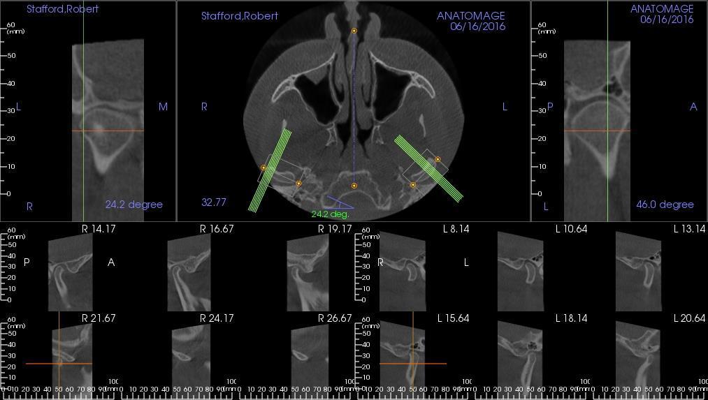
21 13x15 CM FOV Fields of View Orthognathics Airway TMJ Nasal Airway Trauma 21


22 OP300 Maxio Confident diagnostics for entire maxillofacial region OP300 Maxio 8x15 OP300 Maxio 13x15 The perfect FOV for all of todays and any future needs 22
23 CBCT Game Changer Screening & Patient Education 23
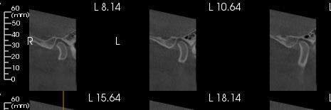
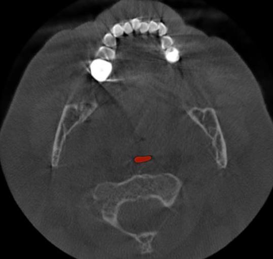
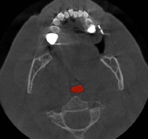
24 Airway Evaluation 24
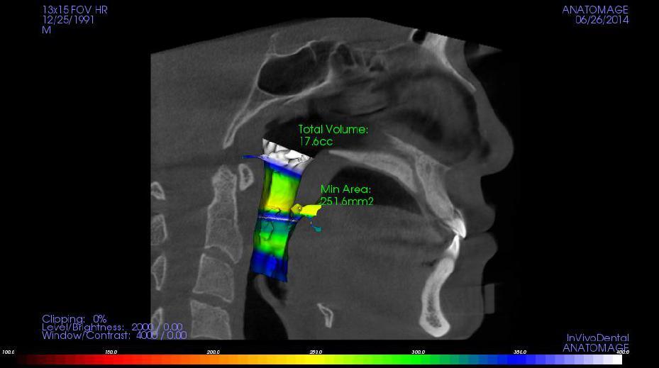
25 Airway Evaluation 25


26 Minimal Cross-Sectional Area Probability of OSA *Avrahami E, Englender M. Relation between CTaxial cross-sectional area of the oropharynx and obstructive sleep apnea syndrome in adults. AJNR Am J Neuroradiol 1995;16(1):
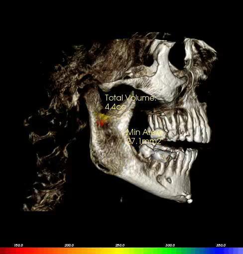
27 Airway Evaluation 27
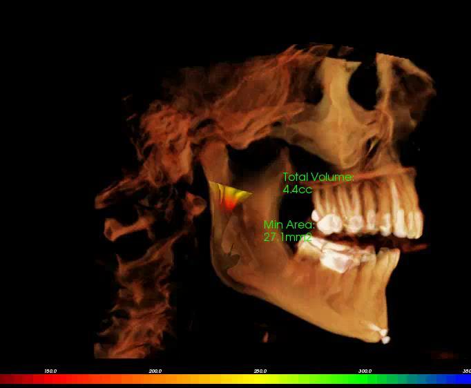
28 Airway Evaluation 28
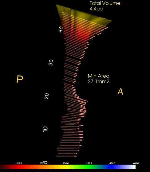
29 Airway Evaluation 29
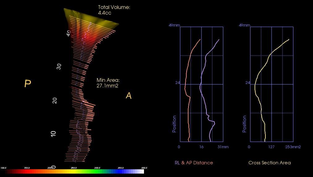
30 Airway Evaluation 30
31 Airway Evaluation 31

32 Airway Evaluation 32
33 Screening & Patient Education Patient Candidacy & predicting factors - Anatomy, other treatment options, etc. TMJ Baseline and Diagnostics Nasal Airway Jaw Position? CBCT Game Changer 33

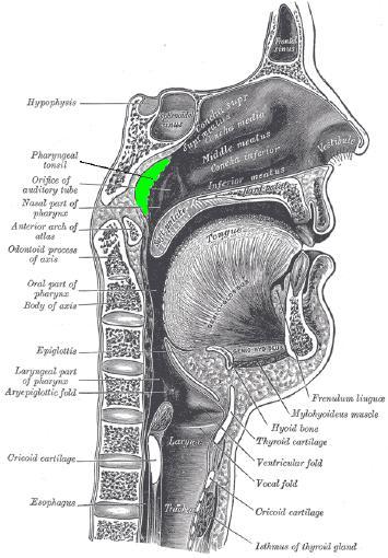
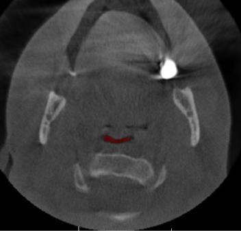
34 Pharyngeal Tonsil (Adenoids) 34

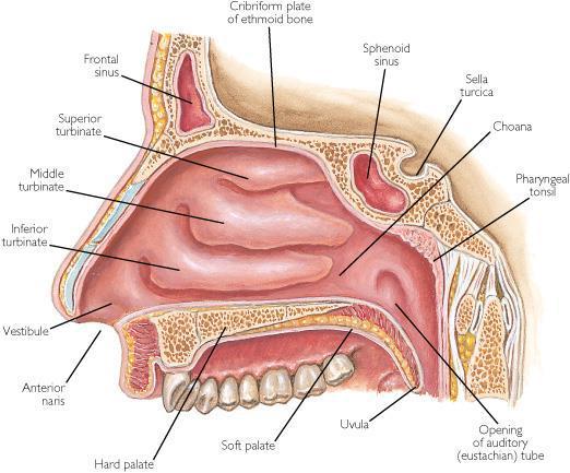
35 Nasal Anatomy 35

36 Normal Nasal Anatomy 36

37 Nasal Deviation 37

38 Nasal Deviation Guess Who? 38

39 Nasal Deviation 39
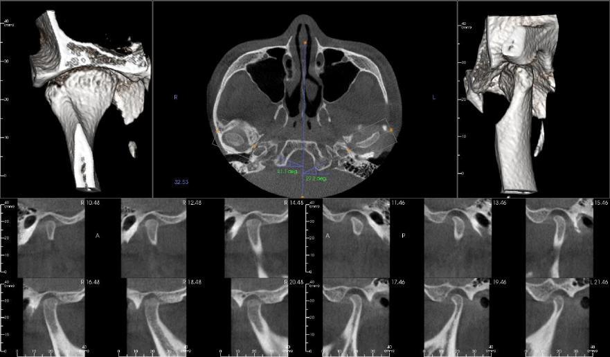
40 CBCT TMJ Considerations 40

41 TMJ Evaluation 41
42 Condylar Head & Fossa Changes 42
43 Patient Case 1 43

44 Patient Case 44


45 Patient Case 45
46 Patient Case 46

47 Patient Case 47
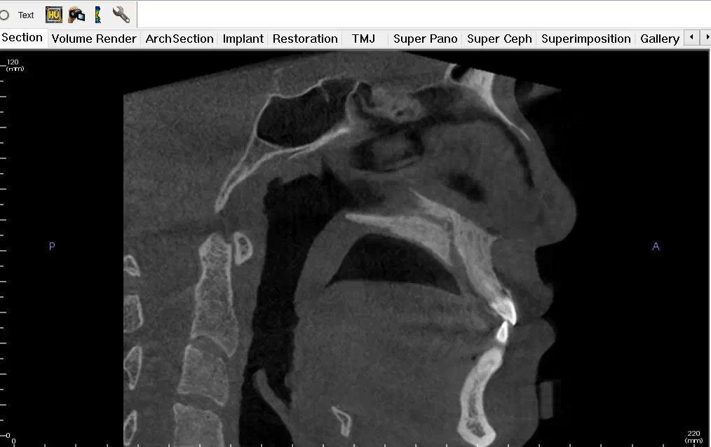
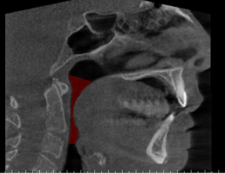
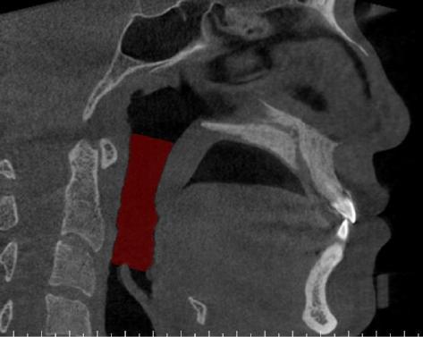
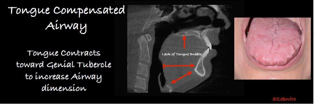
48 Airway Analysis AIRWAY: -The soft palate was approximately 48 mm long. - Nasal septum deviation and small concha bullosa of the right and left middle nasal turbinates was noted. -The most constricted area of the airways corresponded to the area posterior to the tongue and soft palate and it is reduced (approximately 70 mm2). This space is small and should be considered an intermediate risk factor for obstructive sleep apnea (OSA). 48
49 Sinus Evaluation 49
50 Sinus Evaluation Signs of increase in the mucosal thickening were noted from the floor of the maxillary sinuses and from selected ethmoidal air cells. This is consistent with allergies or another condition of inflammatory origin (sinusitis). The antromeatal complexes were patent/clear. - Dome-shaped soft tissue densities were noted from the floor of the maxillary sinuses and sphenoid sinus. They are consistent with mucous retention cysts/sinus polyposis. - A soft tissue density was noted from the superior-anterior wall of the left maxillary sinus. This is consistent with a sinus polyp. 50
51 TMJ Evaluation 51

52 TMJ Evaluation 52
53 TMJ Evaluation 53
54 TMJ Evaluation Left Evidence of mild sclerosis and flattening for the superior/posterior surface of the left condyle and sclerosis for the posterior slope of the left eminence was noted. 54
55 TMJ Evaluation Right The right condyle was small. The reduction in size occurred along the posterior and superior surface of the right condyle (anterior-posterior and vertical dimension). Evidence of sclerosis, flattening and small osteophyte formation for the superior/anterior surface of the right condyle and mild sclerosis for the posterior slope of the right eminence was noted. 55
56 TMJ Evaluation Position: When the mandible was in closed position the right condyle was positioned posterior to the center and the left condyle close to the center of their respective fossa. The right posterior articular spaces were reduced. OTHERS: - Bilateral elongation/partial calcification was noted for the styloid-hyoid ligament/process. 56
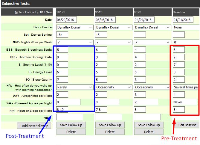
57 Radiologist Impressions Airways The findings described above should be considered risk factors for obstructive sleep apnea (OSA). 57
58 Radiologist Impressions TMJs The findings described above are most consistent with NON-ACTIVE degenerative joint disease (DJD) for the right TMJ and osseous remodeling for the left TMJ. DJD involves the destruction of the articular tissues and may occur when the remodeling capacity of those tissues has been exceeded by the functional demands. The presence of these changes increases the probability of a displaced disc in the right TMJs. Reduction of condylar size maybe associated with changes in occlusion, asymmetries and mandibular posture which may predispose to TMJ dysfunction. The posterior positioned condyles within their fossa may predispose to anterior displaced discs and compression of the posterior surface of the condyles and the adjacent retrodiscal tissues. 58
59 Radiologist Impressions Most of the other findings and their correspondent diagnosis were noted above. Please correlate the sections with the axial and panoramic views for additional diagnosis and treatment planning purposes. Reviewing the remaining available volume, there was no evidence of any other anomaly/pathology in the maxillofacial and surrounding structures available in this study. 59
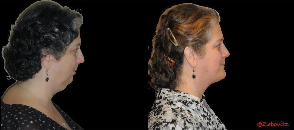
60 Case 1 60

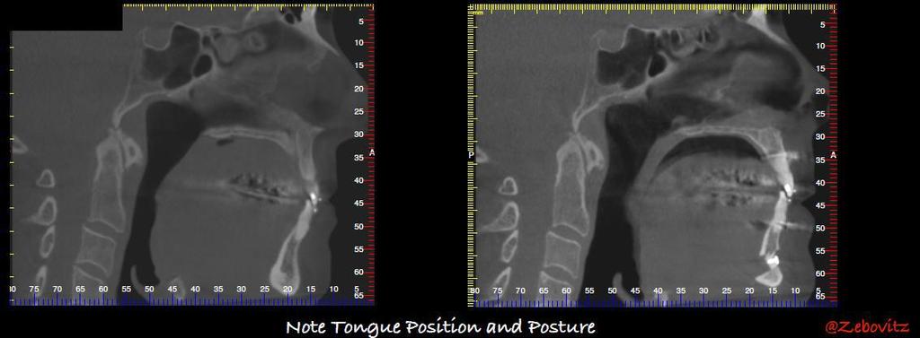
61 So what did we learn? Patient has small airway and is at risk for OSA Nasal airway is compromised. Pt. may have allergies and has sinus abnormalities. TMJ s have existing abnormalities. Teeth are adequate support for Dental Sleep Therapy (DST). 61


62 Advise patient they are at risk for OSA. Advise patient they have increased risks of problems with CPAP and/or Dental Sleep Therapy (DST) due to nasal problems. Advise patient should consider ENT and/or allergist referral. Advise patient they have existing TMJ concerns and they are at higher risks of complications with DST. RB&A of Treatment and informed consent! 62
63 63
64 64
65 65
66 Case 2 86 Events/Hr.! 66
67 Case 2 Subjective 67

68 Case 2 Objective 68

69 Case 2 Pre-Treatment 69

70 Case 2 Post-Treatment 70
71 Case 2 Pre-Treatment Post-Treatment 71

72 CBCT Sleep Medical Insurance billing 72

73 CBCT Billing Key Points Documentation Preauthorization Accreditation 73

74 CBCT Documentation Documenting Medical Necessity is KEY!! 74
75 CBCT Documentation You need to know what questions to ask to gather information specific to the necessity for a CBCT to bill to insurance. History of head or neck trauma Recurrent sinus infections or history of sinus problems History of head or neck surgery Nasal obstruction Problems swallowing TMD signs/symptoms or history of TMD 75
76 CBCT Preauthorization All Pre-Authorization Include a medical necessity statement as to why a CBCT is standard of care for the treatment we provide as well as specific clinical information related to the patient that would support the need for the CBCT. CPT: Maxillofacial sinus CT without contrast Modifier: TC (technical component) We are not billing for interpretation. Must have ICD-10 diagnosis code (OSA G47.33) 76
77 CBCT Accreditation Intersocietal Accreditation Commission Dental Website Requirement of being accredited to receive payment from Medicare as well as many private insurance companies when billing CT scans. 77

78 Dr. Gy Yatros D.M.D SNORE
The future of health is digital
 Dated: XX/XX/XXXX Name: XXXXXXXX XXXXXXXXXXX Birth Date: XX/XX/XXXX Date of scan: XX/XX/XXXX Examination of the anatomical volume: The following structures are reviewed and evaluated for bilateral symmetry,
 www.oralradiologists.com CONE BEAM CT REPORT CASE ---- Case Information Referring Doctor: - Patient Name: - Scan Date: December 1, 2015 Patient DOB: - Reason for Exam: - Study Details: icat Flex, 160x160x112
 www.oralradiologists.com CONE BEAM CT REPORT CASE XXXX Patient information Patient Name: - Referring Doctor: - Patient DOB: - Scan Date: [Start date] Reason for Exam: Maxillary facial pain Doctor Notes:
Clinical details: Details of scan: CONE BEAM CT REPORT: Name: H. B. Gender: Reason for referral: Referred by:
 Name: H. B. Gender: Male DOB: 11/12/1950 Age: 64 Date taken: 16/11/2015 Date reported: 19/11/2015 Clinical details: Reason for referral: Referred by: Investigate symptoms related to left TMJ. Reconstructed
Snoring and Obstructive Sleep Apnea: Patient s Guide to Minimally Invasive Treatments Chapter 2
 Snoring and Obstructive Sleep Apnea: Patient s Guide to Minimally Invasive Treatments Chapter 2 CAUSES OF SNORING AND SLEEP APNEA We inhale air through our nose and mouth. From the nostrils, air flows
ORTHOGNATHIC SURGERY
 Status Active Medical and Behavioral Health Policy Section: Surgery Policy Number: IV-16 Effective Date: 10/22/2014 Blue Cross and Blue Shield of Minnesota medical policies do not imply that members should
TEMPORO-MANDIBULAR JOINT DISORDERS
 Disclaimer This movie is an educational resource only and should not be used to manage your dental health. All decisions about the management of TMJ Disorders must be made in conjunction with your Dental
Snoring And Sleep Apnea in the U.S. Definitions Apnea: Cessation of ventilation for > 10 seconds. Defining Severity of OSA
 Snoring and Obstructive Sleep Apnea: Oral Appliance Therapy Management Midwest Society of Orthodontists October 16-17, 2009 Anthony J DiAngelis DMD, MPH Chief, Department of Dentistry, HCMC Professor,
UNIVERSITY OF MEDICINE AND PHARMACY GR. T. POPA - IASI FACULTY OF DENTAL MEDICINE
 UNIVERSITY OF MEDICINE AND PHARMACY GR. T. POPA - IASI FACULTY OF DENTAL MEDICINE ABSTRACT CONTRIBUTIONS OF THREE-DIMENSIONAL IMAGING TO THE DIAGNOSIS AND MANAGEMENT OF CLEFT LIP AND PALATE PhD ADVISOR,
Planmeca Ultra Low Dose
 OWN THE FUTURE Practice Ultra Low Dose 3D imaging with lower dose than panoramic imaging Ultra Low Dose Pioneering low dose 3D imaging 3D units offer a unique Ultra Low Dose imaging protocol that enables
Obstructive Sleep Apnea- Hypopnea Syndrome and Snoring: Surgical Options
 Obstructive Sleep Apnea- Hypopnea Syndrome and Snoring: Surgical Options Joshua L. Kessler, MD, FACS Boston ENT Associates Clinical Instructor, Otology and Laryngology Harvard Medical School Why Consider
3D Cone beam CT & Digital Radiography Dedicated to Otorhinolaryngology
 3D Cone beam CT & Digital Radiography Dedicated to Otorhinolaryngology Multi-functional imaging solution3 RAYSCAN m is an unique 2-in-1 imaging solution, combining Cone Beam CT and Digital Radiography,
Horizontal Jaw Relation
 Horizontal Jaw Relation Horizontal Jaw Relation It is the relationship of the mandible to the maxilla in a horizontal plane. It can also be described as the relationship of the mandible to the maxilla
Medicare C/D Medical Coverage Policy
 Orthognathic Surgery Origination: June 1998 Review Date: February 15, 2017 Next Review: February 2019 Medicare C/D Medical Coverage Policy DESCRIPTION OF PROCEDURE SERVICE Orthognathic surgery is a class
often the opposing teeth will manifest symptoms as well, due to extrusion of the tooth from increased pressure from the cyst.
 Mucous Retention Cysts of the Maxillary Sinus and Superiority of 3D Cone Beam CT Scans versus Traditional Panoramic Imaging Rebecca L Griffiths, BS, DMD Mucous retention cysts of the maxillary sinus are
Ibelieve the time has come for the general dentists to
 EARLY ORTHODONTIC TREATMENT Brock Rondeau, D.D.S. I.B.O., D.A.B.C.P., D-A.C.S.D.D., D.A.B.D.S.M., D.A.B.C.D.S.M. Ibelieve the time has come for the general dentists to get serious and educated in an effort
The Quality Leader in 3D Cone Beam CT
 The Quality Leader in 3D Cone Beam CT The Complete 2-in-1 or 3-in-1 Multi-modality Solution PreXion, with over 15 years of innovation in the medical and dental fields, introduces the PreXion3D Eclipse.
Basic Functional Anatomy of the Gnathostomatic System and its Clinical Implications. Eugene Santucci DDS, MA, FACD
 Basic Functional Anatomy of the Gnathostomatic System and its Clinical Implications Eugene Santucci DDS, MA, FACD NOTHING IS MORE FUNDAMENTAL TO TREATING PATIENTS THAN KNOWING ANATOMY Jeffery P. Okeson
Educational Supplement
 Educational Supplement 2ADA CERP CE Credits Orthotown is pleased to offer you continuing education. You can read the following CE article, take the post-test and claim your CE credits. See instructions
Matching Patients to Sleep Breathing Therapy
 Matching Patients to Sleep Breathing Therapy Steve Carstensen DDS Diplomate, American Board of Dental Sleep Medicine Disclosures I have no financial relationships with any product mentioned in this talk.
A Look at Two Syndromes: TEMPOROMANDIBULAR JOINT & CERVICOCRANIAL DYSFUNCTION IN THE EDS PATIENT. John Mitakides D.D.S., FAACP
 TEMPOROMANDIBULAR JOINT & CERVICOCRANIAL DYSFUNCTION IN THE EDS PATIENT John Mitakides D.D.S., FAACP A Look at Two Syndromes: How TMJ and CCD impact the EDS patient as they occur separately or together
Extraoral Imaging. Chapter 42. Copyright 2018, Elsevier Inc. All Rights Reserved. 1
 Extraoral Imaging Chapter 42 Copyright 2018, Elsevier Inc. All Rights Reserved. 1 Learning Objectives Lesson 42.1: Panoramic Imaging 1. Pronounce, define, and spell the key terms. 2. Discuss panoramic
MAXILLOFACIAL TRAUMA. The on-call maxillofacial surgeons can be contacted through the switchboard at the Southern General Hospital
 MAXILLOFACIAL TRAUMA The on-call maxillofacial surgeons can be contacted through the switchboard at the Southern General Hospital Mandibular Injuries Mechanism of injury Assault, falls, RTA-Direct trauma
NECK MASS. Clinical history and examination: Document detail history of mass. Imaging: US or CT of neck
 ENT ENT Referral Referral Guidelines Guidelines Austin Health ENT Clinic holds fortnightly multidisciplinary meetings with Plastics/ Maxillary Facial and Oncology units to discuss and plan the treatment
OBSTRUCTIVE SLEEP APNEA and WORK Treatment Update
 OBSTRUCTIVE SLEEP APNEA and WORK Treatment Update David Claman, MD Professor of Medicine Director, UCSF Sleep Disorders Center 415-885-7886 Disclosures: None Chronic Sleep Deprivation (0 v 4 v 6 v 8 hrs)
Anatomy and Physiology. Bones, Sutures, Teeth, Processes and Foramina of the Human Skull
 Anatomy and Physiology Chapter 6 DRO Bones, Sutures, Teeth, Processes and Foramina of the Human Skull Name: Period: Bones of the Human Skull Bones of the Cranium: Frontal bone: forms the forehead and the
Orofacial Myofunctional Therapy and it s Role in Dental Health and SDB. By: Jennie Herklotz, MA, CCC-SLP
 Orofacial Myofunctional Therapy and it s Role in Dental Health and SDB By: Jennie Herklotz, MA, CCC-SLP What is an Orofacial Myofunctional Disorder (OMD)? Includes at least one of the following: Open mouth
Extraoral radiography Introduction: Extraoral radiographs (outside the mouth) are taken when large areas of the skull or jaw must be examined or when
 are taken when large areas of the skull or jaw must be examined or when") Extraoral radiography Introduction: Extraoral radiographs (outside the mouth) are taken when large areas of the skull or jaw must be examined or when patients are unable to open their mouths for film placement.
Extraoral radiography Introduction: Extraoral radiographs (outside the mouth) are taken when large areas of the skull or jaw must be examined or when patients are unable to open their mouths for film placement.
CBCT Specific Guidelines for South African Practice as Indicated by Current Literature:
 CBCT Specific Guidelines for South African Practice as Indicated by Current Literature: CF Hoogendijk Maxillo- facial and Oral surgery: Trauma: 1. Facial trauma for the confirmation or exclusion of fractures
Temporomandibular Joint Clicking Noises Caused by a Multilocular Bone Cyst: A Case Report
 Temporomandibular Joint Clicking Noises Caused by a Multilocular Bone Cyst: A Case Report Abstract When diagnosing patients with temporomandibular disorder (TMD) symptoms, the possibility of unusual causes
ENT Referral Guidelines
 ENT Referral Guidelines Austin Health ENT Clinic holds fortnightly multidisciplinary meetings with Plastics/ Maxillary Facial and Oncology units to discuss and plan the treatment of patients with cancerous
Flexible Easy Competitive. SCANORA 3Dx - The in-office large field-of-view Cone Beam CT system for Head and Neck imaging
 Flexible Easy Competitive SCANORA 3Dx - The in-office large field-of-view Cone Beam CT system for Head and Neck imaging SCANORA 3Dx. The solution. SCANORA 3Dx makes advanced 3D imaging easy in the head
Medicare C/D Medical Coverage Policy
 Medicare C/D Medical Coverage Policy Surgical Treatment of Obstructive Sleep Apnea Origination: June 26, 2000 Review Date: January 18, 2017 Next Review January, 2019 DESCRIPTION OF PROCEDURE OR SERVICE
Muscles of mastication [part 1]
![Muscles of mastication [part 1]](/thumbs/76/73586850.jpg "Muscles of mastication [part 1]") Muscles of mastication [part 1] In this lecture well have the muscles of mastication, neuromuscular function, and its relationship to the occlusion morphology. The fourth determinant of occlusion is the
Muscles of mastication [part 1] In this lecture well have the muscles of mastication, neuromuscular function, and its relationship to the occlusion morphology. The fourth determinant of occlusion is the
Extraoral Radiology October 10th, 2008
 Extraoral Radiology October 10th, 2008 Steven R. Singer, DDS srs2@columbia.edu 212.305.5674 November 8 th, 1895 Extraoral Projections Images can be produced in the dental office X-ray source can be Intraoral
Horizontal jaw relations: The relationship of mandible to maxilla in a
 Horizontal relations Horizontal jaw relations: The relationship of mandible to maxilla in a horizontal plane (in anteroposterior and side to side direction). a- Protruded or forward relation. b-lateral
Course Description 343 DDS- Clinical Oral and Maxillofacial Radiology II ( )
") King Saud University College of Dentistry Dept. of Oral Medicine & Diagnostic Sciences Division of Oral & Maxillofacial Radiology Course Description 343 DDS- Clinical Oral and Maxillofacial Radiology II
King Saud University College of Dentistry Dept. of Oral Medicine & Diagnostic Sciences Division of Oral & Maxillofacial Radiology Course Description 343 DDS- Clinical Oral and Maxillofacial Radiology II
Low Dose Excellent Image Quality Rapid Reconstruction
 Low Dose Excellent Image Quality Rapid Reconstruction Efficient 3 in 1 Dental X-ray System CBCT > Precise 3-D Anatomical structures - Accurate diagnosis for doctors - Safe implant for patients > Significant
Preface Introduction Initial Evaluation Patient Interview Review of the "Initial Patient Questionnaire" Clinical Examination Range of Motion TMJ
 Preface Introduction Initial Evaluation Patient Interview Review of the "Initial Patient Questionnaire" Clinical Examination Range of Motion TMJ Noise TMD Palpations Intraoral Examination Occlusal Changes
Treating OSA? Don't Forget the Tongue
 From: ENT Today, January 2008 Treating OSA? Don't Forget the Tongue by Pippa Wysong Although otolaryngologic surgeons commonly focus on the palate when treating patients with obstructive sleep apnea (OSA),
Airway and Airflow Characteristics In OSAS
 Airway and Airflow Characteristics In OSAS 16 th Annual Advances in Diagnostics and Treatment of Sleep Apnea and Snoring February 12-13, 2010 San Francisco, CA Nelson B. Powell M.D., D.D.S. Adjunct Clinical
The agony and ecstasy of buying cone beam technology Part 1: The Ecstasy
 Clinical The agony and ecstasy of buying cone beam technology Part 1: The Ecstasy Dale A. Miles 1 Abstract Background: Since arriving in North America in 2001, cone beam computed tomography (CBCT) has
AIRWAY MANAGEMENT SUZANNE BROWN, CRNA
 AIRWAY MANAGEMENT SUZANNE BROWN, CRNA OBJECTIVE OF LECTURE Non Anesthesia Sedation Providers Review for CRNA s Informal Questions encouraged 2 AIRWAY MANAGEMENT AWARENESS BASICS OF ANATOMY EQUIPMENT 3
Principle of Occlusion
 Principle of Occlusion Mohammed Alfarsi BDS, MDSc(Pros), PhD www.drmohdalfarsi.com com.+*()ا&%$ر"!. www Overview Principle of Occlusion Overview Principle of Occlusion Point centric Long centric Freedom
ORTHOGNATHIC SURGERY
 ORTHOGNATHIC SURGERY MEDICAL POLICY Effective Date: February 1, 2017 Review Dates: 1/93, 7/95, 10/97, 4/99, 10/00, 8/01, 12/01, 4/02, 2/03, 1/04, 1/05, 12/05, 12/06, 12/07, 12/08, 12/09, 12/10, 12/11,
Skeletal System -Axial System. Chapter 7 Part A
 Skeletal System -Axial System Chapter 7 Part A Skeleton Learn: Names of the s. Identify specific landmarks that allow: Bones to fit into each other, Organs to fit into the cavities, Muscles to attach,
Bones of the skull & face
 Bones of the skull & face Cranium= brain case or helmet Copyright The McGraw-Hill Companies, Inc. Permission required for reproduction or display. The cranium is composed of eight bones : frontal Occipital
Cranium Facial bones. Sternum Rib
 Figure 7.1 The human skeleton. Skull Thoracic cage (ribs and sternum) Cranium Facial bones Sternum Rib Bones of pectoral girdle Vertebral column Sacrum Vertebra Bones of pelvic girdle (a) Anterior view
Review Article The Prevalence of Concha Bullosa and Nasal Septal Deviation and Their Relationship to Maxillary Sinusitis by Volumetric Tomography
 Hindawi Publishing Corporation International Journal of Dentistry Volume 2010, Article ID 404982, 5 pages doi:10.1155/2010/404982 Review Article The Prevalence of and Nasal Septal Deviation and Their Relationship
Case Presentation #1 for the American Board of Craniofacial Pain July 2013
 Case Presentation #1 for the American Board of Craniofacial Pain July 2013 Case I Summary Presentation Pain in right temporomandibular joint with opening of mouth( 7 out of 10 ). Acute right non-reducing
TMJ Disorder & Sleep Conditions: The Effects on Your Body
 TMJ Disorder & Sleep Conditions: The Effects on Your Body TMJ Disorders: Temporomandibular Joint Disorders The mandible, or jaw, is the movable part of the head involving important functions of daily life,
Sleep Apnea Treatments Cheat Sheet
 Sleep Apnea Treatments Cheat Sheet Thank you once again for taking my Sleep Apnea Treatments e-course! This cheat sheet* is an unadvertised bonus for customers who have taken the e- course, and is meant
Boundaries Septum Turbinates & Meati Lamellae Drainage Pathways Variants
 The Fastest 20 Minutes in Michelle A. Michel, MD Professor of Radiology and Otolaryngology Medical College of Wisconsin, Milwaukee Overview Nasal cavity Anterior skull base Ostiomeatal complex Frontal
Initial Doctor Questionnaire
 Initial Doctor Questionnaire DO NOT enter the patient in this study: if your patient does not have a TMD pain diagnosis if your patient does not need treatment at this time if you are not going to treat
Up Date on TMD WHAT IS TMD? Temporomandibular Disorders (TMD)*: Donald Nixdorf DDS, MS
*: Donald Nixdorf DDS, MS") Up Date on TMD Donald Nixdorf DDS, MS Associate Professor Division of TMD and Orofacial Pain WHAT IS TMD? Temporomandibular Disorders (TMD)*: MUSCLE and JOINT DISORDERS * Temporomandibular Muscle and Joint
Up Date on TMD Donald Nixdorf DDS, MS Associate Professor Division of TMD and Orofacial Pain WHAT IS TMD? Temporomandibular Disorders (TMD)*: MUSCLE and JOINT DISORDERS * Temporomandibular Muscle and Joint
Chapter 7. Skeletal System
 Chapter 7 Skeletal System 1 Skull A. The skull is made up of 22 bones: 8 cranial bones, 13 facial bones, and the mandible. B. The Cranium encloses and protects the brain, provides attachments for muscles,
06/12/18. [Note: When orthognathic surgery is not a covered benefit, it is non-covered for any diagnosis, including sleep apnea.]
![06/12/18. [Note: When orthognathic surgery is not a covered benefit, it is non-covered for any diagnosis, including sleep apnea.]](/thumbs/84/91182117.jpg "06/12/18. [Note: When orthognathic surgery is not a covered benefit, it is non-covered for any diagnosis, including sleep apnea.]") Reference #: MC/B002 Page: 1 of 5 PRODUCT APPLICATION: PreferredOne Community Health Plan (PCHP) PreferredOne Administrative Services, Inc. (PAS) ERISA PreferredOne Administrative Services, Inc. (PAS)
Reference #: MC/B002 Page: 1 of 5 PRODUCT APPLICATION: PreferredOne Community Health Plan (PCHP) PreferredOne Administrative Services, Inc. (PAS) ERISA PreferredOne Administrative Services, Inc. (PAS)
Introduction to Occlusion and Mechanics of Mandibular Movement
 Introduction to Occlusion and Mechanics of Mandibular Movement Dr. Pauline Hayes Garrett Department of Endodontics, Prosthodontics, and Operative Dentistry University of Maryland, Baltimore Assigned reading
Increased Tongue Space. Option 1: Short Hook Single Point Midline Adjustment. turns the dsm world upside down! DDS TO MD COMMUNICATION
 Insider MAGAZINE DENTAL SLEEP MEDICINE MARCH 2016 Issue 10 the new dreamtap Greater Adjustment Range Increased Tongue Space Custom-formed Soft Liners Superior Retention Option 1: Short Hook Single Point
ANATOMY & PHYSIOLOGY I Laboratory Version B Name Section. REVIEW SHEET Exercise 10 Axial Skeleton
 ANATOMY & PHYSIOLOGY I Laboratory Version B Name Section REVIEW SHEET Exercise 10 Axial Skeleton 1 POINT EACH. THE SKULL MULTIPLE CHOICE 1. The major components of the axial skeleton include the 7. The
TRAUMA TO THE FACE AND MOUTH
 Dr.Yahya A. Ali 3/10/2012 F.I.C.M.S TRAUMA TO THE FACE AND MOUTH Bailey & Love s 25 th edition Injuries to the orofacial region are common, but the majority are relatively minor in nature. A few are major
The frequency of nasal septal deviation and concha bullosa and their relationship with maxillary sinusitis based on CBCT finding
 Available online at www.ijmrhs.com ISSN No: 2319-5886 International Journal of Medical Research & Health Sciences, 2016, 5, 11:152-156 The frequency of nasal septal deviation and concha bullosa and their
Blair Radiology Exam Examination Packet
 Blair Radiology Exam Examination Packet This packet is made of up five sections: Examiner s Instructions, Applicant Requirements, Analysis Rubric, Overall Result and Comments and Exam Form. The Exam Form
Conventional radiograph verses CT for evaluation of sagittal fracture of mandibular condyle
 Case Report: Conventional radiograph verses CT for evaluation of sagittal fracture of mandibular condyle Dr Anjali Wadhwa, Dr Gaurav Shah, Dr Shweta Sharma, Dr Anand Bhatnagar, Dr Pallavi Malaviya NIMS
A Rare Case of Cheerleader Syndrome, Case Report
 47 A Rare Case of Cheerleader Syndrome, Case Report Dr. Ayad AL mudarris, FIBMS (1) and Dr. Shifaa Hussain, MSc (2) (1)Head of Maxillofacial Surgical Department. Al Imamain Al Kadhemain Medical City; (2)
Dr Mohammed Alfarsi Page 1 9 December Principles of Occlusion
 Dr Mohammed Alfarsi Page 1 9 December 2013 Principles of Occlusion Overview: The occlusion is a very large, yet easy to manage once properly understood, topic. Thus, no one handout is enough to fully understand
Arthrogenous disorders of the TMJ
 Arthrogenous disorders of the TMJ Seena Patel DMD, MPH Assistant Professor, Associate Director of Oral Medicine Arizona School of Dentistry & Oral Health, A.T. Still University, Mesa, AZ Southwest Orofacial
Temporal region. temporal & infratemporal fossae. Zhou Hong Ying Dept. of Anatomy
 Temporal region temporal & infratemporal fossae Zhou Hong Ying Dept. of Anatomy Temporal region is divided by zygomatic arch into temporal & infratemporal fossae. Temporal Fossa Infratemporal fossa Temporal
11/19/2012 ก! " Varies 5-86% in men 2-57% in women. Thailand 26.4% (Neruntarut et al, Sleep Breath (2011) 15: )
 15: )") Snoring ก Respiratory sound generated in the upper airway during sleep that typically occurs during inspiration but may occur during expiration ICSD-2, 2005..... ก ก! Prevalence of snoring Varies 5-86%
Snoring ก Respiratory sound generated in the upper airway during sleep that typically occurs during inspiration but may occur during expiration ICSD-2, 2005..... ก ก! Prevalence of snoring Varies 5-86%
Artigo Original / Original Article
 Artigo Original / Original Article IJD ISSN:1806-146X Mandibular condyle morphology on panoramic radiographs of asymptomatic temporomandibular joints Christiano Oliveira 1 Renata Tarnoschi Bernardo 2 Ana
Chapter 7 Part A The Skeleton
 Chapter 7 Part A The Skeleton Why This Matters Understanding the anatomy of the skeleton enables you to anticipate problems such as pelvic dimensions that may affect labor and delivery The Skeleton The
Sleep Apnoea. The Story of a Pause
 Sleep Apnoea The Story of a Pause There is almost zero awareness in India that many amongst us maybe living with Sleep Apnoea, which left untreated could be life threatening tomorrow. This largely undiagnosed
Oral cavity landmarks
 By: Dr. Ahmed Rabah Oral cavity landmarks The knowledge of oral anatomy and physiology will help the operator and provides enough landmarks to act as positive guide during denture construction. This subject
Nasotracheal Intubation for Head and Neck Surgery
 Nasotracheal Intubation for Head and Neck Surgery Dr A J Cartwright Introduction History Anatomy Indications for Technique of Complications Contraindications Conclusions History First described in 1902
Corporate Medical Policy
 Corporate Medical Policy File Name: Origination: Last CAP Review: Next CAP Review: Last Review: orthodontics_for_pediatric_patients 2/2014 10/2017 10/2018 10/2017 Description of Procedure or Service Children
FOR CMS (MEDICARE) MEMBERS ONLY NATIONAL COVERAGE DETERMINATION (NCD) FOR MAGNETIC RESONANCE IMAGING:
 MEMBERS ONLY NATIONAL COVERAGE DETERMINATION (NCD) FOR MAGNETIC RESONANCE IMAGING:") National Imaging Associates, Inc. Clinical guidelines TEMPOROMANDIBULAR JOINT (TMJ) MRI Original Date: May 23, 2003 Page 1 of 5 CPT Code: 70336 Last Review Date: May 2016 NCD 220.2 MRI Last Effective Date:
National Imaging Associates, Inc. Clinical guidelines TEMPOROMANDIBULAR JOINT (TMJ) MRI Original Date: May 23, 2003 Page 1 of 5 CPT Code: 70336 Last Review Date: May 2016 NCD 220.2 MRI Last Effective Date:
Structure Location Function
 Frontal Bone Cranium forms the forehead and roof of the orbits Occipital Bone Cranium forms posterior and inferior portions of the cranium Temporal Bone Cranium inferior to the parietal bone forms the
o Diaphysis o Area where red marrow is found o Area where yellow marrow is found o Epiphyseal plate AXIAL SKELETON Skull
 64 Anatomy & Physiology Coloring Workbook 7. Figure 5-2A is a midlevel, cross-sectional view of the diaphysis of the femur. Label the membrane that lines the cavity and the membrane that covers the outside
Removable appliances
 Removable appliances Melinda Madléna DMD, PhD associate professor Department of Pedodontics and Orthodontics Faculty of Dentistry Semmelweis University Budapest Classification of the orthodontic anomalies
APPENDICULAR SKELETON 126 AXIAL SKELETON SKELETAL SYSTEM. Cranium. Skull. Face. Skull and associated bones. Auditory ossicles. Associated bones.
 SKELETAL SYSTEM 206 AXIAL SKELETON 80 APPENDICULAR SKELETON 26 Skull Skull and associated s 29 Cranium Face Auditory ossicles 8 4 6 Associated s Hyoid Thoracic cage 25 Sternum Ribs 24 Vertebrae 24 column
K Don Bigelow DDS PC DASBA
 K Don Bigelow DDS PC DASBA Graduate Medical College of Virginia School of Dentistry Diplomate American Sleep and Breathing Academy AASM Dental Sleep Masters International Academy of Dental Sleep Utah Sleep
Obstructive sleep apnea (OSA)
") Obstructive sleep apnea (OSA) In a healthy sleeping child, the mouth is typically closed, the oral cavity is collapsed, and the nasopharynx and hypopharynx are patent with minimal wall motion Obstructive
Obstructive sleep apnea (OSA) In a healthy sleeping child, the mouth is typically closed, the oral cavity is collapsed, and the nasopharynx and hypopharynx are patent with minimal wall motion Obstructive
Head & Neck Clinical Sub Group. Network Agreed Imaging Guidelines for UAT and Thyroid Cancer. Measure Nos: 11-1C-105i & 11-1C-106i
 Greater Manchester, Lancashire & South Cumbria Strategic Clinical Network & Senate Head & Neck Clinical Sub Group Network Agreed Imaging Guidelines for UAT and Thyroid Cancer Measure Nos: 11-1C-105i &
ASK US. A Study for Obstructive Sleep Apnea Patients Using a New At-Home Sleep Test ARE YOU ABOUT THE STUDY #
 #15420040.0 A Study for Obstructive Sleep Apnea Patients Using a New At-Home Sleep Test STUDY DENTIST Dr. Michael Simmons DMD, MSc ARE YOU Struggling with or given up on CPAP and don t know what to do?
Methods of determining vertical dimension of occlusion
 Methods of determining vertical dimension of occlusion 1) Pre-extraction records a) Willis gauge This device could used to measure V D O before teeth extraction and then recorded in the patient record.
Commen Nose Diseases
 Commen Nose Diseases Symptoms List: Nasal obstruction. Nasal discharge: Anterior (Rhinorrhea). Posterior (Postnasal discharge). Epistaxis. Hyposmia and Anosmia. Headache. Snoring. Nasal Obstruction Definition:
Oberoi et al Mandibular deviation with MRI
 Original Article DOI: 10.21276/ijchmr.2017.3.1.04 Correlation of Mandibular Deviation with Temporomandibular Joint (MRI) Dimensions between Deviated and Non Deviated Side: An original study Inderpreet
Corporate Medical Policy Septoplasty
 Corporate Medical Policy Septoplasty File Name: Origination: Last CAP Review: Next CAP Review: Last Review: septoplasty 4/1999 8/2018 8/2019 8/2018 Description of Procedure or Service There are many potential
Normal Radiographic Anatomy Maxillary Lateral Area. Carmen Elena Georgescu1, Gabriela Tãnase 2, Augustin Mihai 3. Objectives.
 Normal Radiographic Anatomy Maxillary Lateral Area Carmen Elena Georgescu1, Gabriela Tãnase 2, Augustin Mihai 3 Bucharest, Romania Summary Intraoral examinations are the backbone of dental radiography.
The goal of diagnosis and treatment planning
 Three-dimensional diagnosis & treatment planning: The use of 3D facial imaging and 3D cone beam CT in orthodontics and dentistry By William E. Harrell, Jr, DMD Threedimensional imaging and its use over
Sample page. OMS An essential coding, billing and reimbursement resource for oral and maxillofacial surgery CODING & PAYMENT GUIDE
 CODING & PAYMENT GUIDE 2019 OMS An essential coding, billing and reimbursement resource for oral and maxillofacial surgery Power up your coding optum360coding.com Contents Getting Started with Coding Guide...1
Course Description 343 DDS- Clinical Oral and Maxillofacial Radiology II ( )
") King Saud University College of Dentistry Dept. of Oral Medicine & Diagnostic Sciences Division of Oral & Maxillofacial Radiology Course Description 343 DDS- Clinical Oral and Maxillofacial Radiology II
King Saud University College of Dentistry Dept. of Oral Medicine & Diagnostic Sciences Division of Oral & Maxillofacial Radiology Course Description 343 DDS- Clinical Oral and Maxillofacial Radiology II
Office location Objectives 5/1/2017. Pneumopedics and Craniofacial Epigenetics. Do I snore?
 Pneumopedics and Craniofacial Epigenetics American Sleep and Breathing Academy Office location 2017 Dr. G. Dave Singh DDSc PhD DMD 2017 Vivos BioTechnologies, Inc. Professor Dr. G. Dave Singh DMD PhD DDSc
Upper arch. 1Prosthodontics. Dr.Bassam Ali Al-Turaihi. Basic anatomy & & landmark of denture & mouth
 1Prosthodontics Lecture 2 Dr.Bassam Ali Al-Turaihi Basic anatomy & & landmark of denture & mouth Upper arch Palatine process of maxilla: it form the anterior three quarter of the hard palate. Horizontal
Course Description 343 DDS- Clinical Oral and Maxillofacial Radiology II ( )
") King Saud University College of Dentistry Dept. of Oral Medicine & Diagnostic Sciences Division of Oral & Maxillofacial Radiology Course Description 343 DDS- Clinical Oral and Maxillofacial Radiology II
King Saud University College of Dentistry Dept. of Oral Medicine & Diagnostic Sciences Division of Oral & Maxillofacial Radiology Course Description 343 DDS- Clinical Oral and Maxillofacial Radiology II
Lips and labial mucosa
 Lips and labial mucosa External portion of the lips: the vermilion border and the skin Vermilion border : the exposed red portion of the lip, covered by mucous membrane, no mucous glands Boundary: the
This pamphlet has been designed as
 This pamphlet has been designed as an educational resource for patients with Obstructive Sleep Apnea (OSA). The successful use of CPAP requires no further intervention; however, not every patient is able,
Conventional Sinus Surgery Vs Fess
 IOSR Journal of Dental and Medical Sciences (IOSR-JDMS) e-issn: 2279-0853, p-issn: 2279-0861.Volume 16, Issue 7 Ver. III (July. 2017), PP 44-51 www.iosrjournals.org Conventional Sinus Surgery Vs Fess *
Alexandria Workshop on
 Alexandria Workshop on 1 Snoring & OSA Surgery Course Director: Yassin Bahgat MD Claudio Vicini MD Course Board: Filippo Montevecchi MD Pietro Canzi MD Snoring & Obstructive ti Sleep Apnea The basic information