Skeletal System -Axial System. Chapter 7 Part A
|
|
|
- Dominick Vincent Gilmore
- 5 years ago
- Views:
Transcription


1 Skeletal System -Axial System Chapter 7 Part A
2 Skeleton Learn: Names of the s. Identify specific landmarks that allow: Bones to fit into each other, Organs to fit into the cavities, Muscles to attach, Blood vessels and nerves to pass. Practice Practice Practice Practice Practice!! Functions: Skeleton forms the framework of the body. Along with muscles, it helps in movement of the body and body parts. It protect many internal organs.

3 Skeleton - Divisions Divided into two major subdivisions: Axial system.blue Appendicular system.tan Axial system: Composed of s that form the longitudinal axis of the body/ s that make up the head and trunk. 80 s. Appendicular system: Composed of s that form The limbs. The s that attach the limbs to the axial. 126 s. Total number of s in adult human body: 206
 Vertebral column:")

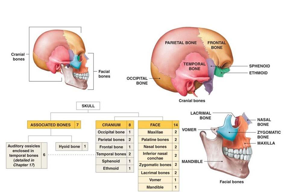
4 Axial Skeleton Axial system: 1) Skull 22 s 2) Bones associated with skull: Hyoid 1 Auditory ossicles 6 s 3) Vertebral column: Vertebrae 24 s Sacrum 1 Coccyx 1 4) Thorax: Sternum Ribs 1 24 s 80 s auditory ossicles U-shaped in the neck that supports the tongue

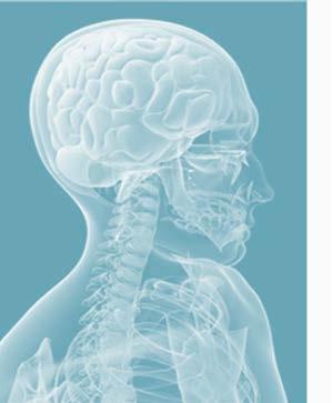
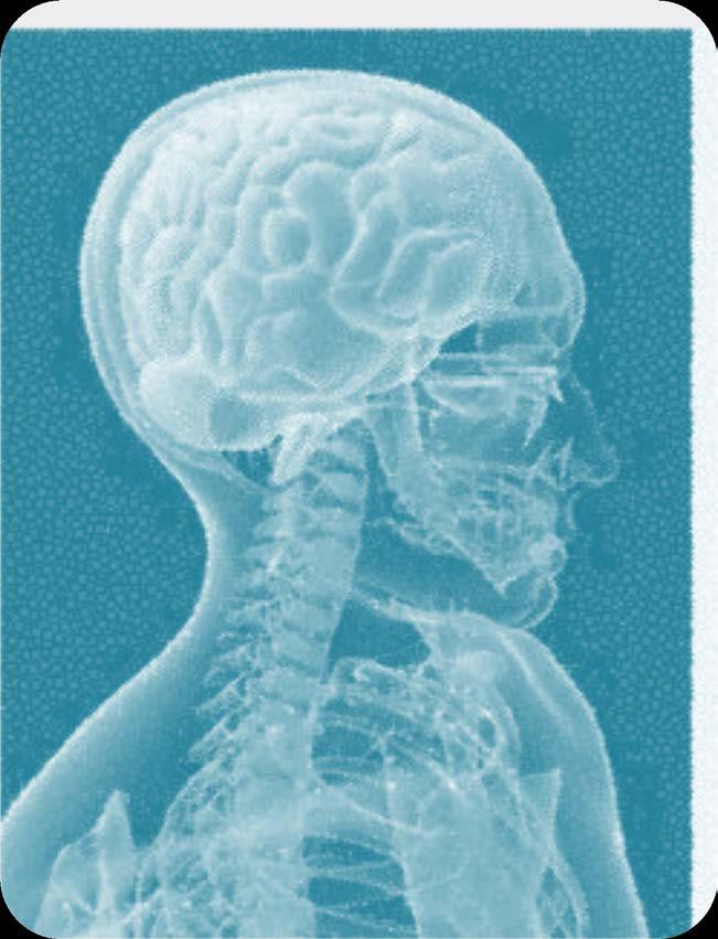
5 Skull


6 Skull Cranial Bones Horizontal section Posterior Roof Floor Anterior Lateral Cranial s form a box cranial cavity to hold the brain: Roof of the cranium Floor of the cranium Anterior, posterior and lateral walls of the cranium

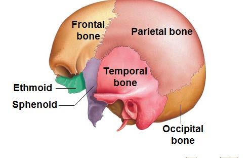
7 Skull Ethmoid Sphenoid
8 Skull Cranial Bones-Frontal Frontal Supraorbital foramen Frontal (1): Form the forehead, part of the roof of cranium and orbital cavity. Two s at birth fuse by age 6-8 years. Lateral view Supraorbital margin- thickened portion that forms the superior margin of the orbital cavity. Frontal Supraorbital foramen Supraorbital margin Supraorbital foramen- a hole in the margin blood vessels and nerves enter the.

9 Skull Cranial Bones-Frontal Frontal Supraorbital foramen Frontal sinus- air filled space in the. Mucus from sinus helps flush the nasal cavity. Makes the skull lighter. Lateral view Frontal sinus Anterior view Sagittal section

10 Skull Cranial Bones-Parietal s Occipital Lateral view Parietal Temporal Frontal Sphenoid Parietal s (2): Form part of the roof, lateral wall and posterior wall of the cranium. Articulations- Parietal s articulate with one another by sagittal suture. Parietal s also articulate with frontal, temporal, sphenoid and occipital. Q. A fracture in the right superior lateral surface of cranium. Which Superior view

11 Skull Cranial Bones-Temporal s Temporal s (2): Form the lateral wall and floor of the cranium. Lateral view Parietal Frontal External auditory/acoustic meatus: a tubelike opening forms the external ear to direct sound into the ear. External acoustic meatus Occipital Temporal Zygomatic process: a projection of the temporal that connects with the cheek (zygomatic ) form zygomatic arch (cheek arch). Zygomatic arch Zygomatic process of temporal Temporal process of zygomatic Zygomatic arch Temporal Temporal Horizontal section Inferior view
12 Skull Cranial Bones-Temporal s Lateral view Mandibular fossa: two depressions to fit lower jaw (mandible). *Pearson-slide 12 Temporal Mastoid process: Site for attachment of muscles that can rotate/extend head Mastoid process Styloid process Styloid process: - Attached to ligaments that support the hyoid. - Attached to tendon of several muscles associated with hyoid, tongue and pharynx Zygomatic arch Mandibular fossa Styloid process Mastoid process U-shaped in the neck that supports the tongue Inferior view
13 Skull Cranial Bones-Occipital Lateral view Occipital Occipital (1): Forms the floor and posterior wall of the cranium. External occipital protuberance: a bump for attachment of ligaments from cervical vertebrae to support head. Superior and inferior nuchal lines: lines extending from protuberance for attachment of muscles and ligaments to support head. Foramen magnum: a big hole for spinal cord to exit. Occipital condyles: curved surfaces lateral to foramen magnum to articulate with 1 st cervical vertebra. *Pearson-slide 11 Occipital condyle Foramen magnum Occipital Superior nuchal line External Occipital protuberance External Occipital protuberance Inferior view Posterior view Inferior nuchal line


14 Skull Cranial Bones-Sphenoid Sella turcica Lesser wing Greater wing Horizontal section Sphenoid (1): Forms the floor of the cranium. Articulates with all other cranial s to hold them together keystone. Sella turcica: a depression to accommodate pituitary gland. Lesser wings: Extend horizontally anteriorly to sella turcica. Greater wings: Extend laterally from body and forms floor of cranium. Sphenoidal sinuses: drains mucus into nasal cavity. *Pearson- Slide 7 Sella turcica Sphenoidal sinus Sagittal section
 d) Superior orbital fissure-orbit (slide 8) e) Optic canal-optic nerve (slide 8) Superior orbital fissure")
15 Skull Cranial Bones-Sphenoid a) Foramen ovale-jaws (slide 23) b) Foramen spinosum-cranial cavity membranes (slide 23) c) Foramen rotundum-face (slide 8) d) Superior orbital fissure-orbit (slide 8) e) Optic canal-optic nerve (slide 8) Superior orbital fissure Optic canal


16 Cribriform plate Horizontal section Skull Cranial Bones-Ethmoid Ethmoid (1): Sponge-like vertically placed in the center of the skull. Cribriform plate: forms the roof of the nasal cavity and floor of the cranium. Has perforations-olfactory foramina- to allow olfactory nerves-provides sense of smell. Crista galli-ridge of that projects from the cribriform plate. Allows the attachment of a membrane that stabilizes the position of the brain. Olfactory foramina Cribriform plate Sagittal section


17 Skull Cranial Bones-Ethmoid Middle nasal concha Ethmoid (1): Perpendicular plate: forms part of the nasal septum. Superior and middle nasal conchae: projections in the nasal cavity cause turbulence of the inhaled air filter, moisten and warm the air. Ethmoidal sinuses: drains mucus into nasal cavity.

18 Squamous suture Lambdoid suture Occipital Parietal Lateral view Temporal Frontal Skull Sutures Coronal suture Sutures: - Seam-lines that connect cranial and facial s to each other dense fibrous connective tissue. - Characterized by zigzag appearance Squamous suture: Temporal Parietal. Coronal suture: Frontal Parietals. Sagittal suture: Parietal Parietal. Lambdoid suture: Occipital Parietals. Superior view
19 Review Cranial Bones! Lateral view Anterior view Cranial floor Inferior view Posterior view Superior view Frontal Parietal Occipital Temporal Sphenoid Frontal Parietal Temporal Occipital Frontal Ethmoid Temporal Sphenoid Occipital Parietal Occipital Temporal Frontal Parietal Occipital
20 Lateral view Lamboid suture Squamous suture External auditory meatus Mastoid process Coronal suture Zygomatic process of temporal Styloid process Review Cranial Landmarks! Anterior view Supraorbital foramen Supraorbital margin Inferior view Zygomatic arch Mandibular fossa Occipital condyle Foramen magnum Mastoid process Cranial floor Cribriform plate Greater wing Sella turcica Posterior view Superior view Sagittal suture Foramen magnum External occipital protuberance Superior nuchal line Inferior nuchal line

21 Skull Fontanelles Sphenoidal fontanelle Posterior fontanelle Mastoid fontanelle Anterior fontanelle Lateral view Superior view Fontanelles: - Soft spots - Non-ossified areas between cranial s-made of tough fibrous membrane - Allows the brain and skull to expand - Enable the bony plates of the skull to flex at birth Craniostenosis: premature closure of one or more fontanelles distortion of the skull can affect proper growth of the brain.

22 Skull Facial s form the face.cheeks, jaws, nose, orbital cavity, and protect/support the entrances to the digestive and respiratory tracts. Nasal Lacrimal Vomer Zygomatic Maxilla Mandible Ethmoid Sphenoid Frontal Temporal Cranial s surround and protect the brain. Parietal Occipital

23 Skull Facial Bones-Nasal s Nasal Nasal s (2): - Forms only the superior part of nose bridge.connected to cartilage that forms the distal portion of the. - Articulates with one another, with ethmoid, with frontal and maxilla. Lateral view Nasal Anterior view
24 Zygomatic Lateral view Skull Facial Bones-Zygomatic s Zygomatic s (2): Cheek s. - Temporal process: a projection of the zygomatic that connects with the temporal form zygomatic arch (cheek arch). Zygomatic arch Zygomatic process of temporal Temporal process of zygomatic Zygomatic arch Inferior view

25 Skull Facial Bones-Maxillae Maxillae (2): - Form the upper jaw. - Two s fuse before birth. - Largest facial. Anterior view Infraorbital foramen Maxilla Palatine process of maxilla Maxillary sinuses: spaces that secrete mucus and drain into the nasal cavity. Alveolar processes: projecting ridge on body of maxilla form alveolar sockets to accommodate teeth. Infraorbital foramen: hone for blood vessel and nerve to enter maxilla. Alveolar process Inferior view
.")

26 Skull Facial Bones-Maxillae Palatine process of maxilla Palatine process of maxilla: horizontal part that forms the anterior part of the hard palate (roof of oral cavity). *Slide 11-Rotate Cleft palate: birth defect when maxillae fail to meet along the midline of the hard palate oral and nasal cavities are continuous; lips are split; difficulty feeding. Inferior view

27 Ramus Angle Body Skull Facial Bones-Mandible Anterior view Mandible (1): Lower jaw the strongest facial and the only movable of the skull. Develops as 2 s fuse before birth. Body: horizontal part of the mandible. Ramus: vertical parts of the mandible. Mandibular/condylar process: rounded projection fits into mandibular fossa of temporal forms temporomandibular joint (TMJ). Coronoid process: Insertion point for temporalis muscle. Mandibular notch: Separates the condylar and coronoid processes. Lateral view TMJ Coronoid process Mandibular notch Head Alveolar process Condylar process Ramus Body Mandible Angle
28 Anterior view Skull Facial Bones-Mandible Mandible: TMJ syndrome: malfunction of the joint headache, referred pain in the ear, muscle pain, noise and reduced movement of the joint physical therapy, anti-inflammatory drug therapy. Four holes: Mental foramen: Opening (in the body) for nerves that carry sensory information from lips and chin to the brain. Ramus Angle Mandibular foramen: Opening (in the ramus)-entrance for mandibular canal-serves lower teeth. Body Mental foramen Mandibular foramen Ramus Angle Body Mental foramen

29 Lacrimal Lateral view Skull Facial Bones-Lacrimal s Lacrimal s (2): Smallest facial. Resembles a fingernail. Part of the medial wall of the orbital cavity. Lacrimal fossa: houses lacrimal sac Lacrimal foramen: passage way for lacrimal artery Lacrimal Lacrimal Lacrimal Anterior view Orbital cavity
30 Skull Facial Bones Palatine s Palatine s (2): Form the posterior part of the hard palate next to palatine process of maxilla. Palatine Inferior view Orbital cavity

31 Skull Facial Bones Vomer (1): A triangular, vertical that forms inferior part of the nasal septum. * Pdf file in Learning Web-Position of vomer-incorrect Vomer Inferior view

32 Skull Facial Bones Inferior nasal conchae (2): The largest of the 3 conchae. (* Superior and middle nasal conchae are part of ethmoid). Functions-Create turbulence in air. - Warms and humidifies internal ear Inferior nasal concha Anterior view Superior and middle nasal conchae of ethmoid Inferior nasal concha Sagittal section
33 Review Skull Bones! maxilla maxilla nasal zygomatic sphenoid palatine vomer mandible maxilla temporal mandible occipital temporal mandible
34 Review Skull Landmarks! Supraorbital foramen Supraorbital margin Zygomatic arch Palatine process-maxilla Temporal process of zygomatic Zygomatic process of temporal Ramus Angle Infraorbital foramen Mental foramen Lambdoid suture Occipital condyle Foramen magnum Sagittal suture Coronal suture External occipital protuberance

35 Skull Orbital Cavity Frontal Sphenoid Superior orbital fissure Optic canal Ethmoid Lacrimal Inferior orbital fissure Zygomatic Orbital cavity: space to accommodate eyeball. Formed by a total of 7 cranial and facial s. Cranial s- frontal, sphenoid, ethmoid. Facial s- zygomatic, maxilla, lacrimal, palatine. Maxilla Palatine Optic canal, superior & inferior orbital fissures for passage of optic and other nerves. Q. Which contains optic canal

36 Skull Paranasal Sinuses Paranasal sinus: space in the s around the nose open into the nasal cavities. Examples: Frontal, Ethmoidal, Sphenoidal and Maxillary sinuses. Functions: Make the skull lighter. Have extensive mucous epithelium that secretes mucus into the nasal cavity provides moisture and traps dust and microorganisms. Sinusitis: viral, bacterial or fungal infection of the sinuses mucous membrane swells openings of sinuses (ostia) narrow mucus accumulates in the sinuses pressure builds headache. Antihistamines, vasoconstrictors and painkillers relieve the pressure and pain.
37 Axial Skeleton-Skull
38 Axial Skeleton Axial system: 1) Skull 22 s 2) Bones associated with skull: Hyoid 1 Auditory ossicles 6 s 3) Vertebral column: Vertebrae 24 s Sacrum 1 Coccyx 1 4) Thorax: Sternum 1 Ribs 24 s 80 s


39 Skull Associated Bones Auditory ossicles: Six smallest s 3 in each ear. Malleus, Incus and Stapes Function: amplify sound stimulus. Auditory ossicles Body Greater horn Lesser horn Hyoid: Single, U-shaped. Located between mandible and larynx. Often fractures during strangulation. Does not articulate with any other floats. Body: horizontal part. Greater and lesser horns/cornua: projections for muscle attachment. Function: supports tongue and attach some of the neck and tongue muscles. An anterior view of the hyoid

40 Axial Skeleton Axial system: Skull Hyoid Auditory ossicles Vertebral column Thorax: Sternum Ribs 22 s 1 6 s 26 s 1 24 s 80 s
Chapter 7 Part A The Skeleton
 Chapter 7 Part A The Skeleton Why This Matters Understanding the anatomy of the skeleton enables you to anticipate problems such as pelvic dimensions that may affect labor and delivery The Skeleton The
Bones of the skull & face
 Bones of the skull & face Cranium= brain case or helmet Copyright The McGraw-Hill Companies, Inc. Permission required for reproduction or display. The cranium is composed of eight bones : frontal Occipital
APPENDICULAR SKELETON 126 AXIAL SKELETON SKELETAL SYSTEM. Cranium. Skull. Face. Skull and associated bones. Auditory ossicles. Associated bones.
 SKELETAL SYSTEM 206 AXIAL SKELETON 80 APPENDICULAR SKELETON 26 Skull Skull and associated s 29 Cranium Face Auditory ossicles 8 4 6 Associated s Hyoid Thoracic cage 25 Sternum Ribs 24 Vertebrae 24 column
Biology 218 Human Anatomy. Adapted from Martini Human Anatomy 7th ed. Chapter 6 The Skeletal System: Axial Division
 Adapted from Martini Human Anatomy 7th ed. Chapter 6 The Skeletal System: Axial Division Introduction The axial skeleton: Composed of bones along the central axis of the body Divided into three regions:
AXIAL SKELETON SKULL
 AXIAL SKELETON SKULL CRANIAL BONES (8 total flat bones w/ 2 paired) 1. Frontal forms forehead & upper portion of eyesocket (orbital) 2. Parietal paired bones; form superior & lateral walls of cranium 3.
Chapter 7. Skeletal System
 Chapter 7 Skeletal System 1 Skull A. The skull is made up of 22 bones: 8 cranial bones, 13 facial bones, and the mandible. B. The Cranium encloses and protects the brain, provides attachments for muscles,
Anatomy and Physiology. Bones, Sutures, Teeth, Processes and Foramina of the Human Skull
 Anatomy and Physiology Chapter 6 DRO Bones, Sutures, Teeth, Processes and Foramina of the Human Skull Name: Period: Bones of the Human Skull Bones of the Cranium: Frontal bone: forms the forehead and the
Cranium Facial bones. Sternum Rib
 Figure 7.1 The human skeleton. Skull Thoracic cage (ribs and sternum) Cranium Facial bones Sternum Rib Bones of pectoral girdle Vertebral column Sacrum Vertebra Bones of pelvic girdle (a) Anterior view
Skeletal system. Prof. Abdulameer Al-Nuaimi. E. mail:
 Skeletal system Prof. Abdulameer Al-Nuaimi E-mail: a.al-nuaimi@sheffield.ac.uk E. mail: abdulameerh@yahoo.com Functions of Bone and The Skeletal System Support: The skeleton serves as the structural framework
Human Anatomy and Physiology - Problem Drill 07: The Skeletal System Axial Skeleton
 Human Anatomy and Physiology - Problem Drill 07: The Skeletal System Axial Skeleton Question No. 1 of 10 Which of the following statements about the axial skeleton is correct? Question #01 A. The axial
Structure Location Function
 Frontal Bone Cranium forms the forehead and roof of the orbits Occipital Bone Cranium forms posterior and inferior portions of the cranium Temporal Bone Cranium inferior to the parietal bone forms the
THE SKELETAL SYSTEM. Focus on the Skull
 THE SKELETAL SYSTEM Focus on the Skull Review Anatomical Terms Anterior/Posterior Dorsal/Ventral Medial/Lateral Superior/Inferior Bone Markings - Review Projections for attachment of muscles, ligaments
ANATOMY & PHYSIOLOGY I Laboratory Version B Name Section. REVIEW SHEET Exercise 10 Axial Skeleton
 ANATOMY & PHYSIOLOGY I Laboratory Version B Name Section REVIEW SHEET Exercise 10 Axial Skeleton 1 POINT EACH. THE SKULL MULTIPLE CHOICE 1. The major components of the axial skeleton include the 7. The
Anatomy and Physiology 1 Chapter 7 self quiz Pro, Dima Darwish,MD.
 Anatomy and Physiology 1 Chapter 7 self quiz Pro, Dima Darwish,MD. 1) How many bones make up the axial skeleton? A) 50 B) 60 C) 70 D) 80 E) 90 2) Which of the following is a function of the axial skeleton?
An Introduction to the Axial Skeleton. Copyright 2009 Pearson Education, Inc., publishing as Pearson Benjamin Cummings
 An Introduction to the Axial Skeleton Copyright 2009 Pearson Education, Inc., publishing as Pearson Benjamin Cummings Terms: Structures of Bones Articulations: Contacts with other bones Landmarks (Bone
ACTIVITY 3: AXIAL SKELETON AND LONG BONE DISSECTION COW BONE DISSECTION
 ACTIVITY 3: AXIAL SKELETON AND LONG BONE DISSECTION Objectives: 1) How to get ready: Read Chapter 7, McKinley et al., Human Anatomy, 4e. All text references are for this textbook. Learning the meanings
SKULL AS A WHOLE + ANTERIOR CRANIAL FOSSA
 SKULL AS A WHOLE + ANTERIOR CRANIAL FOSSA LEARNING OBJECTIVES At the end of this lecture, the student should be able to know: Parts of skeleton (axial and appendicular) Parts of skull Sutures of skull
The Axial Skeleton. C h a p t e r. PowerPoint Lecture Slides prepared by Jason LaPres Lone Star College - North Harris
 C h a p t e r 7 The Axial Skeleton PowerPoint Lecture Slides prepared by Jason LaPres Lone Star College - North Harris Copyright 2009 Pearson Education, Inc., publishing as Pearson Benjamin Cummings An
YOU MUST BRING YOUR OWN GLOVES FOR THIS ACTIVITY.
 ACTIVITY 3: AXIAL SKELETON AND LONG BONE DISSECTION Objectives: 1) How to get ready: Read Chapter 7, McKinley et al., Human Anatomy, 5e. All text references are for this textbook. Learning the meanings
o Diaphysis o Area where red marrow is found o Area where yellow marrow is found o Epiphyseal plate AXIAL SKELETON Skull
 64 Anatomy & Physiology Coloring Workbook 7. Figure 5-2A is a midlevel, cross-sectional view of the diaphysis of the femur. Label the membrane that lines the cavity and the membrane that covers the outside
Chapter 8A. The Skeletal System: The Axial Skeleton. The Skeletal System: The Axial Skeleton. Types of Bones. Types of Bones
 Chapter 8A The Skeletal System: The Axial Skeleton The Skeletal System: The Axial Skeleton 206 named bones Axial Skeleton 80 bones lie along longitudinal axis skull, hyoid, vertebrae, ribs, sternum, ear
Exercise 10. The Axial Skeleton
 Exercise 10 The Axial Skeleton The Axial Skeleton Consists of the skeletal structures found along the midline of the body. Includes the skull, hyoid, vertebrae, ribs, sternum, and sacrum. The cartilages
The Skeletal System: Axial Skeleton
 The Skeletal System: Axial Skeleton The Big Idea The Axial Skeleton & Homeostasis The bones of the axial skeleton contribute to homeostasis by protecting many of the body s organs such as the brain, spinal
Skull-2. Norma Basalis Interna Norma Basalis Externa. Dr. Heba Kalbouneh Associate Professor of Anatomy and Histology
 Skull-2 Norma Basalis Interna Norma Basalis Externa Dr. Heba Kalbouneh Associate Professor of Anatomy and Histology Norma basalis interna Base of the skull- superior view The interior of the base of the
Human Anatomy & Physiology I Dr. Sullivan Unit VIIIa The Axial Skeleton Chapter 8 (Sections )
") Human Anatomy & Physiology I Dr. Sullivan Unit VIIIa The Axial Skeleton Chapter 8 (Sections 8.1-8.3) I. Divisions of the skeletal system a) An adult human skeleton has 206 named bones b) Most are paired
Human Anatomy & Physiology I Dr. Sullivan Unit VIIIa The Axial Skeleton Chapter 8 (Sections 8.1-8.3) I. Divisions of the skeletal system a) An adult human skeleton has 206 named bones b) Most are paired
Chapter 7: Skeletal System: Gross Anatomy
 Chapter 7: Skeletal System: Gross Anatomy I. General Considerations A. How many bones in an average adult skeleton? B. Anatomic features of bones are based on II. Axial Skeleton A. Skull 1. Functionally
Bone Flashcards for 10a
 Bone Flashcards for 0a CLAVICLE (collar bone). Sternal extremity (end) flat end. Acromial extremity (end) rounded end. SCAPULA (shoulder blade). Right or left scapula?. Superior border (superior margin).
Dr. Sami Zaqout, IUG Medical School
 The skull The skull is composed of several separate bones united at immobile joints called sutures. Exceptions? Frontal bone Occipital bone Vault Cranium Sphenoid bone Zygomatic bones Base Ethmoid bone
Skeletal System: Skull.
 Skeletal System: Skull www.fisiokinesiterapia.biz Bones of the Skull SPLANCHNOCRANIUM Nasal (2) Maxilla (2) Lacrimal (2) Zygomatic (2) Palatine (2) Inferior concha (2) Vomer Mandible NEUROCRANIUM Frontal
in compact bone, large vertical canals carrying blood vessels and nerves. in compact bone, large horizontal canals carrying blood vessels and nerves.
 Carl Christensen, PhD Skeletal System (Bones`) Bio. 2304 Human Anatomy 1. Identify a term for each of the following: shaft of a long bone ends of a long bone ossified remnant of the "growth plate" connective
Chapter 7: Head & Neck
 Chapter 7: Head & Neck Osteology I. Overview A. Skull The cranium is composed of irregularly shaped bones that are fused together at unique joints called sutures The skull provides durable protection from
BIO 137 AXIAL SKELETON BONE STUDY THE HUMAN SKELETON
 BIO 137 THE AXIAL SKELETON MARY CATHERINE FLATH, Ph.D. THE HUMAN SKELETON AXIAL SKULL HYOID THORACIC CAGE VERTEBRAL COLUMN APPENDICULAR PECTORAL GIRDLE UPPER LIMBS PELVIC GIRDLE LOWER LIMBS AXIAL SKELETON
The Skull DANIL HAMMOUDI.MD
 The Skull DANIL HAMMOUDI.MD summary of bones/structures in Chapter 15 of the manual need tp be print as soon as possible http://www.mnsu.edu/emuseum/biology/humananatomy/skeletal/skul l/frontal/frontal.html
Biology 210 Chapter 8: Skeletal Tissues Supplement 1
 Biology 210 Chapter 8: Skeletal Tissues Supplement 1 By John McGill Material contributed by Beth Wyatt & Jack Bagwell DIVISIONS OF THE SKELETAL SYSTEM AXIAL SKELETON (80 BONES) Bones of the Head, Neck,
Biology 2401 The Skeletal System
 Biology 2401 The Skeletal System Purpose: The lab will describe the microscopic and gross anatomy of bone, identify bones of the body, and identify important bone markings. I. Overview of the Skeleton
CHAPTER 7, PART II (BONES)
") Anatomy Name: CHAPTER 7, PART II (BONES) Entry #: INSTRUCTIONS: 1) READ Chapter 7, pg. 140-161. 2) Using the outline, make a note card for each underlined bone name or phrase. 3) On each note card, put
Anatomy Name: CHAPTER 7, PART II (BONES) Entry #: INSTRUCTIONS: 1) READ Chapter 7, pg. 140-161. 2) Using the outline, make a note card for each underlined bone name or phrase. 3) On each note card, put
BONE CHALLENGE DANIL HAMMOUDI.MD
 BONE CHALLENGE DANIL HAMMOUDI.MD Bone Basic functions? A. support B. protection C. movement assistance in D. RBC formation-hemopoiesis E. mineral homeostasis +importance of calcium F. energy supply -yellow
Crafton Hills College Human Anatomy & Physiology Axial Skeleton
 A. Major Divisions Crafton Hills College Human Anatomy & Physiology Axial keleton 1. Axial: Part of skeleton lies along long axis of body 2. Appendicular: Bones & features of the appendages B. AXIAL KELETON
Axial skeleton bones and markings
 Axial skeleton bones and markings Skull Cranial bones Frontal x 1 Supraorbital foramen Occipital x 1 Foramen magnum Occipital condyles Superior nuchal line Inferior nuchal line Anterior cranial fossa External
11/25/2012. Chapter 7 Part 2: Bones! Skeletal Organization. The Skull. Skull Bones to Know Cranium
 Chapter 7 Part 2: Bones! 5) Distinguish between the axial and appendicular skeletons and name the major parts of each 6) Locate and identify the bones and the major features of the bones that compose the
Musculoskeletal System (Part A-1) Module 7 -Chapter 10 Overview. Functions
 Module 7 -Chapter 10 Overview. Functions") Musculoskeletal System (Part A-1) Module 7 -Chapter 10 Overview Susie Turner, M.D. 1/8/13 Muscles Attachments Bones Bone types Surface features of bones Divisions of the skeletal system Joints or Articulations
Musculoskeletal System (Part A-1) Module 7 -Chapter 10 Overview Susie Turner, M.D. 1/8/13 Muscles Attachments Bones Bone types Surface features of bones Divisions of the skeletal system Joints or Articulations
Skull basic structures. Neurocranium
 Assoc. Prof. Květuše Lovásová, M.V.D., PhD. Skull basic structures Skull consists of two groups of bones: neurocranium (bones forming the brain box) splanchnocranium (bones forming the facial skeleton)
Spring Written By: J. E. Sutton. Contents: I. Overview of the Skeleton: II. Appendicular Skeleton III. Axial Skeleton IV.
 Spring 2012 Written By: J. E. Sutton Contents: I. Overview of the Skeleton: II. Appendicular Skeleton III. Axial Skeleton IV. Articulations Overview of the Skeleton: I. Orientation to Human Skeleton: a.
External Acoustic Meatus. Mastoid Process. Zygomatic Process. Temporal Bone
 Bone lab review 1. Frontal Bone 2. Supra-Orbital Foramen 3. Orbit (Orbital Cavity) 4. Superior Orbital Fissure 5. Inferior Orbital Fissure 6. Zygomatic Bone 7. Infra-Orbital Foramen 8. Maxilla 9. Mandible
Anatomy Made Easy MSS
 Anatomy Made Easy MSS part #1 هذا الملف يشمل تفريغ المحاضرة الثانية لعون بدءا من الصفحة 11 وحتى األخير Done By :MohamedA. Diabat Edited by Awn Academic team The Axial Skeleton The axial skeleton consist
University of Palestine. Midterm Exam 2013/2014 Total Grade:
 Course No: DNTS2208 Course Title: Head and Neck Anatomy Date: 09/11/2013 No. of Questions: (50) Time: 1hour Using Calculator (No) University of Palestine Midterm Exam 2013/2014 Total Grade: Instructor
Skeletal system overview. Classification of Bones
 Skeletal system overview BIOL241 Lab #9 Classification of Bones Bone are identified by: shape internal tissues bone markings 1 1. Flat bones 2. Long bones 3. Short bones 4. Irregular bones 5. Sutural bones
Labs 9 and 10. Classification of Bones. Bone Shapes 1/05/13. Skeletal system overview. Bone are identified by:
 Labs 9 and 10 Skeletal system overview Classification of Bones Bone are identified by: shape internal tissues bone markings 1. Flat bones 2. Long bones 3. Short bones 4. Irregular bones 5. Sutural bones
TEST YOURSELF- Chapter 7
 TEST YOURSELF- Chapter 7 Cranial Bones 1. Give the name of the bone for each of the following markings. Some of the markings are found on more than one bone. List all that apply. Cranium a. Frontal squama:
Lab Exercise #04 The Skeletal System Student Performance Objectives
 Lab Exercise #04 The Skeletal System Student Performance Objectives The material that you are required to learn in this exercise can be found in either the lecture text or the supplemental materials provided
Bones Ethmoid bone Inferior nasal concha Lacrimal bone Maxilla Nasal bone Palatine bone Vomer Zygomatic bone Mandible
 splanchnocranium - Consists of part of skull that is derived from branchial arches - The facial bones are the bones of the anterior and lower human skull Bones Ethmoid bone Inferior nasal concha Lacrimal
The SKELETAL System. The framework of bones and cartilage which protect organs, and provides a lever system that allows locomotion.
 The SKELETAL System The framework of bones and cartilage which protect organs, and provides a lever system that allows locomotion. Functions of the Skeletal System Support Protection Movement Facilitation
Introduction to Local Anesthesia and Review of Anatomy
 5-Sep Introduction and Anatomy Review 12-Sep Neurophysiology and Pain 19-Sep Physiology and Pharmacology part 1 26-Sep Physiology and Pharmacology part 2 Introduction to Local Anesthesia and Review of
Anatomy images for MSS practical exam- 2019
 Anatomy images for MSS practical exam- 2019 Ilium Ischium Pubis Acetabulaum Iliac crest Iliac tubercle ASIS (muscle and ligament attached) AIIS (muscle attached) PSIS PIIS Ischial spine Ischial tuberosity
Dr.Noor Hashem Mohammad Lecture (5)
") Dr.Noor Hashem Mohammad Lecture (5) 2016-2017 If the mandible is discarded, the anterior part of this aspect of the skull is seen to be formed by the hard palate. The palatal processes of the maxillae
Dr.Noor Hashem Mohammad Lecture (5) 2016-2017 If the mandible is discarded, the anterior part of this aspect of the skull is seen to be formed by the hard palate. The palatal processes of the maxillae
Bio 5/6 5 The Skeletal System Study Guide
 Name: THE SKELETAL SYSTEM: 5 The Skeletal System Study Guide Period: The skeleton is constructed of two of the most supportive tissues found in the human body - cartilage and bone. Besides supporting and
Chapter 7 The Skeletal System:The Axial Skeleton
 Chapter 7 The Skeletal System:The Axial Skeleton Axial Skeleton 80 bones lie along longitudinal axis skull, hyoid, vertebrae, ribs, sternum, ear ossicles Appendicular Skeleton 126 bones upper & lower limbs
Infratemporal fossa: Tikrit University college of Dentistry Dr.Ban I.S. head & neck Anatomy 2 nd y.
 Infratemporal fossa: This is a space lying beneath the base of the skull between the lateral wall of the pharynx and the ramus of the mandible. It is also referred to as the parapharyngeal or lateral pharyngeal
SKULL / CRANIUM BONES OF THE NEUROCRANIUM (7) Occipital bone (1) Sphenoid bone (1) Temporal bone (2) Frontal bone (1) Parietal bone (2)
 Occipital bone (1) Sphenoid bone (1) Temporal bone (2) Frontal bone (1) Parietal bone (2)") Important! 1. Memorizing these pages only does not guarantee the succesfull passing of the midterm test or the semifinal exam. 2. The handout has not been supervised, and I can not guarantee, that these
Important! 1. Memorizing these pages only does not guarantee the succesfull passing of the midterm test or the semifinal exam. 2. The handout has not been supervised, and I can not guarantee, that these
UNIT 4 - SKELETAL SYSTEM LECTURE NOTES
 UNIT 4 - SKELETAL SYSTEM LECTURE NOTES 4.01 FUNCTIONS OF THE SKELETAL SYSTEM A. Support 1. Provides a framework for the body 2. Supports soft tissue 3. Serves as a point of attachment for ligaments, tendons,
Nervous & Skeletal Systems. Virtual Science University
 Nervous & Skeletal Systems Virtual Science University 1 Nervous & Skeletal Systems Texas TEK B.10(A) The student will interpret the function of systems in organisms (humans) including the nervous and skeletal
Skull-2. Norma Basalis Interna. Dr. Heba Kalbouneh Assistant Professor of Anatomy and Histology
 Skull-2 Norma Basalis Interna Dr. Heba Kalbouneh Assistant Professor of Anatomy and Histology Norma basalis interna Base of the skull- superior view The interior of the base of the skull is divided into
Human Anatomy - Problem Drill 06: The Skeletal System Axial Skeleton & Articualtions
 Human Anatomy - Problem Drill 06: The Skeletal System Axial Skeleton & Articualtions Question No. 1 of 10 Instructions: (1) Read the problem and answer choices carefully, (2) Work the problems on paper
University of Palestine. Midterm Exam 2013/2014 Total Grade:
 [ Course No: DNTS2208 Course Title: Head and Neck Anatomy Date: 17/11/1024 No. of Questions: (52) Time: 2hours Using Calculator (No) University of Palestine Midterm Exam 2013/2014 Total Grade: Instructor
Important Parts of Bones
 Important Parts of Bones For 2015 Know: Humerus (posterior) Clavical Femur (Anterior) Foot Hand Mandible Os Coxa Scapula Skull (Anterior, Inferior, Lateral) Sternum Humerus (posterior) A. olecranon fossa
Anatomy Skull and Spinal Cord
 1 Anatomy Skull and Spinal Cord 1. Skull The skull is anterior to the spinal column and is the bony structure that encases the brain. Its purpose is to protect the brain and allow attachments for the facial
Anatomic Relations Summary. Done by: Sohayyla Yasin Dababseh
 Anatomic Relations Summary Done by: Sohayyla Yasin Dababseh Anatomic Relations Lecture 1 Part-1 - The medial wall of the nose is the septum. - The vestibule lies directly inside the nostrils (Nares). -
Lab 6, 7, 8: Skeletal System
 107 Lab 6, 7, 8: Skeletal System Adult Skull Bony orbit (FLEZMS) Frontal bone supraorbital foramen frontal sinus Lacrimal bone Ethmoid bone perpendicular plate of ethmoid middle nasal conchae cribriform
Bones of the Skull Lateral View
 Bones of the Skull Lateral View Frontal Bone Parietal Bone Occipital Bone Temporal Bone Sphenoid Bone Pterion Sutures of the Skull Lateral View Coronal Suture Lambdoid Suture Squamous Suture Sutures of
View of a Skull, 1489 by Leonardo Da Vinci. Kaan Yücel M.D., Ph.D Tuesday
 View of a Skull, 1489 by Leonardo Da Vinci Kaan Yücel M.D., Ph.D. 26.11.2013 Tuesday 1.SKULL skeleton of the head cranium 22 bones excluding ossicles of the ear 1.SKULL Mandible Lower jaw bone Neurocranium
The Skeletal System. Parts of the skeletal system. Bones (Skeleton) Joints Cartilages Ligaments
 Joints Cartilages Ligaments") The Skeletal System Parts of the skeletal system Bones (Skeleton) Joints Cartilages Ligaments Functions of the Bones Support Internal framework of the body Protection Skull and vertebrae protect brain
The Skeletal System Parts of the skeletal system Bones (Skeleton) Joints Cartilages Ligaments Functions of the Bones Support Internal framework of the body Protection Skull and vertebrae protect brain
the Skeletal System provided by Academic Web Services Grand Canyon University
 Anatomy Resource Center Study Guides the Skeletal System HEAD & NECK REGIONAL VIEW SKULL BONES CRANIUM FACE SKULL LANDMARKS ANTERIOR SIDE SUPERIOR/INFERIOR VERTEBRAL COLUMN VERTEBRAL REGIONS CERVICAL C1
Perpendicular Plate Zygomatic Bone. Mental Foramen Mandible
 Glabella Frontal Middle Nasal Concha Nasal Lacrimal Perpendicular Plate Zygomatic Inferior Nasal Concha Maxilla Mental Mandible Skull (anterior view) Squamosal Suture Coronal Suture Frontal Parietal Nasal
Major Anatomic Components of the Orbit
 Major Anatomic Components of the Orbit 1. Osseous Framework 2. Globe 3. Optic nerve and sheath 4. Extraocular muscles Bony Orbit Seven Bones Frontal bone Zygomatic bone Maxillary bone Ethmoid bone Sphenoid
Anatomy and Physiology II. Review Spine and Neck
 Anatomy and Physiology II Review Spine and Neck Spine regions How many cervical vertibrae are there? 7 The curvature is the cervical region posterior? Concave posterior How many thoracic? And curvature?
Skeletal System. It s all about the bones!!!
 Skeletal System It s all about the bones!!! The Skeletal System in Action!! The Skeletal System in Action! https://www.youtube.com/watch?v=icwllrqkv cg&list=plzile25upgebvru0jneppcabh0fhktgt Q 1. FYI 5
SKELETON FUNCTIONS OF BONE:
 SKELETON FUNCTIONS OF BONE: SKELETON: 1. Performs a mechanical function in forming the skeletal support of the body and in forming a leverage system whereby work and movement are possible. 2. Serves as
Bisection of Head & Nasal Cavity 頭部對切以及鼻腔. 解剖學科馮琮涵副教授 分機
 Bisection of Head & Nasal Cavity 頭部對切以及鼻腔 解剖學科馮琮涵副教授 分機 3250 E-mail: thfong@tmu.edu.tw Outline: The structure of nose The concha and meatus in nasal cavity The openings of paranasal sinuses Canals, foramens
The Axial Skeleton Hyoid Bone. Lecture Overview. Marieb s Human Anatomy and Physiology. Chapter 7 The Axial and Appendicular Skeleton Lecture 14
 Marieb s Human Anatomy and Physiology Marieb Hoehn Chapter 7 The Axial and Appendicular Skeleton Lecture 14 1 Axial Skeleton Hyoid bone Bones of the orbit Paranasal sinuses Infantile skull Vertebral column
Lab Unit One Flashcards
 CLAVICLE (collar bone). Sternal extremity (end) flat end. Acromial extremity (end) rounded end.. Conoid tubercle near round end SCAPULA (shoulder blade). Right or left scapula?. Superior border (superior
Skeletal System - Prelab 1
 Skeletal System - Prelab 1 1. Which bones contain the paranasal sinuses? What function do the sinuses serve? 2. What two areas are separated from each other by the hard palate? Name the two bones that
A. Incorrect! The axial skeleton includes bones of the skull, inner ear, chest and spinal column.
 Anatomy and Physiology - Problem Drill 07: The Skeletal System II No. 1 of 10 1. Which of the following statements about the axial skeleton is correct? A. The axial skeleton includes bones of the skull,
Omran Saeed. Luma Taweel. Mohammad Almohtaseb. 1 P a g e
 2 Omran Saeed Luma Taweel Mohammad Almohtaseb 1 P a g e I didn t include all the photos in this sheet in order to keep it as small as possible so if you need more clarification please refer to slides In
Axial Skeleton BONE TERMINOLOGY FEATURES
 Axial Skeleton BONE TERMINOLOGY FEATURES Tuberosity Rounded area on bone often roughened for muscle attachment. Tubercle Rounded projection on bone. This is called a tuberosity on the femur. Crest Ridgeline
PTERYGOPALATINE FOSSA
 PTERYGOPALATINE FOSSA Outline Anatomical Structure and Boundaries Foramina and Communications with other spaces and cavities Contents Pterygopalatine Ganglion Especial emphasis on certain arteries and
Labs 6, 7, 8: Skeletal System
 153 Labs 6, 7, 8: Skeletal System Unit 6: Skeletal System: Bone tissue, Bones and Joints (p. 105-152) Ex. 6-1: Histology of Osseous Tissue, p. 113 Model: Osteon Tiss Lamella Osteocyte Lacunae Canaliculi
Mohammad Hisham Al-Mohtaseb. Lina Mansour. Reyad Jabiri. 0 P a g e
 2 Mohammad Hisham Al-Mohtaseb Lina Mansour Reyad Jabiri 0 P a g e This is only correction for the last year sheet according to our record. If you already studied this sheet just read the yellow notes which
The cribriform plate. ethmoid bone. Ethmoid bone consists from: 1) A horizontal cribriform plate. 2) A perpendicular plate. 3) Two lateral labyrinths.
 A horizontal cribriform plate. 2) A perpendicular plate. 3) Two lateral labyrinths.") ethmoid bone Ethmoid bone consists from: 1) A horizontal cribriform plate. 2) A perpendicular plate. 3) Two lateral labyrinths. The cribriform plate 1) Connect the two labyrinths to the perpendicular plate.
ethmoid bone Ethmoid bone consists from: 1) A horizontal cribriform plate. 2) A perpendicular plate. 3) Two lateral labyrinths. The cribriform plate 1) Connect the two labyrinths to the perpendicular plate.
Objectives continued- Answer each of the objectives on a separate sheet of paper to demonstrate content mastery. Attach answers to back of packet.
 Anatomy and Physiology Chapter 5: The Skeletal System Name: Objectives- By the end of this chapter I will be able to: 1. Identify the subdivisions of the skeleton as axial or appendicular. 2. List at least
Temporal region. temporal & infratemporal fossae. Zhou Hong Ying Dept. of Anatomy
 Temporal region temporal & infratemporal fossae Zhou Hong Ying Dept. of Anatomy Temporal region is divided by zygomatic arch into temporal & infratemporal fossae. Temporal Fossa Infratemporal fossa Temporal
Trigeminal Nerve Worksheets, Distributions Page 1
 Trigeminal Nerve Worksheet #1 Distribution by Nerve Dr. Darren Hoffmann Dental Gross Anatomy, Spring 2013 We have drawn out each of the branches of CN V in lecture and you have an idea now for their basic
Maxilla, ORBIT and infratemporal fossa. Neophytos C Demetriades MD, DDS, MSc Associate professor European University of Cyprus School of Medicine
 Maxilla, ORBIT and infratemporal fossa Neophytos C Demetriades MD, DDS, MSc Associate professor European University of Cyprus School of Medicine MAXILLA Superior, middle, and inferior meatus Frontal sinus
Dr. Sami Zaqout Faculty of Medicine IUG
 The Nose External Nose Nasal Cavity External Nose Blood and Nerve Supplies of the External Nose Blood Supply of the External Nose The skin of the external nose Branches of the ophthalmic and the maxillary
Dr.Ban I.S. head & neck anatomy 2 nd y جامعة تكريت كلية طب االسنان مادة التشريح املرحلة الثانية أ.م.د. بان امساعيل صديق 6102/6102
 جامعة تكريت كلية طب االسنان مادة التشريح املرحلة الثانية أ.م.د. بان امساعيل صديق 6102/6102 Pterygopalatine fossa: The pterygopalatine fossa is a cone-shaped depression, It is located between the maxilla,
SHORT ANSWER. Write the word or phrase that best completes each statement or answers the question.
 Exam Name SHORT ANSWER. Write the word or phrase that best completes each statement or answers the question. Figure 7.1 Using Figure 7.1, match the following: 1) Articulates with hip bones of the pelvis.
Parotid Gland. Parotid Gland. Largest of 3 paired salivary glands (submandibular; sublingual) Ramus of Mandible. Medial pterygoid.
 Ramus of Mandible. Medial pterygoid.") Parotid region Parotid Gland Largest of 3 paired salivary glands (submandibular; sublingual) Ramus of Mandible Medial pterygoid Cross section of mandible Masseter D S SCM Parotid Gland Mastoid Process
Parotid region Parotid Gland Largest of 3 paired salivary glands (submandibular; sublingual) Ramus of Mandible Medial pterygoid Cross section of mandible Masseter D S SCM Parotid Gland Mastoid Process
NEUROCRANIUM VISCEROCRANIUM VISCEROCRANIUM VISCEROCRANIUM
 LECTURE 4 SKULL NEUROCRANIUM VISCEROCRANIUM VISCEROCRANIUM VISCEROCRANIUM CRANIUM NEUROCRANIUM (protective case around brain) VISCEROCRANIUM (skeleton of face) NASOMAXILLARY COMPLEX MANDIBLE (DESMOCRANIUM)
Anatomy & Physiology Skeletal System Worksheet
 1. Name the five functions of the skeleton. c) d) e) Anatomy & Physiology Skeletal System Worksheet 2. The term for the shaft of a bone is:. 3. The bony struts found in spongy bone are called. 4. In ossification,
Anatomy #1; Respiratory Nose and the Nasal Cavity December 1st, 2013
 Note #1: the doctor skipped some slides in the lecture. Those slides are not included in this sheet and so you will have to review the slides to study them. The reason they were not included is because