Epithelial tumors. Dr. F.F. Khuzin, PhD Dr. M.O. Mavlikeev
|
|
|
- Randolf Montgomery
- 6 years ago
- Views:
Transcription

1 Epithelial tumors Dr. F.F. Khuzin, PhD Dr. M.O. Mavlikeev
2 Epithelial tumors Tumors from the epithelium are the most frequent among tumors. There are 2 group features of these tumors: The presence in most of them a clear boundary between parenchyma complexes (strata) and the stroma, The possibility of malignization of many benign epithelial tumors.
3 Classification of epithelioma Depending on the histogenesis (type of epithelium): Tumors from the integumentary epithelium (stratified squamous and transitional), Tumors of glandular epithelium. According to the clinical course (degree of differentiation): Benign, Malignant. Depending on the organ specificity: Organ-specific tumors, Non-specific (tumors without specific localization).

4 Benign epithelioma Benign epitheliomas include: Papilloma, Adenoma.
5 Papilloma Benign tumor from stratified squamous or transitional epithelium. Often occurs in the skin, larynx, oral cavity, esophagus, bladder, kidney, kidney, ureter, vagina etc. Sometimes the papilloma can be multiple (papillomatosis of the larynx). In rare cases, the papilloma recurs and maligns (larynx, bladder), so it is of great clinical importance.
6 Morphology of papilloma Macroscopic picture: Spherical mobile formation on a wide base or thin stem, Soft-elastic consistency, The surface is covered with small papillae ("cauliflower"). Microscopic picture: Papillary growths of stratified squamous (or transitional) epithelium, which, like a glove, cover the connective tissue stroma containing the vessels. The stratified squamous epithelium lies on the basal membrane, retains polarity and complexity. Uneven increase in the layers of the epithelium, increased its cornification (tissue atypism).

7 Larynx papillomas

8 Urinary bladder papilloma
9 Adenoma Benign tumor from prismatic and glandular epithelium. Occurs on mucous membranes lined with prismatic epithelium, and in glandular organs. Adenomas of the mucous membrane of the colon and stomach are often maligns.
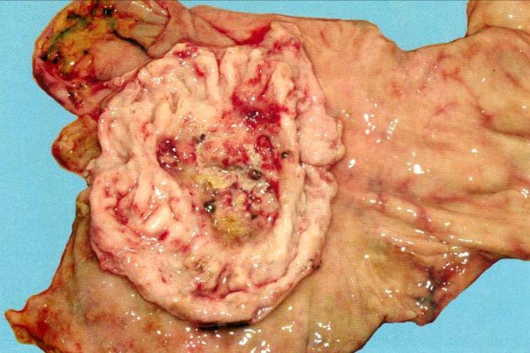
10 Morphological variants of adenoma Alveolar adenoma is from the alveolar epithelium of the glands. Tubular adenoma - from the epithelium of gland ducts. Trabecular adenoma - has a beam structure. Fibroadenoma is an adenoma in which the stroma predominates over the parenchyma. Adenomatous (glandular) polyp - adenoma of the mucous membranes, protruding above the surface in the form of a polyp. Cystic adenoma (cystadenoma) - an adenoma containing cavities.

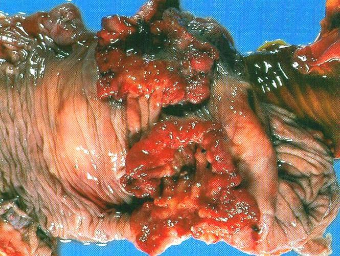
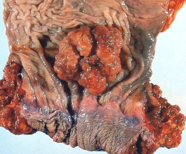
11 Glandular polyps of colon

12 Glandular polyp of colon

13 Cystic adenoma of ovary

14 Liver adenoma

15 Pituitry adenoma

16 Adrenal cortex adenoma
17 Malignant epitheliomas Malignant epithelial tumors are called cancer, or carcinoma. General characteristics: They occur much more often than all other malignant tumors. Often associated with previous diseases and conditions, which are called precancerous. The development of many carcinomas (morphogenesis) is associated with previous changes in the epithelium - hyperplasia, metaplasia, dysplasia. The only precancerous process currently considered epithelial dysplasia, which, progressing from mild to moderate and severe, can lead to the development of carcinoma in situ and further invasive cancer that germinates surrounding tissues.
18 Malignant epitheliomas General characteristic (continued): For invasive cancer, all signs of a malignant tumor are characteristic. Cancer metastasizes primarily lymphogenically: the first metastases occur in the regional lymph nodes, later hematogenous and implantation metastases may occur.
19 Classification of cancer According to the histological structure: from the integument epithelium: Squamous (epidermal) cancer, Basal cell carcinoma, Transitional cell carcinoma, Undifferentiated (small cell, large cell, etc.) from glandular epithelium: Adenocarcinoma (glandular cancer), A solid cancer. By the ratio of parenchyma and stroma: Medullary (cerebral) cancer, Simple (vulgar) cancer, Skyrr (fibrous cancer).
20 Squamous cell carcinoma It develops from stratified squamous epithelium in the skin, cervix, esophagus, larynx, etc. In the lungs, it occurs from squamous cell metaplasia (epidermisation) of the bronchial epithelium. Can be high-, moderate- and low-grade.
21 Squamous cell carcinoma with keratinization This is a highly differentiated cancer, which is characterized by: Slightly expressed cellular atypism, The zoning of the structure is observed, resembling the basal, prickly and granular layers of the epidermis, Formation of extracellular keratogialin in the form of "cancer pearls" (eosinophilic layered masses).

22 Squamous cell carcinoma with keratinization
23 Squamous cell carcinoma without keratinization This is a low-grade squamous cell carcinoma that is characterized by: A high level of cellular atypism, The absence of a zonal structure of the layers of the tumor, Absence of "cancer pearls".

24 Squamous cell carcinoma without keratinization

25 Squamous cell cancer of larynx

26 Squamous cell cancer of lung

27 Squamous cell cancer of cervix
28 Basal-cell carcinoma (basalioma) It is characterized by the formation of polymorphous tumor epithelial complexes consisting of cells that resemble the cells of the basal layer of stratified squamous epithelium. The cells are small, prismatic, or polygonal, with hyperchromatic nuclei and a narrow rim of the cytoplasm. The cells are located in the form of a palisade perpendicular to the basal membrane, mitoses are not uncommon. When localized on the skin it grows slowly, often ulcerates with the formation of a deep ulcer. It is characterized by a slow course, expressed by destructive growth, late gives metastases.

29 Basalioma

30 Lip basalioma
31 Transitional cell carcinoma Highly differentiated cancer, according to the histological picture, are often very difficult to distinguish from transitional cell papilloma. A distinctive feature is the destruction of the basal membrane and the infiltration of the tumor cells of lamina propria of the mucous membrane.

32 Transitional cell carcinoma of urinary bladder

33 Transitional cell carcinoma of pelvis
34 Small cell cancer The form of undifferentiated cancer, which consists of monomorphic lymphocyte-like cells that do not form any structures. Stroma is of little amount. The tumor has many mitoses, extensive areas of necrosis. It grows rapidly with early and widespread metastasis.

35 Small cell lung carcinoma
36 Adenocarcinoma It develops from a prismatic epithelium lining the mucous membranes, as well as from the glandular epithelium of various organs. In the urinary tract occurs after tumorous glandular metaplasia. A characteristic microscopic feature is the presence of invasively growing glandular structures. Depending on the degree of differentiation, high-, moderateand low-grade adenocarcinoma is isolated.
37 Adenocarcinoma With a high degree of differentiation: cancerous epithelium forms glandular structures in which many cells resemble their normal counterparts (mildly expressed atypism), in the lumen of the cancer glands - the corresponding secretory product.
38 Adenocarcinoma With a low degree of differentiation, glandular complexes may look different: If the secretion of mucus does not occur, then not the dimensions, but the number of these complexes in the power field increases. If the function of glandular structures is developed strongly, then they can greatly expand and become overfilled with mucus ( mucus lakes"). This form of a low-grade adenocarcinoma is called mucous, or colloidal, cancer. There is a variant of a low-grade adenocarcinoma in which mucus lakes" are absent, but a lot of randomly located cancer cells filled with mucus, with a nucleus pushed to the plasmalemma (ring-shaped cells). This variant is called cricoid carcinoma.

39 Cricoid cancer

40 Stomach adenocarcinoma
41 Stomach adenocarcinoma

42 Liver adenocarcinoma
43 Colon adenocarcinoma
44 Rectum adenocarcinoma
45 Solid cancer One form of glandular undifferentiated cancer. The tumor has many massive layers of parenchyma, and the stroma is relatively small.
46 Medullar cancer Characterized by the predominance of the parenchyma above the stroma. The tumor is soft, white and pink, resembling the brain tissue.
47 Skyrr (fibrous cancer) One form of low-grade cancer. The tumor is dominated by the stroma, the parenchyma is poor. It occurs mainly in the stomach.

48 Stomach skyrr
NEOPLASIA-I CANCER. Nam Deuk Kim, Ph.D.
 NEOPLASIA-I CANCER Nam Deuk Kim, Ph.D. 1 2 Tumor in the hieroglyphics of the Edwin Smith papyrus (1,600 B.C., Breasted s translation 1930) 3 War on Cancer (National Cancer Act, 1971) 4 Cancer Acts in Korea
Neoplasia part I. Dr. Mohsen Dashti. Clinical Medicine & Pathology nd Lecture
 Neoplasia part I By Dr. Mohsen Dashti Clinical Medicine & Pathology 316 2 nd Lecture Lecture outline Review of structure & function. Basic definitions. Classification of neoplasms. Morphologic features.
Neoplasia literally means "new growth.
 NEOPLASIA Neoplasia literally means "new growth. A neoplasm, defined as "an abnormal mass of tissue the growth of which exceeds and is uncoordinated with that of the normal tissues and persists in the
Salivary Glands 3/7/2017
 Salivary Glands 3/7/2017 Goals and objectives Focus on the entities unique to H&N Common board type facts Information for your future practice Salivary Glands Salivary Glands Major gland. Paratid. Submandibular.
Neoplasia 2018 Lecture 2. Dr Heyam Awad MD, FRCPath
 Neoplasia 2018 Lecture 2 Dr Heyam Awad MD, FRCPath ILOS 1. List the differences between benign and malignant tumors. 2. Recognize the histological features of malignancy. 3. Define dysplasia and understand
Diseases of the breast (1 of 2)
") Diseases of the breast (1 of 2) Introduction A histology introduction Normal ducts and lobules of the breast are lined by two layers of cells a layer of luminal cells overlying a second layer of myoepithelial
Diseases of the breast (1 of 2) Introduction A histology introduction Normal ducts and lobules of the breast are lined by two layers of cells a layer of luminal cells overlying a second layer of myoepithelial
ONCOLOGY. Csaba Bödör. Department of Pathology and Experimental Cancer Research november 19., ÁOK, III.
 ONCOLOGY Csaba Bödör Department of Pathology and Experimental Cancer Research 2018. november 19., ÁOK, III. bodor.csaba1@med.semmelweis-univ.hu ONCOLOGY Characteristics of Benign and Malignant Neoplasms
Tissues. tissue = many cells w/ same structure and function. cell shape aids function tissue shape aids function. Histology = study of tissues
 Tissues tissue = many cells w/ same structure and function cell shape aids function tissue shape aids function Histology = study of tissues 4 types of tissues Epithelial coverings contact openings Connective
number Done by Corrected by Doctor Maha Shomaf
 number 16 Done by Waseem Abo-Obeida Corrected by Zeina Assaf Doctor Maha Shomaf MALIGNANT NEOPLASMS The four fundamental features by which benign and malignant tumors can be distinguished are: 1- differentiation
BREAST PATHOLOGY. Fibrocystic Changes
 BREAST PATHOLOGY Lesions of the breast are very common, and they present as palpable, sometimes painful, nodules or masses. Most of these lesions are benign. Breast cancer is the 2 nd most common cause
SESSION 1: GENERAL (BASIC) PATHOLOGY CONCEPTS Thursday, October 16, :30am - 11:30am FACULTY COPY
 PATHOLOGY CONCEPTS Thursday, October 16, :30am - 11:30am FACULTY COPY") SESSION 1: GENERAL (BASIC) PATHOLOGY CONCEPTS Thursday, October 16, 2008 9:30am - 11:30am FACULTY COPY GOAL: Describe the basic morphologic (structural) changes which occur in various pathologic conditions.
SESSION 1: GENERAL (BASIC) PATHOLOGY CONCEPTS Thursday, October 16, 2008 9:30am - 11:30am FACULTY COPY GOAL: Describe the basic morphologic (structural) changes which occur in various pathologic conditions.
Dr. Abeer.c.Yousif. Histology -2 nd stage. What is histology?
 What is histology? Histology is the science of microscopic anatomy of cells and tissues, in Greek language Histo= tissue and logos = study and it's tightly bounded to molecular biology, physiology, immunology
Histology Notes -Part 1: Epithelial Tissues
 Introduction Group of cells w/ similar structure & function = TISSUE Four Basic Tissue Types 1. Epithelial-covers 2. Connective-supports 3. Muscular*-produces movement (will discuss in the muscular system
Epithelial Tissue. Functions include: 1. Protection 4. Absorption 2. Secretion 5. Filtration 3. Sensory reception
 Tissues There are 4 primary tissue types in the human body: 1. Epithelial (covering/lining) 2. Connective (support) 3. Muscle (movement) 4. Nervous (control) Epithelium Epithelial Tissue Covers the surface
Basic Tissue Types and Functions
 Tissues Histology Basic Tissue Types and Functions 1) Epithelial tissue covering 2) Connective tissue support 3) Muscle tissue movement 4) Nervous tissue control Epithelial Tissue 1) Covers a body surface
Unit I Problem 9 Histology: Basic Tissues of The Body
 Unit I Problem 9 Histology: Basic Tissues of The Body - What is the difference between cytology and histology? Cytology: it is the study of the structure and functions of cells and their contents. Histology:
Tissues. tissue = many cells w/ same structure and function. cell shape aids its function tissue shape aids its function
 Tissues tissue = many cells w/ same structure and function cell shape aids its function tissue shape aids its function Histology = study of tissues 4 types of tissues Epithelial coverings contact openings
Urinary Bladder, Ureter, and Renal Pelvis
 Urinary Bladder, Ureter, and Renal Pelvis Protocol applies to all carcinomas of the urinary bladder, ureter, and renal pelvis. Protocol revision date: January 2005 Based on AJCC/UICC TNM, 6th edition Procedures
Tissue: The Living Fabric: Part A
 PowerPoint Lecture Slides prepared by Janice Meeking, Mount Royal College C H A P T E R 4 Tissue: The Living Fabric: Part A Tissues Groups of cells similar in structure and function Types of tissues Epithelial
PRACTICAL HISTOLOGY LAB
 PRACTICAL HISTOLOGY LAB.1 ----------------------------------------------------------------------------- INTRODUCTION Cells are the smallest units of life, and are named according to their function. Cells
ATLAS OF HEAD AND NECK PATHOLOGY METAPLASIA
 Metaplasia is the conversion of one adult differentiated cell type to another. Generally it is the result of persistent cellular trauma and serves as a protective mechanism. Thus anteriorly along the nasal
Histology: Epithelial tissue
 Histology: Epithelial tissue Epithelial Tissue is presented in two forms: 1. Covering Epithelia: 2. Glandular Epithelia: 1. Simple Epithelium: contain only one layer of cells. 2. Stratified Epithelium:
Histopathology: Cervical HPV and neoplasia
 Histopathology: Cervical HPV and neoplasia These presentations are to help you identify basic histopathological features. They do not contain the additional factual information that you need to learn about
Mody. AIS vs. Invasive Adenocarcinoma of the Cervix
 Common Problems in Gynecologic Pathology Michael T. Deavers, M.D. Houston Methodist Hospital, Houston, Texas Common Problems in Gynecologic Pathology Adenocarcinoma in-situ (AIS) of the Cervix vs. Invasive
Epithelial Tissue. By the end of this lecture, you should be able to: different types of epithelial membranes.
 Epithelial Tissue Objectives: By the end of this lecture, you should be able to: n Describe general characteristics of epithelial tissue. n Discuss microscopic structure and distribution of different types
5/21/2018. Prostate Adenocarcinoma vs. Urothelial Carcinoma. Common Differential Diagnoses in Urological Pathology. Jonathan I.
 Common Differential Diagnoses in Urological Pathology Jonathan I. Epstein Prostate Adenocarcinoma vs. Urothelial Carcinoma 1 2 NKX3.1 NKX3.1 3 4 5 6 Proposed ISUP Recommendations Option to use PSA as a
Lecture Overview. Chapter 4 Epithelial Tissues Lecture 9. Introduction to Tissues. Epithelial Tissues. Glandular Epithelium
 Visual Anatomy & Physiology First Edition Martini & Ober Chapter 4 Lecture 9 Lecture Overview Introduction to Tissues Location General characteristics Functions Classification Glandular Epithelium 2 Where
DUSTURBANCES OF GROWTH. MLS Basic histological diagnosis MLS HIST 422 Semester 8- batch 7 L8 Uz: Musa
 DUSTURBANCES OF GROWTH MLS Basic histological diagnosis MLS HIST 422 Semester 8- batch 7 L8 Uz: Musa Agnesia: means complete absence of an organ (Kidney). Aplasia: s defined in general as "defective development
Urinary Bladder: WHO Classification and AJCC Staging Update 2017
 Urinary Bladder: WHO Classification and AJCC Staging Update 2017 Houston Society of Clinical Pathologists 58 th Annual Spring Symposium Houston, TX April 8, 2017 Jesse K. McKenney, MD Classification
Synonyms. Nephrogenic metaplasia Mesonephric adenoma
 Nephrogenic Adenoma Synonyms Nephrogenic metaplasia Mesonephric adenoma Definition Benign epithelial lesion of urinary tract with tubular, glandular, papillary growth pattern Most frequently in the urinary
INTEGUMENTARY 1-Epidermis, 2-Dermis, Structure of thick and thin skin I- Epidermis . Stratum basale
 INTEGUMENTARY The skin (integument, cutis ) and its derivatives constitute the integumentary system. It form the external covering of the body and is the largest organ of the body. The skin consists of
Biology. Dr. Khalida Ibrahim
 Dr. Khalida Ibrahim Biology Histology: Histology: is the study of the tissues of the body. Tissue: group of similar cells combined to perform a common function. The human body is composed of only 4 basic
Cell and Tissue Types. Epithelial, Connective, Muscle, Nerve
 Cell and Tissue Types Epithelial, Connective, Muscle, Nerve Objectives Explain the major stages of the cell cycle and cellular division (mitosis). Describe specific events occurring in each of the phases
Lecture Overview. Marieb s Human Anatomy and Physiology. Chapter 4 Tissues: The Living Fabric Epithelial Tissues Lecture 9. Introduction to Tissues
 Marieb s Human Anatomy and Physiology Marieb Hoehn Chapter 4 Tissues: The Living Fabric Epithelial Tissues Lecture 9 Lecture Overview Introduction to Tissues Epithelial Tissues Location General characteristics
TUMOR,NEOPLASM. Pathology Department, Zhejiang University School of Medicine,
 TUMOR,NEOPLASM Pathology Department, Zhejiang University School of Medicine, 马丽琴,maliqin198@zju.edu.cn Precancerous lesions, dysplasia, IN and carcinoma in situ 1. precancerous lesions: certain clinical
Tissues. Tissues - Overview. Bio 101 Laboratory 3. Epithelial Tissues and Integument
 Bio 101 Laboratory 3 Epithelial Tissues and Integument 1 Tissues Tissues to be examined under the microscope Epithelial Tissue Integument Connective Tissue **We will be doing muscle and nervous tissues
Epithelium Characteristics cont. 2. Apical Surface
 Epithelium Characteristics cont. 2. Apical Surface always has one exposed (apical) surface Some surfaces are smooth & slick, others may have: microvilli fingerlike extensions of the plasma membrane; increase
Epithelial Lecture Test Questions
 Epithelial Lecture Test Questions 1. Which of the following free surfaces lack(s) epithelia: a. lung alveoli (air sacs) b. hard palate c. joint cavities d. abdominal cavity e. salivary gland ducts 2. Which
SHN-1 Human Digestive Panel Test results
 SHN-1 Human Digestive Panel Test results HN-30 tongue HN-24 salivary gland HN-12 larynx HN-28 esophagus HN-29 stomach HN-20 pancreas HN-13 liver HN-14 gall bladder HN-27-1 duodenum HN-27-2 ileum HN-27-3
A adipose cells. B capillary. C epithelium
 EPITHELIA Objective The objective of this class is to observe how different epithelia vary in terms of cell shape, size and number of cell layers enabling them to be well adapted for functions in different
Tissue: The Living Fabric
 PowerPoint Lecture Slide Presentation by Vince Austin Human Anatomy & Physiology FIFTH EDITION Elaine N. Marieb Chapter 4 Tissue: The Living Fabric Part A Tissues Groups of cells similar in structure and
Tumour Structure and Nomenclature. Paul Edwards. Department of Pathology and Cancer Research UK Cambridge Institute, University of Cambridge
 Tumour Structure and Nomenclature Paul Edwards Department of Pathology and Cancer Research UK Cambridge Institute, University of Cambridge Malignant Metastasis Core idea of cancer Normal Cell Slightly
A neoplasm is defined as "an abnormal tissue proliferation, which exceeds that of adjacent normal tissue. This proliferation continues even after
 NEOPLASIA Neoplasia is a very important topic in pathology because neoplasms are both common and serious diseases. A neoplasm literally means a new growth, and this term is used interchangeably with a
Histology = the study of tissues. Tissue = a complex of cells that have a common function
 { EPITHELIAL TISSUE Histology = the study of tissues Tissue = a complex of cells that have a common function The Four Primary Tissue Types: Epithelium (epithelial tissue) covers body surfaces, lines body
Abid Irshad, MD Director Breast Imaging. Medical University of South Carolina Charleston
 Abid Irshad, MD Director Breast Imaging Medical University of South Carolina Charleston Cases Financial disclosure: I or my family have no financial interest related to the material discussed in this presentation
Basaloid carcinoma of the anal canal
 J. clin. Path. (1967), 0, 18 Basaloid carcinoma of the anal canal LILLIAN S. C. PANG AND B. C. MORSON From the Research Department, St. Mark's Hospital, London SYNOPSIS The pathology and results of treatment
Histology review. Histology. Slides. Epithelial tissue. Another example - kidney. Simple cuboidal epithelium. What to look for
 Histology review Histology What to look for Histology Practical = 50 pts Some slides set up on scopes (~10) Some Powerpoint pictures on the projector Questions I will ask: What kind of tissue? General
Epithelia will be discussed according to the following scheme: Type Number of layers Shape Line drawing. Squamous Cuboidal Columnar
 Epithelia Epithelia will be discussed according to the following scheme: Type Number of layers Shape Line drawing Simple Squamous Cuboidal Columnar Covering and Lining epithelium Pseudostratified Stratified
LYMPHATIC DRAINAGE AXILLARY (MOSTLY) INTERNAL MAMMARY SUPRACLAVICULAR
 INTERNAL MAMMARY SUPRACLAVICULAR") BREAST LYMPHATIC DRAINAGE AXILLARY (MOSTLY) INTERNAL MAMMARY SUPRACLAVICULAR HISTOLOGY LOBE: (10 in whole breast) LOBULE: (many per lobe) ACINUS/I, aka ALVEOLUS/I: (many per lobule) DUCT(S): INTRA- or
BREAST LYMPHATIC DRAINAGE AXILLARY (MOSTLY) INTERNAL MAMMARY SUPRACLAVICULAR HISTOLOGY LOBE: (10 in whole breast) LOBULE: (many per lobe) ACINUS/I, aka ALVEOLUS/I: (many per lobule) DUCT(S): INTRA- or
Histology. There are four basic tissue types in the body are :-
 Histology Lab.I There are four basic tissue types in the body are :- 1- Epithelial tissues (Epithelium) 2- Connective tissues 3- Muscular tissues 4- Nervous tissues 1-Epithelial tissues epithelial tissues
Normal thyroid tissue
 Thyroid Pathology Overview Normal thyroid tissue Normal thyroid tissue with follicles filled with colloid. Thyroid cells form follicles, spheres of epithelial cells (always single layered in health, usually
EPITHELIUM 3/12/2018 د. درويش بدران د. ماهر الحديدي د.امجد الشطرات و احسان العمري
 EPITHELIUM 1 2 3 1- SIMPLE SQUAMOUS EPITHELIUM It is a single layer of flat cells that resembles a tiled floor when viewed from apical surface; centrally located nucleus that is flattened and oval or spherical
2 to 3% of All New Visceral Cancers Peak Incidence is 6th Decade M:F = 2:1 Grossly is a Bright Yellow, Necrotic Mass with a Pseudocapsule
 GENITOURINARY PATHOLOGY Kathleen M. O Toole, M.D. Renal Cell Carcinoma 2 to 3% of All New Visceral Cancers Peak Incidence is 6th Decade M:F = 2:1 Grossly is a Bright Yellow Necrotic Mass Grossly is a Bright
A classification of epithelial tissues
 A classification of epithelial tissues Ramray Bhat Molecular Reproduction Development and Genetics ramray@iisc.ac.in Textbooks for my portion Molecular Biology of the Cell (Bruce Alberts) 6 th Edition
Epithelium. Four primary tissue types:
 Epithelium Four primary tissue types: Epithelial (covering) Connective (support) Nervous (control) Muscular (movement) Smooth muscle Cardiac muscle Skeletal muscle 1 Epithelial Tissue Features Epithelial
Skin and Body Membranes Body Membranes Function of body membranes Cover body surfaces Line body cavities Form protective sheets around organs
 Skin and Body Membranes Body Membranes Function of body membranes Cover body surfaces Line body cavities Form protective sheets around organs Classification of Body Membranes Epithelial membranes Cutaneous
Pathology of the skin. 2nd Department of Pathology, Semmelweis University
 Pathology of the skin 2nd Department of Pathology, Semmelweis University Histology of the skin Epidermis: Stratum corneum Stratum granulosum Stratum spinosum Stratum basale Dermis: papillary and reticular
TUMOR,NEOPLASM. Pathology Department, Zhejiang University School of Medicine,
 TUMOR,NEOPLASM Pathology Department, Zhejiang University School of Medicine, 马丽琴,maliqin198@zju.edu.cn The points in this chapter What is a neoplasm (conception) Morphology of neoplasm Macroscopy of Neoplasm
Differential Diagnosis of Oral Masses. Palatal Lesions
 Differential Diagnosis of Oral Masses Palatal Lesions Palatal Masses Periapical Abscess Torus Palatinus Mucocele Lymphoid Hyperplasia Adenomatous Hyperplasia Benign Salivary Neoplasms Malignant Salivary
Papillary Lesions of the breast
 Papillary Lesions of the breast Emad Rakha Professor of Breast Pathology The University of Nottingham Papillary lesions of the breast are a heterogeneous group of disease, which are characterised by neoplastic
Kidney Case 1 SURGICAL PATHOLOGY REPORT
 Kidney Case 1 Surgical Pathology Report February 9, 2007 Clinical History: This 45 year old woman was found to have a left renal mass. CT urography with reconstruction revealed a 2 cm medial mass which
Epithelial Tissue. SAC Request. Epithelial Tissue 27/06/12. Linings and? BIOL241
 Epithelial Tissue Linings and? BIOL241 SAC Request From Audrey Rose Cabinet Coordinator Student Administrative Council SAC is looking for dedicated students to apply for the Student Cabinet, Fee Board,
Tissues. Group of cells that are similar in structure and function. 4 primary types. Epithelium (covering) Connective (support) Nervous(control)
 Connective (support) Nervous(control)") Tissues Tissues Group of cells that are similar in structure and function 4 primary types Epithelium (covering) Connective (support) Nervous(control) Epithelial tissue (epithelium) Lining, covering, and
Tissues Tissues Group of cells that are similar in structure and function 4 primary types Epithelium (covering) Connective (support) Nervous(control) Epithelial tissue (epithelium) Lining, covering, and
Carcinoma of the Urinary Bladder Histopathology
 Carcinoma of the Urinary Bladder Histopathology Reporting Proforma (Radical & Partial Cystectomy, Cystoprostatectomy) Includes the International Collaboration on Cancer reporting dataset denoted by * Family
Lesions Mimicking Adenoid Cystic Carcinoma. Diagnostic Problems in Salivary Gland Pathology An Update 5/29/2009
 Diagnostic Problems in Salivary Gland Pathology An Update Lesions Mimicking Adenoid Cystic Carcinoma Stacey E. Mills, M.D. W.S. Royster Professor of Pathology Director of Surgical and Cytopathology University
Pathology Slides. [Pathology]
![Pathology Slides. [Pathology]](/thumbs/94/120604575.jpg "Pathology Slides. [Pathology]") Pathology Slides MedicoNotes provides real laboratory pathological slides to aid you to differentiate between different pathological structures under microscope. www.mediconotes.com Histology slides example
Pathology Slides MedicoNotes provides real laboratory pathological slides to aid you to differentiate between different pathological structures under microscope. www.mediconotes.com Histology slides example
Body Tissues Pearson Education, Inc.
 Body Tissues Tissues Groups of cells with similar structure and function Four primary types: Epithelial tissue (epithelium).1 Connective tissue.2 Muscle tissue.3 Nervous tissue.4 Epithelial Tissues Locations:
Bladder Case 1 SURGICAL PATHOLOGY REPORT. Procedure: Cystoscopy, transurethral resection of bladder tumor (TURBT)
") Bladder Case 1 February 17, 2007 Specimen (s) received: Bladder Tumor Pre-operative Diagnosis: Bladder Cancer Post operative Diagnosis: Bladder Cancer Procedure: Cystoscopy, transurethral resection of
Bladder Case 1 February 17, 2007 Specimen (s) received: Bladder Tumor Pre-operative Diagnosis: Bladder Cancer Post operative Diagnosis: Bladder Cancer Procedure: Cystoscopy, transurethral resection of
Maram Abdaljaleel, MD Dermatopathologist and Neuropathologist University of Jordan, School of Medicine
 Maram Abdaljaleel, MD Dermatopathologist and Neuropathologist University of Jordan, School of Medicine The most common non-skin malignancy of women 2 nd most common cause of cancer deaths in women, following
Basement membrane in lobule.
 Bahram Memar, MD Basement membrane in lobule. Normal lobule-luteal phase Normal lobule-follicular phase Lactating breast Greater than 95% are adenocarcinomas in situ carcinomas and invasive carcinomas.
Epithelial Tissue. Simple Cuboidal Function: secretion and absorption. Simple Squamous
 Epithelial Tissue General Functions: Lines and covers organs Absorbs / secretes substances Gas exchange Protection Special Characteristics: - have an apical surface on top - have a basement membrane below
Biliary tract tumors
 Short Course 2010 Annual Fall Meeting of the Korean Society for Pathologists Biliary tract tumors Joon Hyuk Choi, M.D., Ph.D. Professor, Department of Pathology, Yeungnam Univ. College of Medicine, Daegu,
K. M. Sorensen Utah State University, Logan, Utah
 K. M. Sorensen Utah State University, Logan, Utah T. E. Doyle, B. D. Borget, M. Cervantes, J. A. Chappell, B. J. Curtis, M. A. Grover, J. E. Roring, J. E. Stiles, and L. A. Thompson Utah Valley University,
Muco-epidermoid tumours of the anal canal
 J. clin. Path. (1963), 16, 200 Muco-epidermoid tumours of the anal canal B. C. MORSON AND H. VOLKSTADT From the Research Department, St. Mark's Hospital, London SYNOPSIS The pathology of 21 cases of muco-epidermoid
Tongue In the buccal cavity of the digestive system
 Tongue In the buccal cavity of the digestive system same layers as those of tubular organs Mucosa, submucosa, and muscularis muscularis = the muscularis externa no muscularis mucosa 1 Tongue ling = tongue
DISORDERS OF THE SALIVARY GLANDS Neoplasms Dr.M.Baskaran Selvapathy S IV
 DISORDERS OF THE SALIVARY GLANDS Neoplasms Dr.M.Baskaran Selvapathy S IV NEOPLASMS A) Epithelial I. Benign Pleomorphic adenoma( Mixed tumour) Adenolymphoma (Warthin s tumour) Oxyphil adenoma (Oncocytoma)
Alimentary Canal (I)
") Alimentary Canal (I) Esophagus and Stomach (Objectives) By the end of this lecture, the student should be able to discuss the microscopic structure in correlation with the function of the following organs:
Alimentary Canal (I) Esophagus and Stomach (Objectives) By the end of this lecture, the student should be able to discuss the microscopic structure in correlation with the function of the following organs:
Breast pathology. 2nd Department of Pathology Semmelweis University
 Breast pathology 2nd Department of Pathology Semmelweis University Breast pathology - Summary - Benign lesions - Acute mastitis - Plasma cell mastitis / duct ectasia - Fat necrosis - Fibrocystic change/
!! 2 to 3% of All New Visceral Cancers.!! Peak Incidence is 6th Decade!! M:F = 2:1
 !! Kathleen M. O Toole, M.D.!! 2 to 3% of All New Visceral Cancers!! Peak Incidence is 6th Decade!! M:F = 2:1!! Grossly is a Bright Yellow, Necrotic Mass with a Pseudocapsule 1 !!Conventional RCC! Clear
RENAL CELL CARCINOMA 2 to 3% of All New Visceral Cancers Peak Incidence is 6th Decade M:F = 2:1 Grossly is a Bright Yellow, Necrotic Mass with a Pseud
 GENITOURINARY PATHOLOGY Kathleen M. O Toole Toole, M.D. RENAL CELL CARCINOMA 2 to 3% of All New Visceral Cancers Peak Incidence is 6th Decade M:F = 2:1 Grossly is a Bright Yellow, Necrotic Mass with a
CINtec p16 INK4a Staining Atlas
 CINtec p16 INK4a Staining Atlas Rating Rating Positive The rating positive will be assigned if the p16 INK4a -stained slide shows a continuous staining of cells of the basal and parabasal cell layers of
Histology. Marcello Malpighi ( ) is regarded as Father of Histology.
 is regarded as Father of Histology.") Histology The branch of biology which deals about tissue is called Histology. Marcello Malpighi (1628 1694) is regarded as Father of Histology. Tissue:- Group of identical or, unidentical cells which associate
Histology The branch of biology which deals about tissue is called Histology. Marcello Malpighi (1628 1694) is regarded as Father of Histology. Tissue:- Group of identical or, unidentical cells which associate
PowerPoint Lecture Slide Presentation by Patty Bostwick-Taylor, Florence-Darlington Technical College Skin and Body Membranes
 PowerPoint Lecture Slide Presentation by Patty Bostwick-Taylor, Florence-Darlington Technical College Skin and Body Membranes 4 Body Membranes Function of body membranes Cover body surfaces Line body cavities
PSA. HMCK, p63, Racemase. HMCK, p63, Racemase
 Case 1 67 year old male presented with gross hematuria H/o acute prostatitis & BPH Urethroscopy: small, polypoid growth with a broad base emanating from the left side of the verumontanum Serum PSA :7 ng/ml
Dr Narmeen S. Ahmad. Lab 1
 Dr Narmeen S. Ahmad Lab 1 1 Tissues are groups of cells with a common structure (form) and function (job). There are (4) types of tissue: 1. Epithelial 2. Connective 3. Muscle 4. Nervous 2 Epithelial cells
Tissue = groups of cells that are similar in structure and function
 Tissue = groups of cells that are similar in structure and function Types Epithelial - covering Connective - support Muscle - movement Nervous - control Membranes line body cavities and hold organs together
Chapter 4 - Epithelial Tissues
 Chapter 4 - Epithelial Tissues Tissues Definition A group of closely associated cells that work together to perform a specific function Types Epithelial - covering Connective - support Muscle - movement
GI Histology Lab 1. Prepared by: Zeina Kalaji
 GI Histology Lab 1 Prepared by: Zeina Kalaji Lip ORAL MUCOSA -Arrow shows labial salivary glands in the submucosa. VERMILLION transitional zone. SKIN Stratified Squamous epithelium, keratinized -Arrow
Oncocytic-Appearing Salivary Gland Tumors. Oncocytic, Cystic, Mucinous, and High Grade Salivary Gland Tumors SALIVARY GLAND FNA: PART II
 William C. Faquin, MD, PhD Professor of Pathology Harvard Medical School Director of Head and Neck Pathology Massachusetts Eye and Ear Massachusetts General Hospital SALIVARY GLAND FNA: PART II Oncocytic,
XX. Tumours of the nasal cavity *
 XX. Tumours of the nasal cavity * H. STONZI 1 & B. HAUSER2 Tumours of the nasal cavity are rare in domestic animals, most cases occurring in the dog. Epithelial tumours are the most common type in carnivores
Disorders of Cell Growth & Neoplasia. Histopathology Lab
 Disorders of Cell Growth & Neoplasia Histopathology Lab Paul Hanna April 2010 Case #84 Clinical History: 5 yr-old, West Highland White terrier. skin mass from axillary region. has been present for the
Unit 4 - The Skin and Body Membranes 1
 Unit 4 - The Skin and Body Membranes 1 I. Unit 4: Skin and Body Membranes A. Body Membranes 1. Function of body membranes a) Cover body surfaces b) Line body cavities c) Form protective sheets around organs
04/09/2018. Squamous Cell Neoplasia and Precursor Lesions. Agenda. Squamous Dysplasia. Squamo-proliferative lesions. Architectural features
 Squamous Cell Neoplasia and Precursor Lesions Jennifer L. Hunt, MD, MEd Aubrey J. Hough Jr, MD, Endowed Professor of Pathology Chair of Pathology and Laboratory Medicine University of Arkansas for Medical
Prelab #4 BLOOD; BONE MARROW; RESPIRATORY; INTEGUEMENT Page 1
 Prelab #4 BLOOD; BONE MARROW; RESPIRATORY; INTEGUEMENT Page 1 Blood Slide 101 This a classic slide of blood cells using a Wright stain. Inspect red blood cells and their appearance. Note the approximate
Gross appearance of nodular hyperplasia in material obtained from suprapubic prostatectomy. Note the multinodular appearance and the admixture of
 Tiền liệt tuyến Tiền liệt tuyến Gross appearance of nodular hyperplasia in material obtained from suprapubic prostatectomy. Note the multinodular appearance and the admixture of solid and microcystic areas.
Index 179. Genital tract contaminants, 17, 20, 22, 150 papilloma virus-infected cells, 47 squamous cells, sources of, 7
 Index Accuracy of urinary cytology, 166 Acute inflammatory cells, 38 catheter sample, 39 herpes simplex infections, 44 carcinomas, 104, 105 non-viral inclusions, 52, 53 voided urine, 17 Adenocarcinoma
B. Environmental Factors. a. The major risk factor to papillary thyroid cancer is exposure to ionizing radiation, during the first 2 decades of life.
 B. Environmental Factors. a. The major risk factor to papillary thyroid cancer is exposure to ionizing radiation, during the first 2 decades of life. b. Deficiency of dietary iodine: - Is linked with a
CYTOMORPHOLOGY MODULE 28.1 INTRODUCTION OBJECTIVES 28.2 GENERAL GUIDELINES. Notes
 28 CYTOMORPHOLOGY 28.1 INTRODUCTION Light microscopic examination of stained cells in smears is the method of choice of diagnostic cytology. It allows classification of most normal cells as to type and
Integumentary System. Packet #12
 Integumentary System Packet #12 Introduction Skin/Integument Skin, considered an organ, is the major component of the integumentary system. The integumentary system is also composed of other accessory
ACTIVITY 2: HISTOLOGY AND INTEGUMENT
 ACTIVITY 2: HISTOLOGY AND INTEGUMENT Objectives: 1) How to get ready: Read Chapter 4 and 5, McKinley et al., Human Anatomy, 4e. All text references are for this textbook. 2) Identify each tissue (26 tissues)