Functional MRI study of gender effects in brain activations during verbal working
|
|
|
- Lorena Sherman
- 5 years ago
- Views:
Transcription
1 Functional MRI study of gender effects in brain activations during verbal working memory task Zbyněk Tüdös 1, Pavel Hok 2, Petr Hluštík 2, Aleš Grambal 3 1 Department of Radiology, Faculty of Medicine and Dentistry, Palacky University and University Hospital, Olomouc, Czech Republic 2 Department of Neurology, Faculty of Medicine and Dentistry, Palacky University and University Hospital, Olomouc, Czech Republic 3 Department of Psychiatry, Faculty of Medicine and Dentistry, Palacky University and University Hospital, Olomouc, Czech Republic Corresponding author: Zbyněk Tüdös, Department of Radiology, University Hospital, I.P. Pavlova 6, 77900, Olomouc, Czech Republic. Tel , Fax , zbynek.tudos@seznam.cz Short title: Searching for gender effects in verbal working memory networks Summary: Neuroimaging methods have been used to study differences of brain function between males and females. Differences in working memory have been also investigated, but results of such 1
2 studies are mixed with respect to behavioral data, reaction times and activated brain areas. We tried to analyze functional MRI data acquired during the working memory task and search for differences of brain activation between genders. 20 healthy right-handed volunteers (10 male and 10 females) participated in the study. All of them were university students or fresh graduates. Subjects underwent block designed verbal working memory task (Item Recognition Task) inside the MRI scanner. Standard single-subject pre-processing and group fmri analyses were performed using the FEAT software from FSL library. In the behavioral data, there was no statistically significant difference in the number of correct responses during the task. The task activated similar bilateral regions of frontal, parietal, temporal and occipital lobes, basal ganglia, the brainstem and in the cerebellum, which corresponds to the previous verbal working memory neuroimaging research. In direct comparison, there was no statistically significant difference in brain activation between small samples of male and female young healthy volunteers. Keywords: fmri; verbal working memory; brain mapping; gender differences; sex differences Main body of the text Research of differences in higher cognitive functions between men and women using imaging methods is a dynamically evolving area. Frequently searched features include working 2
3 memory. Although there are several neuroimaging studies on the subject, the results are not consistent. Some of the studies report differences in behavioral data (Speck et al., 2000; Zilles et al., 2016), some differences in laterality of activations or different activation areas (Bell et al., 2006; Goldstein et al., 2005; Speck et al., 2000; Zilles et al., 2016), and some papers have not shown significant differences in either aspect (Haut et Barch, 2006; Schmidt et al., 2009). In order to contribute to this topic, we decided to re-analyze previously published data and to directly compare activations in males and females during the verbal memory task using functional MRI (Tüdös et al., 2011). Twenty volunteers (ten women, ten men with a mean age of 22.5 ± 1.7, resp ± 2.9 years, p = 0.38, Mann-Whitney U test) participated in this study. All participants were university students or recent graduates, all were right-handed. Local ethical committee approved research on healthy volunteers and all subjects participated after providing written informed consent. Modified item-recognition task in block-design was used to engage the verbal working memory (E. E. Smith et Jonides, 1998). In our setting, we used rear projection and a mirror mounted on the top of head coil to first display a slide with four target words; participants had to remember the words and their position on the screen. The target words were displayed for 4 seconds, then there was 7-second blank delay interval followed by single probe word in one of four possible positions displayed for 3 seconds. Subject had to respond, whether the probe word corresponded to one of the target words and whether it had the same position. A control condition with the same timing was used to subtract visual processing and to keep subject aware. The control task had the same timing but instead of target words we projected ordinal numbers in four positions ("first", "second", "third", "fourth"), which subjects did not have to remember and the probe word ("first" or "second") was always correct. Each single trial lasted 15 seconds. Two memory trials alternated with two control trials, thus providing 30-seconds 3
4 timing of block design. Six such blocks were one 6-minutes functional run, subjects underwent 2 such runs. MRI data were acquired on 1.5-Tesla scanner (Siemens Avanto, Erlangen, Germany) with a standard head coil. The subject s head was immobilized with cushions to assure maximum comfort and minimize head motion. The MR imaging protocol covered the whole brain with 30 axial slices parallel to the anterior commissure - posterior commissure (AC-PC) line, 5-mm thick, including anatomical T1-weighted images to provide an immediate overlay with functional data, fluid attenuated inversion recovery (FLAIR) images to screen for unsuspected brain lesions and functional T2*-weighted BOLD images during task performance and control state. BOLD images were acquired with gradient echo EPI (TR/TE = 2500/41 ms, flip angle 80, FOV = 220 mm, matrix 64 64) to provide 3.4-mm 3.4-mm 5.0-mm resolution. In total, 144 images were acquired per each 6-minute functional run. For better anatomical reference, a high-resolution 3-dimensional scan (magnetization-prepared rapid acquisition gradient echo, MPRAGE) was performed as well. Numbers of correct responses in males and females was compared using two-tailed t- test. FMRI data processing was carried out using FEAT (6.00 version, FMRI Expert Analysis Tool), part of FSL (FMRIB's Software Library) (S. M. Smith et al., 2004). Standard preprocessing was applied: motion correction; slice-timing correction; non-brain tissue removal; spatial smoothing using a Gaussian kernel of FWHM 8.0 mm; and high-pass temporal filtering with sigma=30.0s. Time-series statistical analysis was carried out using FMRIB's Improved Linear Model (FILM) with local autocorrelation correction. Registration to standard space images was carried out using FMRIB's Linear Image Registration Tool (FLIRT). Higher-level analyses of average group activation and between-gender differences were carried out using one-sample t-tests and a two-sample t-test as implemented in FMRIB's Local Analysis of Mixed Effects (FLAME) stage 1 and 2 (Woolrich, 2008). Z (Gaussianised 4
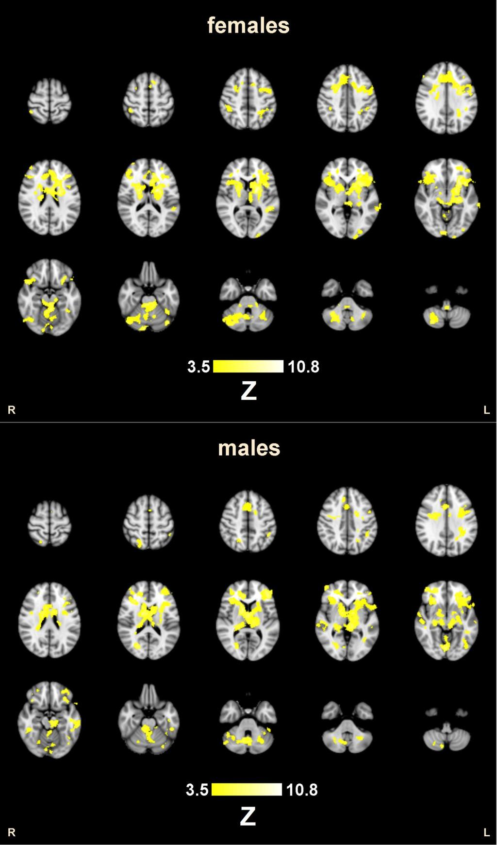
5 T/F) statistic images were thresholded using clusters determined by Z>3.5 and a (corrected) cluster significance threshold of P=0.05 (Worsley K.J., 2001). Performance of the task was good in all subjects, mean number of correct responses out of the 24 possible was 22.5±1.6 in males and 22.4±1.3 in females, this difference was not statistically significant (p = 0.88). Such a result on behavioral level is consistent with most previous studies on neuroimaging of verbal working memory (Bell et al., 2006; Goldstein et al., 2005; Haut et Barch, 2006; Schmidt et al., 2009), studies that have found differences in behavioral domain are not consistent, one reporting a better score for males (Zilles et al., 2016), another for females (Speck et al., 2000). The results of fmri analyses by gender subgroups revealed similar bilateral regions of activation in the frontal, parietal, temporal and occipital lobes, basal ganglia, the brainstem and in the cerebellum (Fig.1) which are areas usually activated during verbal working memory tasks (E. E. Smith et Jonides, 1998; Tüdös et al., 2014; Wager et Smith, 2003). Local maxima were localized to the left BA 9 and 46, in the inferior frontal gyrus (BA 44 and 47), in the left putamen and head of caudate bilaterally. Activations were also clearly visible in the left BA 44, BA13 and BA6. All of these areas were activated also in the right hemisphere, but left-sided dominance was obvious. Bilateral symmetrical activations were in the anterior cingulate cortex (BA 32) and the medial portion of the premotor area BA 6 (SMA). Parietal regions of activation involved bilateral intraparietal sulcus and superior parietal lobule (BA 7/40) with a predominance in the left hemisphere. The temporal lobes showed apparent activity in the posterior division of superior temporal gyrus (BA 21/22) and in both hippocampi. The occipital lobes were activated within Brodmann areas 18 and 19. Areas of activation in both cerebellar hemispheres and vermis were large and symmetric. Betweengender analysis revealed no statistically significant differences. 5
6 Traditionally, there are assumptions about differences between cognitive functions between males and females. Some previous neuroimaging studies report statistically significant differences between males and females during the verbal working memory task, however, these results are inconsistent there are reports of increased activation in occipital and parietal areas in 23 males compared to 10 females during the Sternberg item recognition task (Bell et al., 2006), increased bilateral activation in frontal areas in 7 females compared to 7 males during the auditory continuous performance test (similar to the 3-back task) (Goldstein et al., 2005), differences in the laterality index and overall increased activation in 9 males compared to 8 females during the 2-back task (Speck et al., 2000), and finally increased activation in the midbrain and right-sided Broca area in 139 females compared to 92 males (Zilles et al., 2016). This broad range of findings, therefore, does not indicate the existence of convincing gender differences; in general, it can also be concluded that in direct comparison the differences between the genders were significant in smaller or gender unbalanced study groups, and their power is therefore questionable. Published meta-analysis of neuroimaging studies of working memory between the genders also reports the differences; according to their results, females consistently activate more limbic and prefrontal structures and males activate a distributed network including more parietal regions (Hill et al., 2014). Among the drawbacks of the meta-analysis is unbalanced number of subjects (701 male subjects in 44 studies and 200 female subjects in 15 studies). The analysis also pooled results of working memory studies regardless of the nature of the material (e.g., verbal, spatial, etc.), modality of the stimuli (e.g., visual, auditory, cross-modal, other) and difficulty of tasks (e.g., 2-back, 3- back, task timing, etc.), which can lead to misleading results. Contrary to the above-mentioned inter-gender reports, in our group, there was no statistically significant difference in brain tissue activation between males and females. Schmidt et al (Schmidt et al., 2009) have also come to the same result using 1-, 2- and 3-back task in 25 6
7 males and 25 females. Similarly, another team failed to replicate the statistically significant differences in activation between the genders in two different study groups (the first with 49 subjects and the second with 61 subjects) using verbal 2-back task (Haut et Barch, 2006). Our negative result can be further supported by a study that did not show any significant difference in behavioral performance between men and women in both the verbal and spatial working memory task (Kaufman, 2007). In a recently published study, gender differences in behavioral data were observed only at very high loads of working memory tasks, i.e., with increasing number of objects to maintain (Reed et al., 2017). In this perspective, working memory load in our task with four objects to remember in each trial was relatively low, which should also be considered when interpreting our negative result. Our work has several potential limitations. The main limiting factor of our study is the low number of subjects which reduces the statistical power to detect significant differences. Nevertheless, even the above cited previous studies with larger sample sizes (Haut et Barch, 2006; Schmidt et al., 2009) either did not detect any reliable gender effects, or reported them only at an uncorrected statistical threshold (Zilles et al., 2016). Second, we acknowledge that the current standard for functional neuroimaging studies is the 3T MRI scanner, whereas the 1.5T field strength used in our study provides a lower spatial and temporal resolution and lower signal-to-noise ratio compared to 3T field. To compensate for this, we acquired two functional runs per subject. Finally, the recruited subjects had a narrow range of age and uniform education level, so our results cannot be generalized to the general population. In conclusion, our data obtained on a small sample of young healthy volunteers does not indicate a significantly different brain function in the verbal working memory task between the genders. Whereas the brain networks implementing other cognitive functions 7
8 may be different in males and females, verbal working memory seems to be localized similarly in both genders. Acknowledgements Supported by Palacky University grant IGA_LF_2018_002 and Ministry of Health, Czech Republic - conceptual development of research organization (LF UP ). References BELL, E. C., WILLSON, M. C., WILMAN, A. H., DAVE, S., SILVERSTONE, P. H Males and females differ in brain activation during cognitive tasks. NeuroImage, 30(2): GOLDSTEIN, J. M., POLDRACK, R., BREITER, H. C., MAKRIS, N., GOODMAN, J. M., JERRAM, M., ANAGNOSON, R., TSUANG, M. T., SEIDMAN, L. J Sex differences in prefrontal cortical brain activity during fmri of auditory verbal working memory. Neuropsychology, 19(4): HAUT, K. M., BARCH, D. M Sex influences on material-sensitive functional lateralization in working and episodic memory: Men and women are not all that different. NeuroImage, 32(1): HILL, A. C., LAIRD, A. R., ROBINSON, J. L Gender differences in working memory networks: A BrainMap meta-analysis. Biological Psychology, 102: KAUFMAN, S. B Sex differences in mental rotation and spatial visualization ability: Can they be accounted for by differences in working memory capacity? Intelligence, 35(3): REED, J. L., GALLAGHER, N. M., SULLIVAN, M., CALLICOTT, J. H., GREEN, A. E Sex differences in verbal working memory performance emerge at very high loads of common neuroimaging tasks. Brain and Cognition, 113:
9 SCHMIDT, H., JOGIA, J., FAST, K., CHRISTODOULOU, T., HALDANE, M., KUMARI, V., FRANGOU, S No gender differences in brain activation during the N-back task: an fmri study in healthy individuals. Human Brain Mapping, 30(11): SMITH, E. E., JONIDES, J Neuroimaging analyses of human working memory. Proceedings of the National Academy of Sciences, 95(20): SMITH, S. M., JENKINSON, M., WOOLRICH, M. W., BECKMANN, C. F., BEHRENS, T. E. J., JOHANSEN-BERG, H., BANNISTER, P. R., DE LUCA, M., DROBNJAK, I., FLITNEY, D. E., NIAZY, R. K., SAUNDERS, J., VICKERS, J., ZHANG, Y., DE STEFANO, N., BRADY, J. M., MATTHEWS, P. M Advances in functional and structural MR image analysis and implementation as FSL. NeuroImage, 23:S208 S219. SPECK, O., ERNST, T., BRAUN, J., KOCH, C., MILLER, E., CHANG, L Gender differences in the functional organization of the brain for working memory. NeuroReport, 11(11): TÜDÖS, Z., HLUŠTÍK, P., HOK, P Verbal working memory investigation using functional MRI. Ceska Radiologie, 65(1): TÜDÖS, Z., HOK, P., HRDINA, L., HLUŠTÍK, P Modality effects in paced serial addition task: Differential responses to auditory and visual stimuli. Neuroscience, 272: WAGER, T. D., SMITH, E. E Neuroimaging studies of working memory. Cognitive, Affective, & Behavioral Neuroscience, 3(4): WOOLRICH, M Robust group analysis using outlier inference. NeuroImage, 41(2): WORSLEY K.J Statistical analysis of activation images. In: Jezzard, P., Matthews P.M., Smith S.M. (Eds.). Functional MRI: An Introduction to Methods, Oxford University Press 9
10 ZILLES, D., LEWANDOWSKI, M., VIEKER, H., HENSELER, I., DIEKHOF, E., MELCHER, T., KEIL, M., GRUBER, O Gender Differences in Verbal and Visuospatial Working Memory Performance and Networks. Neuropsychobiology, 73(1): Figure 1 Group analysis for females (top part) and males (bottom part) reveals similar bilateral regions of activation in the frontal, parietal, temporal and occipital neural networks, basal ganglia, the brainstem and in the cerebellum. Between-gender analysis revealed no statistically significant differences. 10
11 11
12 12
Functional MRI Study of Gender Effects in Brain Activations During Verbal Working Memory Task
 Physiol. Res. 67: 825-829, 2018 SHORT COMMUNICATION Functional MRI Study of Gender Effects in Brain Activations During Verbal Working Memory Task Z. TÜDÖS 1, P. HOK 2, P. HLUŠTÍK 2, A. GRAMBAL 3 1 Department
Supplementary Information Methods Subjects The study was comprised of 84 chronic pain patients with either chronic back pain (CBP) or osteoarthritis
 or osteoarthritis") Supplementary Information Methods Subjects The study was comprised of 84 chronic pain patients with either chronic back pain (CBP) or osteoarthritis (OA). All subjects provided informed consent to procedures
Supplementary Information Methods Subjects The study was comprised of 84 chronic pain patients with either chronic back pain (CBP) or osteoarthritis (OA). All subjects provided informed consent to procedures
Procedia - Social and Behavioral Sciences 159 ( 2014 ) WCPCG 2014
 WCPCG 2014") Available online at www.sciencedirect.com ScienceDirect Procedia - Social and Behavioral Sciences 159 ( 2014 ) 743 748 WCPCG 2014 Differences in Visuospatial Cognition Performance and Regional Brain Activation
Available online at www.sciencedirect.com ScienceDirect Procedia - Social and Behavioral Sciences 159 ( 2014 ) 743 748 WCPCG 2014 Differences in Visuospatial Cognition Performance and Regional Brain Activation
Supplementary Online Content
 Supplementary Online Content Green SA, Hernandez L, Tottenham N, Krasileva K, Bookheimer SY, Dapretto M. The neurobiology of sensory overresponsivity in youth with autism spectrum disorders. Published
FMRI Data Analysis. Introduction. Pre-Processing
 FMRI Data Analysis Introduction The experiment used an event-related design to investigate auditory and visual processing of various types of emotional stimuli. During the presentation of each stimuli
Hallucinations and conscious access to visual inputs in Parkinson s disease
 Supplemental informations Hallucinations and conscious access to visual inputs in Parkinson s disease Stéphanie Lefebvre, PhD^1,2, Guillaume Baille, MD^4, Renaud Jardri MD, PhD 1,2 Lucie Plomhause, PhD
GENDER-SPECIFIC SENSITVITY TO TIME-DISCREPANT TASK CONDITIONS OF REASONING DURING fmri
 GENDER-SPECIFIC SENSITVITY TO TIME-DISCREPANT TASK CONDITIONS OF REASONING DURING fmri by Joshua M. Roberts A Thesis Submitted to the Graduate Faculty of George Mason University in Partial Fulfillment
Resistance to forgetting associated with hippocampus-mediated. reactivation during new learning
 Resistance to Forgetting 1 Resistance to forgetting associated with hippocampus-mediated reactivation during new learning Brice A. Kuhl, Arpeet T. Shah, Sarah DuBrow, & Anthony D. Wagner Resistance to
HST.583 Functional Magnetic Resonance Imaging: Data Acquisition and Analysis Fall 2008
 MIT OpenCourseWare http://ocw.mit.edu HST.583 Functional Magnetic Resonance Imaging: Data Acquisition and Analysis Fall 2008 For information about citing these materials or our Terms of Use, visit: http://ocw.mit.edu/terms.
Supporting Online Material for
 www.sciencemag.org/cgi/content/full/324/5927/646/dc1 Supporting Online Material for Self-Control in Decision-Making Involves Modulation of the vmpfc Valuation System Todd A. Hare,* Colin F. Camerer, Antonio
Supplementary Online Content
 Supplementary Online Content Gregg NM, Kim AE, Gurol ME, et al. Incidental cerebral microbleeds and cerebral blood flow in elderly individuals. JAMA Neurol. Published online July 13, 2015. doi:10.1001/jamaneurol.2015.1359.
WHAT DOES THE BRAIN TELL US ABOUT TRUST AND DISTRUST? EVIDENCE FROM A FUNCTIONAL NEUROIMAGING STUDY 1
 SPECIAL ISSUE WHAT DOES THE BRAIN TE US ABOUT AND DIS? EVIDENCE FROM A FUNCTIONAL NEUROIMAGING STUDY 1 By: Angelika Dimoka Fox School of Business Temple University 1801 Liacouras Walk Philadelphia, PA
Final Report 2017 Authors: Affiliations: Title of Project: Background:
 Final Report 2017 Authors: Dr Gershon Spitz, Ms Abbie Taing, Professor Jennie Ponsford, Dr Matthew Mundy, Affiliations: Epworth Research Foundation and Monash University Title of Project: The return of
Supporting online material. Materials and Methods. We scanned participants in two groups of 12 each. Group 1 was composed largely of
 Placebo effects in fmri Supporting online material 1 Supporting online material Materials and Methods Study 1 Procedure and behavioral data We scanned participants in two groups of 12 each. Group 1 was
Brain diffusion tensor imaging changes in cerebrotendinous xanthomatosis reversed with
 Brain diffusion tensor imaging changes in cerebrotendinous xanthomatosis reversed with treatment Claudia B. Catarino, MD, PhD, 1*, Christian Vollmar, MD, PhD, 2,3* Clemens Küpper, MD, 1,4 Klaus Seelos,
HST.583 Functional Magnetic Resonance Imaging: Data Acquisition and Analysis Fall 2006
 MIT OpenCourseWare http://ocw.mit.edu HST.583 Functional Magnetic Resonance Imaging: Data Acquisition and Analysis Fall 2006 For information about citing these materials or our Terms of Use, visit: http://ocw.mit.edu/terms.
Frontal Contributions to Memory Encoding Before and After Unilateral Medial Temporal Lobectomy
 Frontal Contributions to Memory Encoding Before and After Unilateral Medial Temporal Lobectomy Jeff Ojemann, MD Department of Neurological Surgery University of Washington Children s Hospital & Regional
Classification and Statistical Analysis of Auditory FMRI Data Using Linear Discriminative Analysis and Quadratic Discriminative Analysis
 International Journal of Innovative Research in Computer Science & Technology (IJIRCST) ISSN: 2347-5552, Volume-2, Issue-6, November-2014 Classification and Statistical Analysis of Auditory FMRI Data Using
Investigating directed influences between activated brain areas in a motor-response task using fmri
 Magnetic Resonance Imaging 24 (2006) 181 185 Investigating directed influences between activated brain areas in a motor-response task using fmri Birgit Abler a, 4, Alard Roebroeck b, Rainer Goebel b, Anett
Distinct Value Signals in Anterior and Posterior Ventromedial Prefrontal Cortex
 Supplementary Information Distinct Value Signals in Anterior and Posterior Ventromedial Prefrontal Cortex David V. Smith 1-3, Benjamin Y. Hayden 1,4, Trong-Kha Truong 2,5, Allen W. Song 2,5, Michael L.
Supporting Information
 Supporting Information Newman et al. 10.1073/pnas.1510527112 SI Results Behavioral Performance. Behavioral data and analyses are reported in the main article. Plots of the accuracy and reaction time data
Hippocampal brain-network coordination during volitionally controlled exploratory behavior enhances learning
 Online supplementary information for: Hippocampal brain-network coordination during volitionally controlled exploratory behavior enhances learning Joel L. Voss, Brian D. Gonsalves, Kara D. Federmeier,
Human Paleoneurology and the Evolution of the Parietal Cortex
 PARIETAL LOBE The Parietal Lobes develop at about the age of 5 years. They function to give the individual perspective and to help them understand space, touch, and volume. The location of the parietal
Supporting Online Material for
 www.sciencemag.org/cgi/content/full/315/5811/515/dc1 Supporting Online Material for The Neural Basis of Loss Aversion in Decision-Making Under Risk Sabrina M. Tom, Craig R. Fox, Christopher Trepel, Russell
A possible mechanism for impaired joint attention in autism
 A possible mechanism for impaired joint attention in autism Justin H G Williams Morven McWhirr Gordon D Waiter Cambridge Sept 10 th 2010 Joint attention in autism Declarative and receptive aspects initiating
Common Neural Substrates for Ordinal Representation in Short-Term Memory, Numerical and Alphabetical Cognition
 Common Neural Substrates for Ordinal Representation in Short-Term Memory, Numerical and Alphabetical Cognition Lucie Attout 1 *, Wim Fias 2, Eric Salmon 3, Steve Majerus 1 1 Department of Psychology -
Primacy and recency effects as indices of the focus of attention
 Primacy and recency effects as indices of the focus of attention Alexandra B. Morrison, Andrew R. A. Conway and Jason M. Chein Journal Name: Frontiers in Human Neuroscience ISSN: - Article type: Original
Supplementary Online Material Supplementary Table S1 to S5 Supplementary Figure S1 to S4
 Supplementary Online Material Supplementary Table S1 to S5 Supplementary Figure S1 to S4 Table S1: Brain regions involved in the adapted classification learning task Brain Regions x y z Z Anterior Cingulate
Voxel-based morphometry in clinical neurosciences
 Voxel-based morphometry in clinical neurosciences Ph.D. Thesis Ádám Feldmann Department of Behavioural Sciences Leader of Doctoral School: Prof. Dr.Sámuel Komoly, D.Sc. Program leader: Prof. Dr.Sámuel
Supplemental Information. Triangulating the Neural, Psychological, and Economic Bases of Guilt Aversion
 Neuron, Volume 70 Supplemental Information Triangulating the Neural, Psychological, and Economic Bases of Guilt Aversion Luke J. Chang, Alec Smith, Martin Dufwenberg, and Alan G. Sanfey Supplemental Information
CEREBRUM. Dr. Jamila EL Medany
 CEREBRUM Dr. Jamila EL Medany Objectives At the end of the lecture, the student should be able to: List the parts of the cerebral hemisphere (cortex, medulla, basal nuclei, lateral ventricle). Describe
NeuroImage 45 (2009) Contents lists available at ScienceDirect. NeuroImage. journal homepage:
 Contents lists available at ScienceDirect. NeuroImage. journal homepage:") NeuroImage 45 (2009) 614 626 Contents lists available at ScienceDirect NeuroImage journal homepage: www.elsevier.com/locate/ynimg Functional connectivity of the human amygdala using resting state fmri
NeuroImage 45 (2009) 614 626 Contents lists available at ScienceDirect NeuroImage journal homepage: www.elsevier.com/locate/ynimg Functional connectivity of the human amygdala using resting state fmri
Supporting Information
 Supporting Information Braver et al. 10.1073/pnas.0808187106 SI Methods Participants. Participants were neurologically normal, righthanded younger or older adults. The groups did not differ in gender breakdown
Twelve right-handed subjects between the ages of 22 and 30 were recruited from the
 Supplementary Methods Materials & Methods Subjects Twelve right-handed subjects between the ages of 22 and 30 were recruited from the Dartmouth community. All subjects were native speakers of English,
Define functional MRI. Briefly describe fmri image acquisition. Discuss relative functional neuroanatomy. Review clinical applications.
 Dr. Peter J. Fiester November 14, 2012 Define functional MRI. Briefly describe fmri image acquisition. Discuss relative functional neuroanatomy. Review clinical applications. Briefly discuss a few examples
Functional Elements and Networks in fmri
 Functional Elements and Networks in fmri Jarkko Ylipaavalniemi 1, Eerika Savia 1,2, Ricardo Vigário 1 and Samuel Kaski 1,2 1- Helsinki University of Technology - Adaptive Informatics Research Centre 2-
Investigations in Resting State Connectivity. Overview
 Investigations in Resting State Connectivity Scott FMRI Laboratory Overview Introduction Functional connectivity explorations Dynamic change (motor fatigue) Neurological change (Asperger s Disorder, depression)
Title: Resting hyperperfusion of the hippocampus, midbrain and striatum
 Data supplement for Allen et al., Resting Hyperperfusion of the Hippocampus, Midbrain, and Basal Ganglia in People at High Risk for Psychosis. Am J Psychiatry (doi: 10.1176/appi.ajp.2015.15040485) Supplementary
Functional MRI Mapping Cognition
 Outline Functional MRI Mapping Cognition Michael A. Yassa, B.A. Division of Psychiatric Neuro-imaging Psychiatry and Behavioral Sciences Johns Hopkins School of Medicine Why fmri? fmri - How it works Research
Group-Wise FMRI Activation Detection on Corresponding Cortical Landmarks
 Group-Wise FMRI Activation Detection on Corresponding Cortical Landmarks Jinglei Lv 1,2, Dajiang Zhu 2, Xintao Hu 1, Xin Zhang 1,2, Tuo Zhang 1,2, Junwei Han 1, Lei Guo 1,2, and Tianming Liu 2 1 School
Cerebral Cortex 1. Sarah Heilbronner
 Cerebral Cortex 1 Sarah Heilbronner heilb028@umn.edu Want to meet? Coffee hour 10-11am Tuesday 11/27 Surdyk s Overview and organization of the cerebral cortex What is the cerebral cortex? Where is each
Regional and Lobe Parcellation Rhesus Monkey Brain Atlas. Manual Tracing for Parcellation Template
 Regional and Lobe Parcellation Rhesus Monkey Brain Atlas Manual Tracing for Parcellation Template Overview of Tracing Guidelines A) Traces are performed in a systematic order they, allowing the more easily
Text to brain: predicting the spatial distribution of neuroimaging observations from text reports (submitted to MICCAI 2018)
") 1 / 22 Text to brain: predicting the spatial distribution of neuroimaging observations from text reports (submitted to MICCAI 2018) Jérôme Dockès, ussel Poldrack, Demian Wassermann, Fabian Suchanek, Bertrand
1 / 22 Text to brain: predicting the spatial distribution of neuroimaging observations from text reports (submitted to MICCAI 2018) Jérôme Dockès, ussel Poldrack, Demian Wassermann, Fabian Suchanek, Bertrand
Botulinum toxin treatment of freezing of gait in Parkinson s disease patients as reflected in functional magnetic resonance imaging of leg movement
 Botulinum toxin treatment of freezing of gait in Parkinson s disease patients as reflected in functional magnetic resonance imaging of leg movement Miroslav Vaštík 1, Pavel Hok 1, Petr Hluštík 1, Pavel
The neural correlates of conceptual and perceptual false recognition
 The neural correlates of conceptual and perceptual false recognition Rachel J. Garoff-Eaton, Elizabeth A. Kensinger and Daniel L. Schacter Learn. Mem. 2007 14: 684-692 Access the most recent version at
Working Memory: Critical Constructs and Some Current Issues. Outline. Starting Points. Starting Points
 Working Memory: Critical Constructs and Some Current Issues Edward E. Smith Columbia University Outline Background Maintenance: Modality specificity and buffers Interference resolution: Distraction and
Differential contributions of subregions of medial temporal lobe to memory system in. amnestic mild cognitive impairment: insights from fmri study
 Differential contributions of subregions of medial temporal lobe to memory system in amnestic mild cognitive impairment: insights from fmri study Jiu Chen 1, *, Xujun Duan 2, *, Hao Shu 1, Zan Wang 1,
Changes in Default Mode Network as Automaticity Develops in a Categorization Task
 Purdue University Purdue e-pubs Open Access Theses Theses and Dissertations January 2015 Changes in Default Mode Network as Automaticity Develops in a Categorization Task Farzin Shamloo Purdue University
FINAL PROGRESS REPORT
 (1) Foreword (optional) (2) Table of Contents (if report is more than 10 pages) (3) List of Appendixes, Illustrations and Tables (if applicable) (4) Statement of the problem studied FINAL PROGRESS REPORT
Announcement. Danny to schedule a time if you are interested.
 Announcement If you need more experiments to participate in, contact Danny Sanchez (dsanchez@ucsd.edu) make sure to tell him that you are from LIGN171, so he will let me know about your credit (1 point).
SUPPLEMENT: DYNAMIC FUNCTIONAL CONNECTIVITY IN DEPRESSION. Supplemental Information. Dynamic Resting-State Functional Connectivity in Major Depression
 Supplemental Information Dynamic Resting-State Functional Connectivity in Major Depression Roselinde H. Kaiser, Ph.D., Susan Whitfield-Gabrieli, Ph.D., Daniel G. Dillon, Ph.D., Franziska Goer, B.S., Miranda
Functional topography of a distributed neural system for spatial and nonspatial information maintenance in working memory
 Neuropsychologia 41 (2003) 341 356 Functional topography of a distributed neural system for spatial and nonspatial information maintenance in working memory Joseph B. Sala a,, Pia Rämä a,c,d, Susan M.
SUPPLEMENTARY MATERIAL. Table. Neuroimaging studies on the premonitory urge and sensory function in patients with Tourette syndrome.
 SUPPLEMENTARY MATERIAL Table. Neuroimaging studies on the premonitory urge and sensory function in patients with Tourette syndrome. Authors Year Patients Male gender (%) Mean age (range) Adults/ Children
CISC 3250 Systems Neuroscience
 CISC 3250 Systems Neuroscience Levels of organization Central Nervous System 1m 10 11 neurons Neural systems and neuroanatomy Systems 10cm Networks 1mm Neurons 100μm 10 8 neurons Professor Daniel Leeds
fmri and Voxel-based Morphometry in Detection of Early Stages of Alzheimer's Disease
 fmri and Voxel-based Morphometry in Detection of Early Stages of Alzheimer's Disease Andrey V. Sokolov 1,3, Sergey V. Vorobyev 2, Aleksandr Yu. Efimtcev 1,3, Viacheslav S. Dekan 1,3, Gennadiy E. Trufanov
CEREBRUM Dr. Jamila Elmedany Dr. Essam Eldin Salama
 CEREBRUM Dr. Jamila Elmedany Dr. Essam Eldin Salama Objectives At the end of the lecture, the student should be able to: List the parts of the cerebral hemisphere (cortex, medulla, basal nuclei, lateral
How to report my result using REST slice viewer?
 How to report my result using REST slice viewer? Han Zhang Center for Cognition and Brain Disorders, Hangzhou Normal University napoleon1982@gmail.com 2013/12/30 Commonly, you got an activation for functional
Supplementary information Detailed Materials and Methods
 Supplementary information Detailed Materials and Methods Subjects The experiment included twelve subjects: ten sighted subjects and two blind. Five of the ten sighted subjects were expert users of a visual-to-auditory
 Passport control a bit carried away. appreciated the advice forgot to talk to the manager, next thing I know my fmri thankfully, when aroused things back to normal Inattentive impaired children and adolescents:
Do women with fragile X syndrome have problems in switching attention: Preliminary findings from ERP and fmri
 Brain and Cognition 54 (2004) 235 239 www.elsevier.com/locate/b&c Do women with fragile X syndrome have problems in switching attention: Preliminary findings from ERP and fmri Kim Cornish, a,b, * Rachel
The Effects of Music intervention on Functional connectivity. Supplemental Information
 Yang et al. 0 The Effects of Music intervention on Functional connectivity strength of Brain in Schizophrenia Supplemental Information Mi Yang,#, Hui He #, Mingjun Duan,, Xi Chen, Xin Chang, Yongxiu Lai,
The functional neuroanatomy of human working memory revisited Evidence from 3-T fmri studies using classical domain-specific interference tasks
 NeuroImage 19 (2003) 797 809 www.elsevier.com/locate/ynimg The functional neuroanatomy of human working memory revisited Evidence from 3-T fmri studies using classical domain-specific interference tasks
Andy C.H. Lee a,b,, Trevor W. Robbins b, Stephen Smith c, Gemma A. Calvert c, Irene Tracey c, Paul Matthews c, Adrian M. Owen a. 1.
 Neuropsychologia 40 (2002) 2420 2437 Evidence for asymmetric frontal-lobe involvement in episodic memory from functional magnetic resonance imaging and patients with unilateral frontal-lobe excisions Andy
Supplementary materials. Appendix A;
 Supplementary materials Appendix A; To determine ADHD diagnoses, a combination of Conners' ADHD questionnaires and a semi-structured diagnostic interview was used(1-4). Each participant was assessed with
Overt Verbal Responding during fmri Scanning: Empirical Investigations of Problems and Potential Solutions
 NeuroImage 10, 642 657 (1999) Article ID nimg.1999.0500, available online at http://www.idealibrary.com on Overt Verbal Responding during fmri Scanning: Empirical Investigations of Problems and Potential
Supplementary Online Content
 Supplementary Online Content Van Laere K, Vanhee A, Verschueren J, et al. Value of 18 fluorodeoxyglucose positron-emission tomography in amyotrophic lateral sclerosis. JAMA Neurol. Published online March
How do individuals with congenital blindness form a conscious representation of a world they have never seen? brain. deprived of sight?
 How do individuals with congenital blindness form a conscious representation of a world they have never seen? What happens to visual-devoted brain structure in individuals who are born deprived of sight?
AN fmri EXAMINATION OF VISUAL INTEGRATION IN SCHIZOPHRENIA
 Journal of Integrative Neuroscience, Vol. 8, No. 2 (2009) 175 202 c Imperial College Press Research Report AN fmri EXAMINATION OF VISUAL INTEGRATION IN SCHIZOPHRENIA STEVEN M. SILVERSTEIN,,, SARAH BERTEN,,
Sex influences on material-sensitive functional lateralization in working and episodic memory: Men and women are not all that different
 www.elsevier.com/locate/ynimg NeuroImage 32 (2006) 411 422 Sex influences on material-sensitive functional lateralization in working and episodic memory: Men and women are not all that different Kristen
Face-specific resting functional connectivity between the fusiform gyrus and posterior superior temporal sulcus
 HUMAN NEUROSCIENCE Original Research Article published: 24 September 2010 doi: 10.3389/fnhum.2010.00176 Face-specific resting functional connectivity between the fusiform gyrus and posterior superior temporal
HST.583 Functional Magnetic Resonance Imaging: Data Acquisition and Analysis Fall 2008
 MIT OpenCourseWare http://ocw.mit.edu HST.583 Functional Magnetic Resonance Imaging: Data Acquisition and Analysis Fall 2008 For information about citing these materials or our Terms of Use, visit: http://ocw.mit.edu/terms.
Confidence-based Ensemble for GBM brain tumor segmentation
 Confidence-based Ensemble for GBM brain tumor segmentation Jing Huo 1, Eva M. van Rikxoort 1, Kazunori Okada 2, Hyun J. Kim 1, Whitney Pope 1, Jonathan Goldin 1, Matthew Brown 1 1 Center for Computer vision
Effect Assessment of Parkinson Disease on Default Mode Network of the Brain with ICA and SCA Methods in Resting State FMRI Data
 American Journal of Biomedical Engineering 2012, 2(2): 7-12 DOI: 10.5923/j.ajbe.20120202.02 Effect Assessment of Parkinson Disease on Default Mode Network of the Brain with ICA and SCA Methods in Resting
Involvement of both prefrontal and inferior parietal cortex. in dual-task performance
 Involvement of both prefrontal and inferior parietal cortex in dual-task performance Fabienne Collette a,b, Laurence 01ivier b,c, Martial Van der Linden a,d, Steven Laureys b, Guy Delfiore b, André Luxen
Table 1. Summary of PET and fmri Methods. What is imaged PET fmri BOLD (T2*) Regional brain activation. Blood flow ( 15 O) Arterial spin tagging (AST)
 Regional brain activation. Blood flow ( 15 O) Arterial spin tagging (AST)") Table 1 Summary of PET and fmri Methods What is imaged PET fmri Brain structure Regional brain activation Anatomical connectivity Receptor binding and regional chemical distribution Blood flow ( 15 O)
Table 1 Summary of PET and fmri Methods What is imaged PET fmri Brain structure Regional brain activation Anatomical connectivity Receptor binding and regional chemical distribution Blood flow ( 15 O)
Reproducibility is necessary but insufficient for addressing brain science issues.
 1 Reproducibility is necessary but insufficient for addressing brain science issues. GLM promises nothing but the higher amplitude of the experimental trials on the average compared with the control trials.
Supplemental Data. Inclusion/exclusion criteria for major depressive disorder group and healthy control group
 1 Supplemental Data Inclusion/exclusion criteria for major depressive disorder group and healthy control group Additional inclusion criteria for the major depressive disorder group were: age of onset of
independent of comorbid disorders and exposure to maltreatment
 Supplementary material for: Conduct disorder in females is associated with reduced corpus callosum structural integrity independent of comorbid disorders and exposure to maltreatment Philip Lindner 1,2
Supplementary Material. Functional connectivity in multiple cortical networks is associated with performance. across cognitive domains in older adults
 Supplementary Material Functional connectivity in multiple cortical networks is associated with performance across cognitive domains in older adults Emily E. Shaw 1,2, Aaron P. Schultz 1,2,3, Reisa A.
Topographical functional connectivity patterns exist in the congenitally, prelingually deaf
 Supplementary Material Topographical functional connectivity patterns exist in the congenitally, prelingually deaf Ella Striem-Amit 1*, Jorge Almeida 2,3, Mario Belledonne 1, Quanjing Chen 4, Yuxing Fang
Anatomy and Physiology (Bio 220) The Brain Chapter 14 and select portions of Chapter 16
 The Brain Chapter 14 and select portions of Chapter 16") Anatomy and Physiology (Bio 220) The Brain Chapter 14 and select portions of Chapter 16 I. Introduction A. Appearance 1. physical 2. weight 3. relative weight B. Major parts of the brain 1. cerebrum 2.
Anatomy and Physiology (Bio 220) The Brain Chapter 14 and select portions of Chapter 16 I. Introduction A. Appearance 1. physical 2. weight 3. relative weight B. Major parts of the brain 1. cerebrum 2.
Reasoning and working memory: common and distinct neuronal processes
 Neuropsychologia 41 (2003) 1241 1253 Reasoning and working memory: common and distinct neuronal processes Christian C. Ruff a,b,, Markus Knauff a,c, Thomas Fangmeier a, Joachim Spreer d a Centre for Cognitive
QUANTIFYING CEREBRAL CONTRIBUTIONS TO PAIN 1
 QUANTIFYING CEREBRAL CONTRIBUTIONS TO PAIN 1 Supplementary Figure 1. Overview of the SIIPS1 development. The development of the SIIPS1 consisted of individual- and group-level analysis steps. 1) Individual-person
Funding: NIDCF UL1 DE019583, NIA RL1 AG032119, NINDS RL1 NS062412, NIDA TL1 DA
 The Effect of Cognitive Functioning, Age, and Molecular Variables on Brain Structure Among Carriers of the Fragile X Premutation: Deformation Based Morphometry Study Naomi J. Goodrich-Hunsaker*, Ling M.
Prefrontal cortex and recognition memory Functional-MRI evidence for context-dependent retrieval processes
 Brain (1998), 121, 1985 2002 Prefrontal cortex and recognition memory Functional-MRI evidence for context-dependent retrieval processes Anthony D. Wagner, 1 John E. Desmond, 1,2 Gary H. Glover 2 and John
Involvement of human left dorsolateral prefrontal cortex in perceptual decision making is independent of response modality
 Involvement of human left dorsolateral prefrontal cortex in perceptual decision making is independent of response modality H. R. Heekeren*, S. Marrett, D. A. Ruff*, P. A. Bandettini*, and L. G. Ungerleider*
ORIGINAL ARTICLE. Selective Abnormal Modulation of Hippocampal Activity During Memory Formation in First-Episode Psychosis
 ORIGINAL ARTICLE Selective Abnormal Modulation of Hippocampal Activity During Memory Formation in First-Episode Psychosis Amélie M. Achim, PhD; Marie-Claude Bertrand, PhD; Hazel Sutton, BSc; Alonso Montoya,
The neural correlates of visuo-spatial working memory in children with autism spectrum disorder: effects of cognitive load
 Vogan et al. Journal of Neurodevelopmental Disorders 2014, 6:19 RESEARCH Open Access The neural correlates of visuo-spatial working memory in children with autism spectrum disorder: effects of cognitive
Training-Induced Functional Activation Changes in Dual-Task Processing: An fmri Study
 Cerebral Cortex January 2007;17:192-204 doi:10.1093/cercor/bhj137 Advance Access publication February 8, 2006 Training-Induced Functional Activation Changes in Dual-Task Processing: An fmri Study Kirk
Functional Connectivity Measures in Memory Networks Using Independent Component Analysis
 Functional Connectivity Measures in Memory Networks Using Independent Component Analysis Catarina Saiote Ferreira Leite Abstract Memory function often appears to be compromised in several neurological
Cerebrum-Cerebral Hemispheres. Cuneyt Mirzanli Istanbul Gelisim University
 Cerebrum-Cerebral Hemispheres Cuneyt Mirzanli Istanbul Gelisim University The largest part of the brain. Ovoid shape. Two incompletely separated cerebral hemispheres. The outer surface of the cerebral
Title:Atypical language organization in temporal lobe epilepsy revealed by a passive semantic paradigm
 Author's response to reviews Title:Atypical language organization in temporal lobe epilepsy revealed by a passive semantic paradigm Authors: Julia Miro (juliamirollado@gmail.com) Pablo Ripollès (pablo.ripolles.vidal@gmail.com)
Anatomy Lab (1) Theoretical Part. Page (2 A) Page (2B)
 Theoretical Part. Page (2 A) Page (2B)") Anatomy Lab (1) This sheet only includes the extra notes for the lab handout regarding the theoretical part, as for the practical part it includes everything the doctor mentioned. Theoretical Part Page
Anatomy Lab (1) This sheet only includes the extra notes for the lab handout regarding the theoretical part, as for the practical part it includes everything the doctor mentioned. Theoretical Part Page
Chapter 3. Structure and Function of the Nervous System. Copyright (c) Allyn and Bacon 2004
 Allyn and Bacon 2004") Chapter 3 Structure and Function of the Nervous System 1 Basic Features of the Nervous System Neuraxis: An imaginary line drawn through the center of the length of the central nervous system, from the
Chapter 3 Structure and Function of the Nervous System 1 Basic Features of the Nervous System Neuraxis: An imaginary line drawn through the center of the length of the central nervous system, from the
Department of Cognitive Science UCSD
 Department of Cognitive Science UCSD Verse 1: Neocortex, frontal lobe, Brain stem, brain stem, Hippocampus, neural node, Right hemisphere, Pons and cortex visual, Brain stem, brain stem, Sylvian fissure,
Cognitive Neuroscience of Memory
 Cognitive Neuroscience of Memory Types and Structure of Memory Types of Memory Type of Memory Time Course Capacity Conscious Awareness Mechanism of Loss Sensory Short-Term and Working Long-Term Nondeclarative
Supplemental information online for
 Supplemental information online for Sleep contributes to the strengthening of some memories over others, depending on hippocampal activity at learning. Géraldine Rauchs (1,2), Dorothée Feyers (1), Brigitte
Theory of mind skills are related to gray matter volume in the ventromedial prefrontal cortex in schizophrenia
 Theory of mind skills are related to gray matter volume in the ventromedial prefrontal cortex in schizophrenia Supplemental Information Table of Contents 2 Behavioral Data 2 Table S1. Participant demographics
Behavioural Brain Research
 Behavioural Brain Research 197 (2009) 186 197 Contents lists available at ScienceDirect Behavioural Brain Research j o u r n a l h o m e p a g e : www.elsevier.com/locate/bbr Research report Top-down attentional
Auditory fmri correlates of loudness perception for monaural and diotic stimulation
 PROCEEDINGS of the 22 nd International Congress on Acoustics Psychological and Physiological Acoustics (others): Paper ICA2016-435 Auditory fmri correlates of loudness perception for monaural and diotic