Cerebrum-Cerebral Hemispheres. Cuneyt Mirzanli Istanbul Gelisim University
|
|
|
- Amberlynn Hortense Anderson
- 5 years ago
- Views:
Transcription
1 Cerebrum-Cerebral Hemispheres Cuneyt Mirzanli Istanbul Gelisim University
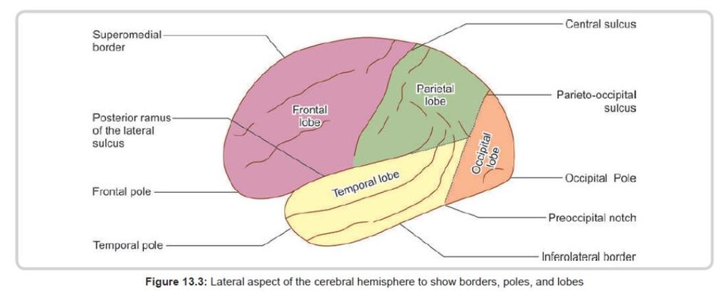
2 The largest part of the brain. Ovoid shape. Two incompletely separated cerebral hemispheres.

3 The outer surface of the cerebral hemisphere is covered with cortex, which is highly folded due to the presence of sulci and gyri.
4 The core of the hemisphere consists of white matter containing a group of nuclei called basal ganglia. The cavity inside each hemisphere is called the lateral ventricle.


5 The median longitudinal fissure of cerebrum intervenes between the medial surfaces of the right and left hemispheres. At the bottom of the fissure lies the corpus callosum, which is a connecting bond between the two hemispheres.
6 The surface of the cerebral hemisphere is covered by a thin layer of grey matter called the cerebral cortex. The cortex follows the irregular contour of the sulci and gyri of the hemisphere and extends into the depths of the sulci.

7 As a result of this folding of the cerebral surface, the cerebral cortex acquires a much larger surface area than the size of the hemispheres would otherwise allow.
8 The greater part of the cerebral hemisphere deep to the cortex is occupied by white matter, within which are embedded certain important masses of grey matter.
9 Borders A coronal section through the cerebral hemispheres shows that each hemisphere has three borders: superomedial, inferolateral, and inferomedial.
10

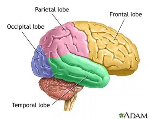
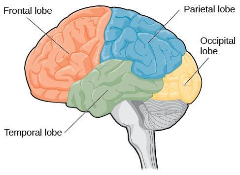
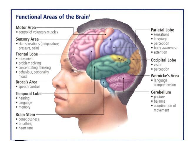
11 Lobes There are four lobes, namely frontal, parietal, occipital, and temporal, which are well demarcated on the superolateral surface.
12
13 Frontal Lobe Located at the front of both cerebral hemispheres Primary motor cortex Pre-motor cortex Broca s Area- motor production of speech Complex Functioning- personality, judgement, insight, reasoning, problem solving, abstract thinking and working memory.
14
15
16 Parietal lobe Located behind frontal lobe. Somatosensory cortex. Spatial orientation, perception and comprehension of language function recognising object by touch. Links visual and somatosensory information together.
17
18
19 Temporal Lobes Located at each side of the brain Involved in receiving and processing auditory information, higher order visual information, complex aspects of memory and language Wernicke s Area- comprehension of speech.
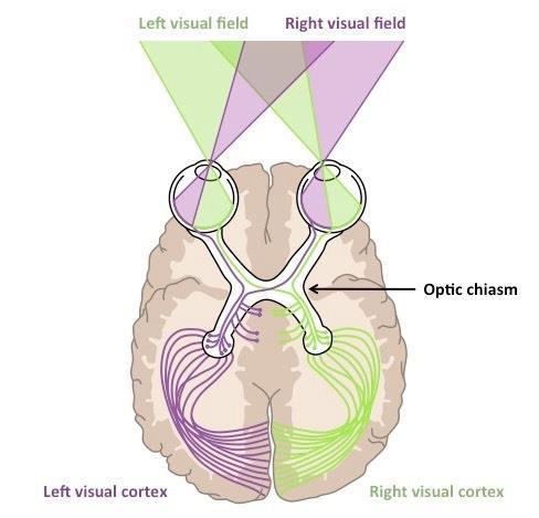
20
21
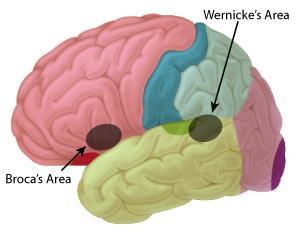
22
23 Speech Werniche s area: concerned with understanding language, left cerebral hemisphere, temporal lobe. Broca s area: concerned with controlling the muscles that produce speech, left cerebrale hemisphere, frontal lobe.
24
25 Occipital lobes Rearmost portion of the brain Visual processing area
26
27 Visual Processing The visual sensory area is at the back of the brain in occipital lobes. Receives sensory input from the optic nerves The 2 hemispheres see slightly different images from the opposite of the visual field, these differences can be used to judge distance
28
29

30 Structure of Cerebral Cortex The cerebral cortex contains the cell bodies of an innumerable number of neurons along with their processes, neuroglia and blood vessels.
31 Pyramidal Cells The most abundant type of cortical neurons. All other neurons in the cortex are referred to as nonpyramidal neurons. Their cell bodies are triangular, The axon arises from the base of the pyramid.
32 The axon of a pyramidal cell may terminate in one of the following ways: It may travel to other regions like the basal ganglia, the brainstem or the spinal cord. It may cross to the opposite side and reach the corresponding region of the opposite hemisphere.
33 It may enter the white matter to travel to another part of the cortex. It may be short and may terminate within the same area of the cortex. The neurotransmitter used by pyramidal cells is either glutamate or aspartate.
34 Stellate Cells The stellate neurons are relatively small and multipolar. They form about one-third of the total neuronal population of the cortex.
35 Fusiform cells The fusiform cells have their long axis vertical to the surface and are concentrated mainly in the deepest cortical layers.
36 Horizontal Cells The horizontal cells are small, fusiform, horizontally oriented cells found in the most superficial layers of the cortex.
37 Martinotti Cells The cells of Martinotti are small,multipolar cells that are present throughout the levels of the cortex.
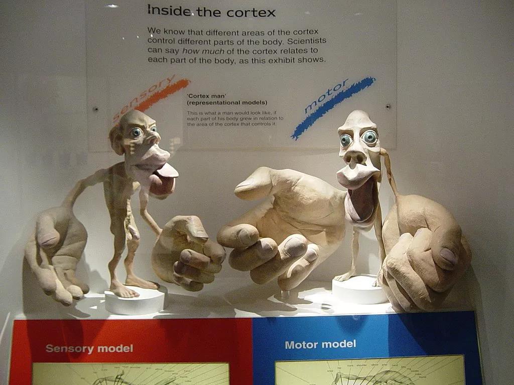
38
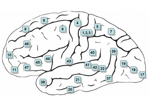
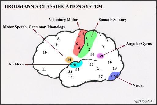
39 Functional Areas of Cerebral Cortex Some areas of the cerebral cortex can be assigned specific functions. These areas can be defined in terms of known sulci and gyri. However, some areas are commonly referred to by numbers. The best known scheme is that of Brodmann who represented different areas by numbers.
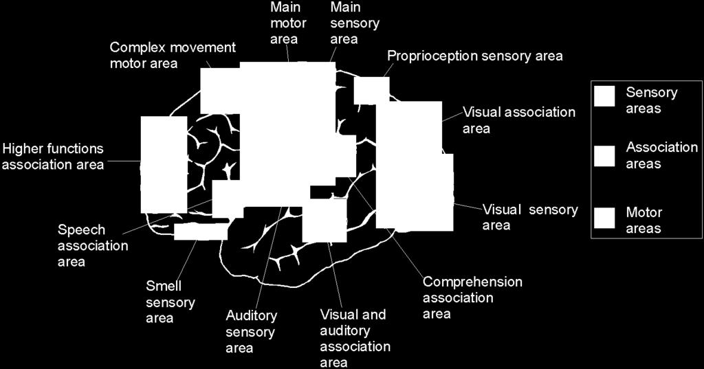
40 The human brain is the site of the major coordination in the nervous system. There are three areas for coordination: Sensory areas Motor areas Association areas
41 Sensory Areas Sensory areas of the cerebral hemispheres receive impulses from sense organs, and transmit them association areas.
42 Motor Areas The motor areas transmit impulses to the effectors. The size of the motor and sensory areas is related to the numbers of receptors in that area.
43 Association Areas The association areas of the cerebral hemispheres receive impulses, interpret them in the light of past experiences, and transmit impulses to the effectors. used to compare sensory input with previous experiences and make decisions
44 These areas are involved in speech, understanding and memory retrieval. The frontal lobes are large in humans and it is thought that they responsible for higher functions like thought, personality & emotion.
45 Sensory & Motor Maps The maps show that regions of the body with many sensory (or motor) neurones have corresponding large areas of the cerebrum linked to them. for example the lips occupy a larger region of the sensory cortex than the shoulder, because there are more sensory neurones in the lips.
46 s ho u ld er s ho u ld er Mapping of the sensory & motor areas to the body toes genitalia legs t ru n k h ip fot Sensory Cortex e lb ow fo r wr i e ar m s t h an d fin ge rs t hum b eye n ose face lip s gum s jaw tr un k h ip legs ankle toes M otor Cortex el bo w w ris t h a n d f ing ers th um b n ec k e ye fac e li ps j aw p haryn x to n gu e s w al owin g a bdo m en c hewing ton gu e
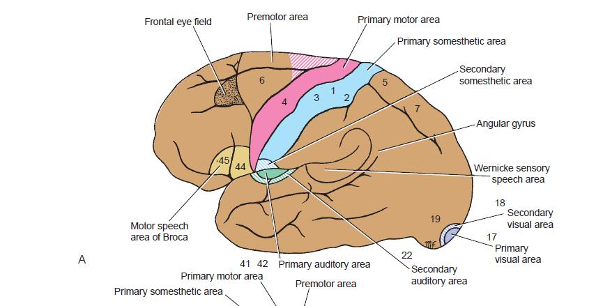
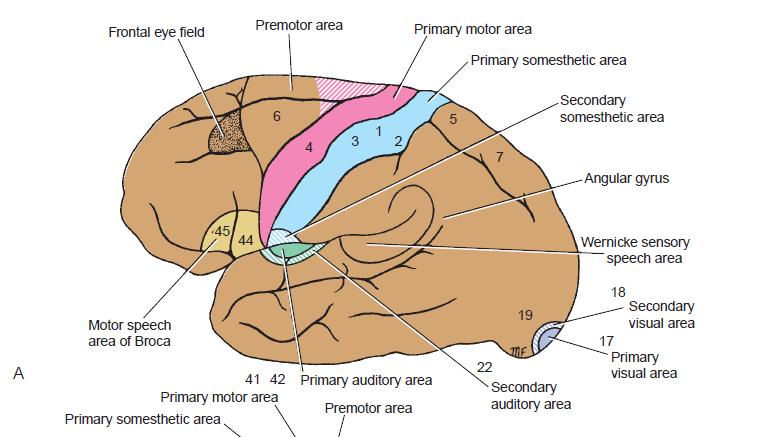
47 CORTICAL AREAS Different areas of the cerebral cortex are functionally specialized. The precise division of the cortex into different areas of specialization is simplified by Brodmann.

48 Korbinian Brodmann
49 Brodmann's areas of the cerebral cortex have unique architectural characteristics in terms of the thickness and layering of the cerebral cortex. His numbering of cortical areas is still used as a shorthand for describing the functional regions of the cortex, particularly those related to sensory functions.
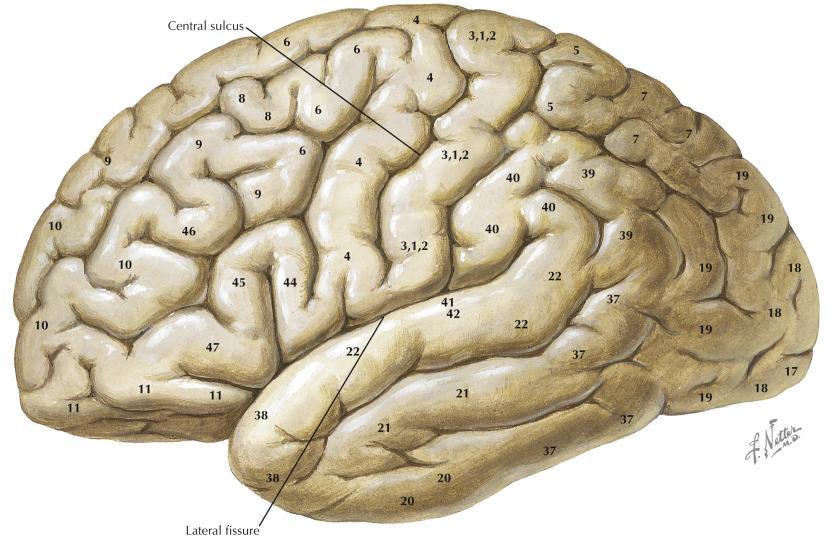
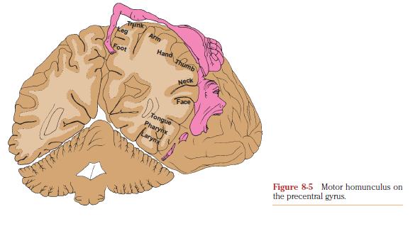
50
51
52
53
54 Frontal Lobe: The precentral area may be divided into posterior and anterior regions. The posterior region which is referred to as the motor area, primary motor area or Brodmann area 4, occupies the precentral gyrus extending over the superior border into the paracentral lobule.
55 The anterior region is known as the premotor area, secondary motor area or Brodmann area 6 and parts of areas 8, 44, and 45. It occupies the anterior part of the precentral gyrus.
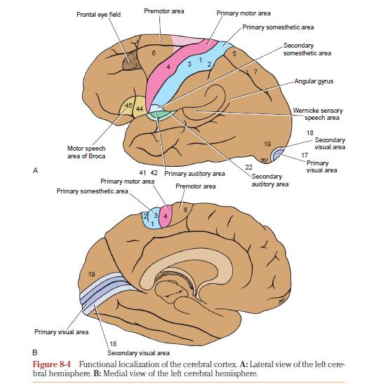
56
57 The primary motor area, if electrically stimulated, produces isolated movements on the opposite side of the body as well as contraction of muscle groups concerned with the performance of a specific movement.
58 The movement areas of the body are represented in inverted form in the precentral gyrus. This is called as motor homunculus.
59 Motor Homunculus Distorted human form, of the somatotopic organization of the motor cortex as originally mapped by U.S. born Canadian neurosurgeon Wilder Penfield ( ). Within this mapping, the size of the brain region associated with a body part reflects the complexity of the activities carried out with that part of the body rather than its actual size.
60
61
62
63 The prefrontal area is concerned with the makeup of the individual s personality. As the result of the input from many cortical and subcortical sources, this area plays a role as a regulator of the person s depth of feeling. It also exerts its influence in determining the initiative and judgment of an individual.
64 Parietal Lobe The primary somesthetic area (primary somatic sensory cortex S1) occupies the postcentral gyrus.
65
 surrounds the primary visual area on the medial")
66 Occipital Lobe Consists the primary visual area (Brodmann area 17). The secondary visual area (Brodmann areas 18 and 19) surrounds the primary visual area on the medial and lateral surfaces of the hemisphere.
67 Temporal Lobe The primary auditory area (Brodmann areas 41 and 42) includes the gyrus of Heschl and is situated in the inferior wall of the lateral sulcus. The secondary auditory area (auditory association cortex) is situated posterior to the primary auditory area in the lateral sulcus and in the superior temporal gyrus (Brodmann area 22).
68
69 CEREBRAL DOMINANCE An anatomical examination of the two cerebral hemispheres shows that the cortical gyri and fissures are almost identical. Nevertheless, certain nervous activity is predominantly performed by one of the two cerebral hemispheres.
70 Handedness, perception of language and speech are functional areas of behavior that in most individuals are controlled by the dominant hemisphere. By contrast, recognition of faces and music are interpreted by the nondominant hemisphere.
71 More than 90% of the adult population is right-handed and, Therefore left hemisphere dominant. About 96% of the adult population is left hemisphere dominant for speech.
CEREBRUM. Dr. Jamila EL Medany
 CEREBRUM Dr. Jamila EL Medany Objectives At the end of the lecture, the student should be able to: List the parts of the cerebral hemisphere (cortex, medulla, basal nuclei, lateral ventricle). Describe
Homework Week 2. PreLab 2 HW #2 Synapses (Page 1 in the HW Section)
") Homework Week 2 Due in Lab PreLab 2 HW #2 Synapses (Page 1 in the HW Section) Reminders No class next Monday Quiz 1 is @ 5:30pm on Tuesday, 1/22/13 Study guide posted under Study Aids section of website
Homework Week 2 Due in Lab PreLab 2 HW #2 Synapses (Page 1 in the HW Section) Reminders No class next Monday Quiz 1 is @ 5:30pm on Tuesday, 1/22/13 Study guide posted under Study Aids section of website
Announcement. Danny to schedule a time if you are interested.
 Announcement If you need more experiments to participate in, contact Danny Sanchez (dsanchez@ucsd.edu) make sure to tell him that you are from LIGN171, so he will let me know about your credit (1 point).
CEREBRUM Dr. Jamila Elmedany Dr. Essam Eldin Salama
 CEREBRUM Dr. Jamila Elmedany Dr. Essam Eldin Salama Objectives At the end of the lecture, the student should be able to: List the parts of the cerebral hemisphere (cortex, medulla, basal nuclei, lateral
Exam 1 PSYC Fall 1998
 Exam 1 PSYC 2022 Fall 1998 (2 points) Briefly describe the difference between a dualistic and a materialistic explanation of brain-mind relationships. (1 point) True or False. George Berkely was a monist.
P. Hitchcock, Ph.D. Department of Cell and Developmental Biology Kellogg Eye Center. Wednesday, 16 March 2009, 1:00p.m. 2:00p.m.
 Normal CNS, Special Senses, Head and Neck TOPIC: CEREBRAL HEMISPHERES FACULTY: LECTURE: READING: P. Hitchcock, Ph.D. Department of Cell and Developmental Biology Kellogg Eye Center Wednesday, 16 March
Essentials of Human Anatomy & Physiology. Seventh Edition. The Nervous System. Copyright 2003 Pearson Education, Inc. publishing as Benjamin Cummings
 Essentials of Human Anatomy & Physiology Seventh Edition The Nervous System Copyright 2003 Pearson Education, Inc. publishing as Benjamin Cummings Functions of the Nervous System 1. Sensory input gathering
Neuroanatomy lecture (1)
") Neuroanatomy lecture (1) Introduction: Neuroanatomy has two parts: the central and peripheral nervous system. The central nervous system is composed of brain and spinal cord. The brain has the following
Neuroanatomy lecture (1) Introduction: Neuroanatomy has two parts: the central and peripheral nervous system. The central nervous system is composed of brain and spinal cord. The brain has the following
CEREBRUM & CEREBRAL CORTEX
 CEREBRUM & CEREBRAL CORTEX Seonghan Kim Dept. of Anatomy Inje University, College of Medicine THE BRAIN ANATOMICAL REGIONS A. Cerebrum B. Diencephalon Thalamus Hypothalamus C. Brain Stem Midbrain Pons
CISC 3250 Systems Neuroscience
 CISC 3250 Systems Neuroscience Levels of organization Central Nervous System 1m 10 11 neurons Neural systems and neuroanatomy Systems 10cm Networks 1mm Neurons 100μm 10 8 neurons Professor Daniel Leeds
FRONTAL LOBE. Central Sulcus. Ascending ramus of the Cingulate Sulcus. Cingulate Sulcus. Lateral Sulcus
 FRONTAL LOBE Central Ascending ramus of the Cingulate Cingulate Lateral Lateral View Medial View Motor execution and higher cognitive functions (e.g., language production, impulse inhibition, reasoning
Neurology study of the nervous system. nervous & endocrine systems work together to maintain homeostasis
 Nervous System Neurology study of the nervous system nervous & endocrine systems work together to maintain homeostasis Nervous System works very fast Uses electrical signals called nerve impulses Short-lived
-Zeina Assaf. -Omar Odeh. - Maha Beltagy
 -3 -Zeina Assaf -Omar Odeh - Maha Beltagy 1 P a g e The Inferior Surface Of The Brain The inferior surface of the brain is divide by the stem of the lateral fissure into 2 parts : The orbital surface and
LEC 1B ANATOMY OF THE NERVOUS SYSTEM. Cogs 17 * UCSD
 LEC 1B ANATOMY OF THE NERVOUS SYSTEM Cogs 17 * UCSD Cerebral Cortex A 6-layer sheet of cells, unfolded = < 1 m square X 3 mm thick Cortex 6 layers of cells Nissl Stain for Cell Bodies Info projected to
The human brain. of cognition need to make sense gives the structure of the brain (duh). ! What is the basic physiology of this organ?
. ! What is the basic physiology of this organ?") The human brain The human brain! What is the basic physiology of this organ?! Understanding the parts of this organ provides a hypothesis space for its function perhaps different parts perform different
The human brain The human brain! What is the basic physiology of this organ?! Understanding the parts of this organ provides a hypothesis space for its function perhaps different parts perform different
Nervous System: Part IV The Central Nervous System The Brain
 Nervous System: Part IV The Central Nervous System The Brain Can you survive when part of your brain is destroyed? 2 Essential Knowledge 3.D.2 2. Cells communicate with each other through direct contact
Anatomy and Physiology (Bio 220) The Brain Chapter 14 and select portions of Chapter 16
 The Brain Chapter 14 and select portions of Chapter 16") Anatomy and Physiology (Bio 220) The Brain Chapter 14 and select portions of Chapter 16 I. Introduction A. Appearance 1. physical 2. weight 3. relative weight B. Major parts of the brain 1. cerebrum 2.
Anatomy and Physiology (Bio 220) The Brain Chapter 14 and select portions of Chapter 16 I. Introduction A. Appearance 1. physical 2. weight 3. relative weight B. Major parts of the brain 1. cerebrum 2.
1. Processes nutrients and provides energy for the neuron to function; contains the cell's nucleus; also called the soma.
 1. Base of brainstem; controls heartbeat and breathing 2. tissue destruction; a brain lesion is a naturally or experimentally caused destruction of brain tissue 3. A thick band of axons that connects the
Anatomy & Physiology Central Nervous System Worksheet
 1. What are the two parts of the CNS? 2. What are the four functions of the CNS Anatomy & Physiology Central Nervous System Worksheet 3. What are the four functions of the meninges? (p430) 4. Starting
The Central Nervous System I. Chapter 12
 The Central Nervous System I Chapter 12 The Central Nervous System The Brain and Spinal Cord Contained within the Axial Skeleton Brain Regions and Organization Medical Scheme (4 regions) 1. Cerebral Hemispheres
Dissection of the Sheep Brain
 Dissection of the Sheep Brain Laboratory Objectives After completing this lab, you should be able to: 1. Identify the main structures in the sheep brain and to compare them with those of the human brain.
meninges Outermost layer of the meninge dura mater arachnoid mater pia mater membranes located between bone and soft tissue of the nervous system
 membranes located between bone and soft tissue of the nervous system meninges Outermost layer of the meninge dura mater middle layer of the meninges, contains no blood vessels arachnoid mater Innermost
Neocortex. Hemispheres 9/22/2010. Psychology 472 Pharmacology of Psychoactive Drugs. Structures are divided into several section or lobes.
 Neocortex Psychology 472 Pharmacology of Psychoactive Drugs 1 Is the most developed in Humans Has many folds and fissures The folds of tissue are called gyri or a gyrus (single) The fissures or valleys
3/20/13. :: Slide 1 :: :: Slide 39 :: How Is the Nervous System Organized? Central Nervous System Peripheral Nervous System and Endocrine System
 :: Slide 1 :: :: Slide 39 :: How Is the Nervous System Organized? Central Nervous System Peripheral Nervous System and Endocrine System The nervous system is organized into several major branches, each
Gives few collaterals, it is mainly a single process surrounded by a myelin sheath
 Lecture 1 - Nerve fiber refers to both axons and dendrites, the dendrites are the afferent fibers (sensory); they receive impulses from neighbouring neurons, and the axon is the efferent fiber (motor);
Chapter 14, Part 2! Chapter 14 Part 2 Brain/Cranial Nerves! The Cerebrum and Cranial Nerves! pp !
 Chapter 14, Part 2! The Cerebrum and Cranial pp. 482 505! SECTION 14-9! The cerebrum, the largest region of the brain, contains motor, sensory, and association areas! 2! White Matter of the Cerebrum! 1.
Motor Functions of Cerebral Cortex
 Motor Functions of Cerebral Cortex I: To list the functions of different cortical laminae II: To describe the four motor areas of the cerebral cortex. III: To discuss the functions and dysfunctions of
Chapter 14, Part 2! The Cerebrum and Cranial Nerves! pp !
 Chapter 14, Part 2! The Cerebrum and Cranial pp. 482 505! SECTION 14-9! The cerebrum, the largest region of the brain, contains motor, sensory, and association areas! 2! 1! ! Chapter 14 Part 2 Brain/Cranial
Biology 3201 Nervous System #2- Anatomy. Components of a Nervous System
 Biology 3201 Nervous System #2- Anatomy Components of a Nervous System In any nervous system, there are 4 main components: (1) sensors: gather information from the external environment (sense organs) (2)
Brain-Behavior Network. Central Nervous System. Cerebral Cortex Gyrus and Sulcus. Nervous System
 Brain-Behavior Network Nervous System Sensory information comes into and decisions come out of the central nervous system (CNS) Central Nervous System The nerves outside the CNS are called the peripheral
Dendrites Receive impulse from the axon of other neurons through synaptic connection. Conduct impulse towards the cell body Axon
 Dendrites Receive impulse from the axon of other neurons through synaptic connection. Conduct impulse towards the cell body Axon Page 22 of 237 Conduct impulses away from cell body Impulses arise from
Outline of the next three lectures
 Outline of the next three lectures Lecture 35 Anatomy of the human cerebral cortex gross and microscopic cell types connections Vascular supply of the cerebral cortex Disorders involving the cerebral cortex
Cerebral Cortex 1. Sarah Heilbronner
 Cerebral Cortex 1 Sarah Heilbronner heilb028@umn.edu Want to meet? Coffee hour 10-11am Tuesday 11/27 Surdyk s Overview and organization of the cerebral cortex What is the cerebral cortex? Where is each
THE CENTRAL NERVOUS SYSTEM. The Brain & Spinal Cord
 THE CENTRAL NERVOUS SYSTEM The Brain & Spinal Cord Review: Nervous System Parallel Distributed Processing Composition of the CNS Nuclei: Clusters of neurons in the CNS ( neighborhoods ) Fiber Tracts/Pathways:
Telencephalon (Cerebral Hemisphere)
") Telencephalon (Cerebral Hemisphere) OUTLINE The Cortex - Lobes, Sulci & Gyri - Functional Subdivisions - Limbic Lobe & Limbic System The Subcortex - Basal Ganglia - White Matter (Internal Capsule) - Relations
Telencephalon (Cerebral Hemisphere) OUTLINE The Cortex - Lobes, Sulci & Gyri - Functional Subdivisions - Limbic Lobe & Limbic System The Subcortex - Basal Ganglia - White Matter (Internal Capsule) - Relations
Anatomy Lab (1) Theoretical Part. Page (2 A) Page (2B)
 Theoretical Part. Page (2 A) Page (2B)") Anatomy Lab (1) This sheet only includes the extra notes for the lab handout regarding the theoretical part, as for the practical part it includes everything the doctor mentioned. Theoretical Part Page
Anatomy Lab (1) This sheet only includes the extra notes for the lab handout regarding the theoretical part, as for the practical part it includes everything the doctor mentioned. Theoretical Part Page
The Nervous System. Divisions of the Nervous System. Branches of the Autonomic Nervous System. Central versus Peripheral
 The Nervous System Divisions of the Nervous System Central versus Peripheral Central Brain and spinal cord Peripheral Everything else Somatic versus Autonomic Somatic Nerves serving conscious sensations
Parts of the Brain. Hindbrain. Controls autonomic functions Breathing, Heartbeat, Blood pressure, Swallowing, Vomiting, etc. Upper part of hindbrain
 Parts of the Brain The human brain is made up of three main parts: 1) Hindbrain (or brainstem) Which is made up of: Myelencephalon Metencephalon 2) Midbrain Which is made up of: Mesencephalon 3) Forebrain
Gross Organization I The Brain. Reading: BCP Chapter 7
 Gross Organization I The Brain Reading: BCP Chapter 7 Layout of the Nervous System Central Nervous System (CNS) Located inside of bone Includes the brain (in the skull) and the spinal cord (in the backbone)
Medical Neuroscience Tutorial Notes
 Medical Neuroscience Tutorial Notes Finding the Central Sulcus MAP TO NEUROSCIENCE CORE CONCEPTS 1 NCC1. The brain is the body's most complex organ. LEARNING OBJECTIVES After study of the assigned learning
Organization of The Nervous System PROF. SAEED ABUEL MAKAREM
 Organization of The Nervous System PROF. SAEED ABUEL MAKAREM Objectives By the end of the lecture, you should be able to: List the parts of the nervous system. List the function of the nervous system.
Sheep Brain Dissection
 Sheep Brain Dissection Mammalian brains have many features in common. Human brains may not be available, so sheep brains often are dissected as an aid to understanding the mammalian brain since he general
BIOL Dissection of the Sheep and Human Brain
 BIOL 2401 Dissection of the Sheep and Human Brain Laboratory Objectives After completing this lab, you should be able to: Identify the main structures in the sheep brain and to compare them with those
Organization of The Nervous System PROF. MOUSAED ALFAYEZ & DR. SANAA ALSHAARAWY
 Organization of The Nervous System PROF. MOUSAED ALFAYEZ & DR. SANAA ALSHAARAWY Objectives At the end of the lecture, the students should be able to: List the parts of the nervous system. List the function
The neurvous system senses, interprets, and responds to changes in the environment. Two types of cells makes this possible:
 NERVOUS SYSTEM The neurvous system senses, interprets, and responds to changes in the environment. Two types of cells makes this possible: the neuron and the supporting cells ("glial cells"). Neuron Neurons
49a A&P: Nervous System -! Synaptic Transmission and Central Nervous System
 49a A&P: Nervous System -! Synaptic Transmission and Central Nervous System 49a A&P: Nervous System -! Synaptic Transmission and Central Nervous System! Class Outline" 5 minutes" "Attendance, Breath of
The Nervous System and the Endocrine System
 The Nervous System and the Endocrine System Neurons: The Building Blocks of the Nervous System Nervous System The electrochemical communication system of the body Sends messages from the brain to the
Chapter 3. Structure and Function of the Nervous System. Copyright (c) Allyn and Bacon 2004
 Allyn and Bacon 2004") Chapter 3 Structure and Function of the Nervous System 1 Basic Features of the Nervous System Neuraxis: An imaginary line drawn through the center of the length of the central nervous system, from the
Chapter 3 Structure and Function of the Nervous System 1 Basic Features of the Nervous System Neuraxis: An imaginary line drawn through the center of the length of the central nervous system, from the
Chapter 18: The Brain & Cranial Nerves. Origin of the Brain
 Chapter 18: The Brain & Cranial Nerves BIO 218 Fall 2015 Origin of the Brain The brain originates from a structure called the neural tube, which arises during a developmental stage called neurulation.
Lecture - Chapter 13: Central Nervous System
 Lecture - Chapter 13: Central Nervous System 1. Describe the following structures of the brain, what is the general function of each: a. Cerebrum b. Diencephalon c. Brain Stem d. Cerebellum 2. What structures
The Nervous System PART B
 7 The Nervous System PART B PowerPoint Lecture Slide Presentation by Jerry L. Cook, Sam Houston University ESSENTIALS OF HUMAN ANATOMY & PHYSIOLOGY EIGHTH EDITION ELAINE N. MARIEB The Reflex Arc Reflex
Central Nervous System. January 7, 2016
 Central Nervous System January 7, 2016 Anatomy of a neuron Cell Body (soma) Receives information from the soma s extensions (dendrites) Passes on information away from the soma towards extensions (axons)
Central Nervous System
 Central Nervous System January 7, 2016 Anatomy of a neuron Cell Body (soma) Receives information from the soma s extensions (dendrites) Passes on information away from the soma towards extensions (axons)
The Brain and Cranial Nerves Pg Three Main Regions of the Brain. Forebrain
 The Brain and Cranial Nerves Pg. 129 Three Main Regions of the Brain Forebrain Cerbral hemispheres Diencephalon Midbrain Brain stem Hindbrain Pons Cerebellum Medulla oblongata Interprets sensory inputs
BRAIN PART I (A & B): VENTRICLES & MENINGES
: VENTRICLES & MENINGES") BRAIN PART I (A & B): VENTRICLES & MENINGES Cranial Meninges Cranial meninges are continuous with spinal meninges Dura mater: inner layer (meningeal layer) outer layer (endosteal layer) fused to periosteum
BRAIN PART I (A & B): VENTRICLES & MENINGES Cranial Meninges Cranial meninges are continuous with spinal meninges Dura mater: inner layer (meningeal layer) outer layer (endosteal layer) fused to periosteum
The Brain and Cranial Nerves Pg. 129
 The Brain and Cranial Nerves Pg. 129 Three Main Regions of the Brain Forebrain Cerbral hemispheres Diencephalon Midbrain Brain stem Hindbrain Pons Cerebellum Medulla oblongata Forebrain Interprets sensory
Disorders affecting region: depression anxiety
 Amygdala Involved in learning, and the processing of emotional memories. Measures sensory input for potential threat level, then hypothalamus Regulates volatile emotions like fear and anger. Disorders
The Nervous system is divided into 2 major divisions: 1) Central Nervous System (CNS): found within bones & consists of:
 Central Nervous System (CNS): found within bones & consists of:") The Nervous system is divided into 2 major divisions: 1) Central Nervous System (CNS): found within bones & consists of: - The Brain: within the skull, composed of cerebrum, cerebellum and brain stem.
The Nervous system is divided into 2 major divisions: 1) Central Nervous System (CNS): found within bones & consists of: - The Brain: within the skull, composed of cerebrum, cerebellum and brain stem.
Chapter 9. Nervous System
 Chapter 9 Nervous System Central Nervous System (CNS) vs. Peripheral Nervous System(PNS) CNS Brain Spinal cord PNS Peripheral nerves connecting CNS to the body Cranial nerves Spinal nerves Neurons transmit
Central Nervous System
 Anatomy of a neuron Cell Body (soma) Receives information from the soma s extensions (dendrites) Central Nervous System January 7, 2016 Passes on information away from the soma towards extensions (axons)
Ch 13: Central Nervous System Part 1: The Brain p 374
 Ch 13: Central Nervous System Part 1: The Brain p 374 Discuss the organization of the brain, including the major structures and how they relate to one another! Review the meninges of the spinal cord and
Basic Brain Structure
 The Human Brain Basic Brain Structure Composed of 100 billion cells Makes up 2% of bodies weight Contains 15% of bodies blood supply Uses 20% of bodies oxygen and glucose Brain Protection Surrounded by
a) Central sulcus- shallow groove that runs across brain sagitally
 Central sulcus- shallow groove that runs across brain sagitally") KEY BRAIN Brain Gross Anatomy Terms 1) Explain each of the following in terms of structure of the brain a) Central sulcus- shallow groove that runs across brain sagitally b) Lateral fissure- deep groove
KEY BRAIN Brain Gross Anatomy Terms 1) Explain each of the following in terms of structure of the brain a) Central sulcus- shallow groove that runs across brain sagitally b) Lateral fissure- deep groove
OBJECTIVES. At the end of the lecture, students should be able to: List the cerebral arteries.
 DR JAMILA EL MEDANY OBJECTIVES At the end of the lecture, students should be able to: List the cerebral arteries. Describe the cerebral arterial supply regarding the origin, distribution and branches.
Anatomy of the Human Brain
 Anatomy of the Human Brain Overview Lobes of the brain (Forebrain) Midbrain/Hindbrain Protection and Blood supply Structure and Function of a neuron Synaptic Transmission Neurotransmitters The brain Most
Lesson 14. The Nervous System. Introduction to Life Processes - SCI 102 1
 Lesson 14 The Nervous System Introduction to Life Processes - SCI 102 1 Structures and Functions of Nerve Cells The nervous system has two principal cell types: Neurons (nerve cells) Glia The functions
Nervous System and Brain Review. Bio 3201
 Nervous System and Brain Review Bio 3201 Dont worry about: glial cells Oligodendrocytes Satelite cells etc Nervous System - Vital to maintaining homeostasis in organisms - Comprised of : brain, spinal
Biological Bases of Behavior. 3: Structure of the Nervous System
 Biological Bases of Behavior 3: Structure of the Nervous System Neuroanatomy Terms The neuraxis is an imaginary line drawn through the spinal cord up to the front of the brain Anatomical directions are
PROPERTY OF ELSEVIER SAMPLE CONTENT - NOT FINAL. Gross Anatomy and General Organization of the Central Nervous System
 3 Gross Anatomy and General Organization of the Central Nervous System C h a p t e r O u t l i n e The Long Axis of the CNS Bends at the Cephalic Flexure Hemisecting a Brain Reveals Parts of the Diencephalon,
Forebrain Brain Structures Limbic System. Brain Stem Midbrain Basil Ganglia. Cerebellum Reticular Formation Medulla oblongata
 Brain structures (1) Cut out the following cards (2) Identify the three major divisions of the brain (as defined by your book). Initially, try this without any form of aid such as your textbook. (3) Organize
The Nervous System. Nerves, nerves everywhere!
 The Nervous System Nerves, nerves everywhere! Purpose of the Nervous System The information intake and response system of the body. Coordinates all body functions, voluntary and involuntary! Responds to
Overview of the Nervous System (some basic concepts) Steven McLoon Department of Neuroscience University of Minnesota
 Steven McLoon Department of Neuroscience University of Minnesota") Overview of the Nervous System (some basic concepts) Steven McLoon Department of Neuroscience University of Minnesota 1 Coffee Hour Tuesday (Sept 11) 10:00-11:00am Friday (Sept 14) 8:30-9:30am Surdyk s
Overview of the Nervous System (some basic concepts) Steven McLoon Department of Neuroscience University of Minnesota 1 Coffee Hour Tuesday (Sept 11) 10:00-11:00am Friday (Sept 14) 8:30-9:30am Surdyk s
WHAT ARE the COMPONENTS OF THE NERVOUS SYSTEM?
 The Nervous System WHAT ARE the COMPONENTS OF THE NERVOUS SYSTEM? The nervous system is made of: the brain & the spinal cord the nerves the senses There are lots of proteins and chemicals in your body
Acetylcholine (ACh) Action potential. Agonists. Drugs that enhance the actions of neurotransmitters.
 Action potential. Agonists. Drugs that enhance the actions of neurotransmitters.") Acetylcholine (ACh) The neurotransmitter responsible for motor control at the junction between nerves and muscles; also involved in mental processes such as learning, memory, sleeping, and dreaming. (See
Acetylcholine (ACh) The neurotransmitter responsible for motor control at the junction between nerves and muscles; also involved in mental processes such as learning, memory, sleeping, and dreaming. (See
Leah Militello, class of 2018
 Leah Militello, class of 2018 Objectives 1. Describe the general organization of cerebral hemispheres. 2. Describe the locations and features of the different functional areas of cortex. 3. Understand
Cortical Organization. Functionally, cortex is classically divided into 3 general types: 1. Primary cortex:. - receptive field:.
 Cortical Organization Functionally, cortex is classically divided into 3 general types: 1. Primary cortex:. - receptive field:. 2. Secondary cortex: located immediately adjacent to primary cortical areas,
Brain and behaviour (Wk 6 + 7)
") Brain and behaviour (Wk 6 + 7) What is a neuron? What is the cell body? What is the axon? The basic building block of the nervous system, the individual nerve cell that receives, processes and transmits
Brain and behaviour (Wk 6 + 7) What is a neuron? What is the cell body? What is the axon? The basic building block of the nervous system, the individual nerve cell that receives, processes and transmits
The Nervous System: Central Nervous System
 The Nervous System: Central Nervous System I. Anatomy of the nervous system A. The CNS & the body by: 1. monitoring of the body 2. & information between parts of the body 3. acting as a to gather, store,
synapse neurotransmitters Extension of a neuron, ending in branching terminal fibers, through which messages pass to other neurons, muscles, or glands
 neuron synapse The junction between the axon tip of a sending neuron and the dendrite of a receiving neuron Building block of the nervous system; nerve cell Chemical messengers that cross the synaptic
The Brain Worksheet Sections 5-7
 The Brain Worksheet Sections 5-7 1. neuroglia 2. autonomic nervous system 3. sensory neurons 4. oligodendrocytes 5. ascending tracts 6. descending tracts 7. saltatory propagation 8. continuous propagation
For more information about how to cite these materials visit
 Author(s): Peter Hitchcock, PH.D., 2009 License: Unless otherwise noted, this material is made available under the terms of the Creative Commons Attribution Non-commercial Share Alike 3.0 License: http://creativecommons.org/licenses/by-nc-sa/3.0/
Composed of gray matter and arranged in raised ridges (gyri), grooves (sulci), depressions (fissures).
, grooves (sulci), depressions (fissures).") PSYC1020 Neuro and Pysc Notes Structure Description Major Functions Brainstem Stemlike portion of the brain, continuous with diencephalon above and spinal cord below. Composed of midbrain, pons, medulla
PSYC1020 Neuro and Pysc Notes Structure Description Major Functions Brainstem Stemlike portion of the brain, continuous with diencephalon above and spinal cord below. Composed of midbrain, pons, medulla
Human Brain. Lateralization of Function. An extension of the spinal cord. Dr. Coulson Cognitive Science Department UCSD
 Lateralization of Function Human Brain An extension of the spinal cord Dr. Coulson Cognitive Science Department UCSD Cerebral Hemispheres Corpus Callosum Cerebral Lobes Neurons Brain composed of neurons
Psychology in Your Life
 Sarah Grison Todd Heatherton Michael Gazzaniga Psychology in Your Life SECOND EDITION Chapter 2 The Role of Biology in Psychology 1 2016 W. W. Norton & Company, Inc. 2.1 How Do Our Nervous Systems Affect
Human Brain. Lateralization of Function. Cortex. Cerebral Hemispheres. An extension of the spinal cord. Dr. Coulson Cognitive Science Department UCSD
 Lateralization of Function Human Brain An extension of the spinal cord Dr. Coulson Cognitive Science Department UCSD Cerebral Hemispheres Two millimeters thick and has area of 1.5 square meters Corpus
Central Nervous System Practical Exam. Chapter 12 Nervous System Cells. 1. Please identify the flagged structure.
 Central Nervous System Practical Exam Chapter 12 Nervous System Cells 1. Please identify the flagged structure. 2. Please identify the flagged structure. 3. Please identify the flagged structure. 4. A
Neuroanatomy. Cerebral Cortex: Movement and Speech
 Neuroanatomy Cerebral Cortex: Movement and Speech Functional Neuroanatomy Phrenology: Pseudoscience Functional neuroanatomy is the study of how different parts of the brain control different aspects of
Name: Period: Test Review: Chapter 2
 Name: Period: Test Review: Chapter 2 1. The function of dendrites is to A) receive incoming signals from other neurons. B) release neurotransmitters into the spatial junctions between neurons. C) coordinate
Department of Cognitive Science UCSD
 Department of Cognitive Science UCSD Verse 1: Neocortex, frontal lobe, Brain stem, brain stem, Hippocampus, neural node, Right hemisphere, Pons and cortex visual, Brain stem, brain stem, Sylvian fissure,
Nervous System, Neuroanatomy, Neurotransmitters
 Nervous System, Neuroanatomy, Neurotransmitters Neurons Structure of neurons Soma Dendrites Spines Axon Myelin Nodes of Ranvier Neurons Structure of neurons Axon collaterals 1 Neurons Structure of neurons
Unit 3: The Biological Bases of Behaviour
 Unit 3: The Biological Bases of Behaviour Section 1: Communication in the Nervous System Section 2: Organization in the Nervous System Section 3: Researching the Brain Section 4: The Brain Section 5: Cerebral
M555 Medical Neuroscience Lab 1: Gross Anatomy of Brain, Crainal Nerves and Cerebral Blood Vessels
 M555 Medical Neuroscience Lab 1: Gross Anatomy of Brain, Crainal Nerves and Cerebral Blood Vessels Anatomical Directions Terms like dorsal, ventral, and posterior provide a means of locating structures
b. The groove between the two crests is called 2. The neural folds move toward each other & the fuse to create a
 Chapter 13: Brain and Cranial Nerves I. Development of the CNS A. The CNS begins as a flat plate called the B. The process proceeds as: 1. The lateral sides of the become elevated as waves called a. The
Medical Neuroscience Tutorial Notes
 Medical Neuroscience Tutorial Notes Lateral Surface of the Brain MAP TO NEUROSCIENCE CORE CONCEPTS 1 NCC1. The brain is the body's most complex organ. LEARNING OBJECTIVES After study of the assigned learning
How We Grow & Change
 How We Grow & Change Neural Development What makes up nerves? Neurons! (single cells) Interesting Facts About Neurons: Average brain has approx 100 billion neurons and we only use 10% (10 billion neurons)!
Principles Arteries & Veins of the CNS LO14
 Principles Arteries & Veins of the CNS LO14 14. Identify (on cadaver specimens, models and diagrams) and name the principal arteries and veins of the CNS: Why is it important to understand blood supply
Functional Organization of the Central Nervous System
 Functional Organization of the Central Nervous System Hierarchical orgnization CNS consists of the brain and the spinal cord The brain analyzes and interprets the information Response messages are
Overview of Brain Structures
 First Overview of Brain Structures Psychology 470 Introduction to Chemical Additions Steven E. Meier, Ph.D. All parts are interrelated. You need all parts to function normally. Neurons = Nerve cells Listen
Brain anatomy tutorial. Dr. Michal Ben-Shachar 459 Neurolinguistics
 Brain anatomy tutorial Dr. Michal Ben-Shachar 459 Neurolinguistics The human brain Left hemisphere Right hemisphere http://www.brainmuseum.org/ Zoom out Zoom in Types of Brain Tissue Gray Matter: Cell
Nervous System. 1. What N.S. division controls skeletal muscles? 3. What kind of neuroglia myelinates axons in the PNS?
 . What N.S. division controls skeletal muscles? Nervous System SRS Review %. Central nervous system %. Peripheral nervous system %. Afferent division %. Somatic division %. Autonomic division %. Sympathetic