A template role of double-stranded RNA in tombusvirus. replication
|
|
|
- Georgiana Briggs
- 5 years ago
- Views:
Transcription
1 JVI Accepts, published online ahead of print on 5 March 2014 J. Virol. doi: /jvi Copyright 2014, American Society for Microbiology. All Rights Reserved A template role of double-stranded RNA in tombusvirus replication Nikolay Kovalev 1,$, Judit Pogany 1,$ and Peter D. Nagy 1* 1 Department of Plant Pathology, University of Kentucky, Lexington, KY $ These authors contributed equally to this work. *To whom correspondence and reprint requests should be addressed at the Department of Plant Pathology, University of Kentucky, 201F Plant Science Building, Lexington, KY 40546; Tel: (859) /ext ; Fax: (859) ; pdnagy2@uky.edu
2 ABSTRACT: Replication of plus-strand RNA viruses of plants is a relatively simple process that involves complementary (-)RNA synthesis and subsequent (+)RNA synthesis. However, the actual replicative form of the (-)RNA template in case of plant (+)RNA viruses is not yet established unambiguously. In this paper, using a cell-free replication assay supporting full cycle of viral replication, we show that replication of Tomato bushy stunt virus (TBSV) leads to the formation of double-stranded (ds)rna. Using ribonuclease digestion, DNAzyme, and RNA mobility-shift assays, we demonstrate the absence of naked (-)RNA templates during replication. Time course experiments showed the rapid appearance of dsrna earlier than the bulk production of new (+)RNAs, suggesting an active role for dsrna in replication. Radioactive nucleotide chase experiments showed that the mechanism of TBSV replication involves the use of dsrna templates in strand displacement reaction, where the newly synthesized (+)strand replaces the original (+)strand RNA in the dsrna. We propose that the use of dsrna as a template for (+)RNA synthesis by the viral replicase is facilitated by recruited host DEAD-box helicases and the viral p33 RNA chaperone protein. Altogether, this replication strategy allows TBSV to separate (-) and (+)-strand synthesis in time and regulate asymmetrical RNA replication that leads to abundant (+)RNA progeny.
3 Importance: Positive-stranded RNA viruses of plants use their RNAs as templates for replication. First, (-)-strand is synthesized by the viral replicase complex (VRC), which then serves as a template for new (+)-strand synthesis. To characterize the nature of the (-)RNA in the membrane-bound viral replicase, the authors performed complete RNA replication of Tomato bushy stunt virus (TBSV) in yeast cell-free extracts and in plant extracts. The experiments demonstrated that the TBSV (-)RNA is present as a double-stranded RNA that serves as template for TBSV replication. During the production of the new (+)-strands, the viral replicase displaces the old (+)-strand in the dsrna template, leading to asymmetrical RNA synthesis. The presented data are in agreement with a model that the dsrna is present in a nuclease-resistant membranous VRCs. This strategy likely allows TBSV to protect the replicating viral RNA from degradation as well as to evade the early detection of viral dsrnas by the host surveillance system.
4 INTRODUCTION Replication of (+)-strand RNA viruses is a relatively simple process driven by the viral replicase complex (VRC). The original viral (+)RNAs are recruited from translation to replication and the (+)RNAs become templates to produce the complementary (-)-stranded RNAs in small amounts while sequestered in membrane-bound VRCs (1-6). Then, the (-)-strand RNAs are used as templates to generate large amounts of new (+)RNA progeny, which are released from the VRCs for new round of translation/replication, cell-to-cell movement and packaging. However, the replicative structure of the (-)RNA within the VRC is currently not yet defined for plant (+)RNA viruses (7, 8). The existence of double-stranded RNA or replicative-intermediate (RI form) with partially annealed one (-)-strand and several (+)RNAs has been shown for several animal (+)RNA viruses after disruption of VRCs via phenol/chloroform extraction (9-15). However, these complete or partial dsrnas might have formed during the removal of membranes and proteins from the RNA samples by the phenol/chloroform extraction, thus not excluding the possibility that these structures formed artificially during RNA sample manipulation. Although dsrna forms are detected via using monoclonal anti-dsrna antibody in cell or tissue samples (9, 16-18), these experiments used denatured (fixed) samples that could promote the artifactual annealing between free (-) and (+)RNA strands during sample preparation. In addition, dsrnas could be the dead-end products of the replication process, mostly accumulating at the end of the RNA replication and not used as templates for new RNA synthesis. Indeed, the earliest characterized (+)RNA virus replication system based on Qβ phage showed the generation of free (-)-strand RNA that was used to generate the abundant (+)RNA
5 progeny by the viral replicase (19-21). Similarly, it has been shown that the yeast ssrna viruses utilize free (-)RNAs as templates for (+)RNA synthesis during their replication (22). However, replication of plant and animal (+)RNA viruses is more complex than Qβ phage replication and also it takes place in membranous structures where the viral RNAs and proteins are sequestered (1, 3, 4, 6). What would make the naked viral (-)RNA prevent to anneal with the (+)RNA in such microenvironment, where the viral (+) and (-)RNAs are present in close proximity and high concentrations? Indeed, the structure of the poliovirus (PV) RNA-dependent RNA polymerase (RdRp) : RNA complex does not support the separation of the (-) and (+)-strands prior to exit of these RNAs from the PV RdRp (23). Overall, after 50 years of research, we still do not know the replicative structure of the viral RNA for plant (+)RNA viruses. In this paper, we have addressed the replicative structure of the viral RNA using Tomato bushy stunt virus (TBSV), a tombusvirus infecting plants and capable of replication in yeast (Saccharomyces cerevisiae) surrogate host (24, 25). Based on the cell-free extract (CFE) prepared from yeast that supports complete TBSV replication in vitro (26, 27), we show the accumulation of dsrna in the viral replicase complex. Importantly, we could not detect free (- )RNAs before phenol/chloroform extraction, thus reducing the possibility that dsrna was formed artificially via annealing of naked viral (-) and (+) RNAs during RNA extraction. Time course radiolabeled nucleotide chase experiments revealed that the dsrna accumulated at early time points before the bulk synthesis of (+)-strands. The data obtained support a stranddisplacement mechanism, where the (+)-strand portion of the dsrna is displaced by the newly synthesized (+)-strand. These results suggest that the replicative structure of a plant (+)RNA virus is different from that shown for Qβ phage, indicating that the membranous microenvironment used by eukaryotic (+)RNA viruses might provide different circumstances
6 and mechanisms for viral RNA replication in comparison with the cytosolic environment exploited by (+)RNA phages MATERIALS AND METHODS In vitro TBSV replication assay in cell-free yeast extract. Three different kinds of yeast cell-free yeast extracts (CFE) capable of supporting TBSV replication in vitro were prepared as described earlier (26, 27). The differences among the yeast CFEs were based on the starting yeast materials used to prepare the CFEs, namely: (i) the yeast cells did not express any tombusvirus component; (ii) the yeast cells expressed p33 and p92 replication proteins (supplementary text); or (iii) the yeast cells expressed p33 and p92 replication proteins and DI-72 reprna. Briefly, the method for (i) CFE assay is the following: the in vitro TBSV replication assays were performed using 2 µl of CFE, 0.25 µg DI-72 (+)reprna T7-made transcripts, 200 ng affinity-purified MBP-p33, 200 ng MBP-p92 pol, buffer A (30 mm HEPES-KOH, ph 7.4, 150 mm potassium acetate, 5 mm magnesium acetate, 0.13 M sorbitol), 0.4 µl actinomycin D (5 mg/ml), 2 µl of 150 mm creatine phosphate, 0.2 µl of 10 mg/ml creatine kinase, 0.2 µl of RNase inhibitor, 0.2 µl of 1 M dithiothreitol (DTT), 2 µl of rntp mixture (10 mm ATP, CTP, and GTP and 0.25 mm UTP) and 0.1 µl of [ 32 P]UTP in 20 µl total volume. The CFE assay was performed at 25 C for 3 hours and stopped by addition of 1/10 volume of 1% SDS and 50 mm EDTA. This was followed by phenol/chloroform extraction and RNA precipitation. The 32 P-labeled reprna products from the CFE assays were separated by electrophoresis in 0.5x Tris-borate-EDTA (TBE) buffer in a 5% polyacrylamide gel (PAGE) containing 8 M urea. The CFEs obtained from yeast expressing viral components were prepared as described (27).
7 For detection of the double-stranded RNA (dsrna) in the CFE assay, the obtained 32 P- labeled reprna products from the CFE assays were divided into two halves: one was loaded onto the gel without heat treatment in the presence of 50% formamide, while the other one was heat denatured at 85 C for 5 min in the presence of 50% formamide (28). To remove the excess amounts of (+)RNA and thus, increase the sensitivity of RNA probing assay, the CFE was fractionated by centrifugation at 14,000 rpm at room temperature for 10 min to separate the soluble (supernatant) and membrane (pellet) fractions at the end of the CFE-based TBSV replication assay. The membrane fraction was re-suspended in buffer A (30 mm HEPES-KOH ph 7.4, 150 mm potassium acetate, 0.13 M sorbitol and 5 mm magnesium acetate). The tombusvirus replicase assembly assay was performed in 20 µl volume using 2 µl of 10 mm ATP and 10 mm GTP mixture followed by incubation for 1h at room temperature (26). After centrifugation at 14,000 rpm at room temperature for 10 min, the pellet (i.e., the membranous fraction) was re-suspended in buffer A. This was done to synchronize the start of RNA synthesis after the addition of rntps and 32 P-UTP to the CFE as described (26). RNA-RNA hybridization with unlabeled RNA probes. The replication assay was performed using 32 P-UTP and the reaction products were centrifuged at room temperature at 14,000 rpm for 10 min. The membranous fraction (pellet) was then re-suspended in buffer A containing different unlabeled T7 pol -made transcripts (0.3 μg) complementary to one of the four regions in DI-72 RNA in the presence of 0.1% Triton X-100. The mixture was incubated at room temperature for 10 min. Then, 5x volume of 1% SDS and 50 mm EDTA was added to stop the reaction, followed by phenol/chloroform extraction and RNA precipitation.
8 In the second replication assay, the samples from the CFE-based replication assay with 32 P-UTP was centrifuged at room temperature at 14,000 rpm for 10 min. The membrane fraction (pellet) was then re-suspended in buffer A containing 0.1% Triton X-100. After incubation at room temperature for 10 min, 5x volume of 1% SDS and 50 mm EDTA was added, followed by phenol/chloroform extraction and RNA precipitation. The samples were dissolved in 1x RNA loading dye, incubated at 85 C for 10 min and immediately cooled down at 4 C. Time-course experiments. To measure ssrna versus dsrna amounts in the CFEbased replication assay, the RNA products were let to become 32 P-labeled to various time points at 25 C. At the end of the replication assay, the reprnas in one half of the sample were hybridized with unlabeled R1(-) transcripts (0.3 μg) in the presence of 0.1% Triton X-100. Another half was untreated. After addition of 5x volume of 1% SDS and 50 mm EDTA, we performed phenol/chloroform extraction and RNA precipitation, and then the samples were dissolved in 1x RNA loading dye. Cleavage of DI-72 (-)reprna with a DNAzyme. A single-stranded DNAzyme, named (with the sequence of 5 -TCCTGTTTAGGCTAGCTACAACGAGAAAGTTAG-3 ) was constructed to cleave DI-72 (-)reprna at position 120 (counted from the 5 -end minus-strand). The CFE-based replication assay was performed as described above for 90 minutes, followed by collecting the membrane fraction by centrifugation. The obtained membrane fractions were resuspended in 1x RdRp buffer (50 mm Tris ph 8.0, 10 mm MgCl 2, 10 mm DTT) in the presence or absence of 0.1% Triton X-100. Then, the DNAzyme (250 pmol final concentration dissolved in water) was added to the membrane fractions and incubated for 2 h at room
9 temperature. The reaction was stopped by addition of 50 mm EDTA and 1% SDS. The control assay contained the full-length 32 P-labeled DI-72(-) reprna T7 pol -made transcript. RNase H digestion of the CFE-based in vitro products. The CFE-based TBSV replication assay was conducted as described (26, 27). After 3 hours incubation, 0.1 μl RNase I was added to the reactions, followed by 15 min incubation at room temperature. Treatment A was performed as follows: 100 pmol oligo and the stop solution (0.05M EDTA ph 8.0 and 1% SDS) was added to the CFE samples, followed by phenol/chloroform extraction and ethanol precipitation. The pellet was then dissolved in water and the RNase H digestion was carried out in a 100 μl final volume in the presence of 20 mm Tris ph 8.0, 50 mm NaCl, and 10 mm MgCl 2 with 1 U of RNase H at 30 ºC for 15 min. The products were purified by phenol/chloroform extraction, and ethanol-precipitated. Treatment B was performed as follows (29): After RNase I digestion, samples were extracted with phenol/chloroform, and ethanol precipitated. Following 70% ethanol wash and drying, each sample was dissolved in 10 μl of sterile water. 10 μl of 2x STE buffer (20 mm Tris ph 8.0, 4 mm EDTA ph 8.0 and 100 mm NaCl) and 100 pmol oligo was added to each sample. The samples were heated to 94 ºC in a PCR machine and gradually cooled to room temperature in 15 min. The RNase H digestion was carried out in a 100 μl final volume in the presence of 20 mm Tris ph 8.0, 50 mm NaCl, and 10 mm MgCl 2 with 1 U of RNase H at 30 ºC for 15 min. Then, each sample was phenol/chloroform extracted, and ethanolprecipitated. Pulse-chase experiments. The CFE-based TBSV replication assay was performed for 1 hour at 25 C in the presence of ATP, CTP, GTP and 32 P-UTP. Then, the samples were centrifuged for 10 min at 14,000 rpm at room temperature, followed by re-suspension of the pellet (membranous fraction) in buffer A. We then added 0.4 µl actinomycin D (5 mg/ml), 2 µl
10 of 150 mm creatine phosphate, 0.2 µl of 10 mg/ml creatine kinase, 0.2 µl of RNase inhibitor, 0.2 µl of 1 M dithiothreitol (DTT), 2 µl of rntp mixture were added to continue the replication assay. For the cold assay, we used 10 mm ATP, 10 mm CTP, 10 mm GTP and 10 mm UTP; while for the "labeling" assay, we used 10 mm ATP, 10 mm CTP, 10 mm GTP, 0.25 mm UTP and 0.1 µl of [ 32 P]UTP. The assay was incubated for 10 to 50 min at 25 C. After that, 5x volume of 1% SDS and 50 mm EDTA was added, followed by phenol/chloroform extraction and RNA precipitation. The RNA samples were dissolved in 1x RNA loading dye, and then, one half of the sample volume was loaded onto the gel directly, while the other half of the RNA samples was annealed with unlabeled R1(-) T7 pol -made RNA transcript as follows. After incubation at 85 C for 5 min, followed by immediate cooling to 4 C, the RNA samples were incubated at room temperature for 5 min with R1(-) (0.3 μg). TBSV replication assay using plant extracts. N. benthamiana plants were inoculated with sap obtained from TBSV-infected plants. The newly emerging systemically-infected leaves were harvested 5 days post-inoculation. The same-sized leaf pieces (~1 cm 2 ) were ground with a pestle in the presence of 0.2 ml buffer F (30 mm HEPES-KOH ph 8.0, 50 mm potassium acetate, 250 mm sorbitol, 5 mm magnesium acetate and 2 mm DTT) followed by centrifugation at 500 X g for 2 min at 4 ºC. The supernatant was centrifuged again at 1,000 X g for 5 min at 4ºC. 1 μl supernatant from the last centrifugation step was used immediately for the in vitro replication reaction (20 μl). The reaction components were the same as for the yeast CFE-based in vitro reaction, except that buffer F was used in the in vitro reaction for the plant extracts. No external RNA template or recombinant protein was added to the in vitro replication assay.
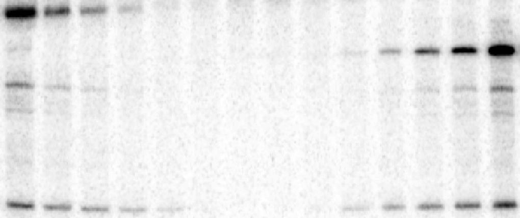
11 RESULTS RNase-resistant viral dsrna is produced during TBSV replication in cell-free extract. To test the nature of the viral RNAs produced during TBSV replication, we utilized a yeast cellfree extract (CFE) that is capable of supporting a single full cycle of authentic TBSV replication, starting with the in vitro assembly of the membrane-bound TBSV replicase, then followed by production of (-)RNA and abundant (+)RNA progeny in a membrane-dependent manner (26, 27, 30). As the final product of replication, the newly synthesized (+)RNAs are released from the VRC to the buffer, while the (-)RNA stays in the nuclease-resistant membrane-bound VRCs (26, 27). First, we assembled the TBSV VRC in the CFE using purified recombinant TBSV p33 and p92 pol replication proteins plus DI-72 (+)replicon (rep)rna (Fig. 1A). The VRC assembly assay contained only ATP and GTP that are needed for VRC assembly, but lacked CTP and UTP to prevent initiation of reprna synthesis (26, 27). After the VRC assembly, we treated the samples with RNases (both ssrna-specific RNase A and dsrna-specific V1 nuclease) to destroy the RNAs not protected by the VRCs (Fig. 1A). After addition of ATP/CTP/GTP and 32 P-labeled UTP, we measured the accumulation of new RNase-resistant viral RNA products in the CFEbased assay. Interestingly, we observed the accumulation of a ribonuclease-resistant dsrna product after 10 min of incubation (Fig. 1B, lane 2), and the amount of this RNA was doubled after min, but did not increase further after 30 min (Fig. 1B, lanes 5-6 versus 4). This dsrna product likely represents annealed viral (-) and (+)RNAs, since it migrated as full-length ssrna after heat denaturation (Fig. 1C, lanes 4 and 6). We also observed the accumulation of a
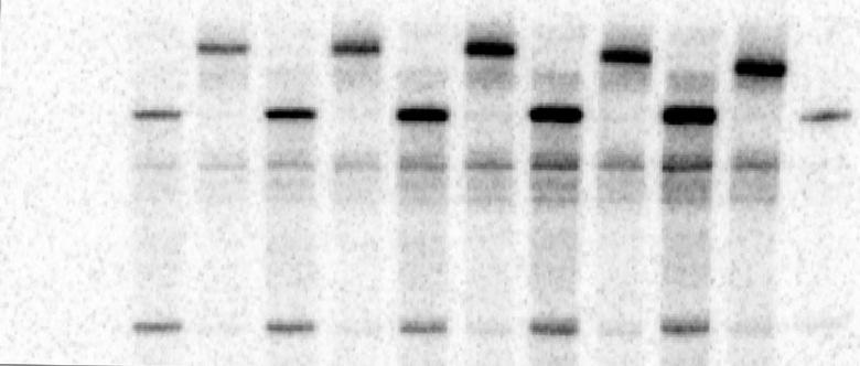
12 small amount of RNase-resistant ssrna (~20 % of the dsrna product) at the end of the assay (3 h time point, Fig. 1C, lanes 3 and 5). A similar CFE-based assay, but performed in the absence of RNases, showed the rapid accumulation of both ssrna and dsrna products (Fig. 1B, lanes 7-11). In contrast to the dsrna, which reached a plateau after 30 min, the amount of newly synthesized ssrna increased continuously. Comparison of the results of the CFE assays in Fig. 1B and 1C suggests that the dsrna product is RNase-resistant and part of the VRC, while most of the ssrnas become RNase-sensitive during replication in vitro, likely due to their release from the VRCs (see below) (26). To confirm that the VRC-bound and RNase-protected viral RNA is indeed present as dsrna species, we treated the CFE samples with RNases after disruption of the membranes in the CFE with low amounts of nonionic detergents (Fig. 2B, lanes 5-6 and 9-10). Treatment with dsrna-specific V1 nuclease destroyed the dsrna only in the presence of the detergent (compare Fig. 2B, lanes 5-6 versus 3-4). On the other hand, treatment with ssrna-specific RNase I did not affect the fast migrating band in the presence of detergent (Fig. 2B, lanes 9-10 versus 7-8), suggesting that this band represents dsrna, formed between (-) and (+)RNAs. The V1 RNase did not destroy the dsrna in the absence of detergent (lanes 3-4, Fig. 2B), likely due to the protection provided by the membrane-bound VRCs. We also repeated these experiments with two other types of CFE-based assays, where the VRCs were pre-assembled in yeast (Fig. S1C-D) or only the protein components of VRCs were pre-assembled in yeast and the reprna was added only during the CFE assay (Fig. S1A-B). Altogether, these assays also confirmed the presence of dsrnas in the VRCs. Moreover, we observed that only tiny amount (0.05% Triton X-100 or 0.01% Nonidet P40) of detergent is needed to make the ssrna in the membrane-bound
13 VRC sensitive to RNase I (Fig. S2A). This amount of detergent is not enough to solubilize membrane-bound proteins (such as TBSV p33 and p92, which are integral membrane proteins), but likely enough to disrupt the membrane-protected structure of VRCs and renders VRCs accessible to RNases. Also, the detergent did not interfere with binding of the p33 replication protein to the viral RNA in vitro (Fig. S2B), suggesting that the replicating RNA is still bound by the replication proteins within the VRCs after the treatment with low amount of detergents. The presence of small amount of Triton X-100 did not promote the annealing between (-) and (+)reprnas under the test conditions (Fig. S2D-E). The dsrna was resistant to RNase I before and also after phenol/chloroform extraction (Fig. S3). Altogether, these CFE-based assays confirmed the accumulation of viral dsrna product during TBSV replication. The early accumulation of V1 nuclease sensitive (in the presence of detergent), but RNase I insensitive, VRC-bound dsrna product during TBSV replication suggests that the dsrna might participate as a template for (+)RNA synthesis. Importantly, we detected the viral dsrna before phenol/chloroform extraction, thus reducing the possibility that dsrna was formed artificially via annealing of naked viral (-) and (+) RNAs during RNA extraction. Free (-)RNA is not produced during TBSV replication in cell-free extract. To further test what form (-)RNA is present in the VRCs, we probed the 32 P-labeled VRC products with short unlabeled RNAs prior to phenol/chloroform extraction (exp. 1, Fig. 3A). Interestingly, annealing of the (+)reprna specific R1(-) RNA, but not the (-)reprna specific R1(+) RNA, to the 32 P-labeled VRC products resulted in a shift in migration of the ssrna products (Fig. 3B, lanes 1 versus 3). After heat denaturation of the reprna samples (exp. 2, Fig. 3A), both (+)RNA
14 and (-)RNA specific R1(-) and R1(+) resulted in annealing and partial duplex formation with ssrnas, confirming that R1(-) and R1(+) anneal efficiently to the complementary sequences in the 32 P-labeled ssrnas under the experimental conditions (Fig. 3B, lanes 2 and 4). We also repeated this experiment with two other sets of short unlabeled RNAs annealing to the 32 P- labeled 3 UTR (Fig. 3C, lanes 5 versus 1) or to a 32 P-labeled internal position of either (+) or (-)- strands with similar results (Fig. S4 lanes 1, 8 versus 5, 12). In addition, we have studied the presence of free (-)-strands of reprnas with short unlabeled RNAs at various time points (40, 60 and 120 min) that showed only free (+)ss reprna and ds reprna were present in the CFEbased replication assay (Fig. 3C, lanes 8 and 10; Fig. S4). Importantly, we could not detect free (-) reprnas within the VRCs in these CFE-based replication assays. To further test if free (-) reprnas are present in the VRCs, we used a (-)-strand-specific DNAzyme, which can cleave the (-) reprna upon annealing (Fig. 3D). The DNAzyme was added at the end of the assay to the detergent-treated membrane fraction of the CFE, prior to phenol/chloroform extraction. We could not observe a DNAzyme-cleaved 32 P-labeled (-) reprna product in the VRC assay (Fig. 3E, lane 3 and 6), albeit the control (-) reprna transcripts were cleaved by this DNAzyme (Fig. 3E, lane 4). This suggests that the (-) reprna was present in the VRCs as part of ds reprna, which is not targeted by the DNAzyme due to the lack of efficient annealing to the duplexed reprna. Since the VRCs contained a small amount of free RNase I-resistant ss reprna (in the absence of detergent, Fig. 1C, lanes 3 and 5), we analyzed the polarity of this reprna species using strand-specific oligos and RNase H, which cleaves RNA/DNA hybrids (Fig. 3F). This assay revealed that the VRCs contained a small amount of free (not base-paired) (+)ss reprna, but not (-)ss reprna (Fig. 3G, lanes 15, 17 versus 11, 13).
15 All the above data suggested that the tombusvirus VRCs contain the (-) reprna annealed to (+)reprna forming dsrna, which is used as template by the VRC to produce (+)RNAs. On the other hand, the VRCs do not seem to contain detectable amount of free/naked (-) reprna, thus making unlikely that free (-) reprna could serve as a template for (+)RNA synthesis during tombusvirus replication. Time course experiments reveal that dsrna could be a template for production of new (+)RNA in cell-free extract. To examine if dsrna is used as a template for (+)RNA synthesis or it is a dead-end product of RNA synthesis during TBSV replication, we performed timecourse experiments (Fig. 4A). We observed that ~70% of the ds reprna was produced within the first min period of the CFE-based assay (Fig. 4B, lanes 10-12), while the bulk of the ss reprna was produced within min period of the assay (Fig. 4B, lanes 13-14). The short unlabeled RNA probe-based mobility shift assay revealed that all the detectable free ss reprna was of (+)-polarity (Fig. 4B, lanes 1-4). Similar time-course experiments characterizing the RNA content of the membrane-bound VRCs (after the removal of the released new reprna progeny from the samples) also revealed that the dsrna accumulated within min period of the assay and most of the ss reprna content of the VRCs accumulated at latter time points ( min) and represented only (+) reprna progeny (Fig. 4C-D). Based on these data, we suggest that the ds reprna in the membrane-bound VRCs serves as a template for (+)RNA synthesis during replication and it is unlikely that the ds reprna is dead-end product of replication. TBSV RNA is replicated by strand-displacement mechanism on dsrna template in the cell-free extract. To test the mechanism of utilization of ds reprna as a template for
16 (+)RNA synthesis during TBSV replication, we used radioactive chase experiments (Fig. 5A-B). First, we produced unlabeled ds reprna in the CFE by allowing replication for 2 hours. During this period, ds reprna reaches maximum level (data not shown). Then, we added 32 P-labeled UTP in three different sets of experiments as shown schematically (Fig. 5B). In experiment #1, we treated the CFE at the 2 hour time point with micrococcal nuclease to remove excess (+)reprna or in experiment #2, we collected the membrane fraction of the CFE (Fig. 5B). The micrococcal nuclease treatment or collection of the membrane fraction by centrifugation removes the released (+)reprna from the VRCs and thus, these treatments exclude the possibility that the (+)reprna could participate in the assembly of new VRCs during the labeling phase of the assay. This design helps obtaining synchronized VRC activities during the labeling phase of the assay. Interestingly, the ds reprna became 32 P-labeled during all three CFE-based assays (Fig. 5C). This observation is consistent with the (+)-strand displacement model (model 1 described in Fig. 5A), while excluding the conservative model as the mechanism of replication (model 2 in Fig. 5A, in which the ds reprna should not be labeled due to lack of new minus-strand synthesis). To further analyze this model, we performed time-course experiments in combination with the above radioactive chase approach as shown in Fig. 5B, exp. #3 and 5D. After production of unlabeled ds reprna in the CFE for 2 hours, we added 32 P-labeled UTP and continued with the CFE-based assay. This time-course study revealed that ds reprna became rapidly labeled (after 10 min of incubation in the presence of 32 P-labeled UTP) and the amount of ds reprna did not change after the first min during the experiment (Fig. 5D, odd number lanes). In contrast, the synthesis of new ss reprnas continued for at least another 40 min. Moreover, as predicted by model 1, all the newly-labeled reprnas, present in either dsrna or ssrna forms, are of (+)
17 RNA polarity based on the complete shift in reprna migration after annealing with the short (+)-strand-specific unlabeled R1(-) RNA probes (Fig. 5D, even number lanes). Therefore, our data demonstrate the lack of new (-) reprna production after the addition of 32 P-labeled UTP to the assay. Thus, the rapid labeling of ds reprna must be due to replacing the unlabeled (+)- strand reprna portion of ds reprna with new 32 P-labeled (+) reprna as predicted by stranddisplacement model (model 1 in Fig. 5A). Another important observation is that the ds reprna becomes labeled at the beginning of the assay, while the bulk of the new labeled (+) reprna is being produced at a latter time point (Fig. 5D). This could be interpreted as an evidence that the ds reprna serves as a template for plus-strand reprna synthesis during replication, while the ss (+)reprna is the final product of replication. In other words, it is unlikely that the ds reprna would be a dead-end product in TBSV replication, since then the ds reprna should mostly accumulate at the end of the assay, which is not the case here. We also performed similar chase experiments after isolation of the membrane fraction of CFE (Fig. S5), which should completely prevent the formation of new VRCs during the labeling step. This time-course study also revealed that ds reprna became labeled after 10 min of incubation in the presence of 32 P-labeled UTP and the amount of ds reprna did not change during further incubation. Also, all the newly-labeled reprnas were of (+)RNA polarity (Fig. S5, even numbered lanes). To obtain further evidence on the mechanism of TBSV replication, we performed additional radioactive chase experiments (Fig. 6A). Here, we first produced 32 P-labeled ds reprna in the CFE by allowing replication to occur for 1 hour, followed by nonradioactive chase assay as shown in experiment #1 in Fig. 6B. As predicted by model 1, the amount of 32 P-label in the ds
18 reprna was reduced continuously, reaching to ~50% by the end of the experiment (Fig. 6C, lanes 10-11). The easiest interpretation of this observation is that during on-going replication, the 32 P-labeled (+) reprna component of the ds reprna template was replaced by an unlabeled newly synthesized (+) reprna (see model 1 in Fig. 6A). In a parallel set of experiment, after the production of 32 P-labeled ds reprna in the CFE for 1 hour, the replication assay was performed in the presence of 32 P-labeled UTP (experiment #2 in Fig. 6B). Increasing levels of (+)reprna during the time-course was observed, suggesting on-going new (+) reprna synthesis (Fig. 6C, lanes 1-5). Also, the level of 32 P-labeled ds reprna did not change during experiment #2 [Fig. 6C, lanes 1-5, due to the new 32 P-labeled (+)reprna replacing the old 32 P-labeled (+)reprna], showing that there was no new generation of ds reprnas under these conditions. Overall, the data obtained in both types of chase replication experiments support the stranddisplacement model (model 1 in Fig. 5A) and suggest that the dsrna is used as a template for (+)RNA synthesis during TBSV replication in the CFE-based assay. RNase-resistant tombusviral dsrna is also present in a plant extract. Since the above CFEbased studies involved yeast CFE and a short reprna, we wanted to test if similar RNA forms are also present in plant extracts containing the infectious TBSV genomic (g)rna. To this end, we prepared a plant extract from Nicotiana benthamiana leaves infected with TBSV grna (Fig. 7A). The plant extract contained the membrane-bound VRCs, including the viral RNAs. Then, we added ATP/CTP/GTP and 32 P-labeled UTP, followed by measuring the accumulation of new TBSV RNAs synthesized in vitro in the plant extract (Fig. 7B). As expected, the plant extracts accumulated RNase I- and RNase V1-resistant TBSV RNAs, which were mostly present as slow migrating partial dsrnas (RI product, Fig. 7B, lanes 1 and 3) as suggested by changes in their
19 migration after heat-denaturation (lanes 2 and 4). Interestingly, treatment with low amount of Triton X-100 made the 32 P-labeled TBSV RNAs RNase V1-sensitive (Fig. 7B, lanes 9-12), but the RNA products were still, at least partially, RNase I-resistant (lanes 5-8). However, RNase I treatment did change the migration pattern of the TBSV RNAs, which moved as completely base-paired dsrnas (grna, sgrna1 and sgrna2), instead of the slowly migrating smeary band in the untreated sample (Fig. 7B, compare lane 5 with 13). These data suggest that TBSV dsrnas are present as replication intermediates (RI, forming a duplex between one minus- and two or more plus-strands, see model 1 in Fig. 5A). Indeed, completely base-paired dsrnas of grna, sgrna1 and sgrna2 were detected in larger amount only after RNase I/detergent treatment (Fig. 7C, lane 1), while these RNAs were removed by the RNase V1/detergenttreatment (Fig. 7C, lanes 3, 5 and 7). Since the detergent/nuclease treatment was performed before the phenol/chloroform extraction (Fig. 7B-C), the data suggest that the replicating TBSV RNAs were mostly present as dsrnas (likely in the form of replication intermediates) in plant extracts. Discussion The (-)RNA is present only in dsrna form during CFE-based TBSV replication. The (-)- stranded RNAs are produced in small amounts and sequestered in membrane-bound VRCs during TBSV (+)-strand RNA virus replication (26, 31). This makes it difficult to study if free (- )RNA strands are present or the (-)RNA is part of dsrna formed by annealing of (+) and (-)- strands within the VRC during replication. By using abundant free (-)RNA-sensing short complementary (+)RNA probes and a (-)RNA-specific DNAzyme during the disruption of VRCs
20 via low concentration of detergent, we show that free (-)RNA is not present at a detectable amount in the VRCs. Therefore, free/naked TBSV (-)RNA is unlikely to serve as a template or as a replicative intermediate for (+)RNA synthesis (Fig. 3 and S4). In addition, we find that V1 dsrna-specific nuclease (in the presence of low amount of detergent) could destroy, while the ssrna-specific RNase I could not eliminate the (-)RNA present in the membrane-bound VRCs (Fig. 2 and S1). Based on these finding and others (Fig. 3), we propose that the TBSV (-)RNA is present in dsrna form during replication in vitro. The dsrna is unlikely to form artifactually during the above in vitro studies, because we performed our assays prior to phenol/chloroform extraction, which could facilitate annealing of naked (-) and (+)-strands due to removal of lipids and proteins and providing non-polar environment. Also, we used only small amount of nonionic detergents (as low as 0.01%, Fig. S2) that could be enough to distort membranes and disrupt the structure of the VRC, but it is not enough to solubilize the viral replication proteins or disrupt p33:reprna interactions (Fig. S2C). Based on these data, we suggest that the TBSV (-)RNA is sequestered into dsrna during replication in membrane-bound VRCs. Another interesting observation in this work is the presence of small amount of free (+)RNAs in the membrane-bound VRCs (Fig. 3G). We propose that these (+)RNAs represent the recently synthesized ssrnas that are waiting to be released to the solution from the VRCs. Accordingly, a small amount of (+)RNAs can be co-purified with the affinity-purified tombusvirus replicase (Panaviene and Nagy, unpublished). Experiments with plant extracts also indicated that the replicating TBSV RNAs are present as V1 nuclease-sensitive dsrnas (in the presence of detergent, Fig. 7B-C). The difference between the yeast CFE and plant extracts was the abundant presence of complete dsrna form in
21 the yeast CFE, while the plant extract contained mostly the replication intermediate (indeed RNase I/detergent-treatment was needed to convert the RI into completely base-paired dsrnas, Fig. 7B-C), which was less abundant in the yeast CFE. This is likely due to the difference in the replicating RNAs: the CFE contained the 621 nt DI-72 reprna, which likely replicates rapidly, making it more difficult to visualize the RI form. In contrast, the plant extract contained the 8- times longer grna and subgenomic RNAs, for which the replicase likely needs much longer time to complete the (+)strand synthesis. This favors the isolation of RI-like dsrnas in the plant extract. Nevertheless, TBSV replication likely follows the same mechanism we established for TBSV reprna replication in yeast CFE. dsrna likely serves as template for (+)RNA synthesis during CFE-based TBSV replication. Although the above experiments supported the existence of dsrna during TBSV replication, it is possible that dsrnas only accumulate as dead-end products of replication. However, time-course experiments (Fig. 1B; Fig. 4) indicate that dsrna is produced prior to the synthesis of the bulk (+)RNAs. This finding is more in line with a model that dsrnas are not final, dead-end products, but instead used as templates for (+)RNA synthesis during TBSV replication. This model is further supported by results from the chase experiments (Figs. 4-6 and Fig. S5), which showed that 32 P-labeled UTP was incorporated during the first 10 min of incubation into the dsrna fraction, while the bulk of 32 P-labeled UTP was incorporated to the newly made (+)-strand RNA at latter time points. In addition, the chase experiments with prelabeled dsrna present in the VRC showed ~50% decrease of 32 P-labeled dsrna at the end of the assay, suggesting that one strand [the (+)-strand] was replaced by an unlabeled new (+)RNA during replication (Fig. 6). Altogether, our data is more consistent with an active template role
22 for dsrnas for (+)RNA synthesis and in disagreement with the model that dsrnas are only dead-end products during TBSV replication The mechanism of dsrna-based replication. Time course chase experiments revealed that 32 P-labeled UTP was incorporated only into the newly-made (+)-strands (Figs. 4-5, S5), while the unlabeled UTP only replaced ~50% of the original 32 P-label during the course of replication (Fig. 6). The strand-displacement model, in which the (-)-strand in the dsrna is used to make new (+)-strands that replace the original (+)-strand in the duplex could explain the obtained data (Fig. 5-6). This model also explains asymmetrical replication if sequential initiation events always take place on the (-)RNA component of the dsrna template to produce (+)RNA products (Fig. 8). Accordingly, we have shown previously that dsrna templates could be used by the tombusvirus replicase to generate (+)-strands in vitro (32, 33). The initiation of (+)RNA synthesis at the 3 end of the (-)RNA component within the dsrna template is likely facilitated by the AU-rich nature of this part of the dsrna template. The weak base-pairs within the AU-rich stretch might promote the limited opening of the dsrna structure prior to initiation (32). Moreover, the role of the RNA chaperone function of the tombusvirus p33 replication protein and co-opted host DEAD-box RNA helicases, such as yeast Ded1p and Dbp2p and the Arabidopsis RH20, in opening the AU-rich containing portion of the dsrna structure to render the 3 -terminus of (-)RNA component of the dsrna accessible to the viral replicase has been shown before (33-35). The p33 chaperone and the recruited host DEADbox helicases are part of the tombusviral replicase, and they could greatly facilitate the utilization of the viral dsrna as template for (+)-strand synthesis (Fig. 8) (33-35). Therefore, the viral dsrna seems to behave as a dynamic structure (alternating between closed/base-paired and
23 partially open forms) due to the presence of co-opted cellular helicases and p33 RNA chaperone in the viral replicase. What is the advantage for the virus to use dsrna as template for (+)RNA synthesis instead of the naked (-)RNA? We speculate that the formation of dsrna templates allows more effective regulation of (-) versus (+)-strand synthesis since the dsrnas can only be used to generate (+)-strands (32-35). This would effectively separate the timing of (-)RNA and (+)RNA synthesis, which could be useful for regulation of asymmetrical replication. The use of dsrna template for (+)RNA synthesis would also favor the generation of a limited number of (-)-strand RNA [possibly one dsrna per one (+)RNA template] since the original (+)-strand RNA would be sequestered into the dsrna right after the (-)RNA synthesis step. The formation of dsrnas within the VRCs could also limit the number of cis-acting RNA elements that are accessible to the viral replicase after (-)RNA synthesis. For example, cis-acting RNA elements located on the (+)-strand, such as the internal p33 recognition element (p33re), the 3 -proximal silencer element and the 3 -terminal (-)strand initiation promoter (gpr), consist of secondary structures needed for function, but these hairpin structures are unlikely to exist and are not functional when part of dsrna structures (32, 33, 36-38). Thus, by masking cis-acting elements located on the (+)-strand through the formation of dsrna structures prevents competition between (+) and (-)RNAs for the replicase and it allows the tombusvirus replicase become committed to plus-strand synthesis at latter time points. Also, formation of dsrnas would limit the possibility of internal initiation and possibly 3 -terminal extensions by the viral replicase, which are rather common processes on naked (-)RNA, resulting in generation of nonfunctional viral RNAs in vitro with purified replicase preparations (39-41).
24 The dsrna nature of the template could also prevent that the template is accidentally lost/released from the membrane-bound VRCs or exposed to the cytosol due to the large size of the dsrna. In contrast, the thinner (+)RNA product could exit VRCs, ending up in the cytosol (buffer in case of the CFE assay) (Fig. 8). This is plausible since the tombusvirus VRCs, similar to many other (+)RNA virus VRCs, form spherule-like structures/vesicles that are connected with the cytosol only via a narrow opening, called neck (42, 43). We propose that the bulky dsrna might not be able to exit through this narrow neck structure of the spherule, essentially trapping the dsrna and thus the (-)RNA component in the VRCs during the entire replication cycle. This strategy could serve dual purpose: avoidance of recognition by the host foreign RNA surveillance system and protection against degradation by host ribonucleases. The possible disadvantage for the formation of dsrnas during (+)RNA virus replication is the prompt recognition of dsrnas by the host anti-dsrna surveillance system, based on Dicer RNase III enzymes for gene silencing in plants and animals, dsrna protein kinase PKR, or RIG-I and MDA5 RNA sensors in mammals (44-51). The viral dsrnas could be destroyed by cellular RNase III-like nucleases or other induced host responses, such as PKR or interferon responses (44-48, 50, 51). However, sequestration of dsrna templates into virus-induced spherule-like structures/vesicles containing the VRCs could greatly reduce the ability of the host cells to sense the presence of viral dsrnas. Thus, the viral dsrna templates hidden within the membrane-bound VRCs might be well-protected against effective antiviral responses for several hours after the start of viral infection. Similarities between TBSV and dsrna virus replication. The formation of dsrnas within the VRCs of tombusviruses adds another piece of evidence for the similarity between (+)-strand
25 RNA viruses and other viruses such as dsrna and retroviruses as noted by Ahlquist before (52). The presence of small amount of recently synthesized (+)RNAs within the tombusvirus VRCs is another shared feature with dsrna viruses. These viruses, TBSV, dsrna and retroviruses, use RNA-binding proteins, such as RNA chaperones and helicases, within the replicase complex to facilitate RNA or cdna synthesis (33-35, 53-56) ACKNOWLEDGEMENTS The authors thank Dr. Daniel Barajas for valuable comments. This work was supported by the National Institute of Health (NIAID, 1R21AI096323). References: 1. den Boon JA, Ahlquist P Organelle-like membrane compartmentalization of positive-strand RNA virus replication factories. Annu Rev Microbiol 64: Denison MR Seeking membranes: positive-strand RNA virus replication complexes. PLoS Biol 6:e Novoa RR, Calderita G, Arranz R, Fontana J, Granzow H, Risco C Virus factories: associations of cell organelles for viral replication and morphogenesis. Biol Cell 97: Salonen A, Ahola T, Kaariainen L Viral RNA replication in association with cellular membranes. Curr Top Microbiol Immunol 285: Bartenschlager R, Cosset FL, Lohmann V Hepatitis C virus replication cycle. Journal of Hepatology 53: Nagy PD, Pogany J The dependence of viral RNA replication on co-opted host factors. Nature Reviews Microbiology 10: Buck KW Replication of tobacco mosaic virus RNA. Philos Trans R Soc Lond B Biol Sci 354: Buck KW Comparison of the replication of positive-stranded RNA viruses of plants and animals. Adv Virus Res 47: Targett-Adams P, Boulant S, McLauchlan J Visualization of double-stranded RNA in cells supporting hepatitis C virus RNA replication. J Virol 82: Weber F, Wagner V, Rasmussen SB, Hartmann R, Paludan SR Doublestranded RNA is produced by positive-strand RNA viruses and DNA viruses but not in detectable amounts by negative-strand RNA viruses. J Virol 80:
26 Westaway EG, Khromykh AA, Mackenzie JM Nascent flavivirus RNA colocalized in situ with double-stranded RNA in stable replication complexes. Virology 258: Westaway EG, Mackenzie JM, Khromykh AA Kunjin RNA replication and applications of Kunjin replicons. Adv Virus Res 59: Sawicki D, Wang T, Sawicki S The RNA structures engaged in replication and transcription of the A59 strain of mouse hepatitis virus. J Gen Virol 82: Sawicki DL, Silverman RH, Williams BR, Sawicki SG Alphavirus minus-strand synthesis and persistence in mouse embryo fibroblasts derived from mice lacking RNase L and protein kinase R. J Virol 77: Baltimore D, Becker Y, Darnell JE Virus-Specific Double-Stranded Rna in Poliovirus-Infected Cells. Science 143: Belov GA, Nair V, Hansen BT, Hoyt FH, Fischer ER, Ehrenfeld E Complex dynamic development of poliovirus membranous replication complexes. J Virol 86: Welsch S, Miller S, Romero-Brey I, Merz A, Bleck CK, Walther P, Fuller SD, Antony C, Krijnse-Locker J, Bartenschlager R Composition and threedimensional architecture of the dengue virus replication and assembly sites. Cell Host Microbe 5: Bamunusinghe D, Seo JK, Rao AL Subcellular localization and rearrangement of endoplasmic reticulum by Brome mosaic virus capsid protein. J Virol 85: Blumenthal T, Carmichael GG RNA replication: function and structure of Qbeta-replicase. Annu Rev Biochem 48: Takeshita D, Tomita K Assembly of Q{beta} viral RNA polymerase with host translational elongation factors EF-Tu and -Ts. Proc Natl Acad Sci U S A 107: Kidmose RT, Vasiliev NN, Chetverin AB, Andersen GR, Knudsen CR Structure of the Qbeta replicase, an RNA-dependent RNA polymerase consisting of viral and host proteins. Proc Natl Acad Sci U S A 107: Fujimura T, Solorzano A, Esteban R Native replication intermediates of the yeast 20 S RNA virus have a single-stranded RNA backbone. J Biol Chem 280: Gong P, Peersen OB Structural basis for active site closure by the poliovirus RNA-dependent RNA polymerase. Proc Natl Acad Sci U S A 107: Nagy PD Yeast as a model host to explore plant virus-host interactions. Annu Rev Phytopathol 46: Panavas T, Nagy PD Yeast as a model host to study replication and recombination of defective interfering RNA of Tomato bushy stunt virus. Virology 314: Pogany J, Stork J, Li Z, Nagy PD In vitro assembly of the Tomato bushy stunt virus replicase requires the host Heat shock protein 70. Proc Natl Acad Sci U S A 105: Pogany J, Nagy PD Authentic replication and recombination of Tomato bushy stunt virus RNA in a cell-free extract from yeast. J Virol 82:
27 Li Z, Pogany J, Tupman S, Esposito AM, Kinzy TG, Nagy PD Translation elongation factor 1A facilitates the assembly of the tombusvirus replicase and stimulates minus-strand synthesis. PLoS Pathog 6:e Pogany J, Nagy PD p33-independent Activation of a Truncated p92 RNA- Dependent RNA Polymerase of Tomato Bushy Stunt Virus in Yeast Cell-Free Extract. J Virol 86: Xu K, Huang TS, Nagy PD Authentic in vitro replication of two tombusviruses in isolated mitochondrial and endoplasmic reticulum membranes. J Virol 86: Panaviene Z, Panavas T, Nagy PD Role of an internal and two 3'-terminal RNA elements in assembly of tombusvirus replicase. J Virol 79: Panavas T, Stork J, Nagy PD Use of double-stranded RNA templates by the tombusvirus replicase in vitro: Implications for the mechanism of plus-strand initiation. Virology 352: Stork J, Kovalev N, Sasvari Z, Nagy PD RNA chaperone activity of the tombusviral p33 replication protein facilitates initiation of RNA synthesis by the viral RdRp in vitro. Virology 409: Kovalev N, Pogany J, Nagy PD A Co-Opted DEAD-Box RNA Helicase Enhances Tombusvirus Plus-Strand Synthesis. PLoS Pathog 8:e Kovalev N, Barajas D, Nagy PD Similar roles for yeast Dbp2 and Arabidopsis RH20 DEAD-box RNA helicases to Ded1 helicase in tombusvirus plus-strand synthesis. Virology 432: Monkewich S, Lin HX, Fabian MR, Xu W, Na H, Ray D, Chernysheva OA, Nagy PD, White KA The p92 polymerase coding region contains an internal RNA element required at an early step in Tombusvirus genome replication. J Virol 79: Pathak KB, Pogany J, Xu K, White KA, Nagy PD Defining the Roles of cis- Acting RNA Elements in Tombusvirus Replicase Assembly In Vitro. J Virol 86: Pogany J, White KA, Nagy PD Specific binding of tombusvirus replication protein p33 to an internal replication element in the viral RNA is essential for replication. J Virol 79: Panavas T, Pogany J, Nagy PD Internal initiation by the cucumber necrosis virus RNA-dependent RNA polymerase is facilitated by promoter-like sequences. Virology 296: Panavas T, Pogany J, Nagy PD Analysis of minimal promoter sequences for plus-strand synthesis by the Cucumber necrosis virus RNA-dependent RNA polymerase. Virology 296: Nagy PD, Pogany J Partial purification and characterization of Cucumber necrosis virus and Tomato bushy stunt virus RNA-dependent RNA polymerases: similarities and differences in template usage between tombusvirus and carmovirus RNAdependent RNA polymerases. Virology 276: Barajas D, Jiang Y, Nagy PD A unique role for the host ESCRT proteins in replication of Tomato bushy stunt virus. PLoS Pathog 5:e McCartney AW, Greenwood JS, Fabian MR, White KA, Mullen RT Localization of the tomato bushy stunt virus replication protein p33 reveals a peroxisome-to-endoplasmic reticulum sorting pathway. Plant Cell 17:
28 DeWitte-Orr SJ, Mehta DR, Collins SE, Suthar MS, Gale M, Jr., Mossman KL Long double-stranded RNA induces an antiviral response independent of IFN regulatory factor 3, IFN-beta promoter stimulator 1, and IFN. J Immunol 183: Garcia MA, Meurs EF, Esteban M The dsrna protein kinase PKR: virus and cell control. Biochimie 89: Kawai T, Akira S Innate immune recognition of viral infection. Nat Immunol 7: Marques JT, Carthew RW A call to arms: coevolution of animal viruses and host innate immune responses. Trends Genet 23: Pflugheber J, Fredericksen B, Sumpter R, Jr., Wang C, Ware F, Sodora DL, Gale M, Jr Regulation of PKR and IRF-1 during hepatitis C virus RNA replication. Proc Natl Acad Sci U S A 99: Pichlmair A, Schulz O, Tan CP, Naslund TI, Liljestrom P, Weber F, Reis e Sousa C RIG-I-mediated antiviral responses to single-stranded RNA bearing 5'-phosphates. Science 314: Samuel CE ADARs: viruses and innate immunity. Curr Top Microbiol Immunol 353: Schmidt A, Rothenfusser S, Hopfner KP Sensing of viral nucleic acids by RIG- I: from translocation to translation. Eur J Cell Biol 91: Ahlquist P Parallels among positive-strand RNA viruses, reverse-transcribing viruses and double-stranded RNA viruses. Nat Rev Microbiol 4: Chong JL, Chuang RY, Tung L, Chang TH Ded1p, a conserved DExD/H-box translation factor, can promote yeast L-A virus negative-strand RNA synthesis in vitro. Nucleic Acids Res 32: Jeang KT, Yedavalli V Role of RNA helicases in HIV-1 replication. Nucleic Acids Res 34: Silvestri LS, Taraporewala ZF, Patton JT Rotavirus replication: plus-sense templates for double-stranded RNA synthesis are made in viroplasms. J Virol 78: Li Z, Nagy PD Diverse roles of host RNA binding proteins in RNA virus replication. RNA Biol 8: Figure legends: Figure 1. Cell-free TBSV replication assay shows the accumulation of RNase protected viral dsrna. (A) Scheme of the CFE-based TBSV replication assay. Purified recombinant p33 and p92 pol replication proteins of TBSV, in vitro transcribed TBSV DI-72 (+)reprna, GTP and ATP were added to the whole cell extract prepared from BY4741 yeast strain. After the replicase assembly on the membranes present in the CFE, ssrna-specific ribonuclease RNase A and dsrna-specific V1 nuclease were added to the assay to destroy unprotected RNAs. (B) Left
29 panel: PAGE analysis of the 32 P-labeled TBSV dsrna products produced in the CFE-based TBSV replication assay in the presence of RNase A and RNase V1. Right panel: Nondenaturing PAGE detection of ssrna and dsrna products produced in the CFE-based TBSV replication assay in the absence of RNase A and V1. The % of dsrna in the samples are shown. (C) Characterization of dsrna products at the end of the CFE assay. ssrna-specific RNase I treatment was applied at the end of the replication assay, prior to phenol/chloroform extraction. The odd numbered lanes represent replicase products, which were not heat-treated (thus both ssrna and dsrna products are present), while the even numbered lanes show the heat-treated replicase products (only ssrna is present). Note that, in the nondenatured samples, the dsrna product represents the annealed (-)RNA and the (+)RNA, while the ssrna products represents the newly made (+)RNA products. Each experiment was repeated at least three times and the data were used to calculate standard deviation. Figure 2. Detection of dsrna-specific V1 nuclease sensitive dsrna products in the CFE-based TBSV replication assay. (A) Scheme of the CFE-based TBSV replication assay. Purified recombinant p33 and p92 pol replication proteins of TBSV and in vitro transcribed TBSV DI-72 (+)reprna were added to the whole cell extract prepared from BY4741 yeast strain. We added V1 or RNase I in the presence or absence of 0.2% Triton X-100 prior to phenol/chloroform extraction. (B) PAGE analysis of the 32 P-labeled TBSV reprna products obtained in the CFE assays. See further details in Figure 1C. Note that RNAse V1 can cleave highly-structured hairpin regions in the ssrnas, which likely causes some loss in the ssrna bands in some samples. Each experiment was repeated at least three times.
30 Figure 3. Lack of free TBSV (-)reprna among the TBSV RNAs produced in the CFE assay. (A) Scheme of the CFE-based TBSV replication assay. In experiment 1, at the end of the replication assay, we added unlabeled short complementary RNAs to the membrane-fraction of the CFE assay in the presence of 0.1% Triton X-100 prior to phenol/chloroform extraction and RNA analysis. In experiment 2, at the end of the replication assay, we added 0.1% Triton X-100 to the membrane-fraction of the CFE assay, performed phenol/chloroform extraction, heat denatured the RNAs, and then added unlabeled short complementary RNAs. Note that experiment 2 tests the ability of the short complementary RNAs to specifically anneal to the target RNA in the assay. (B) Top: Scheme of the annealed unlabeled short complementary RNAs to the 32 P-labeled reprna products. Note that the annealed RNA duplex changes the migration of the RNA in PAGE. Bottom: Representative PAGE analysis of 32 P-labeled reprna products synthesized by the tombusvirus replicase in the CFE assay. The positions of shifted reprnas (due to the annealing to short complementary RNAs), ss reprnas and ds reprnas are shown. Each experiment was repeated three times. (C) The same assay as shown in panel B, except using a different set of short complementary RNAs. (D) Scheme of the DNAzyme-based cleavage of the 32 P-labeled reprna products. The DNAzyme + 0.1% Triton X-100 were added to the membrane fraction of the CFE assay prior to phenol/chloroform extraction. Note that the DNAzyme must anneal to the free (-)reprna to induce cleavage of the target (-)reprna as shown. (E) Representative denaturing PAGE analysis of the DNAzyme-treated 32 P-labeled reprna products synthesized by the tombusvirus replicase in the CFE assay. Lane 4 shows the DNAzyme cleavage products of the in vitro transcribed (-)DI-72 reprna as a positive control that demonstrates the functionality of the DNAzyme. (F) Identification of the polarity of ssrna present in the membrane-bound VRCs. Scheme of the CFE-based TBSV replication assay. The
31 CFE was prepared from BY4741 yeast expressing tombusvirus p33 and p92 pol replication proteins and the TBSV DI-72 (+)reprna. The RNase I-treatment (in the absence of detergent) at the end of the CFE assay removed the released, unprotected 32 P-labeled (+)reprna, followed by phenol/chloroform extraction. Then, the samples were annealed to strand-specific DNA oligos (as shown schematically) and treated with RNase H. (G) PAGE analysis of the 32 P-labeled TBSV reprna products obtained in the CFE assays after RNase H treatment. Note that the RNase I- protected 32 P-labeled ssrna was cleaved by RNase H in the presence of oligo #1159, demonstrating that the ssrna represents (+)reprna in the membrane-bound VRC. Figure 4. Time-course to detect the appearance of TBSV reprna products generated in the CFE assay. (A) Scheme of the CFE-based TBSV replication assay. At the end of the replication assay, we added unlabeled R1(-) RNA to the CFE assay to test the polarity of the ssrna products. (B) Representative nondenaturing PAGE analysis of 32 P-labeled reprna products synthesized by the tombusvirus replicase in the CFE assay. The positions of shifted reprnas [due to the annealing to R1(-) RNA], ss reprnas and ds reprnas are shown. Note that the production of ds and ssrna products requires longer time in this assay than in Fig. 1 because here we did not pre-assemble the replicase complex prior to the addition of ribonucleotides. Each experiment was repeated three times. (C) Scheme of the CFE-based TBSV replication assay. At the end of the time-course replication assay, we removed the supernatant by centrifugation and added unlabeled R1(-) RNA (in the presence of 0.1% Triton X-100) to the membrane fraction of the CFE to test the polarity of the 32 P-labeled ssrna products. Note that only the membrane-bound 32 P-labeled reprna products were analyzed in order to get rid of the large amount of original (+)reprna transcripts added at the start of the CFE assay. (D) Representative PAGE analysis of
32 P-labeled reprna products synthesized by the tombusvirus replicase in the CFE assay. The positions of shifted reprnas [due to the annealing to R1(-) RNA], ss reprnas and ds reprnas are shown. Note that the bulk amount of dsrna appeared earlier (between min) than that of the (+)ssrna product (between min), suggesting that the dsrna might serve as a template for new (+)RNA synthesis and less likely that the dsrna is a dead-end product of replication. Each experiment was repeated three times. Figure 5. TBSV replicates via using strand-displacement mechanism during (+)strand synthesis. (A) Semiconservative (strand-displacement, model 1) and conservative (model 2) models of (+)RNA synthesis based on dsrna templates. Note the different fate of the newly synthesized 32 P-labeled (+)RNA depending on the mechanism of replication. (B) Schemes of the 32 P-labeled UTP chase experiments. In experiment #1, micrococcal nuclease treatment (lasting for 15 min followed by inactivation) was applied prior to addition of the 32 P-labeled UTP. In experiment #2, the membrane fraction of the prior CFE assay was collected to remove unicorporated nonlabeled ribonucleotides, followed by addition of new buffer and new batch of ribonucleotides, including 32 P-labeled UTP. Then, the reaction assay was continued for another 4 h. In experiment #3, 32 P- labeled UTP was added to the CFE-based replication assay at the 2 h time point, followed by additional 4 h of reaction. Note that the nuclease treatment or centrifugation in exp. #1 and #2 were done to prevent the synthesis of new minus-stranded RNAs in the CFE after the addition of 32 P-labeled UTP. (C) PAGE analysis of the 32 P-labeled TBSV reprna products obtained in the CFE assays described in panel B. The even numbered lanes represent replicase products without heat-treatment (thus, both ssrna and dsrna products are visible), while the odd numbered lanes show the heat-treated replicase products (only ssrna is present). See further details in Fig.
33 (D) RNA shift assay based on annealing of short unlabeled complementary RNA in chase time-course studies. The assay was as described in experiment #3 scheme in panel B, except that the reaction was stopped after min as shown, and after heat-denaturation, short R1(-) RNA complementary to the 5 end of (+)reprna was added to every second sample (the even numbered rows) to form a partial duplex. The PAGE shows the complete shift of the newly synthesized reprnas (derived from both ssrna and dsrna), demonstrating that the new 32 P- labeled RNAs represented (+)-strand reprnas, while 32 P-labeled (-)RNA was under detection level. Figure 6. Additional evidence supporting that TBSV replicates via strand-displacement mechanism. (A) Prediction of the different fate of the newly synthesized unlabeled (+)reprna depending on the mechanism of replication. The 32 P-labeled strands are shown with asterisks. (B) Schemes of the unlabeled UTP chase experiments. In experiment #1, the CFE-based TBSV replication assay contained 32 P-labeled UTP and the additional nonlabeled ribonucleotides to obtain 32 P-labeled ss and dsrnas. Then, we removed the soluble 32 P-labeled (+)reprnas (which were released from the VRCs during replication), the unicorporated unlabeled and 32 P- labeled ribonucleotides by collecting the membrane fraction of the CFE. This was followed by addition of nonlabeled UTP in combination with unlabeled ATP/CTP/GTP to the CFE-based replication assay at the 1 h time point, followed by additional 10-to-50 min of reaction. In experiment #2, we used the same procedure as in experiment #1, except adding the 32 P-labeled UTP during the chase period (10-to-50 min reactions). Thus, this experiment is a positive control that could show if additional RNA synthesis took place during the chase period. (C) PAGE analysis of the 32 P-labeled TBSV reprna products obtained in the CFE assays as described in
34 panel B. See further details in Fig. 1. Time point 0 (lane 6) shows the amount and ss or ds nature of reprnas present in the CFE assay prior to the chase period Figure 7. In vitro TBSV replication assay shows the accumulation of RNase protected viral dsrna in plants. (A) Scheme of the plant extract-based TBSV replication assay. N. benthamiana leaves replicating TBSV genomic RNA were used to prepare plant extracts, which were used in in vitro assay in the presence of rntps and 32 P-labeled UTP. (B) Nondenaturing agarose gel analysis of the 32 P-labeled TBSV ssrna and dsrna products produced in the plant extractbased TBSV replication assay in the presence of RNase I and V1 and 0.1% Triton X-100. The odd numbered lanes represent replicase products, which were not heat-treated (thus both ssrna and dsrna products are present), while the even numbered lanes show the heat-treated replicase products (only ssrna is present). Note that, the dsrna product represents the annealed (-)RNA and the (+)RNA, while the ssrna products represents the newly made (+)RNA products. The replication intermediates (RI) are likely present as a smeary band in the gel, as indicated. Each experiment was repeated three times. (C) PAGE analysis of in vitro TBSV replication products in plant extracts. Nondenaturing PAGE of the 32 P-labeled TBSV ssrna and dsrna products produced in the plant extract-based TBSV replication assay in the presence of RNase I or RNase V1 and 0.1% Triton X-100. The odd numbered lanes represent replicase products, which were not heat-treated (thus both ssrna and dsrna products are present), while the even numbered lanes show the heat-treated replicase products (only ssrna is present). Note that, the dsrna product represents the annealed (-)RNA and the (+)RNA, while the ssrna products represents the newly made (+)RNA products. The replication intermediates (RI) are present in the top of the
35 gel, as indicated. The Note that the ss grna and ss sgrna1 co-migrates in this gel. Each experiment was repeated three times Figure 8. A model on the use of dsrna as a template for (+)RNA synthesis by the tombusvirus replicase. The membrane-bound VRC containing viral and host factors synthesizes (-)RNA (gray line) that becomes part of the dsrna. The formation of dsrna disfavors additional rounds of (- )RNA synthesis by sequestering the original (+)RNA (black line) into dsrna structure as shown. Then, the AU-rich side of the dsrna [within and in the vicinity of the (+)-strand promoter represented by an arrowhead] is opened up with the help of recruited host DEAD-box helicases (Ded1p and Dbp2 in yeast and RH20 in plants) and the viral p33 RNA chaperone (1 st step). Then, the exposed (+)-strand promoter is used by the tombusvirus p92 pol RdRp to synthesize a new (+)-strand RNA (2 nd step) by displacing the original (+)-strand RNA, which is then released from the VRC (3 rd step). Finally, the reformed dsrna within the VRC is re-used for additional rounds of (+)RNA synthesis, leading to asymmetrical RNA replication.


36

37


38

39

40

41


42
Plant Pathology Faculty Publications
 University of Kentucky UKnowledge Plant Pathology Faculty Publications Plant Pathology 4-17-2014 The Expanding Functions of Cellular Helicases: The Tombusvirus RNA Replication Enhancer Coopts the Plant
A Co-Opted DEAD-Box RNA helicase enhances tombusvirus plus-strand synthesis
 University of Kentucky UKnowledge Plant Pathology Faculty Publications Plant Pathology 2-16-2012 A Co-Opted DEAD-Box RNA helicase enhances tombusvirus plus-strand synthesis Nikolay Kovalev University of
Replication of plus-strand ( )RNA viruses is driven by the viral
RNA viruses is driven by the viral") In vitro assembly of the Tomato bushy stunt virus replicase requires the host Heat shock protein 70 Judit Pogany, Jozsef Stork, Zhenghe Li, and Peter D. Nagy 1 Department of Plant Pathology, University
In vitro assembly of the Tomato bushy stunt virus replicase requires the host Heat shock protein 70 Judit Pogany, Jozsef Stork, Zhenghe Li, and Peter D. Nagy 1 Department of Plant Pathology, University
Native Replication Intermediates of the Yeast 20 S RNA Virus Have a Single-stranded RNA Backbone*
 THE JOURNAL OF BIOLOGICAL CHEMISTRY Vol. 280, No. 8, Issue of February 25, pp. 7398 7406, 2005 2005 by The American Society for Biochemistry and Molecular Biology, Inc. Printed in U.S.A. Native Replication
Translation elongation factor 1A facilitates the assembly of the tombusvirus replicase and stimulates minus-strand synthesis
 University of Kentucky UKnowledge Plant Pathology Faculty Publications Plant Pathology 11-4-2010 Translation elongation factor 1A facilitates the assembly of the tombusvirus replicase and stimulates minus-strand
Inactivation of the Host Lipin Gene Accelerates RNA Virus Replication through Viral Exploitation of the Expanded Endoplasmic Reticulum Membrane
 University of Kentucky UKnowledge Plant Pathology Faculty Publications Plant Pathology 2-20-2014 Inactivation of the Host Lipin Gene Accelerates RNA Virus Replication through Viral Exploitation of the
A Novel in Vitro Replication System for Dengue Virus
 THE JOURNAL OF BIOLOGICAL CHEMISTRY Vol. 274, No. 47, Issue of November 19, pp. 33714 33722, 1999 1999 by The American Society for Biochemistry and Molecular Biology, Inc. Printed in U.S.A. A Novel in
Defective Interfering RNAs: Foes of Viruses and Friends of Virologists
 Viruses 2009, 1, 895-919; doi:10.3390/v1030895 OPEN ACCESS viruses ISSN 1999-4915 www.mdpi.com/journal/viruses Review Defective Interfering RNAs: Foes of Viruses and Friends of Virologists Kunj B. Pathak
Product Manual. Omni-Array Sense Strand mrna Amplification Kit, 2 ng to 100 ng Version Catalog No.: Reactions
 Genetic Tools and Reagents Universal mrna amplification, sense strand amplification, antisense amplification, cdna synthesis, micro arrays, gene expression, human, mouse, rat, guinea pig, cloning Omni-Array
Materials and Methods , The two-hybrid principle.
 The enzymatic activity of an unknown protein which cleaves the phosphodiester bond between the tyrosine residue of a viral protein and the 5 terminus of the picornavirus RNA Introduction Every day there
October 26, Lecture Readings. Vesicular Trafficking, Secretory Pathway, HIV Assembly and Exit from Cell
 October 26, 2006 Vesicular Trafficking, Secretory Pathway, HIV Assembly and Exit from Cell 1. Secretory pathway a. Formation of coated vesicles b. SNAREs and vesicle targeting 2. Membrane fusion a. SNAREs
Intrinsic cellular defenses against virus infection
 Intrinsic cellular defenses against virus infection Detection of virus infection Host cell response to virus infection Interferons: structure and synthesis Induction of antiviral activity Viral defenses
Chapter 6- An Introduction to Viruses*
 Chapter 6- An Introduction to Viruses* *Lecture notes are to be used as a study guide only and do not represent the comprehensive information you will need to know for the exams. 6.1 Overview of Viruses
number Done by Corrected by Doctor Ashraf
 number 4 Done by Nedaa Bani Ata Corrected by Rama Nada Doctor Ashraf Genome replication and gene expression Remember the steps of viral replication from the last lecture: Attachment, Adsorption, Penetration,
Characterization of Double-Stranded RNA Satellites Associated with the Trichomonas vaginalis Virus
 JOURNAL OF VIROLOGY, Nov. 1995, p. 6892 6897 Vol. 69, No. 11 0022-538X/95/$04.00 0 Copyright 1995, American Society for Microbiology Characterization of Double-Stranded RNA Satellites Associated with the
Yeast as a Model Host to Explore Plant Virus-Host Interactions
 ANNUAL REVIEWS Further Click here for quick links to Annual Reviews content online, including: Other articles in this volume Top cited articles Top downloaded articles Our comprehensive search Annu. Rev.
A discontinuous RNA platform mediates RNA virus replication: building an integrated model for RNA-based regulation of viral processes
 University of Kentucky UKnowledge Plant Pathology Faculty Publications Plant Pathology 3-6-2009 A discontinuous RNA platform mediates RNA virus replication: building an integrated model for RNA-based regulation
Work-flow: protein sample preparation Precipitation methods Removal of interfering substances Specific examples:
 Dr. Sanjeeva Srivastava IIT Bombay Work-flow: protein sample preparation Precipitation methods Removal of interfering substances Specific examples: Sample preparation for serum proteome analysis Sample
brought to you by and REFERENCES
 brought to you by www.thebacteriophages.org and www.phage.org REFERENCES 1. Butcher, S. J., T. Dokland, P. M. Ojala, D. H. Bamford, and S. D. Fuller. 1997. Intermediates in the assembly pathway of the
7.012 Problem Set 6 Solutions
 Name Section 7.012 Problem Set 6 Solutions Question 1 The viral family Orthomyxoviridae contains the influenza A, B and C viruses. These viruses have a (-)ss RNA genome surrounded by a capsid composed
Encapsidation of Sendai Virus Genome RNAs by Purified
 JOURNAL OF VIROLOGY, Mar. 1988, p. 834-838 22-538X/88/3834-5$2./ Copyright C) 1988, American Society for Microbiology Vol. 62, No. 3 Encapsidation of Sendai Virus Genome RNAs by Purified NP Protein during
Problem-solving Test: The Mechanism of Protein Synthesis
 Q 2009 by The International Union of Biochemistry and Molecular Biology BIOCHEMISTRY AND MOLECULAR BIOLOGY EDUCATION Vol. 37, No. 1, pp. 58 62, 2009 Problem-based Learning Problem-solving Test: The Mechanism
Multiplication of RNA Plant Viruses. C.L. Mandahar
 Multiplication of RNA Plant Viruses C.L. Mandahar MULTIPLICATION OF RNA PLANT VIRUSES Multiplication of RNA Plant Viruses by C. L. MANDAHAR Botany Department, Panjab University, Chandigarh, India A C.I.P.
Human Genome: Mapping, Sequencing Techniques, Diseases
 Human Genome: Mapping, Sequencing Techniques, Diseases Lecture 4 BINF 7580 Fall 2005 1 Let us review what we talked about at the previous lecture. Please,... 2 The central dogma states that the transfer
LESSON 4.4 WORKBOOK. How viruses make us sick: Viral Replication
 DEFINITIONS OF TERMS Eukaryotic: Non-bacterial cell type (bacteria are prokaryotes).. LESSON 4.4 WORKBOOK How viruses make us sick: Viral Replication This lesson extends the principles we learned in Unit
Bi 8 Lecture 17. interference. Ellen Rothenberg 1 March 2016
 Bi 8 Lecture 17 REGulation by RNA interference Ellen Rothenberg 1 March 2016 Protein is not the only regulatory molecule affecting gene expression: RNA itself can be negative regulator RNA does not need
Effects of Mutations of the Initiation Nucleotides on Hepatitis C Virus RNA Replication in the Cell
 JOURNAL OF VIROLOGY, Apr. 2004, p. 3633 3643 Vol. 78, No. 7 0022-538X/04/$08.00 0 DOI: 10.1128/JVI.78.7.3633 3643.2004 Copyright 2004, American Society for Microbiology. All Rights Reserved. Effects of
Template Dimerization Promotes an Acceptor Invasion-Induced Transfer Mechanism during Human Immunodeficiency Virus Type 1 Minus-Strand Synthesis
 JOURNAL OF VIROLOGY, Apr. 2003, p. 4710 4721 Vol. 77, No. 8 0022-538X/03/$08.00 0 DOI: 10.1128/JVI.77.8.4710 4721.2003 Copyright 2003, American Society for Microbiology. All Rights Reserved. Template Dimerization
Section 6. Junaid Malek, M.D.
 Section 6 Junaid Malek, M.D. The Golgi and gp160 gp160 transported from ER to the Golgi in coated vesicles These coated vesicles fuse to the cis portion of the Golgi and deposit their cargo in the cisternae
Diverse roles of host RNA binding proteins in RNA virus replication
 RNA Biology ISSN: 1547-6286 (Print) 1555-8584 (Online) Journal homepage: http://www.tandfonline.com/loi/krnb20 Diverse roles of host RNA binding proteins in RNA virus replication Zhenghe Li & Peter D.
Chapter 18. Viral Genetics. AP Biology
 Chapter 18. Viral Genetics 2003-2004 1 A sense of size Comparing eukaryote bacterium virus 2 What is a virus? Is it alive? DNA or RNA enclosed in a protein coat Viruses are not cells Extremely tiny electron
Journal of Agricultural Technology 2012 Vol. 8(4): Journal of Agricultural
: Journal of Agricultural") Journal of Agricultural Technology 2012 Vol. 8(4): 1389-1395 Journal of Agricultural Available Technology online http://www.ijat-aatsea.com 2012, Vol. 8(4): 1389-1395 ISSN 1686-9141 The effect of the decreased
Journal of Agricultural Technology 2012 Vol. 8(4): 1389-1395 Journal of Agricultural Available Technology online http://www.ijat-aatsea.com 2012, Vol. 8(4): 1389-1395 ISSN 1686-9141 The effect of the decreased
Midi Plant Genomic DNA Purification Kit
 Midi Plant Genomic DNA Purification Kit Cat #:DP022MD/ DP022MD-50 Size:10/50 reactions Store at RT For research use only 1 Description: The Midi Plant Genomic DNA Purification Kit provides a rapid, simple
The U1 snrnp Base Pairs with the 5 Splice Site within a Penta-snRNP Complex
 MOLECULAR AND CELLULAR BIOLOGY, May 2003, p. 3442 3455 Vol. 23, No. 10 0270-7306/03/$08.00 0 DOI: 10.1128/MCB.23.10.3442 3455.2003 Copyright 2003, American Society for Microbiology. All Rights Reserved.
Lecture 2: Virology. I. Background
 Lecture 2: Virology I. Background A. Properties 1. Simple biological systems a. Aggregates of nucleic acids and protein 2. Non-living a. Cannot reproduce or carry out metabolic activities outside of a
Reoviruses. Virion. Genome. Genes and proteins. Viruses and hosts. Diseases. Distinctive characteristics
 Reoviruses Virion Genome Genes and proteins Viruses and hosts Diseases Distinctive characteristics Virion Naked icosahedral capsid (T=13), diameter 60-85 nm Capsid consists of two or three concentric protein
Virus Basics. General Characteristics of Viruses 5/9/2011. General Characteristics of Viruses. Chapter 13 & 14. Non-living entities
 Virus Basics Chapter 13 & 14 General Characteristics of Viruses Non-living entities Not considered organisms Can infect organisms of every domain All life-formsf Commonly referred to by organism they infect
The reovirus genome comprises 10 segments of doublestranded
 Reovirus reverse genetics: Incorporation of the CAT gene into the reovirus genome Michael R. Roner* and Wolfgang K. Joklik *Department of Biological Sciences, Center for Molecular Biology and Biotechnology,
Overview: Chapter 19 Viruses: A Borrowed Life
 Overview: Chapter 19 Viruses: A Borrowed Life Viruses called bacteriophages can infect and set in motion a genetic takeover of bacteria, such as Escherichia coli Viruses lead a kind of borrowed life between
Introduction retroposon
 17.1 - Introduction A retrovirus is an RNA virus able to convert its sequence into DNA by reverse transcription A retroposon (retrotransposon) is a transposon that mobilizes via an RNA form; the DNA element
Use of double- stranded DNA mini- circles to characterize the covalent topoisomerase- DNA complex
 SUPPLEMENTARY DATA Use of double- stranded DNA mini- circles to characterize the covalent topoisomerase- DNA complex Armêl Millet 1, François Strauss 1 and Emmanuelle Delagoutte 1 1 Structure et Instabilité
hemagglutinin and the neuraminidase genes (RNA/recombinant viruses/polyacrylamide gel electrophoresis/genetics)
") Proc. Natl. Acad. Sci. USA Vol. 73, No. 6, pp. 242-246, June 976 Microbiology Mapping of the influenza virus genome: Identification of the hemagglutinin and the neuraminidase genes (RNA/recombinant viruses/polyacrylamide
Proc. Natl. Acad. Sci. USA Vol. 73, No. 6, pp. 242-246, June 976 Microbiology Mapping of the influenza virus genome: Identification of the hemagglutinin and the neuraminidase genes (RNA/recombinant viruses/polyacrylamide
Translation. Host Cell Shutoff 1) Initiation of eukaryotic translation involves many initiation factors
 Initiation of eukaryotic translation involves many initiation factors") Translation Questions? 1) How does poliovirus shutoff eukaryotic translation? 2) If eukaryotic messages are not translated how can poliovirus get its message translated? Host Cell Shutoff 1) Initiation
Translation Questions? 1) How does poliovirus shutoff eukaryotic translation? 2) If eukaryotic messages are not translated how can poliovirus get its message translated? Host Cell Shutoff 1) Initiation
Wilmington, Delaware cells were harvested in the cold and pelleted. The cell. pellet was suspended in 2 ml of cold buffer consisting
 JOURNAL OF VIROLOGY, June 1969, p. 599-64 Vol. 3, No. 6 Copyright 1969 American Society for Microbiology Printed in U.S.A. Sindbis Virus-induced Viral Ribonucleic Acid Polymerasel T. SREEVALSAN' AND FAY
Department of Microbiology 1 and Program in Molecular Biology, 2 School of Medicine, University of Colorado Denver, Aurora, Colorado
 JOURNAL OF VIROLOGY, Mar. 2010, p. 2843 2858 Vol. 84, No. 6 0022-538X/10/$12.00 doi:10.1128/jvi.02620-08 Copyright 2010, American Society for Microbiology. All Rights Reserved. Poly(A) at the 3 End of
Chapter13 Characterizing and Classifying Viruses, Viroids, and Prions
 Chapter13 Characterizing and Classifying Viruses, Viroids, and Prions 11/20/2017 MDufilho 1 Characteristics of Viruses Viruses Minuscule, acellular, infectious agent having either DNA or RNA Cause infections
Viruses Tomasz Kordula, Ph.D.
 Viruses Tomasz Kordula, Ph.D. Resources: Alberts et al., Molecular Biology of the Cell, pp. 295, 1330, 1431 1433; Lehninger CD Movie A0002201. Learning Objectives: 1. Understand parasitic life cycle of
Viruses defined acellular organisms genomes nucleic acid replicate inside host cells host metabolic machinery ribosomes
 The Viruses Viruses Viruses may be defined as acellular organisms whose genomes consist of nucleic acid, obligately replicate inside host cells using host metabolic machinery and ribosomes to form a pool
Mechanisms of alternative splicing regulation
 Mechanisms of alternative splicing regulation The number of mechanisms that are known to be involved in splicing regulation approximates the number of splicing decisions that have been analyzed in detail.
Reverse transcription and integration
 Reverse transcription and integration Lecture 9 Biology 3310/4310 Virology Spring 2018 One can t believe impossible things, said Alice. I dare say you haven t had much practice, said the Queen. Why, sometimes
Virus Basics. General Characteristics of Viruses. Chapter 13 & 14. Non-living entities. Can infect organisms of every domain
 Virus Basics Chapter 13 & 14 General Characteristics of Viruses Non-living entities Not considered organisms Can infect organisms of every domain All life-forms Commonly referred to by organism they infect
The TPR domain in the host Cyp40-like cyclophilin binds to the viral replication protein and inhibits the assembly of the tombusviral replicase
 University of Kentucky UKnowledge Plant Pathology Faculty Publications Plant Pathology 2-9-2012 The TPR domain in the host Cyp40-like cyclophilin binds to the viral replication protein and inhibits the
Chapter 25. 바이러스 (The Viruses)
") Chapter 25 바이러스 (The Viruses) Generalized Structure of Viruses 2 2 Virus Classification Classification based on numerous characteristics Nucleic acid type Presence or absence of envelope Capsid symmetry
Chapter 25 바이러스 (The Viruses) Generalized Structure of Viruses 2 2 Virus Classification Classification based on numerous characteristics Nucleic acid type Presence or absence of envelope Capsid symmetry
Viral reproductive cycle
 Lecture 29: Viruses Lecture outline 11/11/05 Types of viruses Bacteriophage Lytic and lysogenic life cycles viruses viruses Influenza Prions Mad cow disease 0.5 µm Figure 18.4 Viral structure of capsid
Ali Alabbadi. Bann. Bann. Dr. Belal
 31 Ali Alabbadi Bann Bann Dr. Belal Topics to be discussed in this sheet: Particles-to-PFU Single-step and multi-step growth cycles Multiplicity of infection (MOI) Physical measurements of virus particles
2) What is the difference between a non-enveloped virion and an enveloped virion? (4 pts)
 What is the difference between a non-enveloped virion and an enveloped virion? (4 pts)") Micro 260 SFCC Spring 2010 Name: All diagrams and drawings shall be hand drawn (do not photo-copied from a publication then cut and pasted into work sheet). Do not copy other student s answers. Para phase
Micro 260 SFCC Spring 2010 Name: All diagrams and drawings shall be hand drawn (do not photo-copied from a publication then cut and pasted into work sheet). Do not copy other student s answers. Para phase
Chapter 19: Viruses. 1. Viral Structure & Reproduction. 2. Bacteriophages. 3. Animal Viruses. 4. Viroids & Prions
 Chapter 19: Viruses 1. Viral Structure & Reproduction 2. Bacteriophages 3. Animal Viruses 4. Viroids & Prions 1. Viral Structure & Reproduction Chapter Reading pp. 393-396 What exactly is a Virus? Viruses
Recombinant Protein Expression Retroviral system
 Recombinant Protein Expression Retroviral system Viruses Contains genome DNA or RNA Genome encased in a protein coat or capsid. Some viruses have membrane covering protein coat enveloped virus Ø Essential
Phenomena first observed in petunia
 Vectors for RNAi Phenomena first observed in petunia Attempted to overexpress chalone synthase (anthrocyanin pigment gene) in petunia. (trying to darken flower color) Caused the loss of pigment. Bill Douherty
A replication silencer element in a plus-strand RNA virus
 The EMBO Journal Vol. 22 No. 20 pp. 5602±5611, 2003 A replication silencer element in a plus-strand RNA virus Judit Pogany 1, Marc R.Fabian 2, K.Andrew White 2 and Peter D.Nagy 1,3 1 Department of Plant
Coat protein blocks the in vitro transcription of the virion RNAs of alfalfa mosaic virus
 Volume 209, number 2 FEBS 4215 December 1986 Coat protein blocks the in vitro transcription of the virion RNAs of alfalfa mosaic virus Corrie J. Houwing and E.M.J. Jaspars* Department of Biochemistry,
Analysis of small RNAs from Drosophila Schneider cells using the Small RNA assay on the Agilent 2100 bioanalyzer. Application Note
 Analysis of small RNAs from Drosophila Schneider cells using the Small RNA assay on the Agilent 2100 bioanalyzer Application Note Odile Sismeiro, Jean-Yves Coppée, Christophe Antoniewski, and Hélène Thomassin
Molecular Biology (BIOL 4320) Exam #2 May 3, 2004
 Exam #2 May 3, 2004") Molecular Biology (BIOL 4320) Exam #2 May 3, 2004 Name SS# This exam is worth a total of 100 points. The number of points each question is worth is shown in parentheses after the question number. Good
Molecular Biology (BIOL 4320) Exam #2 May 3, 2004 Name SS# This exam is worth a total of 100 points. The number of points each question is worth is shown in parentheses after the question number. Good
Chapter 19: Viruses. 1. Viral Structure & Reproduction. What exactly is a Virus? 11/7/ Viral Structure & Reproduction. 2.
 Chapter 19: Viruses 1. Viral Structure & Reproduction 2. Bacteriophages 3. Animal Viruses 4. Viroids & Prions 1. Viral Structure & Reproduction Chapter Reading pp. 393-396 What exactly is a Virus? Viruses
MicroRNA and Male Infertility: A Potential for Diagnosis
 Review Article MicroRNA and Male Infertility: A Potential for Diagnosis * Abstract MicroRNAs (mirnas) are small non-coding single stranded RNA molecules that are physiologically produced in eukaryotic
Each Other. EDTA), quickly cooled in an ice slurry, and made 3 M KCl. before being bound to the column. Sindbis virus RNAs (49S
, quickly cooled in an ice slurry, and made 3 M KCl. before being bound to the column. Sindbis virus RNAs (49S") JOURNAL OF VIROLOGY, Mar. 1986, p. 917-921 0022-538X/86/030917-05$02.00/0 Vol. 57, No. 3 RNA Virus Genomes Hybridize to Cellular rrnas and to Each Other MARCELLA A. McCLURElt* AND JACQUES PERRAULT'2: Department
JOURNAL OF VIROLOGY, Mar. 1986, p. 917-921 0022-538X/86/030917-05$02.00/0 Vol. 57, No. 3 RNA Virus Genomes Hybridize to Cellular rrnas and to Each Other MARCELLA A. McCLURElt* AND JACQUES PERRAULT'2: Department
7.013 Spring 2005 Problem Set 7
 MI Department of Biology 7.013: Introductory Biology - Spring 2005 Instructors: Professor Hazel Sive, Professor yler Jacks, Dr. Claudette Gardel 7.013 Spring 2005 Problem Set 7 FRIDAY May 6th, 2005 Question
Last time we talked about the few steps in viral replication cycle and the un-coating stage:
 Zeina Al-Momani Last time we talked about the few steps in viral replication cycle and the un-coating stage: Un-coating: is a general term for the events which occur after penetration, we talked about
L I F E S C I E N C E S
 1a L I F E S C I E N C E S 5 -UUA AUA UUC GAA AGC UGC AUC GAA AAC UGU GAA UCA-3 5 -TTA ATA TTC GAA AGC TGC ATC GAA AAC TGT GAA TCA-3 3 -AAT TAT AAG CTT TCG ACG TAG CTT TTG ACA CTT AGT-5 OCTOBER 31, 2006
INSTRUCTION MANUAL. RNA Clean & Concentrator -5 Catalog Nos. R1015 & R1016. Highlights. Contents
 INSTRUCTION MANUAL Catalog Nos. R1015 & R1016 Highlights Quick (5 minute) method for cleaning and concentrating RNA. Ideal for purification of RNA from aqueous phase following an acid phenol extraction.
PRODUCT: RNAzol BD for Blood May 2014 Catalog No: RB 192 Storage: Store at room temperature
 PRODUCT: RNAzol BD for Blood May 2014 Catalog No: RB 192 Storage: Store at room temperature PRODUCT DESCRIPTION. RNAzol BD is a reagent for isolation of total RNA from whole blood, plasma or serum of human
Transfection of Sf9 cells with recombinant Bacmid DNA
 Transposition Bacmid DNA Mini Culturing baculo cells Transfection of Sf9 cells with recombinant Bacmid DNA Amplification of the virus Titration of baculo stocks Testing the expression Transposition 1.
AP Biology Reading Guide. Concept 19.1 A virus consists of a nucleic acid surrounded by a protein coat
 AP Biology Reading Guide Name Chapter 19: Viruses Overview Experimental work with viruses has provided important evidence that genes are made of nucleic acids. Viruses were also important in working out
Geneaid DNA Isolation Kit
 Instruction Manual Ver. 02.21.17 For Research Use Only Geneaid DNA Isolation Kit GEB100, GEB01K, GEB01K+ GEC150, GEC1.5K, GEC1.5K+ GET150, GET1.5K, GET1.5K+ GEE150, GEE1.5K, GEE1.5K+ Advantages Sample:
Article Polyprotein Processing as a Determinant for in Vitro Activity of Semliki Forest Virus Replicase
 Article Polyprotein Processing as a Determinant for in Vitro Activity of Semliki Forest Virus Replicase Maija K. Pietilä 1, Irina C. Albulescu 2, Martijn J. van Hemert 2, * and Tero Ahola 1, * 1 Department
SUPPLEMENTARY MATERIAL
 SUPPLEMENTARY MATERIAL Purification and biochemical properties of SDS-stable low molecular weight alkaline serine protease from Citrullus Colocynthis Muhammad Bashir Khan, 1,3 Hidayatullah khan, 2 Muhammad
AP Biology. Viral diseases Polio. Chapter 18. Smallpox. Influenza: 1918 epidemic. Emerging viruses. A sense of size
 Hepatitis Viral diseases Polio Chapter 18. Measles Viral Genetics Influenza: 1918 epidemic 30-40 million deaths world-wide Chicken pox Smallpox Eradicated in 1976 vaccinations ceased in 1980 at risk population?
ab Membrane Fractionation Kit Instructions for Use For the rapid and simple separation of membrane, cytosolic and nuclear cellular fractions.
 ab139409 Membrane Fractionation Kit Instructions for Use For the rapid and simple separation of membrane, cytosolic and nuclear cellular fractions. This product is for research use only and is not intended
Ribonucleic Acid by a Cell-Free
 JOURNAL OF VIROLOGY, Aug. 1971, p. 19-196 Copyright 1971 American Society for Microbiology Vol. 8, No. 2 Printed in U.S.A. Unilateral Synthesis of Reovirus Double-Stranded Ribonucleic Acid by a Cell-Free
Padmanaban Annamalai, A.L.N. Rao* Department of Plant Pathology, University of California, Riverside, CA , USA
 Virology 338 (2005) 96 111 www.elsevier.com/locate/yviro Replication-independent expression of genome components and capsid protein of brome mosaic virus in planta: A functional role for viral replicase
reads observed in trnas from the analysis of RNAs carrying a 5 -OH ends isolated from cells induced to express
 Supplementary Figure 1. VapC-mt4 cleaves trna Ala2 in E. coli. Histograms representing the fold change in reads observed in trnas from the analysis of RNAs carrying a 5 -OH ends isolated from cells induced
Product Contents. 1 Specifications 1 Product Description. 2 Buffer Preparation... 3 Protocol. 3 Ordering Information 4 Related Products..
 INSTRUCTION MANUAL Quick-RNA MidiPrep Catalog No. R1056 Highlights 10 minute method for isolating RNA (up to 1 mg) from a wide range of cell types and tissue samples. Clean-Spin column technology allows
Co-opted Oxysterol-Binding ORP and VAP Proteins Channel Sterols to RNA Virus Replication Sites via Membrane Contact Sites
 University of Kentucky UKnowledge Plant Pathology Faculty Publications Plant Pathology 10-16-2014 Co-opted Oxysterol-Binding ORP and VAP Proteins Channel Sterols to RNA Virus Replication Sites via Membrane
11/15/2011. Outline. Structural Features and Characteristics. The Good the Bad and the Ugly. Viral Genomes. Structural Features and Characteristics
 Chapter 19 - Viruses Outline I. Viruses A. Structure of viruses B. Common Characteristics of Viruses C. Viral replication D. HIV II. Prions The Good the Bad and the Ugly Viruses fit into the bad category
Yuying Liang and Shirley Gillam 1
 Virology 282, 307 319 (2001) doi:10.1006/viro.2001.0862, available online at http://www.idealibrary.com on Rubella Virus RNA Replication Is cis-preferential and Synthesis of Negative- and Positive- Strand
Virology Journal. Open Access. Abstract. BioMed Central
 Virology Journal BioMed Central Research Stimulation of poliovirus RNA synthesis and virus maturation in a HeLa cell-free in vitro translation-rna replication system by viral protein 3CD pro David Franco
VIRUSES. Biology Applications Control. David R. Harper. Garland Science Taylor & Francis Group NEW YORK AND LONDON
 VIRUSES Biology Applications Control David R. Harper GS Garland Science Taylor & Francis Group NEW YORK AND LONDON vii Chapter 1 Virus Structure and 2.2 VIRUS MORPHOLOGY 26 Infection 1 2.3 VIRAL CLASSIFICATION
Title. Author(s)Ishikawa, Masayuki; Janda, Michael; Ahlquist, Paul. CitationJournal of General Virology, 81: Issue Date Doc URL.
Ishikawa, Masayuki; Janda, Michael; Ahlquist, Paul. CitationJournal of General Virology, 81: Issue Date Doc URL.") Title The 3a cell-to-cell movement gene is dispensable for yeast but retained over 1045-fold amplification Author(s)Ishikawa, Masayuki; Janda, Michael; Ahlquist, Paul CitationJournal of General Virology,
Title The 3a cell-to-cell movement gene is dispensable for yeast but retained over 1045-fold amplification Author(s)Ishikawa, Masayuki; Janda, Michael; Ahlquist, Paul CitationJournal of General Virology,
TECHNICAL BULLETIN. R 2 GlcNAcβ1 4GlcNAcβ1 Asn
 GlycoProfile II Enzymatic In-Solution N-Deglycosylation Kit Product Code PP0201 Storage Temperature 2 8 C TECHNICAL BULLETIN Product Description Glycosylation is one of the most common posttranslational
Hepatitis B Virus Genemer
 Product Manual Hepatitis B Virus Genemer Primer Pair for amplification of HBV Viral Specific Fragment Catalog No.: 60-2007-10 Store at 20 o C For research use only. Not for use in diagnostic procedures
Virus and Prokaryotic Gene Regulation - 1
 Virus and Prokaryotic Gene Regulation - 1 We have discussed the molecular structure of DNA and its function in DNA duplication and in transcription and protein synthesis. We now turn to how cells regulate
Minute TM Plasma Membrane Protein Isolation and Cell Fractionation Kit User Manual (v5)
") Minute TM Plasma Membrane Protein Isolation and Cell Fractionation Kit Catalog number: SM-005 Description Minute TM plasma membrane (PM) protein isolation kit is a novel and patented native PM protein
Minute TM Plasma Membrane Protein Isolation and Cell Fractionation Kit Catalog number: SM-005 Description Minute TM plasma membrane (PM) protein isolation kit is a novel and patented native PM protein
7.012 Quiz 3 Answers
 MIT Biology Department 7.012: Introductory Biology - Fall 2004 Instructors: Professor Eric Lander, Professor Robert A. Weinberg, Dr. Claudette Gardel Friday 11/12/04 7.012 Quiz 3 Answers A > 85 B 72-84
SUPPLEMENTAL DATA RESULTS
 SUPPLEMENTAL DATA RESULTS Ded1-mediated ribosomal scanning is less leaky than scanning promoted by eifs 4A/4B/4F. The efficiency of leaky scanning in the presence of Ded1 or of eifs 4A/4B/4F was investigated
Synthesis of Plus- and Minus-Strand RNA in Rotavirus-Infected Cells
 JOURNAL OF VIROLOGY, Nov. 1987, p. 3479-3484 0022-538X/87/113479-06$02.00/0 Copyright 1987, American Society for Microbiology Vol. 61, No. 11 Synthesis of Plus- and Minus-Strand RNA in Rotavirus-Infected
Induction of Particle Polymorphism by Cucumber Necrosis Virus Coat Protein Mutants In Vivo
 JOURNAL OF VIROLOGY, Feb. 2008, p. 1547 1557 Vol. 82, No. 3 0022-538X/08/$08.00 0 doi:10.1128/jvi.01976-07 Copyright 2008, American Society for Microbiology. All Rights Reserved. Induction of Particle
Replication of Sindbis Virus V. Polyribosomes and mrna in Infected Cells
 JOURNAL OF VIROLOGY, Sept. 1974, p. 552-559 Vol. 14, No. 3 Copyright @ 1974 American Society for Microbiology Printed in U.S.A. Replication of Sindbis Virus V. Polyribosomes and mrna in Infected Cells
Innate Immunity & Inflammation
 Innate Immunity & Inflammation The innate immune system is an evolutionally conserved mechanism that provides an early and effective response against invading microbial pathogens. It relies on a limited
XTRAKT FFPE Kit / Manual
 XTRAKT FFPE Kit / Manual For manual extraction of RNA, mirna and/or DNA from Formalin Fixed Paraffin Embedded (FFPE) tissue samples Catalog No. # XTK2.0-96 For research use only. Not intended for diagnostic
Dawn A. Defenbaugh, Matthew Johnson, Renxiang Chen, Ying Yi Zheng and John L. Casey*
 JVI Accepts, published online ahead of print on February 00 J. Virol. doi:./jvi.0-0 Copyright 00, American Society for Microbiology and/or the Listed Authors/Institutions. All Rights Reserved. 1 HEPATITIS