Identification of the Function of the Vpx Protein of Primate Lentiviruses: A Dissertation
|
|
|
- Asher West
- 5 years ago
- Views:
Transcription

1 University of Massachusetts Medical School GSBS Dissertations and Theses Graduate School of Biomedical Sciences Identification of the Function of the Vpx Protein of Primate Lentiviruses: A Dissertation Xiaonan Zhu University of Massachusetts Medical School Follow this and additional works at: Part of the Amino Acids, Peptides, and Proteins Commons, Cells Commons, Genetic Phenomena Commons, and the Viruses Commons Repository Citation Zhu, X. Identification of the Function of the Vpx Protein of Primate Lentiviruses: A Dissertation. (2009). University of Massachusetts Medical School. GSBS Dissertations and Theses. Paper 447. DOI: /cgm8-q gsbs_diss/447 This material is brought to you by escholarship@umms. It has been accepted for inclusion in GSBS Dissertations and Theses by an authorized administrator of escholarship@umms. For more information, please contact Lisa.Palmer@umassmed.edu.
2 IDENTIFICATION OF THE FUNCTION OF THE VPX PROTEIN OF PRIMATE LENTIVIRUSES A Dissertation Presented By XIAONAN ZHU Submitted to the Faculty of the University of Massachusetts Graduate School of Biomedical Sciences, Worcester in partial fulfillment of the requirement for the degree of DOCTOR OF PHILOSOPHY Dec 14, 2009 PROGRAM IN IMMUNOLOGY AND VIROLOGY
3
4 iii COPYRIGHT NOTICE Parts of this dissertation have appeared in separate publications: Sharova N*, Wu Y*, Zhu X, Stranska R, Kaushik R, Sharkey M, Stevenson M. (2008) Primate lentiviral Vpx commandeers DDB1 to counteract a macrophage restriction. PLoS Pathog. 4 (5):e Kaushik R*, Zhu X*, Stranska R, Wu Y, Stevenson M. (2009) A cellular restriction dictates the permissivity of nondividing monocytes/macrophages to lentivirus and gammaretrovirus infection. Cell Host Microbe. 6 (1): *These authors contributed equally. Collaborators contributed to the study presented in this dissertation: Dr. Natalia Sharova: SIVΔVpx and HIV-1ΔVpr infection in macrophages, which laid the foundation for this study, was performed by Dr. Natalia Sharova (Figure 3-1 and Figure 3-4). The data showing the effect of proteasome inhibitor on HIV-1 infection (Figure 4-4A) was generated by Natalia. Dr. Yuanfei Wu: The fluorescent image of macrophages infected by HIV-1 GFP (Figure 3-16) was generated by Dr. Yuanfei Wu. The study of Vpx ubiquitylation and DDB1 interaction (Figure 4-1 and Figure 4-5) was performed by Dr. Yuanfei Wu. Figure 4-6 was a result of collaboration between Yuanfei and I. Yuanfei performed the sirna knockdown experiment and constructed the Vpr-DDB1 fusion protein. Dr. Ruzena Stranska: The heterokaryon experiments shown in Figure 3-2 and Figure 5-1 were performed by Dr. Ruzena Stranska. Dr. Rajnish Kaushik: The fluorescence image of macrophages infected by MLV shown in figure 3-18B was generated by Dr. Rajnish Kaushik.
5 iv ACKNOWLEDGEMENTS First and foremost, I would like to thank my mentor, Mario Stevenson, for his support and insightful discussions during my time in his lab. He encouraged me to think independently and scientifically, which has been very helpful for my professional development. I would not have been successful in my thesis work without the help and support from all the collaborators Natalia Sharova, Yuanfei Wu, Ruzena Stranska, Rajnish Kaushik and Mark Sharkey. We worked closely together and had lots of great discussions. They also encouraged me a lot and taught me experimental techniques. I am so lucky to have Maria Zapp, Paul Clapham and Celia Schiffer on my thesis research committee. They are the ones who are always there helping and encouraging me, even during the most difficult times. I was always being told how much I have done and those encouraging remarks always kept me motivated. I also enjoyed all the constructive suggestions from them. I would also like to thank Heinrich Gottlinger and Ann Sheehy for being on my dissertation committee, and taking time and effort to evaluate my work. I would also like to thank all the past and present members of Stevenson s lab who have been like my family for the last several years supporting me personally and scientifically. They created a great scientific environment and made my study here full of fun and memorable moments. I would especially like to thank Yuanfei Wu and Patrick Younan for their time in completion of this thesis. Last but not least, I would like to thank my husband and my parents. Without their unconditional love and support, I would not have completed the six years of graduate studies. I am also grateful that I have so many friends who have supported and encouraged me along every step of my way.
6 v ABSTRACT Primate lentiviruses encode four accessory proteins including Vif, Vpu, Nef, and Vpr/ Vpx. Vif and Vpu counteract the antiviral effects of cellular restrictions to early and late steps in the viral replication cycle. The functions of Vpx/ Vpr are not well understood. This study presents evidence that the Vpx proteins of HIV-2/ SIV SM promote HIV-1 infection by antagonizing an antiviral restriction in myeloid cells. Fusion of macrophages in which Vpx was essential for virus infection, with COS cells in which Vpx was dispensable for virus infection, generated heterokaryons that supported infection by wild-type SIV but not Vpx-deleted SIV. The restriction potently antagonized infection of macrophages by HIV-1, and expression of Vpx in macrophages in trans overcame the restriction to HIV-1 and SIV infection. Similarly, the cellular restriction is the obstacle to transduction of macrophages by MLV. Neutralization of the restriction by Vpx rendered macrophages permissive to MLV infection. Vpx was ubiquitylated and both ubiquitylation and the proteasome regulated the activity of Vpx. The ability of Vpx to counteract the restriction to HIV-1 and SIV infection was dependent upon the HIV-1 Vpr interacting protein, damaged DNA binding protein 1 (DDB1), and DDB1 partially substituted for Vpx when fused to Vpr. This study further demonstrates that this restriction prevents transduction of quiescent monocytes by HIV-1. Although terminally differentiated macrophages are
7 vi partially permissive to HIV-1, quiescent monocytes, which are macrophage precursors, are highly refractory to lentiviral infection. Monocyte-HeLa heterokaryons were resistant to HIV-1 infection, while heterokaryons formed between monocytes and HeLa cells expressing Vpx were permissive to HIV-1 infection, suggesting the resistance of quiescent monocytes to HIV-1 transduction is governed by a restriction factor. Encapsidation of Vpx within HIV-1 virions conferred the ability to infect quiescent monocytes. Introduction of Vpx into monocytes by pre-infection also rendered quiescent monocytes permissive to HIV-1 infection. Infection of monocytes by HIV-1 either with or without Vpx did not have an effect on temporal expression of CD71. In addition, Vpx increased permissivity of CD71 and CD71 + cells to HIV-1 infection with no apparent bias. These results confirm that Vpx directly renders undifferentiated monocytes permissive to HIV-1 transduction without inducing their differentiation. The introduction of Vpx did not significantly alter APOBEC3G complex distribution, suggesting a restriction other than APOBEC3G was responsible for the resistance of monocytes to HIV-1. Collectively our results indicate that macrophages and monocytes harbor a potent antiviral restriction that is counteracted by the Vpx protein. The relative ability of primate lentiviruses and gammaretroviruses to transduce non-dividing myeloid-cells is dependent upon their ability to neutralize this restriction.
8 vii TABLE OF CONTENTS COPYRIGHT NOTICE... iii ACKNOWLEDGEMENTS...iv ABSTRACT...v LIST OF TABLES...x LIST OF FIGURES...xi ABBREVIATIONS...xiv CHAPTER I Background and Introduction General Introduction to Primate Lentiviruses Introduction to Restriction Factors Introduction to Vpx and Vpr Proteins Monocyte Infection by HIV CHAPTER II Materials and Methods Clones Cell Culture Production of Virus Stocks Infection Analysis of Viral Infection by Quantitative PCR Proteasome Inhibition COS-Macrophage Cell Staining and Cell Fusion HeLa-Monocyte Cell Staining and Cell Fusion Virion Precipitation, Protein Purifications and Western Blotting Analysis of Vpx Ubiquitylation RNA interference of DDB Vpx-DDB1 co-immunoprecipitation...43
9 viii 2.13 Dose Dependent Analysis of Vpx FACS and Monocyte/Macrophage Immunophenotyping APOBEC3G Analysis...44 CHAPTER III Vpx Promotes Macrophage Infection by HIV-1 and MLV Chapter Summary SIV SM Vpx but not Vpr is Required for Infection of Human Macrophages by SIV SM Macrophages Harbor a Restriction Factor that is Counteracted by the Vpx Protein HIV-1 Vpr is Dispensable for HIV-1 Reverse Transcription in Macrophages The Antiviral Restriction Which Antagonizes SIV SM Infection of Macrophages is Active against HIV Vpx Counters the Restriction and Promotes HIV-1 infection in trans Vpx Renders Macrophages Permissive to MLV Infection...76 CHAPTER IV Proteasome/Ubiquitylation Pathway and DDB1 Interaction are Required for the Ability of Vpx to Counteract the Restriction Chapter Summary Vpx Activity Requires Ubiquitylation and Functional Proteasome Vpx Function Requires DDB HIV-1 Vpr Impairs the Ability of Vpx to Neutralize the Macrophage Restriction Primary HIV-1 Vpr Alleles Fail to Overcome the Macrophage Restriction The Ability of Vpx to Neutralize the Restriction is Dose Dependent...99 CHAPTER V Vpx Renders Primary Monocytes Permissive to HIV-1 Transduction Chapter Summary The Resistance of Quiescent Monocytes to Lentivirus Transduction is Governed by a Restriction Vpx Counteracts a Monocyte Restriction to HIV-1 Infection in trans Vpx Renders Monocytes Permissive to HIV-1 Infection without Inducing Monocyte Differentiation Vpx Promotes HIV-1 Infection of Quiescent Monocytes without Preferably Infecting Differentiated Monocytes...115
10 ix 5.6 Vpx Affects Monocyte Permissivity Independent of APOBEC3G CHAPTER VI Summary and Discussions Myeloid Cell Permissivity to Lentiviruses and Gammaretroviruses is Determined by a Restriction Vpx but not Vpr Removes a Restriction in Myeloid Cells Vpx Function Requires Proteasome/Ubiquitylation Pathway and DDB1 Interaction Characteristics of the Restriction Factor in Myeloid Cells Restrictions as Novel Targets for Antiviral Therapy Future Directions REFERENCES...140
11 x LIST OF TABLES Table 1-1 Proposed functions of HIV-1 Vpr, HIV-2/SIV SM Vpr and HIV-2/SIV SM Vpx based on previous studies. 27 Table 2-1 Primers and probes for quantitative real-time PCR. 38 Table 6-1 Functional comparison of HIV-1 Vpr, HIV-2/SIV SM Vpr and HIV-2/SIV SM Vpx based on this study. 129
12 xi LIST OF FIGURES Figure 1-1 The phylogenetic tree of primate lentiviruses. 3 Figure 1-2 General aspects of the primate lentivirus replication cycle. 9 Figure 1-3 Process of reverse transcription of the lentiviral genome. 12 Figure 1-4 Model of APOBEC3G mediated restriction 15 Figure 1-5 The mechanism of Vif-dependent APOBEC3G degradation 17 Figure 1-6 The structure of TRIM5α and the mechanism of TRIM5α- mediated lentivirus restriction 19 Figure 1-7 Model of tetherin mediated inhibition of HIV-1 virion release 24 Figure 1-8 Predicted 3-helix structure of Vpx and Vpr. 25 Figure 3-1 Differential susceptibility of macrophages and cell lines to infection by wild-type and Vpx-deleted (ΔVpx) or Vpr-deleted (ΔVpr) variants of SIV SM. 49 Figure 3-2 Vpx antagonizes an antiviral restriction in macrophages. 52 Figure 3-3 Model of the function of Vpx in SIV SM infection of macrophages. 54 Figure 3-4 Vpr is dispensable for HIV-1 infection of macrophages at the level of reverse transcription. 56 Figure 3-5 Structure of chimeric clones between HIV-1 and SIV SM. 58 Figure 3-6 Vpx is required for SHIV-MA infection of macrophages. 60 Figure 3-7 Vpx is required for SHIV-CA infection of macrophages. 61 Figure 3-8 Vpx is required for SHIV-NA infection of macrophages. 62 Figure 3-9 Vpx is required for SHIV-RT infection of macrophages. 63 Figure 3-10 A schematic of chimeric proviral clone NL43 (SIV-Vif-Env) and 64 Vpx-deleted variant NL43 (SIV-Vif-EnvΔVpx) Figure 3-11 Vpx promote NL43 (SIV-Vif-Env) infection in macrophages. 66
13 xii Figure 3-12 A schematic of chimeric proviral clone NL43 (SIV-Vif-Vpx) and 67 Vpx-deleted variant NL43 (SIV-Vif-ΔVpx). Figure 3-13 Vpx promote NL43 (SIV-Vif-Vpx) infection in macrophages. 68 Figure 3-14 Vpx enhances HIV-1 spreading infection in macrophages. 70 Figure 3-15 HIV-1 is sensitive to the macrophage restriction and SIV Vpx but 73 not HIV-1 Vpr antagonizes the restriction. Figure 3-16 Vpx enhances transduction of macrophages by GFP- expressing 74 HIV-1. Figure 3-17 SIV SM Vpx promotes NA20 B59/NL43ΔVpr infection in 75 macrophages. Figure 3-18 Vpx permits transduction of macrophages by MLV in trans. 78 Figure 4-1 Identification of ubiquitylated residues in Vpx. 83 Figure 4-2 Packaging of Vpx proteins in wild-type and Vpr-deleted HIV Figure 4-3 Figure 4-4 Ubiquitylation is required by Vpx for enhancement of HIV-1 infectivity in macrophages. The ability of Vpx to enhance permissivity of macrophages to HIV-1 and MLV infection requires a functional proteasome Figure 4-5 Vpx ubiquitylation is required for Vpx interaction with DDB1. 91 Figure 4-6 Figure 4-7 Figure 4-8 Figure 4-9 DDB1 is required for the ability of Vpx to counteract the restriction to macrophage infection by HIV-1 and SIV. HIV Vpr inhibits the ability of SIV SM Vpx to antagonize the macrophage restriction. Vpx delivered to macrophages by SIV WT infection enhances the permissivity to HIV-1 WT and HIV-1ΔVpr infection. Primary HIV-1 Vpr alleles fail to overcome the macrophage restriction
14 xiii Figure 4-10 Dose-dependent ability of packaged Vpx to counteract a 102 Figure 5-1 Figure 5-2 Figure 5-3 Figure 5-4 macrophage restriction. Transduction of primary monocytes by HIV-1 is blocked by a restriction. HIV-1 virions encapsidating Vpx efficiently transduce primary monocytes. Vpx delivered to monocytes by SIV WT infection removes a block to subsequent infection by HIV-1. Vpx Renders Monocytes Permissive to HIV-1 Infection without Inducing Monocyte Differentiation Figure 5-5 HIV-1 virions encapsidating Vpx efficiently transduce CD Figure 5-6 monocytes. HIV-1 with encapsidated Vpx equally transduces undifferentiated (CD71 ) and differentiated (CD71 + ) monocyte populations. 120 Figure 5-7 Vpx enhances late product and 2-LTR cdna production in 122 Figure 5-8 undifferentiated CD71 cell population. Vpx renders monocytes permissive to HIV-1 infection without inducing APOBEC3G redistribution. 124
15 xiv ABBREVIATIONS AID AIDS APC APOBEC3G AZT Activation-Induced Deaminase Acquired Immunodeficiency Syndrome Antigen Presenting Cell Apolipoprotein B mrna-editing Catalytic Polypeptides 3G Azidothymidine BST-2 Bone Marrow Stromal Cell Antigen 2 CA CUL5 CTL Capsid Cullin-5 Cytotoxic T-Lymphocyte DDB1 Damaged DNA Binding Protein 1 FACS FPLC GFP GPCR GPI HIV HMM IN LMM LPS LTR MA MHC MLV Fluorescence-Activated Cell Sorting Fast Protein Liquid Chromatography Green Fluorescent Protein G Protein-Coupled Receptor Glycosylphosphatidylinositol Human Immunodeficiency Virus High Molecular Mass Integrase Low-Molecular-Mass Lipopolysaccharide Long Terminal Repeat Matrix Major Histocompatibility Complex Murine Leukemia Virus
16 xv mrna NC PBMC PBS PEG PIC PPT PR RBX1 RRE RT SIV TAR TM TRIM5α Ub UDG VSV-G WT Micro-RNA Nucleocapsid Peripheral Blood Mononuclear Cell Primer Binding Site Polyethylene Glycol Pre-Integration Complex Polypurine-Track Protease Ring-Box-1 Rev Response Element Reverse Transcriptase Simian Immunodeficiency Virus Transactivation Response Element Transmembrane Tripartite Motif-5α Isoform Ubiquitin Uracil DNA Glycosylase Vesicular Stomatitis Virus G Envelope Protein Wild-type
17 1 CHAPTER I Background and Introduction 1.1 General Introduction to Primate Lentiviruses Human immunodeficiency virus (HIV) is the virus that causes acquired immunodeficiency syndrome (AIDS), which leads to opportunistic infections or malignancies due to CD4 + T cell depletion and the progressive failure of the immune system. According to the World Health Organization, as of Dec 2007, there were 33 million people living with HIV, while 2 million deaths were affiliated to AIDS in 2007 alone (1). AIDS is now one of the leading causes of death and loss of life quality worldwide. Effectively fighting HIV/AIDS is one of the most urgent challenges in the world. HIV and the closely related Simian immunodeficiency virus (SIV) are lentiviruses that belong to the retrovirus family, the Retroviridae. They are RNA viruses that encode reverse transcriptase (RT), which catalyzes the synthesis of cdna from viral genomic RNA. Viral DNA is ultimately integrated into the host genome and the integrated proviral DNA directs gene expression. Below is a primate lentivirus phylogenetic tree. HIV-1 group M, which is responsible for the majority of the global AIDS pandemic, as well as groups N and O evolved from the chimpanzee virus SIV CPZ (2, 3). HIV-2, which is slightly attenuated with regards to disease progression, evolved from SIV SM, which
18 2 naturally infects sooty mangabey (4, 5). SIV SM was used to generate the various rhesus macaque-adapted SIV MAC viruses that are often used for AIDS pathogenesis study. There is another cluster that includes SIV AGM evolved from African green monkeys (Figure 1-1) (6). Lentiviral Pathogenesis and Reservoirs In its natural hosts, SIV does not cause AIDS even with high viral loads. However, SIV infection of rhesus macaques, and HIV-1 infection of human cause progression to AIDS. HIV-1 primarily infects immune cells including CD4 + T lymphocytes, tissue macrophages, and dendritic cells. Infection of these cells accounts for the major aspects of HIV-1 pathogenesis in vivo. The depletion of CD4 + T cells is the main reason for the progression to AIDS in HIV-1 infected patients. Upon initial HIV-1 infection, there is active viral replication but viral replication is limited by a strong immune pressure and this leads to the partial recovery of peripheral CD4 + T cells. Infection then enters an asymptomatic phase characterized by low levels of viral load with no disease symptoms, which ultimately leads to loss of cell-mediated immunity due to a reduction in CD4 + T cell numbers. This is the onset of the AIDS stage in which the patient becomes progressively more susceptible to opportunistic infections that inevitably lead to death in the vast majority of untreated patients (7). This model was primarily based on the study of the peripheral CD4 + T cell counts. Recent studies showed that during acute HIV
19 3 HIV Sequence Compendium 2001 Figure 1-1. The phylogenetic tree of primate lentiviruses (6). Phylogenetic relationships of primate lentiviruses with major clusters shown: HIV-1/SIV CPZ, HIV-2/SIV MAC /SIV SM, and SIV AGM.
20 4 infection, the frequency of infection was especially high in memory CD4 + T cells in the gut, and depletion of these cells led to increased gut permeability with microbial translocation. Higher circulating LPS level drives systemic immune activation and leads to chronic HIV infection (8, 9). HIV-1 infection of macrophages, dendritic cells and other non-t cells may also play essential roles in several critical aspects of HIV disease. In addition to CD4 + T lymphocytes, monocyte-macrophage lineage and dendritic cells may form viral reservoirs in HIV-1 infection. HIV-1 in macrophages is assembled in intracellular vesicles and virions can be transmitted from macrophages to T cells across transient virological synapses (10 12). HIV-1virions that are assembled intracellularly within monocyte-derived macrophages persist and retain infectivity for extended intervals (13), confirming that macrophages serve as a HIV-1 reservoir and contribute to viral persistence. In addition, macrophages are able to cross the blood-brain barrier and spread viruses in the immuno-privileged central nervous system (CNS) ( 14 ). Although monocytes express the required HIV-1 receptors and coreceptors, they are not productively infected by HIV-1 in vitro (15, 16). HIV-1 infection of freshly isolated blood monocytes is blocked prior to the completion of reverse transcription and integration (16). Several studies indicate that peripheral monocytes may also serve as a viral reservoir in vivo. HIV-1 can be detected in blood monocytes. However, whether the
21 5 viruses are produced or are maintained latently in monocytes is unclear (17 20). Other evidence support monocytes as a HIV-1 reservoir by showing that there is continued evolution of HIV-1 sequences in peripheral monocytes following antiviral therapy (21). CD16+ monocytes, which constitute about 5% of monocyte population, are more susceptible to HIV-1 infection and possibly harbor the viruses long-term in vivo (22). Dendritic cells may also play a important role in the dissemination of HIV-1 following primary infection, as they can capture virions at the site of infection and provide HIV-1 access to the CD4 + T cells, by being infected and transmitting newly produced viruses, or by harboring infectious viruses without being infected (23). Studies have suggested that DC-SIGN mediates the capture and internalization of infectious virions that are subsequently transmitted to T cells, via the immunological synapses formed between dendritic cells and T cells (24). HIV-1 genome organization and viral proteins Nine open-reading frames are found in the HIV-1 genome, encoding viral gene products that include structural proteins, enzymes for virus replication, and proteins that regulate viral gene expression. The gag and env genes encode the structural proteins and the pol open-reading frame encodes the enzymatic proteins. They are common to all retroviruses. The functions of HIV-1 gene products are reviewed in Fields virology (7). The
22 6 mature proteins of Matrix (MA, p17), Capsid (CA, p24), Nucleocapsid (NC, p7) and p6 are cleaved from the Gag polyprotein by the virus-encoded protease during maturation. MA is responsible for membrane anchoring and interacts with envelope. MA also contains cytotoxic T-lymphocyte (CTL) epitopes. CA is an important structural protein that forms the conical core of the viral particle. NC binds and incorporates viral RNA into virions. P6 contains a late domain that promotes virus release, and Vpx/Vpr binding domains that mediate the incorporation of Vpx/ Vpr proteins into assembling virions. Several distinct enzymatic proteins are encoded by the pol open-reading frame, including protease (PR, p15), reverse transcriptase (RT, p66/ p51), and integrase (IN, p31). PR is responsible for Gag/ Pol cleavage and maturation. RT has RNA-dependant and DNA-dependant polymerase activity and synthesizes DNA from viral RNA, while the RNase H activity of RT removes the RNA template strand from the RNA/ DNA duplex. IN catalyzes viral DNA integration into the host genome. RT and PR are the most targeted enzymes in antiviral therapies. Env glycoprotein is cleaved by a cellular endopeptidase to yield gp120 surface (SU) and gp41 transmembrane (TM) proteins. Gp120 interacts with the CD4 receptor and chemokine coreceptors. Gp41 traverses the lipid bilayer and non-covalently binds to gp120, thus mediates the fusion of viral and cellular membranes. HIV-1 gene expression is regulated by Tat (transactivator of transcription) and Rev
23 7 (regulator of virion expression) at the transcriptional and posttranscriptional levels, respectively. Tat (p16/ p14) is an essential viral transcriptional activator that promotes efficient HIV-1 long terminal repeat (LTR) specific viral transcription. This requires interaction of Tat with transactivation response element (TAR) which is located in the R region of LTR5. Rev (p19) is an RNA transporter and stabilizer. Rev binds to Rev Response Element (RRE) and mediates the exportation of incompletely spliced mrna that contains RRE to the cytoplasm, thus allows the production viral RNA genome and mrna for viral proteins. The cellular Crm1 nuclear export factor is recruited to HIV-1 transcripts by Rev and thereby facilitates their nuclear egress. The genomes of primate and non-primate lentiviruses also encode accessory proteins from small open reading frames which are absent from the genomes of simple retroviruses. They are Vif (viral infectivity factor), Vpx (viral protein x)/ Vpr (viral protein r), Vpu (viral protein u), and Nef (negative factor). Accessory proteins were so named because they were dispensable for viral replication in some cell lines. However, recent studies have shown that they all play crucial roles in lentiviral replication and persistence (reviewed in (25)). Nef (p27) down regulates the major viral receptor CD4, the major histocompatibility complex (MHC) class I and class II molecules, as well as the T cell receptor TCR-CD3 (26 29). Two of the accessory proteins, Vif and Vpu, have been found to overcome restriction factors. Vif antagonizes the antiviral activity of
24 8 APOBEC3G (30). Vpu, which is not expressed in HIV-2 and SIV SM, antagonizes the activity of tetherin/ BST-2 (Bone marrow stromal cell antigen 2) to promote the release of virions from the cell surface (31). Vpu also mediates CD4 receptor degradation (32). Vpx and Vpr both play important but less well defined roles in viral replication. Vpr has been reported to induce G2 cell cycle arrest and to promote HIV-1 infection in macrophages (33 37). HIV-2/SIV SM encodes an additional accessory protein Vpx, which is derived from an ancestral recombination event (38). Vpx is essential for SIV replication in simian macrophages and for dissemination and pathogenesis of SIV in vivo (38 40). Lentiviral LTRs, which are composed of U3, R, and U5, contain important regulatory regions such as those for transcription initiation and polyadenylation. They are also the sequences the IN uses to insert viral cdna into host genomes. Lentivirus life cycle In the replication cycle of HIV-1, the virions undergo binding, fusion and uncoating, reverse transcription, nuclear import, integration, transcription and translation, assembly and budding to produce new virions (Figure 1-2). Viral entry into the target cells is mediated through sequential interactions of HIV-1 Env with the primary receptor CD4, and coreceptors. CCR5 and CXCR4 appear to be the two major coreceptors for HIV-1 entry into cells. HIV-1 is categorized into R5 and X4 tropic based on the

25 9 Rambaut A et al. Nat Rev Genet Jan; 5(1): Figure 1-2. General aspects of the primate lentivirus replication cycle ( 41 ). Complete lentiviral life cycle is depicted as steps including virus binding, fusion and uncoating, reverse transcription, nuclear import, integration, transcription and translation, assembly and budding.
26 10 coreceptor usage during entry. Viruses that use the CCR5 receptor are termed R5 tropic, which mainly infect macrophages and dendritic cells. While those that use CXCR4 are termed X4 tropic, which mainly infect CD4 + T cells. However, coreceptor usage is not the only determinant for viral tropism and recent studies have shown that HIV-1 R5 viruses vary extensively in macrophage tropism (42). In addition to CCR5 and CXCR4, alternative coreceptors are used by lentiviruses. More than 10 members of seven-transmembrane G protein-coupled receptors (GPCRs) have been shown to support lentiviral entry in vitro, including CCR2b, CCR3, CCR8, D6, CXCR5/BLR1, CXCR6/BONZO et al, although there is no clear evidence that they play a role in vivo (43 447). After fusion, the viral core is released into cytoplasm and subsequently disassembles to liberate the viral genome, during a process called uncoating. The viral genome associates with the viral proteins including RT, IN, MA, NC to form a pre-integration complex (PIC), in which the viral RT catalyzes the synthesis of a linear double-stranded cdna from single- stranded viral RNA. After transportation of PIC into the nucleus, the viral cdna is integrated into the host genome by HIV IN. This integrated provirus may remain latent in an unexpressed form, particularly in resting lymphocytes. Upon activation, host RNA polymerase is used to generate transcripts including new viral genomic RNA and spliced mrna molecules, which are transported to the cytoplasm for protein synthesis. The production of viral RNA and proteins is
27 11 regulated by Rev and Tat proteins. The de novo synthesized viral proteins are assembled with HIV-1 genome and the newly assembled viruses bud from the plasma membrane of infected cells. During particle maturation, the virion associated PR cleaves the immature Gag and Gag Pol precursors into functional viral proteins, leading to the production of infectious virions (7, 48). The reverse transcription of HIV-1 is characterized by two strand transfers and requires two enzymatic activities of RT, including DNA polymerase activity that can use RNA or DNA as a template and RNase H activity. The process is summarized in figure 1-3 (49). The trna binds to the primer binding site (PBS) and initiates the synthesis of minus strand strong-stop DNA. RNase H activity of RT then removes the U5 and R region of the RNA. With the first strand transfer, the minus strand strong-stop DNA jumps to the 3 end of RNA and serves as a primer for minus strand DNA synthesis. During the extension, RNase H removes the most of the viral RNA except for a region called polypurine-tract (PPT), which serves as a primer to initiate plus strand DNA synthesis. After completion of plus strand strong stop DNA, this small region of viral RNA and trna is removed. With the second strand transfer the newly synthesized plus strand strong stop DNA jumps to the opposite end and pairs up with minus strand DNA at PBS sequence. The extension of 3 end on both strands is the last step for the synthesis of linear double-stranded DNA with LTR sequences on both ends. Viral cdna is
28 12 Sarafianos SG et al. EMBO J Mar 15;20(6): Figure 1-3. Process of reverse transcription of the lentiviral genome (49). RNA strand is represented in black and DNA strand in red (A) Minus strand strong stop DNA synthesis is initiated by trna binding to the PBS. RNase H removes the U5 and R region of the RNA strand. (B) First strand transfers to the 3 end of viral RNA and serves as primer for minus strand DNA extension. (C) Minus strand DNA extension, accompanied by RNase H digestion of all viral RNA except PPT. (D) PPT serves as a primer for plus strand strong stop DNA synthesis. (E) RNase H removes the trna and the PPT. (F) Minus and plus strands extend following second strand transfer and results in cdna duplex with LTRs at both ends.
29 13 eventually integrated into the host genome, or forms circles containing one or two LTRs through recombination or direct ligation. Since 2-LTR circles are formed only after completion of reverse transcription, they can be used to determine the efficiency of reverse transcription post virus infection. 1.2 Introduction to Restriction Factors Not all the cells support efficient viral infection. Cells that support efficient viral replication are called permissive cells. Cells that do not support efficient viral replication are called nonpermissive. Lentiviruses interact with various cellular factors throughout the viral life cycle. Nonpermissive cells either have evolved some mechanisms to restrict virus infection, or lack some positive factors that are essential for virus infection. Those host cellular factors whose expression restricts viral replication are named restriction factors. They constitute novel aspects of intrinsic immunity and act at different steps in the viral life cycle. Several restriction factors have been identified recently, including APOBEC3G, TRIM5α, and Tetherin (30, 31, 50, 51). Many lentiviruses, in turn, have evolved mechanisms to inactivate or overcome the blocks to infection. APOBEC3G (Apolipoprotein B mrna-editing catalytic polypeptide 3G) Vif, a 23-kDa basic protein, is encoded by the majority of lentiviruses. As a cytoplasmic protein, Vif exists in a soluble cytosolic form or a membrane-associated form. The latter form of Vif is a peripheral membrane protein that is tightly associated
30 14 with the cytoplasmic side of cellular membranes (52). Vif is dispensable for viral replication in permissive cell lines such as SupT1, CEM-SS, C8166, HeLa and 293T cells. However, Vif is essential for viral replication in nonpermissive cells such as primary CD4 + T cells, monocyte-derived macrophages, and certain T-cell leukemia lines such as CEM (53 55). Vifminus virions produced in nonpermissive cells are poorly infectious, whereas Vif minus virions produced in permissive cells are able to infect both permissive and nonpermissive cell types (56, 57). In 2002, Sheehy and colleagues identified APOBEC3G through a cdna subtraction screen for transcripts specifically expressed in nonpermissive cells (30). APOBEC3G belongs to the APOBEC superfamily, which consists of APOBEC1, APOBEC2, APOBEC3A to H, and the activation-induced deaminase (AID) gene (58, 59). They share a conserved cytidine deaminase motif, which acts as DNA mutators that target the genome of viruses by catalyzing deamination of cytidine (C) to uridine (U) in minus strand reverse transcripts, resulting in guanidine (G) to adenosine (A) hypermutation in plus strand DNA (60-61). 61In 62the absence of Vif, APOBEC3G molecules are packaged into viral particles through interaction with viral RNA and Gag proteins (63, 64) and induce cytidine deamination in newly infected cells. Consequently, the infectivity of the virions produced from nonpermissive cells is dramatically impaired (Figure 1-4) (65). However, there is evidence that the enzymatic activity of APOBEC3G

31 15 Harris RS, Liddament MT. Nat Rev Immunol Nov; 4(11): Figure 1-4. Model of APOBEC3G mediated restriction (65). In the absence of Vif, APOBEC3G molecules are packaged into viral particles and induce G to A hypermutation in newly infected cells. Hypermutation of viral genomes leads to the degradation of nascent viral cdna or mutations in viral genes, which hinders viral replication. Vif binds to the APOBEC3G molecules and eliminates APOBEC3G from producing cells through proteasomal degradation.
32 16 is not the only determinant of its antiviral activity (66 68). Deaminase-independent antiviral mechanisms, such as inhibition of trna primer annealing or strand transfer, may also play a role in the antiviral function of APOBEC3G (69, 70). In an evolutionary conflict, the viral Vif protein antagonizes APOBEC3G by inhibiting its packaging into viral particles and therefore allows production of virions free of APOBEC3G. This involves the degradation of APOBEC3G through a proteasome/ubiquitylation system (65, 71). Vif binds to APOBEC3G before it can be packaged into newly assembled virions. The ability of Vif to antagonize APOBEC3G is species-specific and is decided by how efficiently Vif binds to APOBEC3G. (68). Vif recruits the cellular proteins elongin B and elongin C, which then mediate cullin-5 (CUL5)-ring-box-1 (RBX1)-dependent ligation of ubiquitins (Ub) to APOBEC3G. Ubiquitylated APOBEC3G is subsequently degraded by the proteasome (Figure 1-5) (65). However, a degradation-independent pathway was also suggested by observation that a Vif variant efficiently induced APOBEC3G degradation but was unable to restore viral infectivity (72). Although APOBEC3G expressed in target cells does not generally inhibit virus replication (57), studies have suggested that the expression of APOBEC3G in CD4 + T cells or monocytes/macrophages determines their permissivity to HIV-1 (22, 73, 137). Studies show that APOBEC3G is sequestered in an enzymatically active low-molecular-

33 17 Harris RS, Liddament MT. Nat Rev Immunol Nov; 4(11): Figure 1-5. The mechanism of Vif-dependent APOBEC3G degradation (65). Vif binds to APOBEC3G and recruits the cellular proteins elongin B and elongin C, which then mediate CUL5-RBX1-dependent Ubiquitylation of APOBEC3G. Ubiquitylated APOBEC3G is subjected to proteasomal degradation.
34 18 mass (LMM) ribonucleoprotein complex in resting CD4 + T cells or quiescent monocytes thus restricts infection of these cells by HIV-1. However, in activated CD4 + T cells or macrophages, cytoplasmic APOBEC3G resides in an enzymatically inactive high-molecular-mass (HMM) form (22, 74, 137) that allows HIV-1 infection. Whether deamination is required for APOBEC3G activity in these cells remains unclear (137). TRIM5α (Tripartite Motif-5α isoform) HIV-1 enters old world monkey cells efficiently but is restricted before reverse transcription (75, 76). A dominant negative factor TRIM5α confers the post entry block to HIV-1 in old world monkey cells (51). HIV-1 replication is restricted efficiently by TRIM5α derived from rhesus macaques and TRIMCyp from owl monkeys, but is less sensitive to TRIM5α from humans. SIV, which naturally infects old world monkeys, is less sensitive to restriction by rhesus monkey TRIM5α. Murine leukemia virus (MLV) is not restricted by rhesus TRIM5α, however, human TRIM5α efficiently restricts MLV (50, 51, 77). These studies revealed the restriction activities of TRIM5α and its homologues derived from primate are species-specific. TRIM5α is a trimeric cytoplasmic protein belonging to the TRIM (TRIpartite Motif) family, which was first identified by Reddy in 1992 as proteins containing a RING domain, a B-box, and a coiled-coil region (78). TRIM5α is an isoform containing a carboxy-terminal SPRY (B30.2) domain (Figure 1-6). The coiled-coil domain is
 The structure of TRIM5α is composed of a RING domain, a B-box, a coiled-coil region and a carboxy-terminal B30.2 domain. (B) The mechanism of TRIM5α-mediated restriction.")
35 19 A B Emerman M. Proc Natl Acad Sci U S A Apr 4;103(14): Figure 1-6. The structure of TRIM5α and the mechanism of TRIM5α-mediated lentivirus restriction (79). (A) The structure of TRIM5α is composed of a RING domain, a B-box, a coiled-coil region and a carboxy-terminal B30.2 domain. (B) The mechanism of TRIM5α-mediated restriction. After virus binding and fusion with host cells, viral core disassembles and viral RNA is released for reverse transcription. The CA proteins on viral core are targeted by TRIM5α thus leading to accelerated uncoating. As a result reverse transcription is compromised.
36 20 necessary for TRIM5α multimerization and is required for anti-viral restriction. The sequence in the SPRY domain is highly variable and contributes to the species-specific restriction, while in the TRIMCyp isoform from owl monkeys, this region is substituted by a CypA domain (50, 80 82). A single amino acid change (Arginine 332) in this region has been reported to determine the differential ability of human and monkey TRIM5α to restrict HIV-1 replication (83, 84). The B-box domain is suggested to mediate TRIM5α self-association which is important for the efficient binding of TRIM5α to the retroviral CA proteins (85). TRIM5α-mediated restriction occurs post entry and through interaction with viral CA proteins. SIV containing HIV-1 CA has been shown to be sensitive to the block in old world monkey cells, while HIV-1 containing SIV CA is less sensitive to the block in old world monkey cells (86). The expression of TRIM5α induces a decrease of particulate CA and sometimes an increase of soluble CA in the cytosol. This suggests TRIM5α specifically recognizes the CA proteins and promotes the rapid, premature disassembly of viral core, thereby inhibiting reverse transcription (82) (Figure 1-6). Studies have demonstrated that the TRIM5α ring finger has an E3 ubiquitin ligase activity in vitro and in vivo. A mutation in this domain decreases the restriction by TRIM5α and the treatment with proteasome inhibitors removes the restriction. These results indicate the possible involvement of ubiquitin in TRIM5α-mediated restriction
37 21 (87 90). In addition, studies have suggested that TRIM5α may block HIV-1 infection at nuclear import, and may function at later stages of the viral life cycle by degradation of viral Gag polyproteins (91 93). Tetherin Vpu is an accessory protein unique to HIV-1, SIV CPZ, SIV GSN, SIV MUS, SIV MON and SIV DEN. There is no similar gene in HIV-2, SIV SM or other SIVs. Vpu is a dimeric integral membrane protein of 81 amino acids. Vpu mediates degradation of CD4 in the endoplasmic reticulum and regulates HIV-1 replication at late stages of the viral life cycle (32, 94, 95). The influence of Vpu is cell-type specific, suggesting involvement of a cellular factor. Using a cell fusion strategy, non permissive cells were found to express a cellular restriction factor and cause the retention of viral particles (96). In 2008, this restriction factor was identified as Bone marrow stromal cell antigen 2 (BST-2)/CD317 termed tetherin (31, 97). The expression of tetherin is cell type dependent and is part of the interferon induced antiviral defense system (31, 97, 98). Upon treatment with interferon, cells that are Vpu-independent become Vpu-dependent for efficient viral release (98). Tetherin prevents the release of virions from the cell membrane. In the absence of Vpu, viral particles produced from tetherin-expressing cells complete membrane fusion at the plasma membrane. However, these virions are retained at the cell surface where they
38 22 accumulate and are subsequently transported to endosomes (99). Tetherin prevents HIV-1 particle release by directly tethering virions to cells (100). Other than HIV-1, tetherin affects a large diversity of enveloped viruses including retroviruses (31, 97), filoviruses (Ebola) (98, 101), arenaviruses (102) and herpes viruses (103), indicating a common mechanism for tetherin-virion interaction. Tetherin protein has an N-terminal transmembrane (TM) domain and a C terminal glycosylphosphatidylinositol (GPI) lipid anchor ( 104 ). Additionally there is an extracellular domain that is predicted to form a coiled coil between the two membrane anchors (100). The tetherin molecules are anchored to the viral envelope and the cell membrane, associating through the coiled coil domain and thus prevent virions from detaching (31, 100). Virions trapped at the cell surface are eventually internalized for degradation. This is supported by the observation that with protease treatment, the attached virions can be released from cell surface (98, 99). An artificial tetherin protein assembled from fragments of heterologous proteins inhibited the release of HIV-1 and Ebola viruses. This confirmed the above model and demonstrated that the overall configuration of tetherin, rather than the specific sequence of the protein, is required for its antiviral activity (100). The exact role of each domain and how tetherin tethers virions to the cell membrane remains unclear. Studies on tetherin mutants favor a model in which tetherin molecules form a dimer through the coiled coil domains, and the TM
39 23 domains of the dimer are incorporated into the virion envelope, while the GPI domains are anchored to the host-cell membrane (Figure 1-7) (100). How Vpu counteracts tetherin and permits viral particle release remains unclear. Vpu expressed via transient transfection or in the context of viral genome was found to reduce tetherin expression in HeLa cells (97, 103). In addition, Vpu may block the incorporation of the tetherin TM domain into virion envelopes (100, 105). Vpu may also alter tetherin trafficking and thus interfered with the function of tetherin (106). 1.3 Introduction to Vpx and Vpr Proteins In all HIV and SIV genomes, a central viral region overlapping the Vif and Tat open reading frames encodes at least one protein Vpr. Vpr is a nuclear protein of 96-amino acid, with a molecular mass of 14-kDa (107). Vpr interacts with p6 of the Gag precursor and is incorporated into virions during viral particle production ( 108 ). Members of the HIV-2/SIV SM /SIV MAC lineage contain an additional gene termed Vpx, which was originally derived from the African green monkey vpr gene by an ancestral recombination event (38). Vpx is a protein of 16-kDa that is highly conserved among divergent isolates of HIV-2 and SIV SM (109, 110). Vpx and Vpr proteins share considerable sequence similarity. Both proteins are predicted to have 3 helices (Figure 1-8) and are packaged into virions through

40 24 Perez-Caballero D et al. Cell. 2009, 139 (3): Figure 1-7. Model of tetherin mediated inhibition of HIV-1 virion release (100). Tetherin molecules form a dimer through the coiled coil domain. This dimmer is anchored to the host cell membrane through the TM domains on the N terminal, and is anchored to the envelope of budding virions through the GPI domains, thus preventing viral particle release.
 is characterized by 3 helices and flexible N and C termini. Three α helices are presented in pink (17 33), blue (38 50) and orange (54 77).")
41 25 A Le Rouzic E, Benichou S. Retrovirology Feb 22;2:11. B Figure 1-8. Predicted 3-helix structure of Vpx and Vpr. (A) NMR based structure of Vpr. The NMR-based 3D-structure of Vpr (1 96) is characterized by 3 helices and flexible N and C termini. Three α helices are presented in pink (17 33), blue (38 50) and orange (54 77). The loops and flexible N and C termini are presented in green (111). (B) The Vpx and Vpr sequence alignment was generated by Clustal W 2.0 with consensus secondary structure prediction. H represents helix. C represents random coil. E represents beta strand.
42 26 association with p6 region of the Gag polyprotein ( ). The packaging of Vpx and Vpr proteins into virions indicates they may play a role in the early stages of virus life cycle proceeding de novo production of viral proteins. Despite the similarities, the functions of Vpr and HIV-2/SIV SM Vpx do not appear to be the same. Most of the information regarding the roles of Vpr and Vpx proteins in primate lentivirus replication has been derived from studies with HIV-1 Vpr. The Vpr protein of HIV-1 has been shown to promote the accumulation of cells in the G2 stage of the cell cycle (33, 34, 115, 116), and to associate with the DNA repair enzyme Uracil DNA glycosylase (UDG) (117). The ability of HIV-1 Vpr to induce cell cycle arrest requires its binding partner, the E3 ubiquitin ligase complex scaffolding factor, DDB1 ( ). In addition, Vpr has been shown to promote the infection of terminally differentiated macrophages and dendritic cells (33, 36, 37, 123, 124), possibly by mediating the nuclear import of the viral PIC (125). These HIV-1 Vpr-ascribed activities have been suggested to segregate between the Vpx and Vpr proteins of HIV-2/SIV SM. The Vpr protein of HIV-2/SIV SM induces cell cycle arrest and associates with UDG but is dispensable for macrophage infection, while Vpx neither induces cell cycle arrest nor associates with UDG (38, 39). However, Vpx is essential for infection of simian macrophages by SIV in vitro ( Table 1-1 ). Following the infection of simian
43 27 Vpr of HIV-1 Vpr of HIV-2/SIV SM Vpx of HIV-2/SIV SM G2 cell cycle arrest G2 cell cycle arrest No UDG association UDG association No Promote macrophage infection possibly by facilitating nuclear import of PIC No Promote macrophage infection possibly by facilitating nuclear import of PIC Table 1-1. Proposed functions of HIV-1 Vpr, HIV-2/SIV SM Vpr and HIV-2/SIV SM Vpx based on previous studies (38, 39).
44 28 macrophages by Vpx minus SIV SM, late cdna products are reduced while 2-LTR circles, which are formed only after completion of reverse transcription, are absent (38, 39). In vivo, Vpx is also indispensable for dissemination and pathogenesis of SIV in pigtailed macaques (40). Whether any of these activities relate to the functional role of Vpr/Vpx proteins in primate lentivirus replication is unclear. In order to understand the functions of the Vpr/Vpx proteins in macrophage infection, I have focused on Vpx because of its profound impact on macrophage infection. In addition, its effect can be studied independently of other Vpr/Vpx-assigned activities including UDG association and cell cycle arrest. The purpose of this study is to further investigate the function of Vpx in myeloid cells infected by lentiviruses, and the mechanisms by which Vpx enhances lentiviral replication. 1.4 Monocyte Infection by HIV-1 HIV-1 infects and replicates primarily in CD4 + T cells and tissue macrophages. However, peripheral monocytes may also serve as a HIV-1 reservoir in disease progression. Peripheral blood monocytes are vitally important cells in the immune system as the precursor cells to professional antigen presenting cells (APCs). CD14 is expressed on the cell surface of 80-90% of blood monocytes and monocyte-derived macrophages. CD14 is a lipopolysaccharide (LPS) receptor that constitutively expressed
45 29 during monocyte differentiation (126). Upon differentiation of quiescent monocytes to macrophages, the expression of a macrophage-specific transferrin receptor, CD71, increases (126). The frequency of monocyte infection by HIV-1 in vivo appears to be low. Monocytes are implicated as a viral reservoir based on the detection of infectious viruses from monocytes isolated from HIV-1 positive individuals on antiretroviral therapy. In some cases, infectious viruses can be recovered from monocytes upon appropriate stimulation, although these cells produced undetectable amounts of viral RNA (17, 19, 20, 127). In addition, monocytes harbor latent HIV-1 proviral DNA during the disease progression in spite of antiviral therapy (128). A small population of monocytes which are CD16 + apprears to be more susceptible to infection and preferentially harbors HIV-1 viruses in vivo (22). Although lentiviruses have evolved the ability to infect terminally differentiated macrophages, peripheral blood monocytes are highly refractory to lentivirus transduction in vitro (16, ). Permissivity to HIV-1 infection is coordinated to the state of monocyte differentiation (126, 134, 135). The mechanisms underscoring the block to transduction of quiescent monocytes by lentiviruses are not well understood. A different set of factors has been proposed to regulate infection of quiescent monocytes by lentiviruses. G0 monocytes have low intracellular dntp levels (126, 136), and this
46 30 has been proposed to limit the efficiency of viral cdna synthesis in these quiescent cells. The cytidine deaminase APOBEC3G, which is a target of the viral accessory protein Vif, has been shown to influence the permissivity of quiescent lymphocytes and monocytes to HIV-1 infection (22, 137, 138). The enzymatically active LMM APOBECEG complex, which is the exclusive form in quiescent cells, has been shown to restrict infection of quiescent monocytes by HIV-1 (22, 74, 137). In addition, studies suggest that naturally occurring anti-hiv micro-rna (mirna) suppress HIV-1 in peripheral blood mononuclear cells or purified monocytes ( ). In this study, the hypothesis that a restriction prevents the in vitro transduction of quiescent monocytes by HIV-1 was examined.
47 31 CHAPTER II Materials and Methods 2.1 Clones SIV SM clone PBj1.9 The infectious molecular clone SIV SM PBj1.9 was used for SIV infection in this study. This clone, which is representative of the HIV-2/SIV SM group of viruses, is a primary isolate and was derived from short-term peripheral blood mononuclear cell (PBMC) cultures (143). Unlike many other HIV-2 and SIV SM clones, PBj1.9 has a complete set of uninterrupted accessory genes and replicates efficiently in macrophages and represents a physiologically relevant virus strain. Mutants with deletion of Vpx or Vpr were constructed by Dr. Mark Sharkey as previously described (38). GFP (green fluorescent protein) expressing variants of wild-type and ΔVpx SIV contain an EGFP gene inserted between Bst 1107I sites within the viral envelope gene. HIV-1 clones NL43 is a CXCR4 tropism HIV-1 strain. This clone was derived from Full-length, replication and infection competent chimeric DNA, in which the 5 fragment of proviral NY5 and the 3 fragment of proviral LAV were cloned into puc18 after removal of polylinker sites (144). In most experiments, wild-type and ΔVpr HIV-1 variants were
48 32 studied in the context of HIV-1 NL43. NL43ΔVpr clone was constructed by PCR based mutagenesis to generate mutation on start codon and to introduce early stop codons. NL43 GFP (a gift from Dr. Paul Clapham) contains an EGFP gene inserted between the envelope stop codon and nef within the HIV-1 NL43 backbone. ADA is a R5 HIV-1 strain originally isolated from a seropositive AIDS patient. LAI is an X4 HIV-1 strain which is the first strain of HIV-1 isolated in the laboratory cultured from Human PBMCs or CEM cells NA20 B59 is a NL43 clone with highly macrophage-tropic envelope (a gift from Dr. Paul Clapham s lab). R5 tropic NA20 B59 envelope was amplified from brain tissue of AIDS patients (145), and cloned into pnl43 backbone using a BbsI site at the start of the envelope and an XhoI site downstream from the 3 end of the envelope. Chimeric clones between SIV and HIV-1 In all HIV-1/SIV SM chimeras, SIV SM proviral sequence was from PBj1.9, HIV-1 sequence was from NL43 clone. The different domains of NL43 were precisely substituted for the corresponding domains of PBj1.9. All the chimeric clones were created by overlapping PCR. NL43 (SIV-Vif-Env) and the Vpx-deleted variant: The fragment from vif to env of NL43 was replaced with the corresponding sequence from PBj1.9 to generate chimeric clone NL43 (SIV-Vif-EnvΔVpx). NL43 (SIV-Vif-EnvΔVpx) is the same
49 33 chimera with Vpx deletion, which was introduced by Vpx start codon mutation and early stop codons. All the mutations are silent mutations for vif gene. NL43 (SIV-Vif-Vpx) and the Vpx-deleted variant: the NL43 sequence from vif to vpr was replaced with the sequence of vif to vpx from PBj1.9 to generate NL43 (SIV-Vif-Vpx) clone. NL43 (SIV-Vif-ΔVpx) is the chimera variant in which Vpx was deleted with the same strategy mentioned above. Vpx and M4 mutant The SIV SM Vpx gene was amplified from PBj1.9 proviral clone, and inserted into a pires2-egfp vector (BD) either with or without a N-terminal minimum HA epitope. The upstream primer for each PCR product provided a Kozak sequence. The Vpx lysine mutant M4 was generated using Quikchange XL sitedirected mutagenesis (Stratagene). These clones were generated by Dr. Yuanfei Wu. DDB1-Vpr fusion The DDB1 gene was amplified and subcloned from pbj-hp125 (ATCC, MBA-126) and inserted into pires2-egfp as an in frame fusion with the C-terminal of SIV Vpr. A Flag epitope was added to the N terminal of DDB1 as flanking sequences between Vpr and DDB1. As a control, an N-terminal Flag tagged DDB1 was inserted into pires2-egfp.
50 34 Vpr expressing plasmid and primary Vpr expressing plasmid The HIV-1 Vpr was amplified from NL43 proviral clones, and inserted into a pires2-egfp vector (BD). The primary Vpr sequences were derived from plasma samples of AIDS patients with high viral load (Courtesy from Dr. Katherine Luzuriaga s lab). The RNA was extracted from the plasma sample and first strand DNA was synthesized. The primary Vpr region was amplified from DNA and cloned into pires2-egfp vector to create the primary Vpr expressing vectors. 2.2 Cell Culture 293T, HeLa and COS-1 cells 293T, HeLa, and COS-1 cells were maintained in DMEM containing 10% Fetal Bovine Serum (FBS). Macrophages Peripheral blood monocytes were obtained by elutriation and counter current centrifugation and maintained 2 days in DMEM containing 10% human serum and monocyte colony stimulating factor (MCSF) (RD Systems) and for a further 5 days in medium lacking MCSF prior to use in experiment.
51 35 Monocytes Human monocytes were obtained from healthy donors by countercurrent centrifugal elutriation (146). The monocytes were kept in DMEM containing 10% human serum and used freshly for infection. 2.3 Production of Virus Stocks Unless indicated other way, viruses were pseudotyped with Vesicular Stomatitis Virus G envelope proteins (VSV-G). For the generation of viral stocks, 293T cells were transfected with proviral DNAs using a modified calcium phosphate/dna precipitation method. Viruses were pseudotyped with VSV envelope glycoproteins by cotransfection of proviral DNAs with a plasmid expressing the VSV envelope glycoprotein. For encapsidation of wild-type and mutant Vpx and Vpr proteins, 293T cells were cotransfected with proviral DNAs and plasmids expressing Vpx and Vpr proteins. The DNA ratio for pvsv-g, proviral clones and pires2-egfp-vpx was 1:14:1. HIV-1 and SIV stocks were normalized on the basis of reverse transcriptase activity. Pseudotyped MLV (MLV-G) was prepared by Dr. Rajnish Kaushik. The stocks were obtained by transfecting retroviral-packaging 293A cells with plegfp-c1 and pmd-g. Viral particles in culture supernatants were harvested after 24 and 48 hours, passed through 0.45 mm filter and concentrated by ultra-centrifugation. All virus stocks were treated with DNaseI (Worthington, NJ, USA) to remove
52 36 residual transfection DNA. 2.4 Infection For infection, macrophages or monocytes were plated in 24 well plates and infected with HIV-1 at 1 million RT units/well. After 4 hours, cells were washed with fresh medium and incubated at 37ºC for the remainder of the experiment. Pre-infection studies were performed by first infecting macrophages with pseudotyped wild-type SIV (SIV WT ) or SIVΔVpx variants and 4 hours later, the supernatant was removed and the cells were inoculated with HIV-1 or MLV for another 4 hours. The supernatant was removed and the cells were washed with fresh medium and incubated at 37ºC for the remainder of the experiment. At indicated time points after inoculation, the cells were lysed and DNA extracted from each sample for quantitative real time PCR. When indicator viruses were used for infection, the cells were checked under fluorescent microscope or the numbers of green cells were quantitated by flow cytometry. To control for the carry over DNA from virions, cells were treated with reverse transcriptase inhibitor Azidothymidine (AZT) 4 hours before infection at a final concentration of 10µM. The same concentration of AZT was kept during the whole experiment.
53 Analysis of Viral Infection by Quantitative PCR Viral infection efficiency was gauged from synthesis of viral cdna products at early intervals (24 and 48 h) post-infection. Cells were initially infected with viruses. After 4 hours, cells were washed with fresh medium and incubated at 37 º C for the remainder of the experiment. Pre-infection studies were performed by first infecting macrophages with pseudotyped SIV wild-type or SIVΔVpx variants and 4 hours later, the cells were infected with HIV-1 or MLV for another 4 hours before washing cells with fresh medium. Infected cells were washed with PBS before harvesting samples for DNA analysis. Total DNA was extracted from infected cells using DNAzol reagent (Invitrogen). Quantitative analysis of viral cdna intermediates is as described (38, 147, 148), and primers and probes for quantitative PCR are listed in table 2-1. Copy number estimates of cdna and 2-LTR circles were determined on an ABI Prism 7500 fast machine. The real-time PCR analysis from each sample was carried out in duplicate wells and most of the values shown in the figures are average of independent experiments using macrophages from at least 3 different donors. Viral cdna was expressed on a per cell basis after quantitation of cellular CCR Proteasome Inhibition Macrophages in 24 well plates were directly infected with VSV-G-pseudotyped
54 38 PBj1.9 late products (Plus strand extension) PBj1.9 late f AAGCCAGTGTGTGTTCCCATC U5 PBj1.9 late r CATCTGCTTTCTTCCCTGACAA Gag PBj1.9 late prb 6-FAM-CCTAGCCGCCGCCTGGTCATCT- Iowa Black U5 FQ TM PBj1.9 2-LTR circles pbj4n/f AGAAGCCCCTGGTCTGTTAGGAC U5 pbjc/r TCGTCTTCCTGAGCTTCATCTGA U3 PBj1.9 probe 6-FAM-TGGCAAAATTACACAGCAGGGCCAG-Iowa U3 Black FQ TM HIV late products (Plus strand extension) C1/f TAGACCAGATTTGAGCCTGGGA R LAI 7/r TCTCCTTCTAGCCTCCGCTAGTCAA GLS 2nr4nr 6-FAM-AGCCTCAATAAAGCTTGCCTTGAGTGC-Iowa R Black FQ TM HIV 2-LTR circles C1/f TAGACCAGATCTGAGCCTGGGA R C4/r GTAGTTCTGCCAATCAGGGAAG U3 2nr4nr 6-FAM-AGCCTCAATAAAGCTTGCCTTGAGTGC-Iowa R Black FQ TM MLV late products (Plus strand extension) OJWB45 GCGCCAGTCTTCCGATAGACTG R OJWB48 ACAATCGGACAGACACAGATAAGTTG Gag OJWB38 6-FAM-ATCCGAATCGTGGTCTCGCTGTTC-TAMRA R MLV 2-LTR circles OJWB45 GCGCCAGTCTTCCGATAGACTG R OJWB46 GGGCTCTTTTATTGAGCTCGGAG U3 OJWB38 6-FAM-ATCCGAATCGTGGTCTCGCTGTTC-TAMRA R Macrophage CCR5 CCR5/r CTCACAGCCCTGTGCCTCTTCTTC CCR5/f GCTGTCTTTGCGTCTCTCCCAGGA CCR5 probe 6-FAM-AGCAGCGGCAGGACCAGCCCCAAG-Iowa Black FQ TM Table 2-1. Primers and probes for quantitative real-time PCR.
55 39 viruses (1 million RT/well) in the presence of proteasome inhibitors including Lactacystine (10 µm), ALLN (50 µm) or Proteasome inhibitor 1 (50µM). After 3 5 hours, supernatant was removed and replaced with fresh medium containing proteasome inhibitors. After 24 and 48 h postinfection total DNA was isolated using DNAzol reagent (Invitrogen) and analyzed by real-time PCR assay for 2-LTR circles. 2.7 COS-Macrophage Cell Staining and Cell Fusion For FACS analysis, COS cells and human macrophages were stained with 3.5 mm CellTracker Green CMFDA (5-chloro methylfluorescein chloromethyl- fluorescein diacetate) and 24 mm CellTracker Blue CMAC (7-amino-4- chloromethylcoumarin), respectively. For fluorescence microscopy, COS cells and macrophages were stained with 2.5 mm DiO (3,39-dioctadecyloxacarbo cyanine perchlorate) and 12 mm DiI (1,19-dioctadecyl-3,3,39,39- tetramethylindo- carbocyanine perchlorate) respectively, according to manufacturer s instructions (Molecular Probes). Generation of macrophage homokaryons was achieved by polyethylene glycol (PEG). Briefly, labeled cells, each group, were mixed and centrifuged at 250 g. 50% PEG-1450 was added dropwise to the pellet and cells incubated for 2 min at 37ºC with gentle mixing. 1 ml PBS was then added dropwise to the cells over 1 min, followed by 3 ml of 2% FBS/PBS over another 2 minutes. Cells were washed 3 times with 2% FBS/PBS and plated in a 100 mm culture dish ( cells/dish). COS-macrophage and
56 40 COS-COS cell fusion was achieved using paramyxovirus hemagglutinin-neuraminidase (HN) protein and fusion (F) protein. Briefly, COS cells were transfected with pcaggs-hn and pcaggs-f expression vectors encoding HN and F proteins of Newcastle disease virus (gift of Prof. T. Morrison) (149). Sixteen to twenty hours post-transfection, COS cells were stained, mixed with stained macrophages (ratio 1:1.5) and plated in 100 mm dishes. COS homokaryons were generated at 1:1 ratio. After overnight incubation, cells were infected with either SIV wild-type or SIVΔVpx for 24 h. Cell sorting was performed with a FACSAria flow cytometer using the FACSDiva software (Becton Dickinson). Double-stained cells were sorted. Total DNA was isolated using DNeasy Blood and Tissue Kit (Quiagen) and analyzed by realtime PCR assay for 2-LTR circles. 2.8 HeLa-Monocyte Cell Staining and Cell Fusion HeLa-monocyte fusion was achieved using a GenomeONE-CFEX HVJ envelope fusion kit (Cosmo Bio Co., Ltd.). Manufacturer s instructions for fusion in suspension were followed. Briefly, GFP-expressing HeLa were mixed with monocytes (ratio 1:6) and incubated in the presence of HVJ-E suspension (1.25 ul/1x 10 6 cells) on ice for 5 min and subsequently at 37ºC for 15 min. Cells were plated in 100 mm dishes and infected with HIV-1NL43 Luc or SIV WT for 40 hours. Prior to cell sorting, cells were stained with an APC-conjugated antibody to CD14 (BD Biosciences). Heterokaryons
57 41 were sorted based on GFP and APC double staining. HIV-1 NL43 Luc infection was measured by quantifying luciferase activity. 2.9 Virion Precipitation, Protein Purifications and Western Blotting Supernatants from 293T cells transfected with infectious molecular clones were cleared of cellular debris by low-speed centrifugation (1500g, 10 min) and then filtered (0.45 µm). Virions in clarified supernatants were concentrated at 10,000 g for 2 h with 20% sucrose cushion. The supernatants were removed and the virus pellets were lysed in RIPA buffer (50 mm Tris-Hcl ph 7.5, 150 mm NaCl, 1% NP-40, 0.5% NaDoc, 0.1% SDS and protease inhibitor cocktail). The cells were also lysed in RIPA buffer and clarified by centrifugation at 14,000 rpm for 15 min. Lysates of transfected cells or purified virions were boiled in sample buffer, resolved by SDS/ PAGE and Western blotted. The presence of encapsidated Vpx proteins was examined by an anti-ha antibody (HA, 16B12, Covance).The presence of viral Capsid was examined by anti-capsid antibody (polyclonal, ABI) Analysis of Vpx Ubiquitylation 293T cells were co-transfected with HA-Vpx, HA-Vpx lysine mutants or a pires2-egfp empty vector and prgb4-6his-myc-ubiquitin at a 1:4 ratio using lipofectamine 2000 (Invitrogen). Non-6His tagged Ubiquitin was included as a control
58 42 for Ni-NTA pull-down. 36 h after transfection, the 6His-ubiquitin conjugated proteins were purified using Ni-NTA Magnetic Agarose beads (Qiagen) under native conditions (150). Briefly, cells were lysed in detergent buffer (10 mm Tris-Hcl ph7.5, 150 mm NaCl,1% Triton X-100 and protease inhibitor cocktail) and clarified by centrifugation at 14,000 rpm for 15 min. The cell lysates were incubated with Ni-NTA beads overnight at 4uC in detergent buffer with 300 mm NaCl, 20 mm imidazole and 5 mm MG132. The beads were washed in lysis buffer and attached proteins were eluted in elution buffer (50 mm NaH 2 PO 4, 375 mm NaCl, 1% Triton, 250 mm imidazole ph 8.0) RNA interference of DDB1 The sirna sequences for DDB1 silencing in macrophages, COS-1 or 293T cells were sirna1: GCAAGGACCTGCTGTTTAT sirna2: GCATGCCAGCATTGACTTA sirna3: CCTGCATCCTGGAGTATAA The Scrambled control sirna sequence was CAGTCGCGTTTGCGACTGG Macrophages or COS-1 cells were transfected twice with 60 pmol each sirna using lipofectamine h after sirna transfection, cells were infected with RT-normalized viruses as indicated. The DDB1 protein knockdown levels were examined at the same time point as cdna analysis.
59 Vpx-DDB1 co-immunoprecipitation 293T cells were transfected with Flag-Vpx, Flag-Vpx lysine mutant (Vpx M4 ) or pires2-egfp vector. 36 h after transfection, cells were harvested and lysed in Co-IP lysis buffer (100 mm NaCl, 50 mm Tris-Hcl ph 7.5, 5 mm MgCl 2, 0.5% NP-40, protease inhibitor cocktail) and incubated with Protein A and Protein G beads (Invitrogen) conjugated anti-flag M2 antibody overnight at 4uC. The beads were washed 4 times in a more stringent wash buffer (400 mm NaCl, 50 mm Tris-Hcl ph 7.5, 5 mm MgCl 2, 0.5 % NP-40, and protease inhibitor cocktail). And bound proteins were boiled and eluted in 26 Laemmli s SDS sample buffer Dose Dependent Analysis of Vpx 293T cells were co-transfected with ΔVpx PBj1.9 proviral DNA (9µg) and increasing amount of plasmid expressing Vpx, or empty plasmid. Wild-type PBj1.9 was used as a positive control. All viruses were pseudotyped with VSV envelope glycoprotein. Viral infection efficiency was gauged from synthesis of viral 2-LTR cdna products at 24 and 48 h post-infection. Viral cdna copies were normalized with CCR5 copy numbers. Virions were harvested from supernatant through sucrose density centrifugation and lysed in RIPA buffer. Lysed virions were resolved by SDS/PAGE and western blotted with anti p27 or anti Vpx antibody. The integrated density was measured by Scion Image software.
60 FACS and Monocyte/Macrophage Immunophenotyping Expression of CD14, CD71, or GFP in monocytes/macrophages was monitored by flow cytometry. Cells were collected day 0 to day 6 post-infection and washed twice with buffer (PBS containing 0.1% fetal bovine serum and 2 mm EDTA). The washed cells were incubated with an antibody mixture containing PE conjugated anti-human CD14 (BD Biosciences) and APC conjugated anti-human CD71 (BD Biosciences) for 40 min. Cells were rinsed twice with washing buffer and fixed with 1% paraformaldehyde. Fixed cells were analyzed by cell flow cytometry analysis using a FACSCalibur System (BD Biosciences) and analyzed with Flowjo software (Tree Star, Inc). The percentage of infected CD71 monocytes and CD71 + macrophages were determined from the percentages of GFP + /CD71 or GFP + /CD71 + cells, respectively APOBEC3G Analysis H9 cells, monocytes or macrophages were washed twice with PBS and incubated with lysis buffer containing 50 mm HEPES ph 7.4, 125 mm NaCl, 0.2% NP-40 and EDTA-free protease inhibitor cocktail (Roche). Cell lysates were clarified by centrifugation at rpm for 30 min at 4ºC (Microfuge 22R, Beckman Coulter). Cleared cell lysates were quantitated (Protein assay kit, Bio-Rad) and analyzed by Fast Performance Liquid Chromatography (FPLC). For RNase treatment of HMM complexes from H9 cells, cell lysates was incubated with 50 μg/ml RNase A (Roche, DNase-free) at
61 45 room temperature for 1h before analysis by FPLC. FPLC was run on an ÄKTA FPLC using a Superose 6 10/300 GL gel filtration column (GE healthcare). The running buffer contained 50 mm HEPES ph 7.4, 125 mm NaCl, 0.1%NP-40, 1mM DTT and 10% Glycerol. Fraction size was set at 1ml. 20 μl of each fraction was boiled with Laemmli s 23 SDS-sample buffer (6xreducing, BOSTON Bioproducts) and loaded onto a 10% SDS-PAGE. Proteins were transferred to nitrocellulose membranes and blotted with rabbit anti-apobec3g antibody (Courtesy of Dr. Tariq Rana) using a TROPIX CDP-star system (PerkinElmer).
62 46 CHAPTER III Vpx Promotes Macrophage Infection by HIV-1 and MLV 3.1 Chapter Summary This chapter presents evidence that a restriction in macrophages potently antagonizes infection of macrophages by SIV. Vpx was required for macrophage infection by SIV SM. Viral cdna production was reduced dramatically in macrophages infected with an SIV SM variant lacking Vpx. However, infection of COS or HeLa cells with wild-type or Vpx-deleted SIV SM resulted in comparable cdna levels. This suggests the existence of a cellular factor that regulates SIV SM infection. To determine if COS cells contain a positive factor which is activated by Vpx, or if macrophages contain a negative factor which is counteracted by Vpx, heterokaryons between permissive COS cells and nonpermissive macrophages were generated. Macrophage/COS heterokaryons remained resistant to SIVΔVpx infection, indicating that macrophages harbored a dominant antiviral restriction that is counteracted by the Vpx protein and that this restriction is absent from COS cells. Using an HIV-1 chimera containing SIV SM Vpx, I demonstrated that the antiviral restriction that antagonized SIV SM infection of macrophages was active against HIV-1. Furthermore, neutralization of this restriction by encapsidating Vpx into HIV-1 virions or by preinfecting macrophages with wild-type SIV dramatically enhanced HIV-1
63 47 infection in macrophages, confirming the susceptibility of HIV-1 to this restriction. This also indicated that Vpx functioned in the target cells shortly after infection and that a small amount of Vpx was sufficient to neutralize the restriction. Packaging of Vpr in trans failed to enhance HIV-1 infection in macrophages, supporting that the neutralization activity is specific for Vpx. Macrophages are refractory to transduction by gammaretroviruses such as MLV. To determine whether the same restriction is an obstacle to transduction of macrophages by MLV, I examined whether neutralization of the restriction would render macrophages permissive to MLV. Macrophages preinfected with wild-type but not Vpx-deleted SIV were permissive to MLV transduction. This suggests that Vpx delivered to macrophages removes the block to MLV and that the same restriction that antagonizes SIV/HIV-1 infection of macrophages also antagonizes MLV. In summary, our results indicate that macrophages harbor a restriction that antagonizes SIV, HIV-1, and MLV at the level of reverse transcription. The Vpx protein of HIV-2/SIV SM specifically overcomes this restriction. Therefore, the relative ability of lentiviruses and other retroviruses such as MLV to transduce macrophages is partially or fully dependent upon their ability to neutralize this cellular restriction.
64 SIV SM Vpx but not Vpr is Required for Infection of Human Macrophages by SIV SM. Previous publications have demonstrated that Vpx of HIV-2/SIV SM was essential for early events in SIV SM infection of macaque macrophages yet dispensable for infection of macaque PBMCs or CEM174 cells (38). The first experiment examined whether Vpx or Vpr was required for SIV SM infection of human macrophages and cell lines. Vpx function was studied in the context of SIV SM PBj which represents a primary isolate (143). To increase particle infectivity and facilitate analysis of early events in the viral life cycle, viruses were pseudotyped with VSV-G envelope proteins. Although VSV pseudotyping has been shown to alleviate the defects exhibited by other accessory gene mutants such as Nef, pseudotyping did not alleviate the infectivity defect of Vpx-deleted viruses in macrophages. In order to gauge infection of primary macrophages under single cycle conditions, viral cdnas (mainly 2-LTR cdna) were quantitated by real time PCR. The profound requirement for Vpx but not Vpr in macrophage infection by SIV SM is illustrated in Figure 3-1A. 2-LTR cdna is formed only after completion of viral reverse transcription and translocation of viral cdna to the nucleus where circularization occurs. Levels of 2-LTR cdna in macrophages infected with a wild-type SIV and an SIV variant lacking Vpr were indistinguishable (Figure 3-1A). In contrast,
The profound requirement for Vpx but not Vpr in macrophage infection by SIV SM Virus infection was gauged from the levels of viral 2-LTR cdna at 24 and 48 hours post-infection.")
65 49 A B Figure 3-1. Differential susceptibility of macrophages and cell lines to infection by wild-type and Vpx-deleted (ΔVpx) or Vpr-deleted (ΔVpr) variants of SIV SM. (A)The profound requirement for Vpx but not Vpr in macrophage infection by SIV SM Virus infection was gauged from the levels of viral 2-LTR cdna at 24 and 48 hours post-infection. SIV cdna copy number was normalized to cell number based on CCR5 copy number. (B) Susceptibility of COS or HeLa cells to infection by wild-type, ΔVpx and ΔVpr SIV SM.
66 50 there is a profound block to infection of macrophages by Vpx-deleted SIV at the level of reverse transcription. Viral 2-LTR cdna was reduced at least 100 fold in macrophages infected with an SIV variant lacking Vpx (Figure 3-1A). In COS cells and in HeLa cells, viral cdna synthesis with wild-type and Vpr-deleted or Vpx-deleted viruses were similar (Figure 3-1B). Our original study on the requirement for Vpx in SIV infection of monkey macrophages reported a significant defect in 2-LTR circle formation using non quantitative PCR (38). This is consistent with the defect observed in this study which involves infection of human macrophages with SIV. These results suggest that Vpx protein is required for SIV SM infection in macrophages, but is dispensable for SIV SM infection of COS and HeLa cells. 3.3 Macrophages Harbor a Restriction Factor that is Counteracted by the Vpx Protein Although Vpx was necessary for macrophage infection, Vpx was dispensable for infection of COS/ HeLa cells. This suggested the existence of cellular activities, differentially expressed between macrophages and COS cells or HeLa cells, which impact primate lentivirus infection. One possibility was that COS and HeLa cells contain a cellular activity that promotes virus infection. But in macrophages, this activity must be activated by the Vpx protein. An alternative possibility was that macrophages contain a cellular restriction to infection that is counteracted by the Vpx protein and this cellular
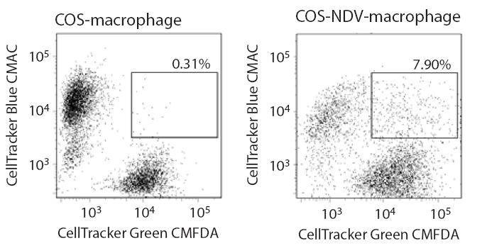
67 51 restriction is not expressed in COS or HeLa cells. To distinguish between these two possibilities, a strategy previously adopted to characterize the mechanism by which Vif promotes viral infection was used (151, 152). Heterokaryons were generated between macrophages and COS cells and the susceptibility of the heterokaryons to infection by wild-type SIV and SIVΔVpx was compared. When the fusogenic proteins of Newcastle Disease Virus (NDV) were expressed in COS cells, these cells readily underwent fusion with primary macrophages (Figure 3-2A). Macrophage/COS heterokaryons (double staining cells) were isolated by fluorescence-activated cell sorting (FACS). Double staining cells were not observed when normal COS cells (not expressing NDV proteins) were mixed with macrophages (Figure 3-2B). As an additional control, macrophage homokaryons were produced using polyethylene glycol (PEG). Both macrophage/cos heterokaryons as well as COS and macrophage homokaryons were infectible by wild-type SIV (Figure 3-2C). In contrast, macrophage homokaryons and macrophage/cos heterokaryons were resistant to SIVΔVpx infection (Figure 3-2C). Since fusion with COS cells did not relieve the block to macrophage infection by SIVΔVpx, this indicated that macrophages harbor an antiviral restriction which is counteracted by the Vpx protein and this restriction is absent from COS and Hela cells (Figure 3-3).

68 52 A B C
69 53 Figure 3-2. Vpx antagonizes an antiviral restriction in macrophages. (A) Heterokaryons were formed between primary macrophages and between COS cells expressing fusogenic HN and F proteins of Newcastle Disease Virus. To visualize heterokaryons by fluorescence microscopy, COS cells were stained with DiO (green) and macrophages were stained with DiI (red). (B) FACS analysis of macrophage-cos heterokaryons. COS cells were cotransfected with NDV HN and F expression vectors (COS-NDV) or with empty, control vectors (COS). COS cells were stained with CellTracker Green CMFDA and macrophages were stained with CellTracker Blue CMAC. Double-stained cells were sorted as indicated by the gate. (C) Susceptibility of macrophage/cos heterokaryons and COS and macrophage homokaryons to infection by SIV WT and SIVΔVpx virus variants. Infection was gauged from levels of late cdnas and 2-LTR circle cdnas.

70 54 Figure 3-3. Model of the function of Vpx in SIV SM infection of macrophages. Macrophages harbor an antiviral restriction blocking SIV SM infection before completion of reverse transcription. This restriction is counteracted by Vpx protein.
71 HIV-1 Vpr is Dispensable for HIV-1 Reverse Transcription in Macrophages HIV-2, SIV SM and SIV MAC encode both Vpx and Vpr proteins. However, Vpr is the only accessory protein in HIV-1 that is closely related to Vpx. Previous studies from our group demonstrate that Vpr proteins of HIV-2/SIV SM induce cell cycle arrest and associate with UDG, but are dispensable for macrophage infection in vitro. Vpx neither induces cell cycle arrest nor associates with UDG, but is required for SIV infection in macrophages (38, 39, 40). This suggests that HIV-1 Vpr-ascribed activities segregate between the Vpx and Vpr proteins of HIV-2/SIV SM (38). I next sought to determine if HIV-1 Vpr has the ability to overcome the restriction factor in macrophages. Vpx-deleted SIV SM was fully attenuated in macrophages, thus whether Vpr-deleted HIV-1 was also attenuated in macrophages was examined. Two HIV-1 strains, LAI and NL43 were used in this study. The transduction efficiency of wild-type or Vpr-deleted HIV-1 in macrophages was similar, when gauged by the level of 2-LTR circles (Figure 3-4). This suggests that HIV-1 Vpr is dispensable for infection of macrophages at the level of reverse transcription. The observation that both wild-type and Vpr-deleted HIV-1 were comparable in single round infection in macrophages can be explained by two possibilities. One possibility is that HIV-1 is not blocked by the same restriction factor that blocks SIV SM infection in macrophages. Therefore, the deletion of Vpr has no effect on HIV-1 2-LTR
72 56 HIV cdna (2LTR copies/cell) LAI LAIΔVpr 24h+AZT 24h 48h+AZT 48h HIV cdna (2LTR copies/cell) NL43 NL43ΔVpr 24h+AZT 24h 48h+AZT 48h Figure 3-4. Vpr is dispensable for HIV-1 infection of macrophages at the level of reverse transcription. Macrophages were infected with wild-type or Vpr-deleted HIV-1. Both LAI (upper panel) and NL43 (lower panel) strains were used for this study. Virus infection was gauged from the levels of viral 2-LTR circles at 24 and 48 hours post infection.
73 57 level. Another possibility is that HIV-1 is partially restricted by the same restriction factor, but HIV-1 Vpr is not able to overcome this restriction. This would account for the indistinguishable infectivity between wild-type and Vpr-deleted HIV-1 in macrophages. 3.5 The Antiviral Restriction Which Antagonizes SIV SM Infection of Macrophages is Active against HIV-1 A significant defect in 2-LTR circle synthesis was observed in macaque and human macrophages infected with Vpx-deleted SIV. In contrast to SIV, wild-type or Vpr-deleted HIV-1 infected human macrophages to a similar level, as evidenced by comparable levels of 2-LTR circle production. Based on these observations, it was initially assumed that HIV-1 was not affected by the macrophage restriction. If SIV SM is restricted in macrophages while HIV-1 is not, replacing the viral component that is targeted by the restriction in SIV SM with the corresponding sequence from HIV-1 would rescue SIV SM ΔVpx infection of macrophages. The same strategy was used to identify CA as the target of TRIM restriction (86). The Gag-Pol products of primate lentiviruses function at several different steps of the lentivirus life cycle. I first examined whether SIV SM Gag-Pol products are involved in determining the antiviral restriction in macrophages. Chimeras between HIV-1 and SIV SM were generated with sequences of MA, CA, NC, p6, PR or RT from HIV-1 inserted into the corresponding region of the SIV SM backbone (Figure 3-5). All the
74 58 HIV-1 SIV SM Figure 3-5. Structure of chimeric clones between HIV-1 and SIV SM. The SIV SM PBj1.9 isolate and HIV-1 NL43 were used to create these chimeric constructs. The different domains of NL43 were precisely substituted for the same domain in the PBj1.9 proviral clone. Sequences from SIV SM were shown in white, while the black area represented components from HIV-1 that were inserted into the corresponding region of the SIV SM proviral clone. Vpx-deleted variant of each chimera was also constructed.
75 59 chimeras in this study were created by overlapping PCR. The chimeric SIV SM containing p6 and PR from HIV-1 were replication-defective in COS cells that support SIV ΔVpx replication. The Vpx-deleted chimeric viruses with MA, CA, NC or RT replaced were capable of single round infection in COS cells. However, those viruses were restricted in macrophages (Figure 3-6 to Figure 3-9), suggesting that the substituted viral components were not the target of the macrophage restriction. I next examined whether the target of the restriction was located on the 5 half or 3 half of the SIV SM genome. Additional chimeric viruses were constructed with an HIV-1 backbone containing the 3 half of the SIV SM genome (Figure 3-10). The junctions between SIV SM and HIV-1 were located at the beginning the vif gene and the end of the env gene. In this chimeric NL43 (SIV-Vif-Env), the LTR and the gag-pol sequences were derived from the HIV-1 clone NL43. Vif (including a small overlapping region between pol and vif), vpx, vpr, env, tat and rev genes were derived from the SIV SM clone PBj1.9 (Figure 3-10). The Vpx-deleted variant of this chimera was created by mutating the vpx start codon and introducing an early stop codon (Figure 3-10). The mutations introduced were silent mutations for vif gene. The susceptibility of COS cells and macrophages to HIV-1/SIV SM chimeric virus infection was examined. The infectivity of NL43 (SIV-Vif-Env) and the Vpx-deleted variant NL43 (SIV-Vif-EnvΔVpx) in COS cells was similar when gauged by HIV-1 cdna levels,
76 60 A B SIV cdna (2LTR copies/cell) SIV SHIV-MA-1 Infection of COS cells SHIV-MA-2 SIVΔVpx SHIVΔVpx-MA-1 SHIVΔVpx-MA-2 Mock d1 AZT d1 d2 AZT d2 C SIV cdna (2LTR copies/cell) SIV SHIV-MA-1 Infection of Macrophages SHIV-MA-2 SIVΔVpx SHIVΔVpx-MA-1 SHIVΔVpx-MA-2 Mock d1 AZT d1 d2 AZT d2 Figure 3-6. Vpx is required for SHIV-MA infection of macrophages. (A) A schematic of chimeric proviral clone with SIV backbone and MA from HIV-1 (SHIV-MA) and the Vpx-deleted variant (SHIVΔVpx-MA). (B) Susceptibility of COS cells to infection by SHIV-MA chimera and Vpx-deleted variant SHIVΔVpx-MA. COS cells were infected and virus infection was gauged from the levels of viral 2-LTR cdna at 24 and 48 h post-infection. (C) Susceptibility of macrophages to infection by SHIV-MA chimera and Vpx-deleted variant.
77 61 A B SIV cdna (2LTR copies/cell) SHIV-CA-1 Infection of COS cells SHIV-CA-2 SIVΔVpx SHIVΔVpx-CA-1 SHIVΔVpx-CA-2 Mock d1 AZT d1 d2 AZT d2 C SIV cdna (2LTR copies/cell) SHIV-CA-1 SHIV-CA-2 Infeciton of Macrophages SIVΔVpx SHIVΔVpx-CA-1 SHIVΔVpx-CA-2 Mock d1 AZT d1 d2 AZT d2 Figure 3-7. Vpx is required for SHIV-CA infection of macrophages. (A) A schematic of chimeric proviral clone with CA and p2 from HIV-1 inserted into SIV backbone (SHIV-CA) and the Vpx-deleted variant (SHIVΔVpx-CA). (B) Susceptibility of COS cells to infection by SHIV-CA chimera and Vpx-deleted variant SHIVΔVpx-CA. (C) Susceptibility of macrophages to infection by SHIV-CA chimera and Vpx-deleted variant.
78 62 A B SIV cdna (2LTR copies) SHIV-NC-1 SHIV-NC-2 Infection of COS cells SHIV-NC-3 SIVΔVpx SHIVΔVpx-NC-1 SHIVΔVpx-NC-2 SHIVΔVpx-NC-3 Mock d1 AZT d1 C SIV cdna (2LTR copies) SHIV-NC-1 SHIV-NC-2 Infection of macrophages SHIV-NC-3 SIVΔVpx SHIVΔVpx-NC-1 SHIVΔVpx-NC-2 SHIVΔVpx-NC-3 Mock d1 AZT d1 Figure 3-8. Vpx is required for SHIV-NC infection of macrophages. (A) A schematic of chimeric proviral clone with NC and p1 from HIV-1 inserted into SIV backbone (SHIV-NC) and the Vpx-deleted variant (SHIVΔVpx-NC). (B) Susceptibility of COS cells to infection by SHIV-NC chimera and Vpx-deleted variant SHIVΔVpx-NC. (C) Susceptibility of macrophages to infection by SHIV-NC chimera and Vpx-deleted variant.
79 63 A B Infeciton of COS cells SIV cdna (2LTR copies) SIV SHIV-RT-1 SIVΔVpx SHIVΔVpx-RT-1 SHIVΔVpx-RT-2 SHIVΔVpx-RT-3 Mock d1 AZT d1 d2 AZT d2 C SIV cdna (2LTR copies) SIV SHIV-RT-1 Infection of Macrophages SIVΔVpx SHIVΔVpx-RT-1 SHIVΔVpx-RT-2 SHIVΔVpx-RT-3 Mock d1 AZT d1 d2 AZT d2 Figure 3-9. Vpx is required for SHIV-RT infection of macrophages. (A) A schematic of chimeric proviral clone with RT from HIV-1 inserted into SIV backbone (SHIV-RT) and the Vpx-deleted variant (SHIVΔVpx-RT). (B) Susceptibility of COS cells to infection by SHIV-RT chimera and Vpx-deleted variant SHIVΔVpx-RT. (C) Susceptibility of macrophages to infection by SHIV-RT chimera and Vpx-deleted variant.
80 64 NL43 (SIV-Vif-Env) NL43 (SIV-Vif-Env ΔVpx) HIV-1 SIV SM Figure A schematic of chimeric proviral clone NL43 (SIV-Vif-Env) and Vpx-deleted variant NL43 (SIV-Vif-EnvΔVpx). The fragment from vif to env of NL43 was precisely substituted for the same domain in the PBj1.9 proviral clone. Sequences from SIV SM were shown in white, while the black area represented the fragment from HIV-1 that was inserted into the corresponding region of the SIV SM proviral clone. NL43 (SIV-Vif-Env ΔVpx) was the same chimera in which the expression of Vpx was eliminated. Vpx was deleted by mutating the start codon of Vpx and introducing an early stop codon. All the mutations introduced were silent mutations for vif gene.
81 65 indicating that these chimeric viruses are capable of single round infection in permissive cells (Figure 3-11B). However, infection of macrophages by NL43 (SIV-Vif-EnvΔVpx) was highly inefficient compared to that by NL43 (SIV-Vif-Env), as evidenced by the levels of viral cdna synthesis (Figure 3-11A), suggesting Vpx dramatically enhanced the infectivity of this chimera in macrophages. Therefore, chimeric NL43 (SIV-Vif-Env) is restricted in macrophages, indicating that the 3 half of SIV SM may contain the target of the macrophage restriction. To confirm this, another chimera between HIV-1 and SIV SM was constructed in which the fragment of HIV-1 vif-vpr was replaced by SIV SM vif-vpx in the context of the NL43 backbone. The junctions between SIV SM and HIV-1 were located at the beginning of vif and the end of vpx (Figure 3-12). The infectivity of these chimeric viruses in COS cells and macrophages was examined. In permissive COS cells, both NL43 (SIV-Vif-Vpx) and Vpx-deleted variant NL43 (SIV-Vif-ΔVpx) underwent comparable reverse transcription with similar levels of 2-LTR circle production (Figure 3-13B). Surprisingly, 2-LTR circle production in macrophages infected with NL43 (SIV-Vif-ΔVpx) dramatically decreased (Figure 3-13A), indicating that this chimera is antagonized by the macrophage restriction. The Vpx-deleted chimeras with the HIV-1 backbone infected macrophages although with lower efficiency, suggesting that they are partially restricted compared to SIV SM. These results led us to consider that HIV-1 is also
82 66 A Macropahges 2LTR cdna copies NL43(SIV-Vif-Env) NL43(SIV-Vif-EnvΔVpx) 24h+AZT 24h 48h+AZT 48h B COS cells 2LTR cdna copies NL43(SIV-Vif-Env) NL43(SIV-Vif-EnvΔVpx) 24h+AZT 24h 48h+AZT 48h Figure Vpx promotes chimeric NL43 (SIV-Vif-Env) infection in macrophages. (A) Susceptibility of macrophages to infection by NL43 (SIV-Vif-Env) chimera and Vpx-deleted variant NL43 (SIV-Vif-EnvΔVpx). Macropahges were infected and virus infection was gauged from the levels of viral 2-LTR cdna at 24 and 48 h post-infection. (B) Susceptibility of COS cells to infection by NL43 (SIV-Vif-Env) chimera and Vpx-deleted variant.
83 67 NL43 (SIV-Vif-Vpx) NL43 (SIV-Vif-ΔVpx) HIV-1 SIV Figure A schematic of chimeric proviral clone NL43 (SIV-Vif-Vpx) and Vpx-deleted variant NL43 (SIV-Vif-ΔVpx). The black area represents the fragment from HIV-1 and the white area represents the fragment from HIV-1. The fragment from vif to vpx of NL43 was precisely substituted for the same domain in the PBj1.9 proviral clone. NL43 (SIV-Vif-ΔVpx) was the same chimera in which the expression of Vpx was eliminated. Vpx was deleted by mutating the start codon of Vpx and introducing an early stop codon. All the mutations introduced were silent mutations for vif gene.
84 68 A Macrophages 2LTR cdna copies NL43(SIV-Vif-Vpx) NL43(SIV-Vif-ΔVpx) 24h+AZT 24h 48h+AZT 48h B COS cells 2LTR cdna copies NL43(SIV-Vif-Vpx) NL43(SIV-Vif-ΔVpx) 24h+AZT 24h 48h+AZT 48h Figure Vpx promotes NL43 (SIV-Vif-Vpx) infection in macrophages. (A) Differential susceptibility of macrophages to infection by NL43 (SIV-Vif-Vpx) and Vpx-deleted variant NL43 (SIV-Vif- ΔVpx). Macropahges were infected and virus infection was gauged from the levels of viral 2-LTR cdna at 24 and 48 hours post infection. (B) Susceptibility of COS cells to infection by by NL43 (SIV-Vif-Vpx) and Vpx-deleted variant. COS cells were infected and virus infection was determined as described in A.
85 69 antagonized by the macrophage restriction that blocks SIV SM infection. To evaluate whether the antiviral restriction which antagonized HIV-2/SIV SM infection was active against HIV-1 and if Vpx indeed promoted HIV-1 infection of macrophages, the spreading infection of macropahges by a HIV-1 variant expressing Vpx was examined. A chimeric construct ADAΔVpr(PBj-Vpx) was created, within which the sequence of Vpr in the ADA backbone was replaced by the sequence of Vpx derived from SIV PBj (Figure 3-14A). Since the expression of Vpr was eliminated in this chimera, Vpr-deleted ADA was used as a control. Macropahges were infected with wild-type ADA, ADAΔVpr and ADAΔVpr(PBj-Vpx) chimera that expressed Vpx. The culture Supernatants were collected and HIV-1 infection was determined by reverse transcriptase activity in the supernatants over 31 days post-infection. The infectivity of ADAΔVpr was lower than that of wild-type ADA in macrophages (Figure 3-14B). However, the infectivity of ADAΔVpr was profoundly enhanced compared to that of ADAΔVpr (Figure 3-14B), indicating that the expression of Vpx promoted HIV-1 infection in macrophages. Therefore, the antiviral restriction that antagonizes SIV SM infection of macrophages is active against HIV Vpx Counters the Restriction and Promotes HIV-1 infection in trans Vpx and Vpr are virion proteins and would thus be predicted to exert their function in the target cell shortly after infection. Therefore, I next examined whether the Vpx
86 70 A ADA vpx ADAΔVpr(PBj-Vpx) B RT activity (million cpm/ml) d1 d4 d7 d10 d13 d16 d19 d22 d25 d28 d31 Days post inoculation ADA ADAΔVpr ADAΔVpr(PBj-Vpx)
87 71 Figure Vpx enhances HIV-1 spreading infection in macrophages. (A) Construction of ADAΔVpr(PBj-Vpx). A schematic of wild-type ADA and chimeric ADA which expresses SIV SM Vpx. Vpx sequence was derived from SIV PBj and was inserted into ADA backbone in place of Vpr. (B) Spreading infection of macrophages by wild-type ADA, ADAΔVpr or ADAΔVpr((PBj-Vpx). Macrophages were infected with RT unites (cpm) of ADA, ADAΔVpr or ADAΔVpr((PBj-Vpx). Culture supernatants were collected and RT activity (10 6 cpm/ml) was measured at 3 day intervals post-infection.
88 72 protein, when packaged in trans within HIV-1 virions, enhanced virus infectivity for primary macrophages. HIV-1 Vpr and SIV SM Vpx proteins were packaged into wild-type or Vpr-deleted HIV-1 viruses by cotransfection. The infectivity of those viruses for COS cells and macrophages was then determined from levels of viral cdna (2-LTR circles) at 24 and 48 hours post-infection. While Vpx had no significant effect on the infectivity of wild-type HIV-1, the infectivity of HIV-1ΔVpr for macrophages was profoundly enhanced by Vpx but not by HIV-1 Vpr (Figure 3-15, lower panel). The infectivity enhancement was also apparent in macrophages infected with an HIV-1 variant expressing GFP (Figure 3-16). Thus, while HIV-1 was infectious for macrophages, its ability to infect these cells was markedly enhanced in the presence of Vpx. Vpx had no effect on the infectivity of wild-type or ΔVpr HIV-1 for COS cells (Figure 3-15, upper panel). In the above experiments, VSV-G pseudotyped HIV-1 was used. To further confirm that Vpx promotes HIV-1 infection in trans, I used a non-pseudotyped HIV-1 virus NA20 B59/NL43, which is a macrophage tropic NL43 variant. The infectivity of NA20 B59/NL43ΔVpr for macrophages was remarkably enhanced by packaging of SIV Vpx (Figure 3-17). Collectively, these results support our hypothesis that although HIV-1 is infectious
89 73 Figure HIV-1 is sensitive to the macrophage restriction and SIV Vpx but not HIV-1 Vpr antagonizes the restriction. HIV-1 Vpr and SIV Vpx proteins were packaged in HIV-1 WT or HIV-1 ΔVpr viruses by cotransfection. For controls, viruses were cotransfected with an empty vector. The infectivity of those viruses for COS-1 cells (upper panel) and macrophages (lower panel) was then determined from levels of viral cdna (2-LTR circles) at 24 and 48 hours post-infection.
 or was not ( ) packaged.")
90 74 Figure Vpx enhances transduction of macrophages by GFP-expressing HIV-1. Infection of macrophages with GFP-expressing HIV-1ΔVpr variants in which Vpx was (+) or was not ( ) packaged. GFP positive macrophages (representative fields) were visualized 48 h post-infection.
91 75 HIV-1 cdna (2LTR copies/cell) Empty vector Vpx Empty vector Vpx 24h+AZT 24h 48h+AZT 48h NA20 B59/NL43 WT NA20 B59/NL43ΔVpr Figure SIV SM Vpx promotes NA20 B59/NL43ΔVpr infection in macrophages. Macrophages were infected with a non-pseudotyped NL43 variant NA20 B59/NL43 (R5 tropic) that had or had not packaged Vpx. Virus infection was gauged from the levels of viral 2-LTR cdna 24 and 48hours post inoculation.
92 76 for macrophages, HIV-1 is partially restricted by the same restriction factor that blocks SIV SM ΔVpx, at the level of reverse transcription. The ability of HIV-1 to infect macrophages is remarkably enhanced in the presence of Vpx. However, HIV-1 Vpr is not able to overcome the restriction 3.7 Vpx Renders Macrophages Permissive to MLV Infection A fundamental characteristic that distinguishes lentiviruses from simple gammaretroviruses is their capacity to infect nondividing cells (153, 154). Primate lentiviruses are able to transduce nondividing cells, and this underscores their ability to transduce terminally differentiated nondividing cells, including macrophages, microglia, and dendritic cells, both in vitro and in vivo (17, 155, 156). In contrast, gammaretroviruses such as murine leukemia virus (MLV) transduce cells in mitosis and nondividing cells (in G1/S/G2) are refractory to gammaretrovirus transduction ( ). Previous experiments have presented evidence that macrophages harbor a restriction that antagonizes HIV-1 and SIV at the level of reverse transcription and that the Vpx protein of SIV SM specifically overcomes this restriction. I next examined whether neutralization of the restriction by Vpx would render macrophages permissive to MLV. Whether introduction of Vpx into macrophages by wild-type SIV infection would render macrophages susceptible to subsequent transduction by MLV was first examined.
93 77 Macrophages were initially infected with SIV WT or SIVΔVpx and subsequently infected by MLV. MLV late cdna copies (Figure 3-18A, upper panel) and 2-LTR circles (Figure 3-18A, lower panel) were determined 24 and 48 hours after MLV infection. The primary block to transduction of macrophages by MLV appeared to be at the level of reverse transcription (Figure 3-18A). Pre-infection of macrophages with a SIV WT but not a Vpx-deleted SIV (SIVΔVpx) resulted in an increased ability of MLV to transduce macrophages, as evidenced by MLV cdna synthesis (Figure 3-18A) and expression of GFP from the MLV genome (Figure 3-18B). Therefore, the restriction that antagonizes lentivirus infection of macrophages also prevents infection of macrophages by MLV and Vpx delivered in trans permits transduction of macrophages by MLV.
 2.5 2 1.5 1 0.")
94 78 A 800 MLV cdna (late copies) h 48h 0 AZT No Pre SIVΔVpx SIVwt Pre-infection 3 MLV cdna (2LTR copies) h 48h 0 AZT No Pre SIVΔVpx SIVwt Pre-infection B
95 79 Figure Vpx permits transduction of macrophages by MLV in trans (A) Vpx delivered to macrophages by wild-type SIV (SIV WT ) infection removes the block to synthesis of MLV cdna in macrophages. Macrophages were initially infected with SIV WT or SIVΔVpx and subsequently infected by MLV-GFP after 4 hours. Macrophages infected with MLV without pre-infection were used as control (no Pre). Synthesis of MLV late cdnas (upper panel) and 2-LTR circles (lower panel) was assessed 24 and 48 hours after MLV infection. (B) A representative field of macrophages transduced by MLV-GFP in an independent experiment.
96 80 CHAPTER IV Proteasome/Ubiquitylation Pathway and DDB1 Interaction are Required for the Ability of Vpx to Counteract the Restriction 4.1 Chapter Summary A possible role for the proteasome/ubiquitin system in the ability of Vpx to neutralize the unknown restriction was evaluated. Vpx was ubiquitylated by over-expressed Histidine-ubiquitin in 293 T cells, as well as by endogenous ubiquitin. The extent of ubiquitylation was reduced in a Vpx mutant (Vpx M4 ) lacking all four lysine ubiquitylation sites. Vpx M4, although packaged within HIV-1 particles, was defective in enhancing HIV-1 infection of macrophages. This suggestes the ability of Vpx to enhance HIV-1 infectivity requires its ubiquitylation. Furthermore, Vpx failed to enhance the permissiveness of macrophages to HIV-1 or MLV infection when proteasome function was disrupted by a proteasome inhibitor. This indicates that a functional proteasome system was necessary for the biological activity of Vpx. This chapter also provides evidence that a cellular binding partner for Vpr, DDB1, is required for the ability of Vpx to counteract the restriction. In 293T cells, endogenous DDB1 associated with a wild-type SIV Vpx protein but not with Vpx M4, which suggests that Vpx ubiquitylation is required for DDB1 interaction. This may explain why ubiquitylation sites are important for Vpx function. Furthermore, fusion of DDB1 to SIV
97 81 Vpr partially restored the ability of Vpx-deleted SIV to infect macrophages. Vpx failed to enhance lentiviral infection in macrophages in the presence of HIV-1 Vpr, suggesting that HIV-1 Vpr impaired the ability of Vpx to overcome the restriction. Vpx was packaged into wild-type HIV-1 as well as Vpr-deleted HIV-1 virions. Therefore, HIV-1 Vpr did not compete with SIV Vpx for virion packaging. Infection of macrophages by SIV harboring a wild-type Vpx gene alleviated the block to subsequent infection by wild-type or ΔVpr HIV-1, suggesting that HIV-1 Vpr competed with SIV Vpx in target cells, possibly for restriction binding. To determine if Vpr from lab-adapted HIV-1 lost its ability to overcome the restriction due to mutations happened during propagation in vitro, I examined if primary Vpr alleles derived from AIDS patients were able to promote HIV-1 infection. All the tested primary HIV-1 Vpr alleles failed to enhance HIV-1 infection. This confirmed that HIV-1 Vpr does not overcome the putative restriction. In addition, I examined the amount of Vpx needed to rescue SIVΔVpx infection in macrophages. The ability of trans-packaged Vpx to counteract a macrophage restriction is dose-dependent, and only a small amount of Vpx packaged in trans is sufficient to overcome the restriction in macrophages. 4.2 Vpx Activity Requires Ubiquitylation and Functional Proteasome Primate lentiviruses have evolved the accessory protein Vif to counteract the
98 82 antiviral activity of cellular APOBEC 3 cytidine deaminases (30). Vif achieves this by promoting ubiquitylation and proteasomal degradation of APOBEC3 proteins (161, 162). Therefore Vpx possibly antagonizes the macrophage restriction through a similar mechanism. Vpx is ubiquitylated To evaluate a possible role for the ubiquitin-proteasome system in the activity of Vpx, whether Vpx itself was ubiquitylated was first examined. A Vpx mutant (Vpx M4 ) containing lysine (K) to arginine (R) substitutions at all four lysine residues was constructed (Figure 4-1A). HA-tagged Vpx and Vpx M4 were co-expressed in 293T cells with 6-Histidine-myc-tagged ubiquitin. Mono and poly ubiquitylated Vpx proteins were purified on nickel beads and western blotted. Immunoblotting with an HA antibody revealed the presence of mono and poly ubiquitylated forms of Vpx (Ub-Vpx, Figure 4-1B, left panel). It was also examined whether Vpx was ubiquitylated by endogenous ubiquitin (as opposed to over expressed and tagged ubiquitin). HA-tagged Vpx was expressed in 293 T cells and cell lysates were directly Western blotted and probed with an HA antibody. This revealed the presence of higher molecular weight ubiquitylated forms of Vpx (Figure 4-1B, right panel). The extent of Vpx ubiquitylation was reduced in Vpx M4 (Figure 4-1B, left panel). Despite mutagenesis of all four lysines in Vpx, polyubiquitylated forms of the protein were still evident (compare GFP signal with
 Wild-type and lysine mutant Vpx M4 (HA tag) were expressed in 293T cells expressing Histidine-tagged ubiquitin. Ubiquitylated Vpx proteins (Ub-Vpx) were purified on nickel beads.")
99 83 A B Figure 4-1. Identification of ubiquitylated residues in Vpx. (A) The positions of the Lysine (K) to Arginine (R) mutations in Vpx M4 mutant. (B) Wild-type and lysine mutant Vpx M4 (HA tag) were expressed in 293T cells expressing Histidine-tagged ubiquitin. Ubiquitylated Vpx proteins (Ub-Vpx) were purified on nickel beads. Immunoblotting with an HA antibody revealed the presence of mono and poly ubiquitylated forms of Vpx (left panel). Vpx was also ubiquitylated by endogenous ubiquitin. HA-tagged Vpx was expressed in 293T cells and cell lysates were directly blotted with anti-ha. The presence of higher molecular weight Ub-Vpx was revealed (right panel).
100 84 Vpx M4 signal, Figure 4-1B, left panel). This suggested an involvement of both lysine and nonlysine residues in Vpx ubiquitylation (163, 164). Vpx M4 does not enhance HIV-1 infection in macrophages. I first examined if Vpx M4 mutant was able to be packaged into HIV-1 virions. Vpx M4 was packaged within HIV-1 particles at levels indistinguishable from wild-type Vpx (Figure 4-2). The packaging of the Vpx M4 in viral particles suggests, at the very least, that this mutant is competent for binding to the p6 domain of the viral Gag polyprotein through which packaging of Vpr and Vpx protein is mediated (112, 113). I next evaluated whether the ability of Vpx to enhance HIV-1 infectivity depended upon its ubiquitylation. Wild-type SIV SM Vpx, Vpx M4 or wild-type HIV-1 Vpr were packaged within HIV-1ΔVpr virions by cotransfection. Empty vector was cotransfected with HIV-1 proviral clone as a control. Single cycle infection of macrophages was evaluated from synthesis of 2-LTR cdnas. Package of Vpx dramatically promoted HIV-1 infection of macrophages (Figure 4-3). VpxM4 did not enhance HIV-1 infectivity when packaged within HIV-1ΔVpr virions (Figure 4-3), indicating the ability of Vpx to enhance the infectivity of HIV-1ΔVpr was compromised by the M4 mutation. To conclude, a Vpx mutant lacking lysine ubiquitylation sites failed to enhance macrophage infection by HIV-1. Therefore, the ability of Vpx to enhance HIV-1 infectivity for macrophages requires its ubiquitylation.

101 85 Figure 4-2. Packaging of Vpx proteins in wild-type and Vpr-deleted HIV-1. HA tagged wild-type Vpx or Vpx M4 proteins were packaged within NL43 WT or NL43 ΔVpr by cotransfection. The virions were collected and pelleted with 20% sucrose cushion. Virion lysates are subject to SDS-PAGE and blotted with anti-p24 (left panel) or anti-ha (right panel).
102 86 Figure 4-3. Ubiquitylation is required by Vpx for enhancement of HIV-1 infectivity in macrophages. Wild-type HIV-1 Vpr, SIV Vpx or lysine mutant Vpx (Vpx M4 ) proteins were packaged in HIV-1 ΔVpr and infectivity of those viruses was assessed in macrophages. Viral cdna synthesis was evaluated 24 and 48 h post-infection.
103 87 A functional proteasome is necessary for the ability of Vpx to enhance HIV-1 and MLV infection Whether the ability of Vpx to regulate HIV-1 and MLV infection of macrophages required proteasome function was then examined. Macrophages were treated with two different proteasome inhibitors and then infected with HIV-1ΔVpr variants that had packaged wild-type Vpx or Vpx M4. 2-LTR cdna was quantitated 24 hours after infection. ALLN and proteasome inhibitor 1 (Prot.1) markedly impaired infection of macrophages by HIV-1ΔVpr containing wild-type Vpx (Figure 4-4A). In contrast, macrophage infection by HIV-1ΔVpr which does not contain wild-type Vpx was not dramatically compromised by the proteasome inhibitors (Figure 4-4A). Since proteasome disruption only impacted macrophage infection by HIV-1ΔVpr containing Vpx, this argued that proteasome inhibition specifically impaired Vpx function rather than impacting virus infection through off-target effects. I also examined if the ability of Vpx to render macrophages permissive to MLV infection required a functional proteasome. Macrophages were treated with the proteasome inhibitor ALLN, or DMSO as control. The cells were then infected with wild-type or Vpx-deleted SIV, and subsequently infected with MLV after 6 hours. MLV cdna production was quantitated by real-time PCR 24 and 48 hours post-infection. Vpx delivered to macrophages by wild-type SIV pre-infection rendered macrophages
104 88 A B MLV in Macrophages 3.5 MLV cdna (late/ccr5) h 48h 0 AZT No-Preinfection SIVwt no treat SIVwt+DMSO SIVwt+ALLN SIVΔVpx no treat SIVΔVpx+DMSO SIVΔVpx+ALLN Pre-infection
105 89 Figure 4-4. The ability of Vpx to enhance permissivity of macrophages to HIV-1 and MLV infection requires a functional proteasome. Effects of proteasome inhibitors on HIV and MLV infection of macrophages are indicated. (A) Macrophages were treated with the proteasome inhibitors ALLN or proteasome inhibitor 1 (Prot. 1) or with DMSO as a control. Those cells were then infected with HIV-1ΔVpr variants which had packaged wild-type or lysine mutant Vpx proteins. The level of HIV-1 infection (2-LTR cdna) was determined 24 hours post-infection by PCR. (B) Macrophages were treated with the proteasome inhibitor ALLN or with DMSO as a control. Those cells were initially infected with wild-type or ΔVpx SIV, and subsequently infected by MLV. The level of MLV infection (late cdna) was determined 24 and 48 hours post-infection.
106 90 permissive to MLV (Figure 4-4B). However, Vpx was not able to rescue MLV infection of macrophages in cells that were treated with a proteasome inhibitor (Figure 4-4B). Vpx failed to enhance permissiveness of macrophages to HIV-1 or MLV infection in macrophages in which proteasome function was disrupted by proteasome inhibitors. Therefore, the ability of Vpx to enhance HIV-1 infectivity or to rescue MLV infection required proteasome function. Collectively, these experiments indicate the presence of a potent antiviral restriction in macrophages that is counteracted by the Vpx protein and that the proteasome/ubiquitin system is required for the ability of Vpx to counteract this restriction. 4.3 Vpx Function Requires DDB1 Recent studies have demonstrated that the ability of HIV-1 Vpr to induce cell cycle arrest requires the E3 ubiquitin ligase complex scaffolding factor, damaged DNA binding protein 1 (DDB1) ( ). Therefore, whether the ability of Vpx to counteract the macrophage restriction to SIV and HIV-1 infection was DDB1-dependent was examined. First experiment investigated whether DDB1 is required for Vpx ubiquitylation or Vpx ubiquitylation is required for DDB1 interaction. In 293T cells, endogenous DDB1 associated with a wild-type SIV Vpx protein but not with Vpx M4 (Figure 4-5A). A
 and non-ubiquitylated Vpx (Vpx M4 ) was evaluated in 293T cells expressing FLAG-tagged Vpx or IRES-GFP as a control.")
107 91 A B Figure 4-5. Vpx ubiquitylation is required for Vpx interaction with DDB1. (A) Association of SIV Vpx with endogenous DDB1. Association of DDB1 with wild-type Vpx (Vpx WT ) and non-ubiquitylated Vpx (Vpx M4 ) was evaluated in 293T cells expressing FLAG-tagged Vpx or IRES-GFP as a control. FLAG immunoprecipitates were immunoblotted with DDB1 or FLAG antibodies (upper panels). Levels of endogenous DDB1 and expressed Vpx in cell lysates were confirmed by Western blotting with a DDB1 antibody and with a FLAG antibodie respectively. (lower panels) (B) Impact of DDB1 silencing on Vpx ubiquitylation. 293T cells were cotransfected with DDB1 or scrambled sirnas and with Histidine-ubiquitin and HA-Vpx or IRES-GFP expression plasmids as outlined in Figure 4-1. Ubiquitin-conjugated proteins were nickel purified and immunoblotted for HA-Vpx (upper panels). Cell lysates were directly blotted for Vpx and DDB1 proteins (lower two panels).
108 92 specific association of SIV Vpx with DDB1 was apparent in coimmunoprecipitation experiments with FLAG-tagged Vpx protein (Figure 4-5A). Although ubiquitylation was necessary for the ability of Vpx to counteract the restriction to HIV-1 and SIV infection of macrophages, DDB1 protein was not required for Vpx ubiquitylation (Figure 4-5B). Mono and poly ubiquitylated forms of Vpx were evident and apparently increased in cells in which DDB1 expression was reduced by RNA interference (Figure 4-5B). Whether DDB1 was required for the ability of Vpx to counteract the restriction to HIV-1 infection was next examined. Since Vpx, when packaged in HIV-1 virions, enhanced macrophage infection, a transpackage strategy was used to examine whether Vpx enhanced HIV-1 infection in DDB1 depleted macrophages. While packaging of Vpx in HIV-1 particles markedly increased infectivity for macrophages transfected with a scrambled sirna (Figure 4-6A), silencing of DDB1 in macrophages significantly reduced the ability of Vpx to enhance HIV-1 infection (Figure 4-6A). However, HIV-1 Vpr did not antagonize a macrophage restriction. The activity of the restriction in HIV-1 was only revealed by the ability of Vpx to enhance HIV-1 infection of macrophages. In agreement with this, DDB1 silencing had no significant effect on the infectivity of HIV-1 which had not packaged Vpx (Figure 4-6A). Therefore, DDB1 appears to be a specific Vpx cofactor in primary macrophages. Since SIV Vpx but not SIV Vpr was essential for macrophage infection (Figure
109 93 Figure 4-6. DDB1 is required for the ability of Vpx to counteract the restriction to macrophage infection by HIV-1 and SIV. (A) SIV Vpx (or GFP as a control) was packaged into HIV-1 ΔVpr virions by co-transfection. Infectivity of those viruses for DDB1-depleted macrophages (DDB1 sirna) or control macrophages (ScrI sirna) were evaluated from levels of viral cdna 24 h later (+, p>0.2; *, p<0.002). (B) DDB1 packaging partially substitutes for Vpx. A Vpx/Vpr deletion mutant of SIV (SIVΔXR) was co-transfected with vectors expressing SIV Vpr, SIV Vpx, DDB1 or a Vpr-DDB1 fusion. Infectivity of the resulting viruses for macrophages was evaluated from levels of SIV cdna at 24 and 48 hours post-infection (*, p<0.005).
110 94 3-1A), I examined whether fusion of DDB1 to SIV Vpr was sufficient to allow SIV Vpr to counteract the macrophage restriction. Packaging of Vpr alone into a Vpr and Vpx-deleted SIV (SIV ΔXR ) did not permit macrophage infection. In contrast, there was a partial and significant restoration of infectivity when a Vpr-DDB1 fusion was packaged relative to infectivity of virions in which the DDB1 protein was not packaged (Figure 4-6B). Collectively, these results suggest that DDB1 is required for the ability of Vpx to antagonize a restriction to infect macrophages by HIV and SIV, but that DDB1 is not required for Vpx ubiquitylation. 4.4 HIV-1 Vpr Impairs the Ability of Vpx to Neutralize the Macrophage Restriction Previous experiments demonstrated that Vpx had the ability to compliment HIV-1ΔVpr but not wild-type HIV-1 (Figure 3-15 and Figure 3-17). Therefore, one possibility is that the ability of Vpx to promote HIV-1 infection is compromised by the presence of HIV-1 Vpr. To assess this hypothesis I packaged SIV SM Vpx or HIV-1 Vpr into a Vpx-expressing HIV-1 variant NL43 (SIV-Vif-Vpx), or NL43 (SIV-Vif-ΔVpx) that does not express Vpx. The infectivity of NL43 (SIV-Vif-Vpx) was dramatically higher than that of NL43 (SIV-Vif-ΔVpx) in macrophages, as evidenced by viral 2-LTR production (Figure 4-7). However, packaging of HIV-1 Vpr into NL43 (SIV-Vif-Vpx) virions profoundly decreased virus infection of macrophages, to a level similar to NL43 (SIV-Vif-ΔVpx) (Figure 4-7). This confirmed that the packaged HIV-1 Vpr protein
111 HIV-1 cdna (2LTR copies) No trans GFP Vpx Vpr No trans GFP Vpx Vpr 24h+AZT 24h 48h+AZT 48h Proteins packaged in trans NL43 (SIV-Vif-Vpx) NL43 (SIV-Vif-ΔVpx) Figure 4-7. HIV Vpr inhibits the ability of SIV SM Vpx to antagonize the macrophage restriction. Macrophages were infected with NL43 (SIV-Vif-Vpx) which had packaged SIV SM Vpx or HIV-1 Vpr proteins. The infectivity of those viruses was gauged from the levels of HIV-1 2-LTR circle 24 and 48 hours post infection.
112 96 inhibited the ability of Vpx to neutralize the macrophage restriction. A possible explanation is that Vpx and HIV-1 Vpr proteins compete for packaging within HIV-1 virions. An alternative possibility is that these proteins do not compete for packaging into virions but compete for interaction with the restriction after infection has occurred. Western blotting analysis revealed that Vpx proteins were packaged into wild-type and Vpr-deleted HIV-1 virions (Figure 4-2). This suggested that HIV-1 Vpr competed with Vpx in the target cell following infection and this competition precluded the ability of Vpx to neutralize the restriction. A prediction of this is that delivery of Vpx to this target cell prior to HIV-1 infection should be sufficient to inactivate the restriction and subsequently enhance macrophage infection by both wild-type and Vpr-deleted HIV-1. To evaluate this, I bypassed the requirement for Vpx packaging by directly introducing Vpx into the target cell by SIV WT infection. The susceptibility of those cells to infection by wild-type or Vpr-deleted HIV-1 variants was then examined. The infectivity of both wild-type and Vpr-deleted HIV-1 variants for macrophages was enhanced when Vpx was first introduced into the cell by SIV WT infection (Figure 4-8). In contrast, prior infection with a SIVΔVpx variant did not enhance subsequent HIV-1 infection of macrophages (Figure 4-8). Therefore, in the absence of competition by packaged Vpr, Vpx greatly enhanced wild-type HIV-1 infectivity for macrophages. In summary, HIV-1 Vpr does not abrogate Vpx packaging into HIV-1 virions.
113 97 Figure 4-8. Vpx delivered to macrophages by SIV WT infection enhances the permissivity to HIV-1 WT and HIV-1ΔVpr infection. Macrophages were first infected (1º infection) with wild-type or ΔVpx SIV variants, left uninfected (none) or treated with AZT. After 8 h, these cells were super-infected (2º infection) with WT or ΔVpr HIV-1 variants and HIV-1 infection (2-LTR cdna synthesis) was determined 24 and 48 h later.
114 98 Instead, HIV-1 Vpr interferes with Vpx function in target cells, possibly by competing for interaction with the restriction factor. Without competition from HIV-1 Vpr, SIV SM Vpx delivered into macrophages by SIV pre-infection antagonizes the restriction and enhances wild-type HIV-1 infection. 4.5 Primary HIV-1 Vpr Alleles Fail to Overcome the Macrophage Restriction Initial experiments demonstrated that both HIV-1 and SIV SM were antagonized by a macrophage restriction factor. Although HIV-1 was only partially restricted in macrophages and infected macrophages to a moderate level, the introduction of Vpx dramatically enhanced its infectivity. One possibility is that HIV-1 Vpr antagonizes the macrophage restriction but the ability of Vpr to overcome the restriction is lost during viral expansion in lymphocyte culture. HIV-1 Vpr induces cell cycle arrest; therefore, it is possible that during viral propagation in vitro, viruses with mutations in vpr that are less efficient in inducing G2 cell cycle arrest are selected. As a result, Vpr from lab-adapted viruses may lose the ability to counteract the restriction factor. To test this hypothesis, I examined if primary Vpr alleles derived from AIDS patients were able to neutralize the macrophage restriction and promote HIV-1 infection. PBMCs were obtained from AIDS patients with high viral load. Primary Vpr sequences were amplified from total cdna and then cloned into a p-ires-egfp expression vector. Primary Vpr proteins were packaged into Vpr-deleted HIV-1 virions by cotransfection.
115 99 While HIV-1 containing wild-type NL43 Vpr moderately infected macrophages, encapsidation of SIV SM Vpx enhanced HIV-1 infectivity in macrophages dramatically (Figure 4-9). However, none of the primary Vpr alleles obtained from clinical samples enhanced HIV-1 transduction in macrophages (Figure 4-9), suggesting that the primary Vpr alleles do not neutralize the macrophage restriction. 4.6 The Ability of Vpx to Neutralize the Restriction is Dose Dependent Previous experiments have demonstrated that Vpx delivered in trans by virus particles enhances the permissivity of macrophages to SIV SM and HIV-1 infection. I next titered the amount of Vpx needed to rescue SIVΔVpx infection in macrophages and whether the ability of packaged Vpx to counteract the macrophage restriction was dose-dependent. Since primary macrophages cannot be transfected with Vpx expression plasmids and expression of Vpx following infection of macrophages with viruses containing packaged Vpx has not been detectable with available Vpx antibodies, I conducted an experiment in which different amounts of Vpx were packaged into SIV SM virions. A Vpx-deleted SIV (SIVΔVpx) variant expressing GFP was used for this experiment. Increasing amount of Vpx was packaged into SIVΔVpx by cotransfection. Wild-type SIV was used as a control. The levels of packaged Vpx in the purified virions were determined by western blotting with a Vpx antibody and were normalized with p27
116 100 HIV-1Δvpr containing primary Vpr HIV-1 cdna (2LTR copies) NL43Δvpr+GFP NL43Δvpr+Vpx NL43Δvpr+Vpr NL43 NL43Δvpr+Vpr 48-5 NL43Δvpr+Vpr 90-7 NL43Δvpr+Vpr 48-6 NL43Δvpr+Vpr 48-7 NL43Δvpr+Vpr 49-4 NL43Δvpr+Vpr h+AZT 24h 48h+AZT 48h Figure 4-9. Primary HIV-1 Vpr alleles fail to overcome the macrophage restriction. Primary Vpr alleles were derived from AIDS patient plasma. NL43 Vpr and primary Vpr proteins were packaged into Vpr deleted HIV-1 (NL43ΔVpr) virions by cotransfection. The infectivity of those viruses for macrophages was then determined from levels of 2-LTR cdna 24 and 48 hours post-infection.
117 101 level. With increasing amount of a co-transfected Vpx-expressing plasmid, an increasing amount of Vpx in viral particles was observed (Figure 4-10A). When 0.5µg Vpx-expressing plasmid was cotransfected with 9µg SIVΔVpx proviral DNA, the amount of Vpx packaged into SIVΔVpx virions reached a level similar to that of wild-type SIV (Figure 4-10A). The infectivity of these viruses in primary macrophages was determined from the levels of SIV 2-LTR circle or late cdna 24 and 48 hours post-infection. The percentage of infected (GFP + ) cells was determined by FACS. An increasing level of 2-LTR production was observed with increasing amount of packaged Vpx (Figure 4-10B). The percentage of GFP + post infection was also proportional to the amount of Vpx packaged into particles (Figure 4-10C). This suggests that the ability of packaged Vpx to counteract a macrophage restriction is dose-dependent. It was also observed that even a small amount of trans-packaged Vpx can counter the restriction present in macrophages (Figure 4-10). Vpx is packaged in molar amounts equivalent to Gag proteins (165). Assuming ~2000 Gag molecules per virion (166), and assuming uniform Vpx:Gag stoichiometry in each viral particle, Vpx packaged at ~10% of wild-type levels still rescued a ΔVpx virus (Figure 4-10), suggesting that as few as 200 Vpx molecules/virion can counteract the restriction.

118 102 A B 7.00E+00 SIV cdna (2LTR/cell) 6.00E E E E E E+00 AZT d1 d2 0.00E+00 X2+0µg vpx X2+0.01µg vpx X µg vpx X2+0.05µg vpx X2+0.1µg vpx X2+0.25µg vpx X2+0.5µg vpx PBj wt C 50 GFP+ Percnetage (%) d2 d3 0 Mock X2+0µg vpx X2+0.01µg vpx X µg vpx X2+0.05µg vpx X2+0.1µg vpx X2+0.25µg vpx X2+0.5µg vpx PBj wt
119 103 Figure Dose-dependent ability of packaged Vpx to counteract a macrophage restriction. Pseudotyped SIVΔVpx (X2) was produced in 293T cells by cotransfecting SIVΔVpx proviral DNA with pvsv-g and increasing amount of a Vpx expression vector. (A) The amount of packaged Vpx in the purified virions was determined by western blotting with a Vpx antibody and normalization with p27 by densitometry. The integrated density was measured by Scion Image software. (B) The infectivity of these viruses in primary macrophages was determined from quantitation of SIV 2-LTR cdna 24 and 48 h post-infection. PBj WT was used as positive control for this experiment. (C) Virus transduction was determined from percentage of GFP + cells 48 and 72 h post-infection by FACS.
120 104 CHAPTER V Vpx Renders Primary Monocytes Permissive to HIV-1 Transduction 5.1 Chapter Summary Quiescent monocytes are highly refractory to HIV-1 and the infection is blocked at an early post-entry step. Susceptibility to infection occurs only upon differentiation of monocytes to macrophages (126, 134, 135). A heterokaryon experiment was used to examine if the resistance of monocytes to infection in vitro was due to the absence of a positive factor or the presence of a restriction factor. The fusion of nonpermissive monocytes with permissive HeLa cells did not remove the block to HIV-1 infection, suggesting the presence of a restriction factor in monocytes. The Vpx protein of SIV SM counteracts a macrophage restriction and enhances HIV-1 infection in macrophages. I therefore examined whether the same restriction is operative in monocytes. Packaging of Vpx within HIV-1 virions conferred the ability to transduce quiescent monocytes, as evidenced by increased cdna production and GFP expression. Additionally, Vpx introduced by pre-infecting monocytes with wild-type SIV also removed the block to infection by HIV-1. To confirm that Vpx rendered undifferentiated monocytes permissive to HIV-1 without inducing monocyte differentiation into macrophages, the expression of CD71 post-infection was examined. Infection of monocytes by HIV-1 either with or without
121 105 Vpx did not have an effect on temporal expression of CD71. I also examined whether HIV-1 transduction was restricted to a small percentage of differentiated CD71 + macrophages in the culture. As the frequency of GFP + cells increased, there was no apparent bias to an increased frequency of CD71 + /GFP + cells, confirming that Vpx directly rendered undifferentiated monocytes permissive to HIV-1 transduction. Previous studies have suggested that an enzymatically active, low-molecular -mass (LMM) APOBEC3G complex contributes to the resistance of monocytes to lentiviral infection. To determine whether Vpx rendered monocytes permissive to HIV-1 by inducing a shift of APOBEC3G into an enzymatically inactive HMM complex, I examined APOBEC3G complexes in monocytes infected with wild-type or Vpx deleted SIV. The majority of APOBEC3G in quiescent monocytes was present as a LMM form. Introduction of Vpx into the monocytes did not significantly shift APOBEC3G into a HMM APOBEC3G complex. This suggested that the impact of Vpx on monocyte permissivity to HIV-1 infection was not due to changes in APOBEC3G distribution. In conclusion, our studies indicate that the ability of primate lentiviruses to infect quiescent monocytes is governed by a cellular restriction. Vpx is necessary and sufficient to remove this restriction and permit the transduction of quiescent monocytes by HIV The Resistance of Quiescent Monocytes to Lentivirus Transduction is
122 106 Governed by a Restriction The first experiment investigated whether the fusion of HeLa cells with monocytes would result in heterokaryons that are permissive to HIV-1 infection. To generate HeLamonocyte heterokaryons, the fusogenic properties of Sendai Virus (hemmaglutinating virus of Japan; HVJ) envelope proteins were exploited. The susceptibility of those heterokaryons to HIV-1 infection was then examined. HIV-1 infection was determined by luciferase activity expressed from the HIV-1 genome (values were expressed as percentages of those obtained with HeLa cells). As with unfused monocytes, HeLa-monocyte heterokaryons were highly refractory to transduction by HIV-1 (Figure 5-1A), suggesting the presence of a negative factor in nonpermissive monocytes. The ability of Vpx to overcome the block to HIV-1 infection of HeLa-monocyte heterokaryons was next examined. Heterokaryons between monocytes and between HeLa cells that expressed the Vpx protein was constructed (Figure 5-1B). In this case, the permissivity of HeLa-monocyte heterokaryons to HIV-1 transduction was increased by Vpx whereas HeLa-monocyte heterokaryons not expressing Vpx remained refractory to HIV-1 transduction (Figure 5-1B). Since Vpx does not increase the efficiency of HIV-1 infection in HeLa cells, this result was not due to infection of unfused HeLa cells. Therefore, the heterokaryons formed between nonpermissive monocytes and permissive HeLa cells are nonpermissive, due to the presence of a dominant restriction, and that
123 107
124 108 Figure 5-1. Transduction of primary monocytes by HIV-1 is blocked by a restriction. Heterokaryons were formed between primary monocytes and HeLa cells using HVJ envelope fusion kit. (A) Hela-monocyte heterokaryons are highly refractory to transduction by HIV-1. FACS analysis of HeLa-monocyte heterokaryons is shown on the left panels. HeLa cells expressed GFP and macrophages were stained with an APC-conjugated anti-cd14. Double-stained cells were sorted as indicated by the gate. HIV-1 infection was gauged from luciferase activity (right panels). Values were expressed relative to those obtained for HeLa cells. (B) Vpx renders HeLa-monocyte heterokaryons permissive to HIV-1 infection. Heterokaryons were formed between primary monocytes and HeLa cells expressing Vpx as described in A). Susceptibility of HeLa-monocyte heterokaryons to HIV-1 infection was examined after expression of Vpx in HeLa cells. FACS analysis of HeLa-Vpx-monocyte heterokaryons is shown on the left panels. Double-stained cells were sorted as indicated by the gate. Infection of monocytes and infection of HeLa-monocyte heterokaryons with and without Vpx was gauged by luciferase activity (right panels).
125 109 Vpx was sufficient to render HeLa-monocyte heterokaryons permissive to HIV-1 infection. 5.3 Vpx Counteracts a Monocyte Restriction to HIV-1 Infection in trans During HIV-1 infection of macrophages, the restriction is neutralized by Vpx proteins that are encapsidated within the virus particle. Therefore, I examined whether packaging of Vpx within virions would be sufficient to confer upon HIV-1 the ability to transduce quiescent monocytes. Vpx proteins were packaged into HIV-1 virions by cotransfecting LAIΔVpr-GFP proviral DNA with a Vpx expression plasmid. HIV-1 containing Vpr or empty vector was used as control. Fresh monocytes were infected with different viruses and viral 2-LTR and late product cdna synthesis was determined 24, 48 and 72 hours post-infection. Transduction by HIV-1 (based on GFP expression) was visualized 72h after monocyte infection. HIV-1 infection of quiescent monocytes was blocked at, or prior to, reverse transcription as evidenced by viral cdna production (Figure 5-2A). The transduction efficiency was similar for HIV-1ΔVpr with or without Vpr packaged in trans (Figure 5-2). Packaging of Vpx within HIV-1ΔVpr virions markedly increased the efficiency of transduction in primary monocytes, in terms of viral cdna synthesis (Figure 5-2A) and GFP expression from the HIV-1 genome (Figure 5-2B). Furthermore, HIV-1 cdna that was detected in these monocytes was synthesized de novo and was inhibited in the presence of AZT (Figure 5-2A). These


126 110 A HIV cdna (late/cell) AZT d1 d2 d3 Days post-infection LAIΔvpr LAIΔvpr+vpr LAIΔvpr+vpx 0.3 HIV cdna (2LTR/cell) AZT d1 d2 d3 Days post-infection LAIΔvpr LAIΔvpr+vpr LAIΔvpr+vpx B
127 111 Figure 5-2. HIV-1 virions encapsidating Vpx efficiently transduce primary monocytes. (A) Monocytes were infected with HIV-1ΔVpr-GFP variants in which Vpx was packaged. Levels of HIV-1 late cdna (upper panel) and 2-LTR circle (lower panel) production were gauged on the indicated intervals post-infection. (B) GFP expression was visualized 72 hours post-infection. Representative fields of primary monocytes following transduction by HIV-1-GFP is shown.
128 112 results suggest that packaging of Vpx within HIV-1 Virions confers the ability to transduce quiescent monocytes. I next examined whether introduction of Vpx into monocytes by pre-infection would remove the monocyte restriction. Since monocytes were partially permissive to SIV WT transduction, Vpx was introduced into monocytes by SIV WT infection, and those monocytes were subsequently examined for permissivity to HIV-1. SIV infection rendered monocytes highly permissive to subsequent HIV-1 infection, as evidenced by an increase in HIV-1 cdna synthesis (Figure 5-3A). In contrast, monocytes that had not been preinfected with SIV remained refractory to HIV-1 (Figure 5-3A). Furthermore, monocytes infected with SIV WT but not SIVΔVpx could be transduced by HIV-1, as evidenced by expression of GFP from the HIV-1 genome (Figure 5-3B). Therefore, Vpx introduced by SIV infection removes the block to Monocyte infection by HIV-1. Collectively these results indicate that Vpx is sufficient to render primary monocytes permissive to HIV-1 infection in trans. 5.4 Vpx Renders Monocytes Permissive to HIV-1 Infection without Inducing Monocyte Differentiation Quiescent monocytes acquire permissivity to HIV-1 transduction once they differentiate into macrophages (126, 134, 135). Vpx may not permit transduction of undifferentiated monocytes by HIV-1, but rather, rapidly induced monocyte
 0.1 0.08 0.06 0.04 0.")
129 113 A HIV cdna (late/cell) No preinfection SIVΔVpx preinfection SIVwt preinfection AZT d1 d2 d3 Days post-infection HIV cdna (2LTR/cell) No preinfection SIVΔVpx preinfection SIVwt preinfection AZT d1 d2 d3 Days post-infection B No pre-infection SIVΔVpx SIV WT
130 114 Figure 5-3. Vpx delivered to monocytes by SIV WT infection removes a block to subsequent infection by HIV-1. (A) Infection of monocytes by wild-type SIV removes a reverse transcription block to subsequent infection by HIV-1. SIV WT -infected monocytes were subsequently infected (6 hours later) by HIV-1. Levels of HIV-1 late cdna (upper panel) and 2-LTR circle (lower panel) production were gauged on the indicated intervals post-infection. (B) Prior infection by SIV WT but not SIVΔVpx renders primary monocytes permissive to subsequent transduction by HIV-1. Monocytes were infected as in A and were visualized 72 hours post-infection by phase and fluorescence microscopy.
131 115 differentiation into macrophages that are permissive to HIV-1 infection. To examine the effect of Vpx protein on monocyte differentiation, Monocytes were infected with HIV-1ΔVpr with or without Vpx, and the percentage of CD14 + (monocyte/macrophage) or CD71 + (macrophage) cells was determined by FACS at indicated intervals post-infection. HIV-1 virions harboring Vpx protein efficiently transduced primary monocytes as evidenced by GFP expression from the HIV-1 genome (Figure 5-4A). CD14 expression remained constant throughout 6 days post-infection (Figure 5-4B, upper panel). CD71 expression increased gradually, indicating ongoing monocyte differentiation in culture (Figure 5-4B, lower panel). Although there were dramatic differences in GFP expression level between monocytes infected with HIV-1 encapsidating Vpx and cells infected with HIV-1 not containing Vpx (Figure 5-4A), there were no significant differences in CD14 or CD71 expression (Figure 5-4B). In conclusion, infection of monocytes by HIV-1 either with or without Vpx did not have an effect on temporal expression of CD14 or CD71, suggesting that Vpx affected monocyte permissivity independent of differentiation status. 5.5 Vpx Promotes HIV-1 Infection of Quiescent Monocytes without Preferably Infecting Differentiated Monocytes Another possibility is that HIV-1 transduction was restricted to a small percentage of differentiated (CD71 + ) macrophages in the culture. To examine this, frequencies of

132 116 A GFP (% positive) Mock HIV-1ΔVpr HIV-1ΔVpr+Vpx d0 d1 d2 d3 d4 d5 d6 Days post-infection B
133 117 Figure 5-4. Vpx Renders Monocytes Permissive to HIV-1 Infection without Inducing Monocyte Differentiation. (A) HIV-1 virions encapsidating Vpx efficiently transduce primary monocytes. Monocytes were infected with HIV-1ΔVpr-GFP variants in which Vpx was or was not packaged. Levels of transduction (percentage of GFP + monocytes) were determined at the indicated intervals after monocyte infection. (B) Vpx does not affect differentiation status of monocytes in culture. Fresh monocytes were infected with HIV-1ΔVpr-GFP that had or had not packaged Vpx, and the percentage of monocyte/macrophage (CD14 +, upper panel) and differentieated monocyte (CD71 +, lower panel) subsets was determined by FACS at the indicated intervals post-infection.
134 118 infected monocytes (CD71 ) and macrophages (CD71 + ) were examined by FACS following infection with a GFP-expressing HIV-1 variant in which Vpx had been packaged. Packaging of Vpx within HIV-1 virions markedly increased their ability to transduce undifferentiated monocytes, as evidenced by percentage of GFP + cells in CD71 subset (Figure 5-5), arguing against the possibility that HIV-1 virions containing Vpx selectively transduced differentiated cells. Furthermore, as the frequency of GFP + cells increased, there was no apparent bias to an increased frequency of CD71 + /GFP + cells (Figure 5-6A). Indeed, the frequencies of infected CD71 monocytes at day 2, 3 and 4 post-infection paralleled those for infected CD71 + cells (Figure 5-6A). In an independent experiment, equivalent transduction of CD71 + and CD71 by HIV-1 over 6 days post-infection was maintained (Figure 5-6B). As infected monocytes do not begin to express GFP until 2-3 days in culture, viral cdna synthesis in CD71 monocytes 24 and 48 hours post infection was examined, to determine if Vpx rendered CD71 monocytes permissive to HIV-1 at early stages of infection. Packaging of Vpx within HIV-1 virions markedly increased 2-LTR circle and late cdna levels in CD71 cells (Figure 5-7). Collectively, these results indicate that Vpx directly renders undifferentiated monocytes permissive to HIV-1 transduction without preferentially infecting differentiated cells.
135 119 GFP (% positive) Percentage of GFP+ in CD71- subset Mock HIV-1ΔVpr HIV-1ΔVpr+Vpx d0 d1 d2 d3 d4 d5 d6 Days post-infection Figure 5-5. HIV-1 virions encapsidating Vpx efficiently transduce CD71 monocytes. Fresh monocytes were infected with HIV-1 ΔVpr -GFP that had or had not packaged Vpx. The frequency of GFP-expressing cells in CD71 subset was determined by FACS at indicated intervals post-infection.

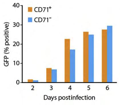
136 120 A B
Fayth K. Yoshimura, Ph.D. September 7, of 7 HIV - BASIC PROPERTIES
 1 of 7 I. Viral Origin. A. Retrovirus - animal lentiviruses. HIV - BASIC PROPERTIES 1. HIV is a member of the Retrovirus family and more specifically it is a member of the Lentivirus genus of this family.
Human Immunodeficiency Virus
 Human Immunodeficiency Virus Virion Genome Genes and proteins Viruses and hosts Diseases Distinctive characteristics Viruses and hosts Lentivirus from Latin lentis (slow), for slow progression of disease
Retroviruses. ---The name retrovirus comes from the enzyme, reverse transcriptase.
 Retroviruses ---The name retrovirus comes from the enzyme, reverse transcriptase. ---Reverse transcriptase (RT) converts the RNA genome present in the virus particle into DNA. ---RT discovered in 1970.
HIV INFECTION: An Overview
 HIV INFECTION: An Overview UNIVERSITY OF PAPUA NEW GUINEA SCHOOL OF MEDICINE AND HEALTH SCIENCES DIVISION OF BASIC MEDICAL SCIENCES DISCIPLINE OF BIOCHEMISTRY & MOLECULAR BIOLOGY PBL MBBS II SEMINAR VJ
HIV & AIDS: Overview
 HIV & AIDS: Overview UNIVERSITY OF PAPUA NEW GUINEA SCHOOL OF MEDICINE AND HEALTH SCIENCES DIVISION OF BASIC MEDICAL SCIENCES DISCIPLINE OF BIOCHEMISTRY & MOLECULAR BIOLOGY PBL SEMINAR VJ TEMPLE 1 What
MedChem 401~ Retroviridae. Retroviridae
 MedChem 401~ Retroviridae Retroviruses plus-sense RNA genome (!8-10 kb) protein capsid lipid envelop envelope glycoproteins reverse transcriptase enzyme integrase enzyme protease enzyme Retroviridae The
Primate lentiviral Vpx commandeers DDB1 to counteract a macrophage restriction
 University of Massachusetts Medical School escholarship@umms Open Access Articles Open Access Publications by UMMS Authors 5-3-2008 Primate lentiviral Vpx commandeers DDB1 to counteract a macrophage restriction
Fayth K. Yoshimura, Ph.D. September 7, of 7 RETROVIRUSES. 2. HTLV-II causes hairy T-cell leukemia
 1 of 7 I. Diseases Caused by Retroviruses RETROVIRUSES A. Human retroviruses that cause cancers 1. HTLV-I causes adult T-cell leukemia and tropical spastic paraparesis 2. HTLV-II causes hairy T-cell leukemia
HIV/AIDS. Biology of HIV. Research Feature. Related Links. See Also
 6/1/2011 Biology of HIV Biology of HIV HIV belongs to a class of viruses known as retroviruses. Retroviruses are viruses that contain RNA (ribonucleic acid) as their genetic material. After infecting a
, virus identified as the causative agent and ELISA test produced which showed the extent of the epidemic
 1 Two attributes make AIDS unique among infectious diseases: it is uniformly fatal, and most of its devastating symptoms are not due to the causative agent Male to Male sex is the highest risk group in
Under the Radar Screen: How Bugs Trick Our Immune Defenses
 Under the Radar Screen: How Bugs Trick Our Immune Defenses Session 7: Cytokines Marie-Eve Paquet and Gijsbert Grotenbreg Whitehead Institute for Biomedical Research HHV-8 Discovered in the 1980 s at the
How HIV Causes Disease Prof. Bruce D. Walker
 How HIV Causes Disease Howard Hughes Medical Institute Massachusetts General Hospital Harvard Medical School 1 The global AIDS crisis 60 million infections 20 million deaths 2 3 The screen versions of
Fig. 1: Schematic diagram of basic structure of HIV
 UNIVERSITY OF PAPUA NEW GUINEA SCHOOL OF MEDICINE AND HEALTH SCIENCES DIVISION OF BASIC MEDICAL SCIENCES DISCIPLINE OF BIOCHEMISTRY & MOLECULAR BIOLOGY PBL SEMINAR HIV & AIDS: An Overview What is HIV?
LESSON 4.6 WORKBOOK. Designing an antiviral drug The challenge of HIV
 LESSON 4.6 WORKBOOK Designing an antiviral drug The challenge of HIV In the last two lessons we discussed the how the viral life cycle causes host cell damage. But is there anything we can do to prevent
VIROLOGY. Engineering Viral Genomes: Retrovirus Vectors
 VIROLOGY Engineering Viral Genomes: Retrovirus Vectors Viral vectors Retrovirus replicative cycle Most mammalian retroviruses use trna PRO, trna Lys3, trna Lys1,2 The partially unfolded trna is annealed
 CDC site UNAIDS Aids Knowledge Base http://www.cdc.gov/hiv/dhap.htm http://hivinsite.ucsf.edu/insite.jsp?page=kb National Institute of Allergy and Infectious Diseases http://www.niaid.nih.gov/default.htm
Acquired immune deficiency syndrome.
 Acquired immune deficiency syndrome 491 Acquired immune deficiency syndrome. The first cases of acquired immune deficiency syndrome (AIDS) were reported in 1981 but it is now clear that cases of the disease
CURRENT DEVELOMENTS AND FUTURE PROSPECTS FOR HIV GENE THERAPY USING INTERFERING RNA-BASED STRATEGIES
 [Frontiers in Bioscience 5, d527-555, May 1, 2000] CURRENT DEVELOMENTS AND FUTURE PROSPECTS FOR HIV GENE THERAPY USING INTERFERING RNA-BASED STRATEGIES Betty Lamothe, Sadhna Joshi Department of Medical
Mayo Clinic HIV ecurriculum Series Essentials of HIV Medicine Module 2 HIV Virology
 Mayo Clinic HIV ecurriculum Series Essentials of HIV Medicine Module 2 HIV Virology Eric M. Poeschla, MD Professor of Medicine College of Medicine Consultant, Division of Infectious Diseases Mayo Clinic
Lentiviruses: HIV-1 Pathogenesis
 Lentiviruses: HIV-1 Pathogenesis Human Immunodeficiency Virus, HIV, computer graphic by Russell Kightley Tsafi Pe ery, Ph.D. Departments of Medicine and Biochemistry & Molecular Biology NJMS, UMDNJ. e-mail:
Herpesviruses. Virion. Genome. Genes and proteins. Viruses and hosts. Diseases. Distinctive characteristics
 Herpesviruses Virion Genome Genes and proteins Viruses and hosts Diseases Distinctive characteristics Virion Enveloped icosahedral capsid (T=16), diameter 125 nm Diameter of enveloped virion 200 nm Capsid
Evidence of HIV-1 Adaptation to HLA- Restricted Immune Responses at a Population Level. Corey Benjamin Moore
 Evidence of HIV-1 Adaptation to HLA- Restricted Immune Responses at a Population Level Corey Benjamin Moore This thesis is presented for the degree of Doctor of Philosophy of Murdoch University, 2002 I
Section 6. Junaid Malek, M.D.
 Section 6 Junaid Malek, M.D. The Golgi and gp160 gp160 transported from ER to the Golgi in coated vesicles These coated vesicles fuse to the cis portion of the Golgi and deposit their cargo in the cisternae
HIV 101: Fundamentals of HIV Infection
 HIV 101: Fundamentals of HIV Infection David H. Spach, MD Professor of Medicine University of Washington Seattle, Washington Learning Objectives After attending this presentation, learners will be able
EVOLUTION OF HUMAN IMMUNODEFICIENCY VIRUS TYPE 1 CLADE C ENVELOPE V1-V5 REGION DURING DISEASE PROGRESSION IN NON- HUMAN PRIMATE MODEL
 University of Nebraska - Lincoln DigitalCommons@University of Nebraska - Lincoln Dissertations and Theses in Biological Sciences Biological Sciences, School of 5-2013 EVOLUTION OF HUMAN IMMUNODEFICIENCY
Let us know how access to this document benefits you. Follow this and additional works at:
 University of Montana ScholarWorks at University of Montana Graduate Student Theses, Dissertations, & Professional Papers Graduate School 2011 INTERACTIONS BETWEEN THE GLYCOSYLATED GAG PROTEIN OF A MURINE
Contribution of PDZD8 to Stabilization of the Human Immunodeficiency Virus (HIV-1) Capsid
 Capsid") Contribution of PDZD8 to Stabilization of the Human Immunodeficiency Virus (HIV-1) Capsid The Harvard community has made this article openly available. Please share how this access benefits you. Your story
Contribution of PDZD8 to Stabilization of the Human Immunodeficiency Virus (HIV-1) Capsid The Harvard community has made this article openly available. Please share how this access benefits you. Your story
October 26, Lecture Readings. Vesicular Trafficking, Secretory Pathway, HIV Assembly and Exit from Cell
 October 26, 2006 Vesicular Trafficking, Secretory Pathway, HIV Assembly and Exit from Cell 1. Secretory pathway a. Formation of coated vesicles b. SNAREs and vesicle targeting 2. Membrane fusion a. SNAREs
Investigation of the C-Terminal Helix of HIV-1 Matrix: A Region Essential for Multiple Functions in the Viral Life Cycle: A Dissertation
 University of Massachusetts Medical School escholarship@umms GSBS Dissertations and Theses Graduate School of Biomedical Sciences 7-10-2011 Investigation of the C-Terminal Helix of HIV-1 Matrix: A Region
Some living things are made of ONE cell, and are called. Other organisms are composed of many cells, and are called. (SEE PAGE 6)
") Section: 1.1 Question of the Day: Name: Review of Old Information: N/A New Information: We tend to only think of animals as living. However, there is a great diversity of organisms that we consider living
Section: 1.1 Question of the Day: Name: Review of Old Information: N/A New Information: We tend to only think of animals as living. However, there is a great diversity of organisms that we consider living
Combining Pharmacology and Mutational Dynamics to Understand and Combat Drug Resistance in HIV
 Combining Pharmacology and Mutational Dynamics to Understand and Combat Drug Resistance in HIV A dissertation submitted for the degree of Doctor of Philosophy by Max von Kleist, B.Sc., M.Sc. Supervisor:
Running Head: AN UNDERSTANDING OF HIV- 1, SYMPTOMS, AND TREATMENTS. An Understanding of HIV- 1, Symptoms, and Treatments.
 Running Head: AN UNDERSTANDING OF HIV- 1, SYMPTOMS, AND TREATMENTS An Understanding of HIV- 1, Symptoms, and Treatments Benjamin Mills Abstract HIV- 1 is a virus that has had major impacts worldwide. Numerous
General information. Cell mediated immunity. 455 LSA, Tuesday 11 to noon. Anytime after class.
 General information Cell mediated immunity 455 LSA, Tuesday 11 to noon Anytime after class T-cell precursors Thymus Naive T-cells (CD8 or CD4) email: lcoscoy@berkeley.edu edu Use MCB150 as subject line
Identification and Characterization of SNAPIN as a Novel Antagonist of HIV-1 Egress: A Dissertation
 University of Massachusetts Medical School escholarship@umms GSBS Dissertations and Theses Graduate School of Biomedical Sciences 4-5-2010 Identification and Characterization of SNAPIN as a Novel Antagonist
VIRUSES. 1. Describe the structure of a virus by completing the following chart.
 AP BIOLOGY MOLECULAR GENETICS ACTIVITY #3 NAME DATE HOUR VIRUSES 1. Describe the structure of a virus by completing the following chart. Viral Part Description of Part 2. Some viruses have an envelope
Introduction retroposon
 17.1 - Introduction A retrovirus is an RNA virus able to convert its sequence into DNA by reverse transcription A retroposon (retrotransposon) is a transposon that mobilizes via an RNA form; the DNA element
HIV-1 SUBTYPE C MOTHER-TO-CHILD TRANSMISSION: GENETIC AND IMMUNOLOGIC CORRELATES. Elizabeth Susan Russell
 HIV-1 SUBTYPE C MOTHER-TO-CHILD TRANSMISSION: GENETIC AND IMMUNOLOGIC CORRELATES Elizabeth Susan Russell A dissertation submitted to the faculty of the University of North Carolina at Chapel Hill in partial
Hepadnaviruses: Variations on the Retrovirus Theme
 WBV21 6/27/03 11:34 PM Page 377 Hepadnaviruses: Variations on the Retrovirus Theme 21 CHAPTER The virion and the viral genome The viral replication cycle The pathogenesis of hepatitis B virus A plant hepadnavirus
A Novel Motif in HIV-1 Nef that Regulates MIP-1β Chemokine Release in Macrophages: A Dissertation
 University of Massachusetts Medical School escholarship@umms GSBS Dissertations and Theses Graduate School of Biomedical Sciences 6-17-2010 A Novel Motif in HIV-1 Nef that Regulates MIP-1β Chemokine Release
Dr. Ahmed K. Ali Attachment and entry of viruses into cells
 Lec. 6 Dr. Ahmed K. Ali Attachment and entry of viruses into cells The aim of a virus is to replicate itself, and in order to achieve this aim it needs to enter a host cell, make copies of itself and
Lecture 2: Virology. I. Background
 Lecture 2: Virology I. Background A. Properties 1. Simple biological systems a. Aggregates of nucleic acids and protein 2. Non-living a. Cannot reproduce or carry out metabolic activities outside of a
There are approximately 30,000 proteasomes in a typical human cell Each proteasome is approximately 700 kda in size The proteasome is made up of 3
 Proteasomes Proteasomes Proteasomes are responsible for degrading proteins that have been damaged, assembled improperly, or that are of no profitable use to the cell. The unwanted protein is literally
Julianne Edwards. Retroviruses. Spring 2010
 Retroviruses Spring 2010 A retrovirus can simply be referred to as an infectious particle which replicates backwards even though there are many different types of retroviruses. More specifically, a retrovirus
Lecture Readings. Vesicular Trafficking, Secretory Pathway, HIV Assembly and Exit from Cell
 October 26, 2006 1 Vesicular Trafficking, Secretory Pathway, HIV Assembly and Exit from Cell 1. Secretory pathway a. Formation of coated vesicles b. SNAREs and vesicle targeting 2. Membrane fusion a. SNAREs
Recombinant Protein Expression Retroviral system
 Recombinant Protein Expression Retroviral system Viruses Contains genome DNA or RNA Genome encased in a protein coat or capsid. Some viruses have membrane covering protein coat enveloped virus Ø Essential
Viral Genetics. BIT 220 Chapter 16
 Viral Genetics BIT 220 Chapter 16 Details of the Virus Classified According to a. DNA or RNA b. Enveloped or Non-Enveloped c. Single-stranded or double-stranded Viruses contain only a few genes Reverse
HIV-1 Viral Infectivity Factor Recruitment of Cellular. Protein Complexes to Counter Host Antiviral Response
 HIV-1 Viral Infectivity Factor Recruitment of Cellular Protein Complexes to Counter Host Antiviral Response By Sean L. Evans A dissertation submitted to Johns Hopkins University in conformity with requirements
HIV Immunopathogenesis. Modeling the Immune System May 2, 2007
 HIV Immunopathogenesis Modeling the Immune System May 2, 2007 Question 1 : Explain how HIV infects the host Zafer Iscan Yuanjian Wang Zufferey Abhishek Garg How does HIV infect the host? HIV infection
Prokaryotic Biology. VIRAL STDs, HIV-1 AND AIDS
 Prokaryotic Biology VIRAL STDs, HIV-1 AND AIDS Prokaryotic Biology FROM THE CDC VIRAL STDs, HIV-1 AND AIDS VIRAL STDs & CONTACT VIRAL DISEASES A. GENITAL HERPES & COLD SORES 1. HERPES SIMPLEX VIRUS-2 (HHV-2)
Centers for Disease Control August 9, 2004
 HIV CDC site UNAIDS Aids Knowledge Base http://www.cdc.gov/hiv/dhap.htm http://hivinsite.ucsf.edu/insite.jsp?page=kb National Institute of Allergy and Infectious Diseases http://www.niaid.nih.gov/default.htm
LTR R U5 PBS. 5' LTR : weak promoter [Inr, TATA-box, and three Sp1 sites] + enhancer elements.
![LTR R U5 PBS. 5' LTR : weak promoter [Inr, TATA-box, and three Sp1 sites] + enhancer elements.](/thumbs/83/87506206.jpg "LTR R U5 PBS. 5' LTR : weak promoter [Inr, TATA-box, and three Sp1 sites] + enhancer elements.") Foreword HIV has a very complex system of transcription regulation and mrna splicing. This produces something that approximates "life-cycle" stages. Expression from the promoter always occurs but at first
Foreword HIV has a very complex system of transcription regulation and mrna splicing. This produces something that approximates "life-cycle" stages. Expression from the promoter always occurs but at first
number Done by Corrected by Doctor Ashraf
 number 4 Done by Nedaa Bani Ata Corrected by Rama Nada Doctor Ashraf Genome replication and gene expression Remember the steps of viral replication from the last lecture: Attachment, Adsorption, Penetration,
Last time we talked about the few steps in viral replication cycle and the un-coating stage:
 Zeina Al-Momani Last time we talked about the few steps in viral replication cycle and the un-coating stage: Un-coating: is a general term for the events which occur after penetration, we talked about
19 Viruses BIOLOGY. Outline. Structural Features and Characteristics. The Good the Bad and the Ugly. Structural Features and Characteristics
 9 Viruses CAMPBELL BIOLOGY TENTH EDITION Reece Urry Cain Wasserman Minorsky Jackson Outline I. Viruses A. Structure of viruses B. Common Characteristics of Viruses C. Viral replication D. HIV Lecture Presentation
Viral Vectors In The Research Laboratory: Just How Safe Are They? Dawn P. Wooley, Ph.D., SM(NRM), RBP, CBSP
, RBP, CBSP") Viral Vectors In The Research Laboratory: Just How Safe Are They? Dawn P. Wooley, Ph.D., SM(NRM), RBP, CBSP 1 Learning Objectives Recognize hazards associated with viral vectors in research and animal
Viral Vectors In The Research Laboratory: Just How Safe Are They? Dawn P. Wooley, Ph.D., SM(NRM), RBP, CBSP 1 Learning Objectives Recognize hazards associated with viral vectors in research and animal
L I F E S C I E N C E S
 1a L I F E S C I E N C E S 5 -UUA AUA UUC GAA AGC UGC AUC GAA AAC UGU GAA UCA-3 5 -TTA ATA TTC GAA AGC TGC ATC GAA AAC TGT GAA TCA-3 3 -AAT TAT AAG CTT TCG ACG TAG CTT TTG ACA CTT AGT-5 OCTOBER 31, 2006
7.012 Quiz 3 Answers
 MIT Biology Department 7.012: Introductory Biology - Fall 2004 Instructors: Professor Eric Lander, Professor Robert A. Weinberg, Dr. Claudette Gardel Friday 11/12/04 7.012 Quiz 3 Answers A > 85 B 72-84
Micro 301 HIV/AIDS. Since its discovery 31 years ago 12/3/ Acquired Immunodeficiency Syndrome (AIDS) has killed >32 million people
 has killed >32 million people") Micro 301 HIV/AIDS Shiu-Lok Hu hus@uw.edu December 3, 2012 Since its discovery 31 years ago Acquired Immunodeficiency Syndrome (AIDS) has killed >32 million people In 2011 34.0 million [31.4 35.9 million]
Micro 301 HIV/AIDS Shiu-Lok Hu hus@uw.edu December 3, 2012 Since its discovery 31 years ago Acquired Immunodeficiency Syndrome (AIDS) has killed >32 million people In 2011 34.0 million [31.4 35.9 million]
Citation for published version (APA): Von Eije, K. J. (2009). RNAi based gene therapy for HIV-1, from bench to bedside
: Von Eije, K. J. (2009). RNAi based gene therapy for HIV-1, from bench to bedside") UvA-DARE (Digital Academic Repository) RNAi based gene therapy for HIV-1, from bench to bedside Von Eije, K.J. Link to publication Citation for published version (APA): Von Eije, K. J. (2009). RNAi based
UvA-DARE (Digital Academic Repository) RNAi based gene therapy for HIV-1, from bench to bedside Von Eije, K.J. Link to publication Citation for published version (APA): Von Eije, K. J. (2009). RNAi based
A review of HIV restriction factors and viral countering mechanisms
 A review of HIV restriction factors and viral countering mechanisms By Qimeng Gao A thesis submitted to the Johns Hopkins University in conformity with the requirements for the degree of Master of Health
NIH Public Access Author Manuscript Expert Rev Mol Med. Author manuscript; available in PMC 2010 April 28.
 NIH Public Access Author Manuscript Published in final edited form as: Expert Rev Mol Med. ; 12: e4. doi:10.1017/s1462399409001343. Interactions of host APOBEC3 restriction factors with HIV-1 in vivo:
HIV-1 Neutralisation and Other Aspects of the Envelope Glycoprotein
 HIV-1 Neutralisation and Other Aspects of the Envelope Glycoprotein Pei-Jen Lai Thesis submitted in fulfilment of the requirements for the degree of Doctor of Philosophy of Imperial College London 2009
Coronaviruses. Virion. Genome. Genes and proteins. Viruses and hosts. Diseases. Distinctive characteristics
 Coronaviruses Virion Genome Genes and proteins Viruses and hosts Diseases Distinctive characteristics Virion Spherical enveloped particles studded with clubbed spikes Diameter 120-160 nm Coiled helical
The Harvard community has made this article openly available. Please share how this access benefits you. Your story matters
 Emergence of simian immunodeficiency virus in rhesus macaques is characterized by changes in structural and accessory genes that enhance fitness in the new host species The Harvard community has made this
ACQUIRED IMMUNODEFICIENCY SYNDROME AND ITS OCULAR COMPLICATIONS
 ACQUIRED IMMUNODEFICIENCY SYNDROME AND ITS OCULAR COMPLICATIONS Acquired immunodeficiency syndrome (AIDS ) is an infectious disease caused by a retrovirus, the human immunodeficiency virus(hiv). AIDS is
Chapter 6. Antigen Presentation to T lymphocytes
 Chapter 6 Antigen Presentation to T lymphocytes Generation of T-cell Receptor Ligands T cells only recognize Ags displayed on cell surfaces These Ags may be derived from pathogens that replicate within
Immunodeficiency. (2 of 2)
") Immunodeficiency (2 of 2) Acquired (secondary) immunodeficiencies More common Many causes such as therapy, cancer, sarcoidosis, malnutrition, infection & renal disease The most common of which is therapy-related
Immunodeficiency (2 of 2) Acquired (secondary) immunodeficiencies More common Many causes such as therapy, cancer, sarcoidosis, malnutrition, infection & renal disease The most common of which is therapy-related
A PROJECT ON HIV INTRODUCED BY. Abdul Wahab Ali Gabeen Mahmoud Kamal Singer
 A PROJECT ON HIV INTRODUCED BY Abdul Wahab Ali Gabeen Mahmoud Kamal Singer Introduction: Three groups of nations have been identified in which the epidemiology of HIV(Human Immunodeficiency Virus) varies:
An Evolutionary Story about HIV
 An Evolutionary Story about HIV Charles Goodnight University of Vermont Based on Freeman and Herron Evolutionary Analysis The Aids Epidemic HIV has infected 60 million people. 1/3 have died so far Worst
Supplementary information. MARCH8 inhibits HIV-1 infection by reducing virion incorporation of envelope glycoproteins
 Supplementary information inhibits HIV-1 infection by reducing virion incorporation of envelope glycoproteins Takuya Tada, Yanzhao Zhang, Takayoshi Koyama, Minoru Tobiume, Yasuko Tsunetsugu-Yokota, Shoji
~Lentivirus production~
 ~Lentivirus production~ May 30, 2008 RNAi core R&D group member Lentivirus Production Session Lentivirus!!! Is it health threatening to lab technician? What s so good about this RNAi library? How to produce
Polyomaviridae. Spring
 Polyomaviridae Spring 2002 331 Antibody Prevalence for BK & JC Viruses Spring 2002 332 Polyoma Viruses General characteristics Papovaviridae: PA - papilloma; PO - polyoma; VA - vacuolating agent a. 45nm
Lecture 11. Immunology and disease: parasite antigenic diversity
 Lecture 11 Immunology and disease: parasite antigenic diversity RNAi interference video and tutorial (you are responsible for this material, so check it out.) http://www.pbs.org/wgbh/nova/sciencenow/3210/02.html
VIRAL ENVELOPE PROTEINS AND THE HIV-1 ACCESSORY GENE VPU MEDIATE SELECTIVITY OF VIRAL AND HOST PROTEINS IN RETROVIRAL ASSEMBLY
 VIRAL ENVELOPE PROTEINS AND THE HIV-1 ACCESSORY GENE VPU MEDIATE SELECTIVITY OF VIRAL AND HOST PROTEINS IN RETROVIRAL ASSEMBLY A Dissertation presented to the Faculty of the Graduate School at the University
11/15/2011. Outline. Structural Features and Characteristics. The Good the Bad and the Ugly. Viral Genomes. Structural Features and Characteristics
 Chapter 19 - Viruses Outline I. Viruses A. Structure of viruses B. Common Characteristics of Viruses C. Viral replication D. HIV II. Prions The Good the Bad and the Ugly Viruses fit into the bad category
Structure of HIV. Virion contains a membrane envelope with a single viral protein= Env protein. Capsid made up of Gag protein
 Structure of HIV Virion contains a membrane envelope with a single viral protein= Env protein Important in receptor recognition Capsid made up of Gag protein (group-specific antigen) Icosahedral Interior
Development of Broadly Reactive HIV-1/AIDS Virus-like Particle Vaccines. Sean Patrick McBurney. B.S. Microbiology, University of Pittsburgh, 2004
 Development of Broadly Reactive HIV-1/AIDS Virus-like Particle Vaccines by Sean Patrick McBurney B.S. Microbiology, University of Pittsburgh, 2004 Submitted to the Graduate Faculty of School of Medicine
Newly Synthesized APOBEC3G Is Incorporated into HIV Virions, Inhibited by HIV RNA, and Subsequently Activated by RNase H
 Newly Synthesized APOBEC3G Is Incorporated into HIV Virions, Inhibited by HIV RNA, and Subsequently Activated by RNase H Vanessa B. Soros 1, Wes Yonemoto 1, Warner C. Greene 1,2,3* 1 Gladstone Institute
RAISON D ETRE OF THE IMMUNE SYSTEM:
 RAISON D ETRE OF THE IMMUNE SYSTEM: To Distinguish Self from Non-Self Thereby Protecting Us From Our Hostile Environment. Innate Immunity Acquired Immunity Innate immunity: (Antigen nonspecific) defense
Role of poly-proline motif in HIV-2 Vpx expression
 Role of poly-proline motif in HIV-2 Vpx expression Ariko Miyake, Yasuyuki Miyazaki, Mikako Fujita, Masako Nomaguchi and Akio Adachi Journal Name: Frontiers in Microbiology ISSN: 1664-302X Article type:
Modeling and Simulation of HIV-1 Intracellular Replication
 Modeling and Simulation of HIV-1 Intracellular Replication MSc Thesis Author Narges Zarrabi Supervisor: Prof. Dr. Peter M.A. Sloot Submitted to Faculty of Science in partial fullfilment of the requirments
INFECTION OF HEMATOPOIETIC PROGENITOR CELLS BY HIV-1. Christoph C. Carter
 INFECTION OF HEMATOPOIETIC PROGENITOR CELLS BY HIV-1 by Christoph C. Carter A dissertation submitted in partial fulfillment of the requirements for the degree of Doctor of Philosophy (Cellular and Molecular
Protein Trafficking in the Secretory and Endocytic Pathways
 Protein Trafficking in the Secretory and Endocytic Pathways The compartmentalization of eukaryotic cells has considerable functional advantages for the cell, but requires elaborate mechanisms to ensure
 CDC site UNAIDS Aids Knowledge Base http://www.cdc.gov/hiv/dhap.htm http://hivinsite.ucsf.edu/insite.jsp?page=kb National Institute of Allergy and Infectious Diseases http://www.niaid.nih.gov/default.htm
Irina Maljkovic Berry
 Thesis for doctoral degree (Ph.D.) 2008 Thesis for doctoral degree (Ph.D.) 2008 Genetic aspects of HIV-1 evolution and transmission Irina Maljkovic Berry Genetic aspects of HIV-1 evolution and transmission
Virus and Prokaryotic Gene Regulation - 1
 Virus and Prokaryotic Gene Regulation - 1 We have discussed the molecular structure of DNA and its function in DNA duplication and in transcription and protein synthesis. We now turn to how cells regulate
Influenza viruses. Virion. Genome. Genes and proteins. Viruses and hosts. Diseases. Distinctive characteristics
 Influenza viruses Virion Genome Genes and proteins Viruses and hosts Diseases Distinctive characteristics Virion Enveloped particles, quasi-spherical or filamentous Diameter 80-120 nm Envelope is derived
MINIREVIEW. Human Immunodeficiency Virus and AIDS: Insights from Animal Lentiviruses
 JOURNAL OF VIROLOGY, August 2000, p. 7187 7195 Vol. 74, No. 16 0022-538X/00/$04.00 0 MINIREVIEW Human Immunodeficiency Virus and AIDS: Insights from Animal Lentiviruses ROGER J. MILLER, J. SCOTT CAIRNS,
Mechanisms by which HIV-1 Nef disrupts the intracellular trafficking of host. proteins. Jolie A. Leonard
 Mechanisms by which HIV-1 Nef disrupts the intracellular trafficking of host proteins by Jolie A. Leonard A dissertation submitted in partial fulfillment Of the requirements for the degree of Doctor of
Reverse transcription and integration
 Reverse transcription and integration Lecture 9 Biology 3310/4310 Virology Spring 2018 One can t believe impossible things, said Alice. I dare say you haven t had much practice, said the Queen. Why, sometimes
Lentiviral gene therapy for HIV-1 using TRIM-Cyclophilin restriction factors
 Lentiviral gene therapy for HIV-1 using TRIM-Cyclophilin restriction factors Emma Chan Institute of Child Health University College London A thesis submitted for the degree of Doctor of Philosophy 2012
Tetherin/BST-2 Antagonism by Nef Depends on a Direct Physical Interaction between Nef and Tetherin, and on Clathrin-mediated Endocytosis
 Tetherin/BST-2 Antagonism by Nef Depends on a Direct Physical Interaction between Nef and Tetherin, and on Clathrin-mediated Endocytosis The Harvard community has made this article openly available. Please
HIV-1: Fifteen Proteins and an RNA
 Annu. Rev. Biochem. 1998. 67:1 25 Copyright c 1998 by Annual Reviews. All rights reserved HIV-1: Fifteen Proteins and an RNA Alan D. Frankel Department of Biochemistry and Biophysics, University of California,
Identification of Mutation(s) in. Associated with Neutralization Resistance. Miah Blomquist
 in. Associated with Neutralization Resistance. Miah Blomquist") Identification of Mutation(s) in the HIV 1 gp41 Subunit Associated with Neutralization Resistance Miah Blomquist What is HIV 1? HIV-1 is an epidemic that affects over 34 million people worldwide. HIV-1
Identification of Mutation(s) in the HIV 1 gp41 Subunit Associated with Neutralization Resistance Miah Blomquist What is HIV 1? HIV-1 is an epidemic that affects over 34 million people worldwide. HIV-1
Human Immunodeficiency Virus
 Human Immunodeficiency Virus 1. Identification of the AIDS Virus a) opportunistic infections observed in homosexual men (all had T4 helper cell depletion) -> termed Acquired Immune Deficiency Syndrome;
Human Genome Complexity, Viruses & Genetic Variability
 Human Genome Complexity, Viruses & Genetic Variability (Learning Objectives) Learn the types of DNA sequences present in the Human Genome other than genes coding for functional proteins. Review what you
HUMAN IMMUNODEFICIENCY VIRUS
 Futuro promisorio de la terapia antirretroviral: Nuevos blancos terapéuticos. María José Míguez, M.D., PhD., Universidad de Miami, EE.UU. HUMAN IMMUNODEFICIENCY VIRUS REVERSE TRANSCRIPTASA REPLICATION
Chapter13 Characterizing and Classifying Viruses, Viroids, and Prions
 Chapter13 Characterizing and Classifying Viruses, Viroids, and Prions 11/20/2017 MDufilho 1 Characteristics of Viruses Viruses Minuscule, acellular, infectious agent having either DNA or RNA Cause infections
Going Wild: Lessons from Naturally Occurring T-Lymphotropic Lentiviruses. VandeWoude & Apretrei Clinical Microbiology Reviews 27:728 (2014)
") In ancient times, citizens sought answers to their most important inquiries by consulting oracles. Myths were part of daily life, and mythological creatures were feared or defeated. Oedipus deciphered
In ancient times, citizens sought answers to their most important inquiries by consulting oracles. Myths were part of daily life, and mythological creatures were feared or defeated. Oedipus deciphered
7.014 Problem Set 7 Solutions
 MIT Department of Biology 7.014 Introductory Biology, Spring 2005 7.014 Problem Set 7 Solutions Question 1 Part A Antigen binding site Antigen binding site Variable region Light chain Light chain Variable