(JPC ) Caprine lungs
|
|
|
- Charla Wood
- 5 years ago
- Views:
Transcription
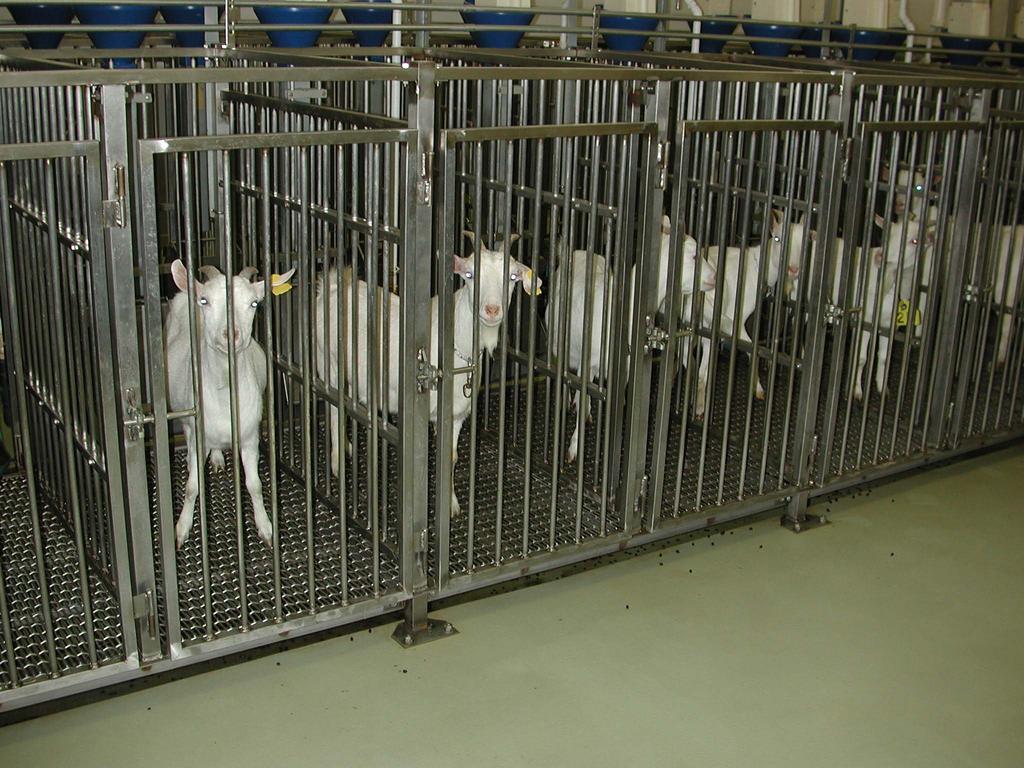
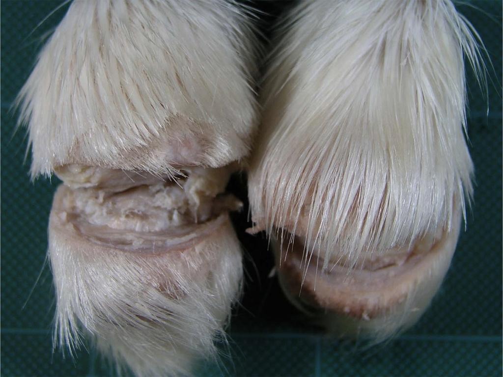
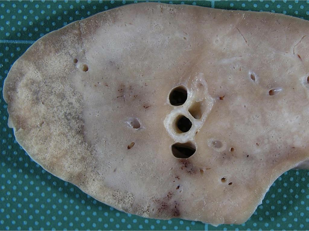
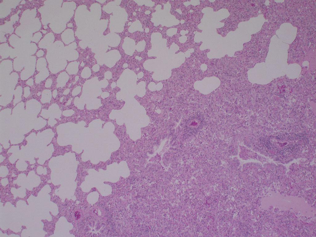
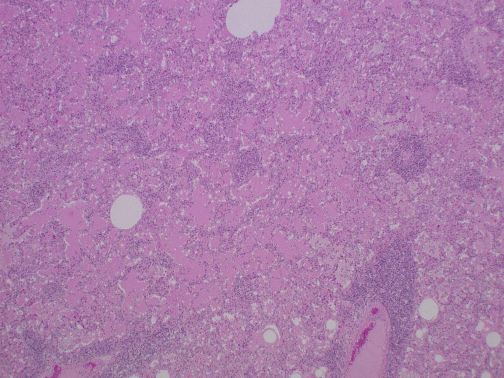
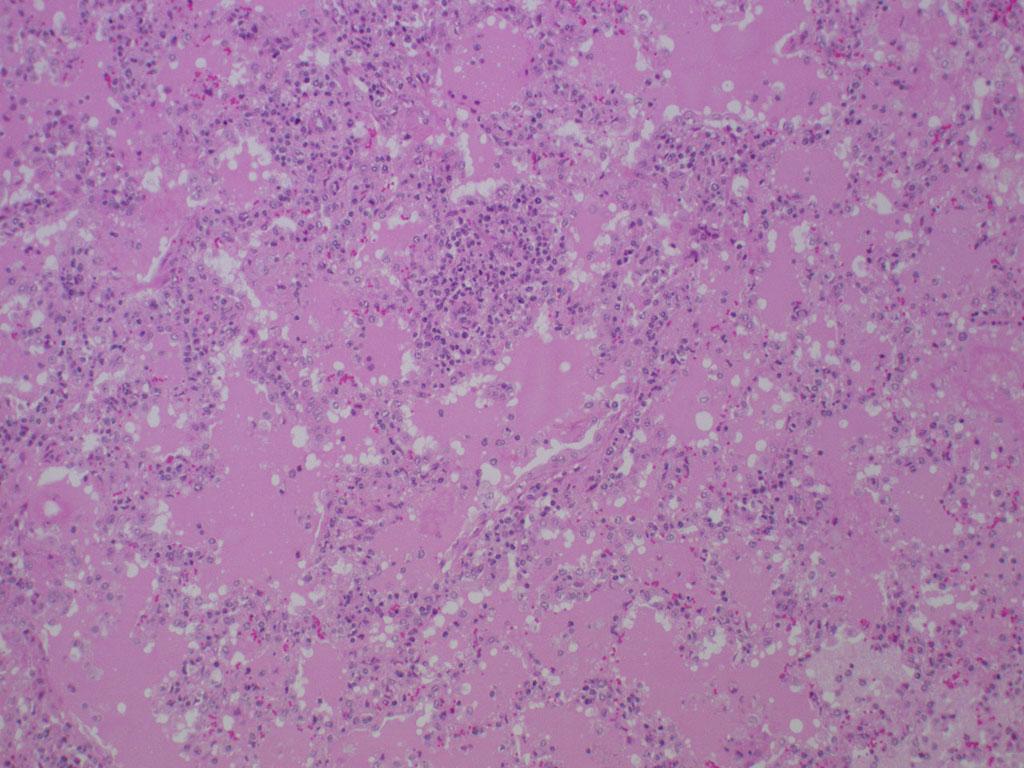
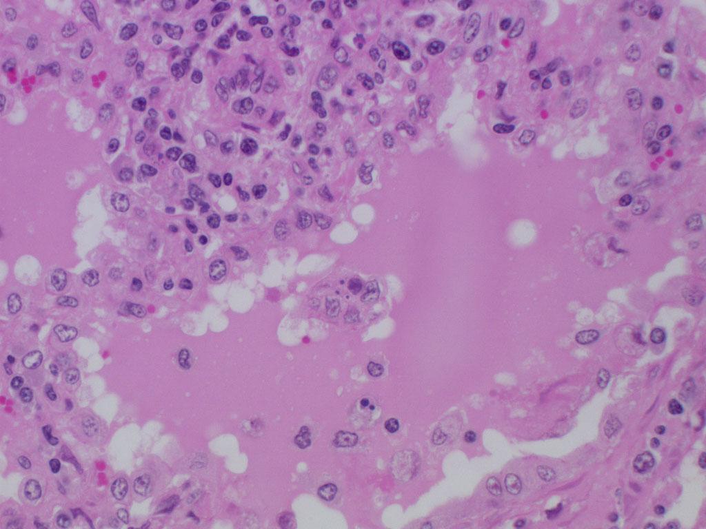
1 (JPC ) Caprine lungs Bat Otgontugs Bovine Pathology Contributor: Natoinal Institute Animal Health, Tsukuba, Japan Signalment: 5-year 3-month old female Japanese native breed goat, (Capra aegagrus). History: A Japanese native breed goat showed respiratory symptoms including tachypnea and cough. Three months later, the goat developed mild carpal swelling. At the time, the serum was negative for caprine arthritis-encephalitis (CAE) virus. Two months later, many nodules resembling abscesses, up to 10 cm in diameter, developed in the mammary gland and lip. The goat was submitted for necropsy. The serum collected at the necropsy was weakly positive for CAE. Gross pathology: The goat was in poor body condition. The lung had distinctive pale firm lesions with ecchymosis throughout the lung. Both carpal joints had mild swelling. Many abscesses were seen in the mammary gland. Histopathologic Description: Lung: The foci in the lung were demarcated from relatively unaffected areas. Distinctive cuffs consisting of lymphocytes and plasma cells were formed around blood vessels and airways. Alveolar septa were thickened with infiltrates of lymphocytes and the hyperplasia of type II pneumocytes. Alveoli were filled with eosinophilic fluid, small amounts of fibrin, neutrophils and macrophages. Occasionally, macrophages phagocytizing eosinophilic fluid were seen.
2
3
4

5
6
7
8
9
10 Contributor s Morphologic Diagnosis: Lung: Pneumonia, interstitial, lymphocytic, with perivascular and peribronchial lymphoplasmacytic infiltration, type II pneumocyte hyperplasia and intra-alveolar eosinophilic proteinaceous materials. Etiology: CAE Contributor s comments: - CAE is a progressive disease of domestic goats caused by the CAE virus and belongs to Lentivirinae of the family Retroviridae. - The first case was identified in USA in (In Japan in 2002) -AGID and ELISA histopathological diagnosis is definitive for CAE. - The lesions of the lung in the present case consisted of those of typical CAE which are characterized by the lymphoplasmacytic infiltration in septa with the type II pneumocyte hyperplasia, infiltration of lymphocytes around blood vessels, bronchi, and bronchioles, and eosinophilic exudates in alveoli. - Distinctive eosinophilic fluid is an important finding for differential diagnosis between CAE and maedi. - The present case had lymphoplasmacytic arthritis and periarthritis with villous proliferation of the synovium, which are suggestive of CAE. - In the mammary gland, many abscesses and periductal cuffs consisting of lymphocytes were seen.
11 JPC Diagnosis: Lung: Pneumonia, interstitial, lymphohistiocytic, diffuse, moderate, with marked pulmonary edema, peribronchial and perivascular lymphohistiocytic infiltrates, and type II pneumocyte hyperplasia. Conference comments: - The virus occurs mainly in improved dairy goat breeds and is spread primarily via infected colostrum and milk. - In this case demonstrates a typical histologic appearance of CAE and shown demarcation between affected and unaffected lung. - The copious proteinaceous fluid filling the alveoli in this section is visually distinct from edema fluid, and is also very characteristic of this disease. - The moderator pointed out that there was no corroborative evidence to attribute the prominent eosinophilic fluid to hydrostatic edema, such as dilated lymphatics and rarefaction around blood vessels. This fluid has been shown to contain pulmonary surfactant, which is consistent with the abundant type II pneumocyte hyperplasia. As the virus destroys type I pneumocytes, and type II pneumocytes proliferate, the local environment becomes hypoxic due to the thickening of the interstitium, which also fills with lymphocytes, plasma cells, and macrophages. As the diffusion capacity of the lung decreases, cells release hypoxia-induced factor-1α, which activates the transcription of vascular endothelial growth factor, increasing vascular permeability and the leakage of high protein plasma fluid.
Histopathology: pulmonary pathology
 Histopathology: pulmonary pathology These presentations are to help you identify basic histopathological features. They do not contain the additional factual information that you need to learn about these
09-Mar-15 PNEUMONIA RESPIRATORY SYSTEM L-3
 RESPIRATORY SYSTEM L-3 Professor Department of Pathology, University of Agriculture, Faisalabad. Email: mtjaved@uaf.edu.pk Web: https://sites.geocities.ws/mtjaved PNEUMONIA The pulmonary inflammatory response
INFLAMMATION & REPAIR
 INFLAMMATION & REPAIR Histopath Laboratory 1 Winter 2013 Chelsea Martin Special thanks to Drs. Hanna and Forzan Goals: Examine Tissue and Identify the Organ Describe the lesion, grossly and histologically
Influenza A virus infection predisposes hosts to secondary infection with different
 Supplementary information Influenza A virus infection predisposes hosts to secondary infection with different Streptococcus pneumoniae serotypes with similar outcome but serotype-specific manifestation.
Replacement of air with fluid, inflammatory. cells or cellular debris. Parenchymal, Interstitial (Restrictive) and Vascular Diseases.
 and Vascular Diseases.") Parenchymal, Interstitial (Restrictive) and Vascular Diseases Alain C. Borczuk, M.D. Dept of Pathology Replacement of air with fluid, inflammatory cells Pulmonary Edema Pneumonia Hemorrhage Diffuse alveolar
Parenchymal, Interstitial (Restrictive) and Vascular Diseases Alain C. Borczuk, M.D. Dept of Pathology Replacement of air with fluid, inflammatory cells Pulmonary Edema Pneumonia Hemorrhage Diffuse alveolar
Wedge Biopsy for Diffuse Lung Diseases
 Chapter VI Wedge Biopsy for Diffuse Lung Diseases Wedge biopsy via thoracoscopic biopsy or open lung biopsy is occasionally performed to obtain tissue for the diagnosis of a diffuse lung disease. A wedge
SESSION IV: MECHANISMS OF HUMAN DISEASE: LABORATORY SESSIONS PULMONARY PATHOLOGY I. December 5, 2012
 SESSION IV: MECHANISMS OF HUMAN DISEASE: LABORATORY SESSIONS PULMONARY PATHOLOGY I December 5, 2012 FACULTY COPY GOAL: Describe the basic morphologic and pathophysiologic changes in various conditions
Unit II Problem 2 Pathology: Pneumonia
 Unit II Problem 2 Pathology: Pneumonia - Definition: pneumonia is the infection of lung parenchyma which occurs especially when normal defenses are impaired such as: Cough reflex. Damage of cilia in respiratory
Disturbances of Circulation, Lab 1: Edema and Congestion/Hyperemia. Shannon Martinson, Feb
 Disturbances of Circulation, Lab 1: Edema and Congestion/Hyperemia Shannon Martinson, Feb 2017 http://people.upei.ca/smartinson/ Case #1 Signalment and History: 6-month old feeder lamb found dead on pasture
Canine Liver Eneku Wilfred Bovine Pathology
 2012-1-3 Canine Liver Eneku Wilfred Bovine Pathology Contributor: New Mexico Department of Agriculture Veterinary Diagnostic Services Signalment: 5 month old male Weimaraner dog (Canis familiaris) History:
Lymphoid System: cells of the immune system. Answer Sheet
 Lymphoid System: cells of the immune system Answer Sheet Q1 Which areas of the lymph node have most CD3 staining? A1 Most CD3 staining is present in the paracortex (T cell areas). This is towards the outside
Infectious Diseases of Small Ruminants. Assoc.Prof. Dr. Theera Rukkwamsuk Faculty of Veterinary Medicine Kasetsart University, Kampangsaen Campus
 Infectious Diseases of Small Ruminants Assoc.Prof. Dr. Theera Rukkwamsuk Faculty of Veterinary Medicine Kasetsart University, Kampangsaen Campus Infectious diseases of small ruminants Caprine arthritis-encephalitis
HISTO-PHYSIOLOGY HISTO-PHYSIOLOGY HISTO-PHYSIOLOGY. 09-Mar-15. Dr. Muhammad Tariq Javed. RESPIRATORY SYSTEM Lec-1
 RESPIRATORY SYSTEM Lec-1 Dr. Muhammad Tariq Javed Professor Department of Pathology, University of Agriculture, Faisalabad. Email: mtjaved@uaf.edu.pk Web: http://www.geocities.ws/mtjaved 1 2 Conducting
Bronkhorst colloquium Interstitiële longziekten. Katrien Grünberg, klinisch patholoog
 Bronkhorst colloquium 2013-2014 Interstitiële longziekten De pathologie achter de CT Katrien Grünberg, klinisch patholoog K.grunberg@vumc.nl Preparing: introduction and 3 cases The introduction on microscopic
HRCT in Diffuse Interstitial Lung Disease Steps in High Resolution CT Diagnosis. Where are the lymphatics? Anatomic distribution
 Steps in High Resolution CT Diagnosis Pattern of abnormality Distribution of disease Associated findings Clinical history Tomás Franquet MD What is the diagnosis? Hospital de Sant Pau. Barcelona Secondary
Pathological Investigations on Bovine Pheumonic Pasteurellosis by Use of Immunoperoxidase Technique
 JARQ 29, 13 1-136 (1995) Pathological Investigations on Bovine Pheumonic Pasteurellosis by Use of Immunoperoxidase Technique Makoto HARITANI Tohoku Branch Laboratory, National Institute of Animal Health
Parenchymal, Interstitial i (Restrictive) i and Vascular Diseases
 i and Vascular Diseases") Pulmonary Diseases: Structure-Function Correlation II Parenchymal, Interstitial i (Restrictive) i and Vascular Diseases Alain C. Borczuk, M.D. Dept of Pathology Pulmonary Diseases: Structure-Function Correlation
Pulmonary Diseases: Structure-Function Correlation II Parenchymal, Interstitial i (Restrictive) i and Vascular Diseases Alain C. Borczuk, M.D. Dept of Pathology Pulmonary Diseases: Structure-Function Correlation
Diseases of the Lung and Respiratory Tract, Part I. William Bligh-Glover M.D. Department of Anatomy, CWRU
 Diseases of the Lung and Respiratory Tract, Part I William Bligh-Glover M.D. Department of Anatomy, CWRU Educational objectives: Distinguish the types of atelectasis and their etiologies Distinguish the
Firm Texture. (chronic) Cut surface: purulent exudate in bronchi Sequels: Abscesses,
 Cut surface: purulent exudate in bronchi Sequels: Abscesses,") 2008 Classification of Pneumonias in Domestic Animals There is no universal classification! Based on texture, distribution of lesions and type of exudate, pneumonias in domestic animals are currently classified
2008 Classification of Pneumonias in Domestic Animals There is no universal classification! Based on texture, distribution of lesions and type of exudate, pneumonias in domestic animals are currently classified
Lower Respiratory Tract (Trachea, Bronchi, Bronchioles) & the Lung
 & the Lung") Lower Respiratory Tract (Trachea, Bronchi, Bronchioles) & the Lung Color code: Important Extra & Doctor notes Editing file Objectives: By the end of this lecture, the student should be able to describe:
Lower Respiratory Tract (Trachea, Bronchi, Bronchioles) & the Lung Color code: Important Extra & Doctor notes Editing file Objectives: By the end of this lecture, the student should be able to describe:
DISEASES OF THE RESPIRATORY SYSTEM 2018 DR HEYAM AWAD LECTURE 3: CHRONIC BRNCHITIS AND BRONCHIECTASIS
 DISEASES OF THE RESPIRATORY SYSTEM 2018 DR HEYAM AWAD LECTURE 3: CHRONIC BRNCHITIS AND BRONCHIECTASIS INTRDUCTION In the last lecture we discussed the difference between restrictive and obstructive lung
Pulmonary Patterns & Correlated Pathology
 Pulmonary Patterns & Correlated Pathology Russell Tucker, DVM, DACVR Washington State University College of Veterinary Medicine Objective: correlate radiographic findings of common lung diseases to actual
Histopathology Description:
 2013-2-1 CANINE HEART Ahmed M. Abubakar BOVINE PATHOLOGY CONTRIBUTING INSTITUTION : The Royal Veterinary college, Dept. of Pathology and Biology Signalment: 11-month-old male Border Collie dog (Canis familiaris)
Acute and Chronic Lung Disease
 KATHOLIEKE UNIVERSITEIT LEUVEN Faculty of Medicine Acute and Chronic Lung Disease W De Wever, JA Verschakelen Department of Radiology, University Hospitals Leuven, Belgium Clinical utility of HRCT To detect
Acute and Chronic Inflammation Pathology 1 - Dr. Gary Mumaugh
 Acute and Chronic Inflammation Pathology 1 - Dr. Gary Mumaugh Introduction Injurious stimuli cause a protective vascular connective tissue reaction called inflammation Acute and chronic forms o Inflame
New lung lesion in a 55 year-old male treated with chemoradiation for non-small cell lung carcinoma
 July 2016 New lung lesion in a 55 year-old male treated with chemoradiation for non-small cell lung carcinoma Contributed by: Laurel Rose, MD, Resident Physician, Indiana University School of Medicine,
Restrictive lung diseases
 Restrictive lung diseases Restrictive lung diseases are diseases that affect the interstitium of the lung. Interstitium of the lung is the very thin walls surrounding the alveoli, it s formed of epithelium
PATHOLOGY OF PASTEURELLA MULTOCIDA INFECTION IN CHICKENS
 Indian J. Anim. Res., 40 (1): 15-19, 2006 PATHOLOGY OF PASTEURELLA MULTOCIDA INFECTION IN CHICKENS Shilpa Sood 1 and P.C. Verma CCS Haryana Agricultural University, Hisar - 125 004, India ABSTRACT The
Imaging in pediatric lung diseases The roles of CT and pathology in diagnosing inherited and developmental lung diseases
 Imaging in pediatric lung diseases The roles of CT and pathology in diagnosing inherited and developmental lung diseases Dr Alistair D Calder Consultant Radiologist We re not so different, you and I. Invasiveness
The crazy-paving pattern: A radiological-pathological correlated and illustrated overview
 The crazy-paving pattern: A radiological-pathological correlated and illustrated overview Poster No.: C-0827 Congress: ECR 2010 Type: Educational Exhibit Topic: Chest Authors: W. F. M. De Wever, J. Coolen,
MORPHOLOGIC DIAGNOSIS: Liver: Hepatitis, necrotizing, multifocal to coalescing, severe, with numerous trichomonads. (3 pt)
") Case 1. Tissue from a pelican. MICROSCOPIC DESCRIPTION: Liver: Approximately 80% (1 pt) of the liver is replaced by multifocal to coalescing areas of coagulative and lytic necrosis. Centrally, within these
Case 1. Tissue from a pelican. MICROSCOPIC DESCRIPTION: Liver: Approximately 80% (1 pt) of the liver is replaced by multifocal to coalescing areas of coagulative and lytic necrosis. Centrally, within these
Lec #2 histology. Bronchioles:
 Lec #2 histology. Last lecture we talked about the upper respiratory tract histology, this one is about the lower part histology. We will discuss the histology of: -bronchioles -respiratory bronchioles
Outline Definition of Terms: Lexicon. Traction Bronchiectasis
 HRCT OF IDIOPATHIC INTERSTITIAL PNEUMONIAS Disclosures Genentech, Inc. Speakers Bureau Tadashi Allen, MD University of Minnesota Assistant Professor Diagnostic Radiology 10/29/2016 Outline Definition of
8/14/2017. Objective: correlate radiographic findings of common lung diseases to actual lung pathologic features
 What is that lung disease? Pulmonary Patterns & Correlated Pathology Dr. Russell Tucker, DACVR Objective: correlate radiographic findings of common lung diseases to actual lung pathologic features Improved
Slide 120, Lobar Pneumonia. Slide 120, Lobar Pneumonia. Slide 172, Interstitial Pneumonia. Slide 172, Interstitial Pneumonia. 53 Year-Old Smoker
 Slide 120, Lobar Pneumonia Slide 120, Lobar Pneumonia Slide 172, Interstitial Pneumonia Slide 172, Interstitial Pneumonia 53 Year-Old Smoker Emphysema Pink puffer Barrel chest Hyperinflation Trapped air
Cellular Pathology. Histopathology Lab #2 (web) Paul Hanna Jan 2018
 Paul Hanna Jan 2018") Cellular Pathology Histopathology Lab #2 (web) Paul Hanna Jan 2018 Slide #91 Clinical History: a necropsy was performed on an aged cat the gross pathological changes included: widespread subcutaneous edema
Cellular Pathology Histopathology Lab #2 (web) Paul Hanna Jan 2018 Slide #91 Clinical History: a necropsy was performed on an aged cat the gross pathological changes included: widespread subcutaneous edema
Causes of Edema That Result From an Increased Capillary Pressure. Student Name. Institution Affiliation
 Running Head: CAUSES OF EDEMA 1 Causes of Edema That Result From an Increased Capillary Pressure Student Name Institution Affiliation CAUSES OF EDEMA 2 Causes of Edema That Result From an Increased Capillary
The lungs as the site of delayed-type hypersensitivity reactions in guinea pigs
 BRIEF COMMUNICATION The lungs as the site of delayed-type hypersensitivity reactions in guinea pigs Terumasa Miyamoto, M.D., and Junzaburo Kobe, M.D. Tokyo, Japan Guinea pigs immunized hy a single intramuscular
How does COPD really work?
 How does COPD really work? by Alex Goodell View online Where does COPD fit in the mix of respiratory diseases? I ve made a map of the major pathologies outlined in Robbins and First Aid (obviously these
Acute pneumonia in a cat
 Acute pneumonia in a cat Elspeth Milne, Anita Schwartz, Alasdair Stuart, Danielle Gunn-Moore, Kerry Simpson and Sionagh Smith, Division of Veterinary Clinical Sciences, University of Edinburgh, United
DISEASES OF THE RESPIRATORY SYSTEM 2017 DR HEYAM AWAD LECTURE 1: RESPIRATORY DISTRESS SYNDROMES
 DISEASES OF THE RESPIRATORY SYSTEM 2017 DR HEYAM AWAD LECTURE 1: RESPIRATORY DISTRESS SYNDROMES This lecture covers two topics: neonatal respiratory distress syndrome (neonatal RDS) and adult respiratory
SESSION 1: GENERAL (BASIC) PATHOLOGY CONCEPTS Thursday, October 16, :30am - 11:30am FACULTY COPY
 PATHOLOGY CONCEPTS Thursday, October 16, :30am - 11:30am FACULTY COPY") SESSION 1: GENERAL (BASIC) PATHOLOGY CONCEPTS Thursday, October 16, 2008 9:30am - 11:30am FACULTY COPY GOAL: Describe the basic morphologic (structural) changes which occur in various pathologic conditions.
SESSION 1: GENERAL (BASIC) PATHOLOGY CONCEPTS Thursday, October 16, 2008 9:30am - 11:30am FACULTY COPY GOAL: Describe the basic morphologic (structural) changes which occur in various pathologic conditions.
Figure 2: Lymph node Cortical follicular (F) and paracortical (PC) atrophy, with narrowing of the cortex relative to the medulla (M).
 and paracortical (PC) atrophy, with narrowing of the cortex relative to the medulla (M).") Figure 1: Lymph node Follicular hyperplasia, with expansion of the follicular germinal centres (F) by large blast cells. Paracortical hyperplasia, with expansion of the paracortex (PC) by small lymphocytes.
Figure 1: Lymph node Follicular hyperplasia, with expansion of the follicular germinal centres (F) by large blast cells. Paracortical hyperplasia, with expansion of the paracortex (PC) by small lymphocytes.
WSC , Conference 9, Case 1. Tissue from a nyala.
 WSC 2009-2010, Conference 9, Case 1. Tissue from a nyala. MICROSCOPIC DESCRIPTION: Heart, atrium (1 pt.): Approximately 40% of the atrial myocardium is replaced by areas of fibrous connective tissue (1
A Change in the Type of Lesion Produced by the Fibroma Virus
 A CHANGE IN RABBIT FIBROMA VIRUS SUGGESTING MUTATION II. BEHAVIOR 0]~ THE VARIANT VIRUS IN COTTONTAIL RABBITS BY RICHARD E. SHOPE, M.D. (From the Department of Animal and Plant Pathology of The Rockefeller
An Image Repository for Chest CT
 An Image Repository for Chest CT Francesco Frajoli for the Chest CT in Antibody Deficiency Group An Image Repository for Chest CT he Chest CT in Antibody Deficiency Group is an international and interdisciplinary
Interstitial Lung Disease in Infants and Children
 Interstitial Lung Disease in Infants and Children David A. Mong, MD SUNDAY Andrew Mong MD Beyond the interstitium (path includes airways/airspace) Radiographic diffuse disease Adult Interstitial Lung Disease
Histology and development of the respiratory system
 Histology and development of the respiratory system Árpád Dobolyi Semmelweis University, Department of Anatomy, Histology and Embryology Outline of the lecture 1. Structure of the trachea 2. Histology
An Introduction to Radiology for TB Nurses
 An Introduction to Radiology for TB Nurses Garold O. Minns, MD September 14, 2017 TB Nurse Case Management September 12 14, 2017 EXCELLENCE EXPERTISE INNOVATION Garold O. Minns, MD has the following disclosures
Pulmonary Diseases. We Move A Lot of Air. Basic Categories. Alveolar Level. Developmental
 Pulmonary Diseases We Move A Lot of Air Alveolar Level Functions Oxygenation CO 2 & ph Basic defenses Nose hairs Cilia Mucus Cough reflex Immune system Basic Categories Congenital Infectious Neoplastic
The Thorax The Ever Challenging Pulmonary Patterns
 The Thorax The Ever Challenging Pulmonary Patterns Lisa G. Britt, DVM, MS, Diplomate American College of Veterinary Radiology, Clinical Assistant Professor @ University of Missouri s College of Veterinary
TB Radiology for Nurses Garold O. Minns, MD
 TB Nurse Case Management Salina, Kansas March 31-April 1, 2010 TB Radiology for Nurses Garold O. Minns, MD April 1, 2010 TB Radiology for Nurses Highway Patrol Training Center Salina, KS April 1, 2010
Diseases of the breast (1 of 2)
") Diseases of the breast (1 of 2) Introduction A histology introduction Normal ducts and lobules of the breast are lined by two layers of cells a layer of luminal cells overlying a second layer of myoepithelial
Diseases of the breast (1 of 2) Introduction A histology introduction Normal ducts and lobules of the breast are lined by two layers of cells a layer of luminal cells overlying a second layer of myoepithelial
Respiratory Physiology
 Respiratory Physiology Dr. Aida Korish Associate Prof. Physiology KSU The main goal of respiration is to 1-Provide oxygen to tissues 2- Remove CO2 from the body. Respiratory system consists of: Passages
According to the etiology, edema may be:
 What is edema? Edema : It refers to the accumulation of excess liquid in the interstitial (extracellular) spaces of a tissue or in pre-existing cavities. It may affect any organ, but most often it appears
I have no relevant conflicts of interest to disclose
 I have no relevant conflicts of interest to disclose Diffuse parenchymal lung disease (DPLD) and its associations Secondary lobular anatomy DPLD History, clinical findings, temporal evolution, and exposures
Prelab #4 BLOOD; BONE MARROW; RESPIRATORY; INTEGUEMENT Page 1
 Prelab #4 BLOOD; BONE MARROW; RESPIRATORY; INTEGUEMENT Page 1 Blood Slide 101 This a classic slide of blood cells using a Wright stain. Inspect red blood cells and their appearance. Note the approximate
Nyamdolgor.U, Usuhgerel.S, Baatarjargal.P, others, Journal of agricultural sciences 15 (02): 51-55, 2015
: 51-55, 2015") 51 HISTOPATHOLOGICAL STUDY FOR USING OF POX INACTIVATED VACCINE IN GOATS Nyamdolgor.U 1*, Usuhgerel.S 2, Baatarjargal.P 1, Altanchimeg.A 1, Odbileg.R 1 1-Institute of Veterinary Medicine, MULS, Mongolia
51 HISTOPATHOLOGICAL STUDY FOR USING OF POX INACTIVATED VACCINE IN GOATS Nyamdolgor.U 1*, Usuhgerel.S 2, Baatarjargal.P 1, Altanchimeg.A 1, Odbileg.R 1 1-Institute of Veterinary Medicine, MULS, Mongolia
Vascular Lung Diseases
 Vascular Lung Diseases SESSION SPECIFIC OBJECTIVES List the major types of vascular lung disease Recognize and describe the pathology of vascular lung disease: Pulmonary embolism, thrombosis, hypertension,
Diseases of the respiratory system/ summary for mid material Dr Heyam Awad FRCPath
 Diseases of the respiratory system/ summary for mid material 2017 Dr Heyam Awad FRCPath notes I have 15 theory questions in the final. 4-5 of which will be from the mid material.. Study the mid material
7/12/2012. Respiratory system. Respiratory Response to Toxic Injury (Lung) Ninth Industrial Toxicology and Pathology Short Course.
 Ninth Industrial Toxicology and Pathology Short Course.") Ninth Industrial Toxicology and Pathology Short Course 23 27 July, 2012 Contemporary Concepts in Target Organ Toxicologic Pathology Respiratory system Respiratory Response to Toxic Injury (Lung) Eric Wheeldon
Ninth Industrial Toxicology and Pathology Short Course 23 27 July, 2012 Contemporary Concepts in Target Organ Toxicologic Pathology Respiratory system Respiratory Response to Toxic Injury (Lung) Eric Wheeldon
I don t need you. Disclosure Statement. Pathology Approach to ILD 11/5/2016. Kirk D. Jones, MD UCSF Dept of Pathology
 Pathology Approach to ILD Disclosure Statement Relevant financial relationships with a commercial interest: Boeringer Ingleheim, speaker Kirk D. Jones, MD UCSF Dept of Pathology kirk.jones@ucsf.edu I don
RESPIRATORY BLOCK. Bronchial Asthma. Dr. Maha Arafah Department of Pathology KSU
 RESPIRATORY BLOCK Bronchial Asthma Dr. Maha Arafah Department of Pathology KSU marafah@ksu.edu.sa Jan 2018 Objectives Define asthma (BA) Know the two types of asthma 1. Extrinsic or atopic allergic 2.
EDEMA. Learning Objectives
 EDEMA Learning Objectives Define edema Recognize and be able to describe the gross and microscopic appearance of edema Know the four pathophysiological mechanisms by which edema develops Understand the
Differential diagnosis
 Differential diagnosis Idiopathic pulmonary fibrosis (IPF) is part of a large family of idiopathic interstitial pneumonias (IIP), one of four subgroups of interstitial lung disease (ILD). Differential
How to identify interstitial pneumonias.
 How to identify interstitial pneumonias. Poster No.: C-0804 Congress: ECR 2014 Type: Educational Exhibit Authors: S. claret loaiza, M. C. Cañete Moslero, R. Carreño Gonzalez, C. de la Torre; Malaga/ES
Radiologic findings of drug-induced lung disease
 Radiologic findings of drug-induced lung disease Poster No.: P-0115 Congress: ESTI 2015 Type: Educational Poster Authors: A. I. C. Santos, A. F. Roque, R. Mamede, L. Oliveira, T. Saldanha; Lisbon/PT Keywords:
CASE REPORTS. Inflammatory Polyp of the Bronchus. V. K. Saini, M.S., and P. L. Wahi, M.D.
 CASE REPORTS V. K. Saini, M.S., and P. L. Wahi, M.D. I n 1932 Jackson and Jackson [l] first reported a number of clinical cases under the title Benign Tumors of the Trachea and Bronchi with Especial Reference
Circulatory Disturbances 1: Introduction and Edema
 Circulatory Disturbances 1: Introduction and Edema Shannon Martinson, January 2016 http://people.upei.ca/smartinson/ VPM 152 General Pathology INTRODUCTION NORMAL CIRCULATORY SYSTEM Important concepts
Hemodynamic Disorders, Thrombosis, and Shock. Richard A. McPherson, M.D.
 Hemodynamic Disorders, Thrombosis, and Shock Richard A. McPherson, M.D. Edema The accumulation of abnormal amounts of fluid in intercellular spaces of body cavities. Inflammation and release of mediators
Observations on the Pathology of Lesions Associated with Stephanofilaria dinniki Round, 1964 from the Black Rhinoceros (Diceros bicornis)
") Journal of Helminthology, ~ol. XXXVIII, Nos. 1/2, 1964, pp. 171-174. Observations on the Pathology of Lesions Associated with Stephanofilaria dinniki Round, 1964 from the Black Rhinoceros (Diceros bicornis)
Journal of Helminthology, ~ol. XXXVIII, Nos. 1/2, 1964, pp. 171-174. Observations on the Pathology of Lesions Associated with Stephanofilaria dinniki Round, 1964 from the Black Rhinoceros (Diceros bicornis)
Respiratory Pathology. Kristine Krafts, M.D.
 Respiratory Pathology Kristine Krafts, M.D. Normal lung: alveolar spaces Respiratory Pathology Outline Acute respiratory distress syndrome Obstructive lung diseases Restrictive lung diseases Vascular
Case 4 History. 58 yo man presented with prox IP joint swelling 2 months later pain and swelling in multiple joints Chest radiograph: bi-basilar
 Case 4 History 58 yo man presented with prox IP joint swelling 2 months later pain and swelling in multiple joints Chest radiograph: bi-basilar basilar infiltrates suggestive of pulmonary fibrosis Open
DISEASES OF THE RESPIRATORY SYSTEM LECTURE 5 DR HEYAM AWAD FRCPATH
 DISEASES OF THE RESPIRATORY SYSTEM LECTURE 5 DR HEYAM AWAD FRCPATH RESTRICTIVE, INTERSTITIAL LUNG DISESAES. FIROSING DISESES. GRANULOMATOUS DISEASES. EOSINOPHILIC. SMOKING RELATED. FIBROSING DISEASES
The labyrinth of neonatal and pediatric ILD
 The labyrinth of neonatal and pediatric ILD Matthias Griese München child-eu Register and biobank child-eu Register and biobank 4-2014 until 7-2017 439 cases entered 378 peer-reviewed and diagnosed into
Fungal Diseases of the Respiratory System
 Fungal Diseases of the Respiratory System Histoplasmosis(cave disease) Dr. Hala Al Daghistani Histoplasmosis is a disease caused by the fungus Histoplasma capsulatum. Histoplasma capsulatum, is usually
Anatomy and Physiology of the Lungs
 The lungs consist of right and left sides. The right lung has three lobes: Upper lobe, Middle lobe, Lower lobe The left lung has two lobes: Upper lobe, Lower lobe Anatomy and Physiology of the Lungs The
Usual Interstitial pneumonia and Nonspecific Interstitial Pneumonia. Nitra and the Gangs.
 Usual Interstitial pneumonia and Nonspecific Interstitial Pneumonia Nitra and the Gangs. บทน ำและบทท ๓, ๑๐, ๑๒, ๑๓, ๑๔, ๑๕, ๑๗ Usual Interstitial Pneumonia (UIP) Most common & basic pathologic pattern
2014 SEVPAC Case #63 (Slide ID: #1)
") 2014 SEVPAC Case #63 (Slide ID: #1) Tuskegee University College of Veterinary Medicine Dr. Ebony Gilbreath Tissues submitted to TUSVM diagnostic services for histopathology Puppies 4 weeks of age From
2014 SEVPAC Case #63 (Slide ID: #1) Tuskegee University College of Veterinary Medicine Dr. Ebony Gilbreath Tissues submitted to TUSVM diagnostic services for histopathology Puppies 4 weeks of age From
Aetiology of unresolved pneumonia
 Thorax, 1978, 33, 307-314 Aetiology of unresolved pneumonia S. R. BULMER, D. LAMB, R. J. M. McCORMACK', AND P. R. WALBAUM' From the Department of Pathology, University of Edinburgh, Medical School, Teviot
an inflammation of the bronchial tubes
 BRONCHITIS DEFINITION Bronchitis is an inflammation of the bronchial tubes (or bronchi), which are the air passages that extend from the trachea into the small airways and alveoli. Triggers may be infectious
11/10/2014. Multi-disciplinary Approach to Diffuse Lung Disease: The Imager s Perspective. Radiology
 Multi-disciplinary Approach to Diffuse Lung Disease: The Imager s Perspective Radiology Pathology Clinical 1 Role of HRCT Diagnosis Fibrosis vs. inflammation Next step in management Response to treatment
June 2013 Pulmonary Case of the Month: Diagnosis Makes a Difference. Lewis J. Wesselius, MD 1 Henry D. Tazelaar, MD 2
 June 2013 Pulmonary Case of the Month: Diagnosis Makes a Difference Lewis J. Wesselius, MD 1 Henry D. Tazelaar, MD 2 Departments of Pulmonary Medicine 1 and Laboratory Medicine and Pathology 2 Mayo Clinic
Inflammation Laboratory 1
 Inflammation Laboratory 1 Lab1 Emphasis: The exudates of acute inflammation Descriptions Morphologic Diagnoses Shannon Martinson: http://people.upei.ca/smartinson VPM 152: February 2012 Describing Lesions
INDEX. surgpath.theclinics.com. Note: Page numbers of article titles are in boldface type. diffuse pleural fibrosis, pleural plaques,
 INDEX Note: Page numbers of article titles are in boldface type. A Adenocarcinoma, minimally invasive. See Minimally invasive adenocarcinoma (MIA). Airway-centered interstitial fibrosis, 183 184 ALK (anaplastic
Radiation Pneumonitis Joseph Junewick, MD FACR
 Radiation Pneumonitis Joseph Junewick, MD FACR 03/19/2010 History 16 year old with history of relapsed stage IV-A Hodgkin disease. Prior pulmonary involvement was irradiated. Diagnosis Radiation Pneumonitis
Fourth Practical Pathology. Circulatory disturbances
 Fourth Practical Pathology Circulatory disturbances 12.12.2018 1 Organ: Lung (40X, low power) 1) The blood capillaries within the alveolar septa are engorged with blood 2) Pinkish proteinaceous fluid,
Congenital anomalies of the lungs. Atelectasis. Acute lung injury
 Congenital anomalies of the lungs Atelectasis Acute lung injury Gábor Smuk M.D. Developmental lung diseases I.a. Bronchogenic cyst: abnormal budding of the tracheobronchial primordium of the primitive
PULMONARY PATHOLOGY FOR FORENSIC PATHOLOGISTS. David Moffat Pathology Update 2019
 PULMONARY PATHOLOGY FOR FORENSIC PATHOLOGISTS David Moffat Pathology Update 2019 This session will be in the form of a slide seminar, reflecting 12 years of referral practice from forensic autopsies. The
The Respiratory System
 The Respiratory System Cells continually use O2 & release CO2 Respiratory system designed for gas exchange Cardiovascular system transports gases in blood Failure of either system rapid cell death from
Imaging Cancer Treatment Complications in the Chest
 Imaging Cancer Treatment Complications in the Chest Michelle S. Ginsberg, MD Objectives Imaging Cancer Treatment Complications in the Chest To understand the mechanisms of action of different classes of
Particle Size and Dust Inhalation
 Pneumoconiosis A disease of the lungs characterized by fibrosis and caused by the chronic inhalation of mineral dusts, especially silica and asbestos. Helen Lang Dept. Geology & Geography West Virginia
Pathology of Hypertension
 2016-03-07 Pathology of Hypertension Honghe Zhang honghezhang@zju.edu.cn Tel:88208199 Department of Pathology ❶ Genetic predisposition ❷ Dietary factors ❸ Environmental factors ❹ Others Definition and
HEMODYNAMIC DISORDERS
 HEMODYNAMIC DISORDERS Normal fluid homeostasis requires vessel wall integrity as well as maintenance of intravascular pressure and osmolarity within certain physiologic ranges. Increases in vascular volume
Lung diseases of Vascular Origin. By: Shefaa Qa qqa
 Lung diseases of Vascular Origin By: Shefaa Qa qqa Pulmonary Hypertension Pulmonary hypertension is defined as a mean pulmonary artery pressure greater than or equal to 25 mm Hg at rest. Based on underlying
Lecture 3. Inflammatory Processes
 Lecture 3 Inflammatory Processes Process: Increased vascular permeability Water and cellular infiltrations Results: Abscess, ulceration, cavitation Penetration, perforation and fistula formation Scarring,
Cystic Lung Disease. Cristopher A. Meyer, MD
 Cystic Lung Disease Cristopher A. Meyer, MD Air filled structure with definable wall typically less than 1 mm thick Cris A. Meyer, M.D. Professor of Radiology University of Wisconsin School of Medicine
Key Difference - Pleural Effusion vs Pneumonia
 Difference Between Pleural Effusion and Pneumonia www.differencebetween.com Key Difference - Pleural Effusion vs Pneumonia Pleural effusion and pneumonia are two conditions that affect our respiratory
Immunocompromised patients. Immunocompromised patients. Immunocompromised patients
 Value of CT in Early Pneumonia in Immunocompromised Patients Nantaka Kiranantawat, PSU Preventative Factors Phagocyts Cellular immunity Humoral immunity Predisposing Factors Infection, Stress, Poor nutrition,