Firm Texture. (chronic) Cut surface: purulent exudate in bronchi Sequels: Abscesses,
|
|
|
- Nora Hudson
- 5 years ago
- Views:
Transcription
1 2008
2 Classification of Pneumonias in Domestic Animals There is no universal classification! Based on texture, distribution of lesions and type of exudate, pneumonias in domestic animals are currently classified as: 1. Suppurative Bronchopneumonia 2. Fibrinous Bronchopneumonia 3. Interstitial Pneumonia 4. Embolic Pneumonia 5. Granulomatous Pneumonia
3
 to grey (chronic) Cut surface: purulent exudate in")

 Bacteria and mycoplasma,")
4 Cranioventral Distribution ib i Firm Texture Color: Red (acute) to grey (chronic) Cut surface: purulent exudate in bronchi Sequels: Abscesses, bronchiectasis Port of entry: Aerogenous Moderate injury Suppurative Bronchopneumonia Neutrophils and macrophages (acute) Goblet cell hyperplasia/metaplasia (chronic) Bacteria and mycoplasma, aspiration of bland material


5 N C Suppurative Bronchopneumonia in a Pig / Diagram of distribution. Example of a suppurative bronchopneumonia. Note cranioventral consolidation while the caudal lung remains unaffected. The texture of consolidated lung (C) would be firmer than normal (N). Typically, on cut surface purulent exudate could be expressed from bronchi (arrow).


.")

6 Suppurative Bronchopneumonia in a Pig / Diagram of distribution. Another example of a suppurative bronchopneumonia. Note once again that the consolidation is restricted to the cranioventral lung while the caudal lung remains unaffected. There is also a lobular pattern typical of suppurative bronchopneumonia (see diagram). The texture of consolidated lung would be firmer than normal and on a cut surface purulent exudate could be expressed from bronchi. The more chronic the lesion, the more mucus in the exudate expressed from bronchi.


 to grey resembling fish-flesh in")

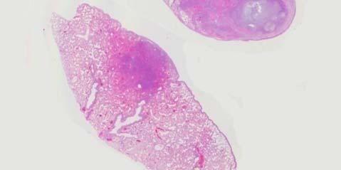
7 Bacteria / Mycoplasma / aspiration of bland material Subacute, suppurative bronchopneumonia / Cut surface with purulent exudate in bronchi. Normal (light color) and a pneumonic lung (dark red). Note purulent exudate coming out of a major bronchus. The appearance of the exudate in suppurative bronchopneumonia varies from purulent in acute cases to more mucoid in chronic cases. The presence of mucus in the exudate is due to severe goblet (mucus) cell hyperplasia in bronchi and goblet cell metaplasia in bronchioles. The color of an affected lung also varies from dark red in acute bronchopneumonia (hyperaemia + exudate) to grey resembling fish-flesh in chronic bronchopneumonia (exudate, fibrosis, reduced blood in capillaries). i If you take a section of bronchopneumonic lung, the main lesion as you may expect, will have accumulation of polymorphonuclear leukocytes in bronchoalveolar spaces (next slide).


8 Subacute, Suppurative bronchopneumonia / H&E stain/ This is the histological section of a lung with suppurative bronchopneumonia. Note a bronchiole plugged with purulent exudate (pe). Also note the large number of neutrophils mixed with edematous fluid in alveoli. The pathogenesis of this lesion is related to release of chemotactic factors on mucosal surface of bronchi, bronchioles and alveoli. This results in migration of neutrophils into the lumen of the airways. In uncomplicated cases, the exudate moves along the mucociliary escalator. In severe cases, the excessive release of leukocytic enzymes damages the mucosa, submucosa, smooth muscle and cartilage, causing bronchiectasis (see next slide).
9 Pulmonary abscesses and bronchiectasis are two important sequels to suppurative bronchopneumonia. Note large abscesses in the consolidated cranial and intermediate lobes. The caudal lobes are essentially normal. Histologically, abscesses are composed of a purulent core surrounded by connective tissue (pyogenic membrane).

.")


10 Chronic, Suppurative Bronchopneumonia with bronchitis and BRONCHIECTASIS / Bovine. Note tubular dilations in the cranioventral lung (arrow). These tubular structures are distended bronchi filled with purulent exudate that has destroyed the bronchial walls (insert). Bronchiectasis is also a common sequel to chronic bronchopneumonia. These lesions look like abscesses. However, microscopically instead of being lined by a capsule, in bronchiectasis the exudate is contained by remnants of bronchial walls.
11 Fibrinous Bronchopneumonia Cranioventral distribution Hard texture Color: Red yellow gray Fibrins on pleura Cut surface: fibrin / necrosis Sequels: Sequestrum, pleural adhesions Port of entry: Aerogenous Severe injury Fibrosis (chronic) Bacteria + toxins, aspiration of harsh material
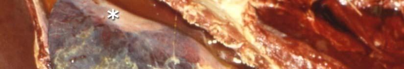
12 Fibrinous Bronchopneumonia Note cranioventral consolidation. Affected lung is covered with fibrin. Only a small portion of the lung appears grossly normal (asterisk)
13 Fibrinous Bronchopneumonia Note cranioventral consolidation. Affected lung is covered with fibrin. Only the dorsocaudal lung appears normal. The texture of consolidated lungs in fibrinous bronchopneumonia is typically hard.


14 Fibrinous bronchopneumonia / Cut surface of lung / Shipping Fever / Bovine. Fibrinous bronchopneumonia generally implies severe injury to the lung with leakage of fibrin into the airspaces. On cut surface, fibrinous bronchopneumonia often has a mosaic appearance due to distention of the interlobular septa in areas of coagulation necrosis. Note in this photograph p a mosaic appearance of the lung. The arrows delineate distended interlobular septa that results from lymphatic thrombosis and edema. Highly pathogenic bacteria Toxins Mannheimia haemolytica Haemophilus pleuropneumoniae Note also areas of coagulation necrosis (asterisks) caused by the toxins of Mannheimia haemolytica A1


15 Acute, fibrinous bronchopneumonia / Histopathology th H&E. Fibrinous exudation results from severe inflammatory response and leakage of plasma from vasculature into the bronchiolar and alveolar spaces. Note loss of airspaces due to exudation of fibrin and to a lesser extent t of leukocytes. Since fibrin predominates over polymorphonuclear leukocytes, the diagnosis i of fibrinous i is preferred to that of suppurative bronchopneumonia. R b th t fib i i h t ti Remember that fibrin is chemotactic for neutrophils and therefore any fibrinous pneumonia has many neutrophils microscopically.


16 4 More examples of Fibrinous Bronchopneumonia


17 Fibrous Adhesions Another important sequel to bronchopneumonia is the fibrous adhesions between visceral and parietal pleura. Note a thick band of connective tissue (arrows) forming an adhesion between the visceral pleura lining, the lung and the parietal pleura lining the thoracic cavity. Pleural adhesions are often found in postmortem examinations. It is generally an incidental finding that reveals the animal has survived an episode of bronchopneumonia.
18 Interstitial Pneumonia Diffuse Distribution Elastic Texture Rib imprints Cut surface: meaty Port of entry: Aerogenous Hematogenous Lungs failed to collapse when thorax is opened Injury to the alveolar wall (endothelium / pneumocytes) Thickening of the alveolar walls Often difficult to diagnose grossly
19 Interstitial Pneumonia Virus Toxins Sepsis Allergic Acute, severe, diffuse interstitial pneumonia / Horse. Note rib imprints on lung surfaces which result from lack of deflation at the time when the negative pressure is removed by opening the thorax.

.")


20 Interstitial Pneumonia Acute, severe, diffuse interstitial pneumonia / Dog. This is another example of an interstitial pneumonia. Note again rib imprints on the pleural surfaces (arrows) of this dog that died acutely with canine distemper (morbillivirus). Remember that the pleural imprints result from lack of deflation at the time when the negative pressure is removed by opening the thorax. Changes are often subtle, difficult to diagnose grossly and generally require histopathological confirmation. On cut surface, the lung tissue in interstitial pneumonia has an elastic texture and a meaty appearance. The primary lesion is centered in the alveolar wall The primary lesion is centered in the alveolar wall. Thickening of alveolar walls results from interstitial infiltration of mononuclear cells or from proliferation of type II pneumocytes. In chronic interstitial pneumonia, there is alveolar fibrosis.


21 Interstitial Pneumonia Subacute, severe, diffuse interstitial pneumonia / Steer. This lung belonged to a steer that died a few days after severe respiratory distress. The lungs failed to collapse when the thorax was opened and there were rib imprints on pleural surface. The texture of this lung was notably elastic. Note some edematous distention ti of the interlobular septa (arrows) and "meaty appearance of the parenchyma. Interstitial titi pneumonia is perhaps the most difficult type of pneumonia to diagnose grossly. In most cases it is imperative to do histopathology. The basic lesion in interstitial titi pneumonia is the thickening i of alveolar septa which is what gives it the elastic texture and prevents alveoli from collapsing. Microscopic view of the basic interstitial pneumonia is illustrated in the next slide.
 due to increased")


22 Interstitial Pneumonia Interstitial pneumonia / Steer. HE Note thick alveolar walls (arrows) due to increased cellularity of the alveolar interstitium (asterisks). Also note that alveoli are empty, containing no inflammatory cells. Thickening of alveolar septa results from the influx of inflammatory cells or fluid into the walls. Sometimes when there has been extensive necrosis of the alveolar epithelium, the alveoli are lined by hyperplastic type II pneumonocytes. In chronic cases, there may be fibrosis of interstitium and the lesion is often called "fibrosing alveolitis. Inflammation of alveolar interstitium is commonly caused by injury and destruction of type-i pneumocytes. It may be the result of viral infections, deposition of antigen-antibody complexes, inhalation of toxic gases (nitrogen dioxide, sulfur dioxide, ozone, oxygen toxicity), or ingestion of herbicides such as paraquat.

.")


23 Embolic Pneumonia Note multifocal hemorrhagic lesions randomly distributed in all pulmonary lobes (arrows). Also note that the center of the hemorrhagic foci is often white, suggesting neutrophilic inflammation. Naturally, the port of entry in embolic pneumonia is necessarily hematogenous. The distribution is typically multifocal and the texture nodular. Among the most common causes of embolic pneumonia are: Rupture of hepatic abscesses into the vena cava in cattle Vegetative endocarditis (right side of the heart) Jugular thrombosis Embolic foreign body (hair, septic emboli, etc).
24 Embolic Pneumonia Note numerous foci of inflammation scattered throughout the lungs (arrows)
25 See Figure 9-41 Pathologic Basis of Veterinary Diseases


26 Embolic Pneumonia / Foal. Sometimes the embolic lesions are very small and difficult to see as in the lungs of this foal. Look closely in the inset and you will notice small dark foci with white center (arrows).


27 Embolic Pneumonia Dog


28 Endocarditis (arrow) is a common cause of embolic pneumonia Embolic Pneumonia

 of the heart.")


29 Embolic Pneumonia Chronic, multifocal embolic pneumonia with abscesses Numerous small abscesses resulting form septic embolisms due to vegetative endocarditis affecting the tricuspid valve (right side) of the heart. Septic emboli are easily trapped in pulmonary vasculature causing embolic pneumonia and it s sequel pulmonary abscesses Bronchopneumonia may also have abscesses as sequels, except that in this later type of pneumonia the distribution is cranioventral and not random as in embolic pneumonia.
30 Multifocal Distribution Nodular texture Cut surface: granulomas Port of entry: Aerogenous Hematogenous Etiology: Myocbacterium spp Systemic mycoses Parasitic ova Trapped food particles Granulomatous Pneumonia

31 Granulomatous Pneumonia Chronic, severe, multifocal granulomatous pneumonia / Canine. Note entire lung parenchyma filled with medium size, often confluent granulomas. The distribution of granulomatous pneumonia is random multifocal affecting all pulmonary lobes. The port of entry could be either aerogenous or hematogenous. Texture is typically nodular.
32 Granulomatous Pneumonia in Bovine Tuberculosis Note numerous focal to coalescing granulomas scattered throughout the lung parenchyma. The port of entry in granulomatous pneumonia could be either aerogenous or hematogenous. The texture is nodular


 with tuberculosis are not seen in")
33 Chronic, severe, multifocal granulomatous pneumonia. Equine Tuberculosis / Lung - Cut surface. Numerous focal to coalescing granulomas in lung parenchyma. Tuberculosis can affect any tissue including bones, bone marrow, gonads, kidneys, meninges, etc. Mycobacterium tuberculosis generally affects humans, but cross infections with M. bovis and M. avium can also occur. Pigs can be affected by all three species. Remember that typical granulomatous lesions seen in cows and sheep (caseous necrosis and calcification) with tuberculosis are not seen in horses, cats and dogs. In these other animals tubercles have the appearance of tumoral growths (sarcomatous). Acid fast stain reveals the TB bacillus
 surrounded by macrophages")
 infiltrated")
.")


")
34 Note typical granuloma formed by a necrotic center (asterisks) surrounded by macrophages and then by an external band of fibrous connective tissue (double arrows) infiltrated by lymphocytes and plasma cells (not seen at this magnification). In most granulomatous pneumonias, the etiologic agent can be detected with special stains (to be shown later). Among the most common causes of granulomatous pneumonia in animals are: Tuberculosis Systemic mycosis (Cryptococcus neoformans, Blastomyces dermatitides, Coccidiodes immits) Aberrant parasitic larvae, foreign body (food particles) Granulomatous pneumonia. Histopathology Feline Infectious Peritonitis in cats, etc.


35 The five gross morphologic types of Pneumonia in Domestic Animals Suppurative Bronchopneumonia Fibrinous Bronchopneumonia Embolic Pneumonia Interstitial Pneumonia Granulomatous Pneumonia

36 THE END
09-Mar-15 PNEUMONIA RESPIRATORY SYSTEM L-3
 RESPIRATORY SYSTEM L-3 Professor Department of Pathology, University of Agriculture, Faisalabad. Email: mtjaved@uaf.edu.pk Web: https://sites.geocities.ws/mtjaved PNEUMONIA The pulmonary inflammatory response
Pathology of the Respiratory System 4: Pneumonia
 Pathology of the Respiratory System 4: Pneumonia Shannon Martinson, March 2016 http://people.upei.ca/smartinson/ VPM 222 Systemic Pathology LUNG PNEUMONIA Review Classification of Pneumonia Diffuse LUNG
Respiratory Pathology Lab 2: Lung. Shannon Martinson,
 Respiratory Pathology Lab 2: Lung Shannon Martinson, 2017 http://people.upei.ca/smartinson/ Case 1 Signalment: 9 month old DSH cat History: Poor doer with stunted growth One month of lethargy one day the
INFLAMMATION & REPAIR
 INFLAMMATION & REPAIR Histopath Laboratory 1 Winter 2013 Chelsea Martin Special thanks to Drs. Hanna and Forzan Goals: Examine Tissue and Identify the Organ Describe the lesion, grossly and histologically
Unit II Problem 2 Pathology: Pneumonia
 Unit II Problem 2 Pathology: Pneumonia - Definition: pneumonia is the infection of lung parenchyma which occurs especially when normal defenses are impaired such as: Cough reflex. Damage of cilia in respiratory
Histopathology: pulmonary pathology
 Histopathology: pulmonary pathology These presentations are to help you identify basic histopathological features. They do not contain the additional factual information that you need to learn about these
Inflammation Laboratory 2. Shannon Martinson: VPM 152: March 2012
 Inflammation Laboratory 2 Shannon Martinson: http://people.upei.ca/smartinson VPM 152: March 2012 Reminder - Creating a Morphologic Diagnosis for Inflammatory Lesions Organ and Process Exudate Distribution
Pathology of Pneumonia
 Pathology of Pneumonia Dr. Atif Ali Bashir Assistant Professor of Pathology College of Medicine Majma ah University Introduction: 5000 sq meters of area.! (olympic track) Filters >10,000 L of air / day!
Inflammation Laboratory 1
 Inflammation Laboratory 1 Lab1 Emphasis: The exudates of acute inflammation Descriptions Morphologic Diagnoses Shannon Martinson: http://people.upei.ca/smartinson VPM 152: February 2012 Describing Lesions
(JPC ) Caprine lungs
 Caprine lungs") 2011-7-2 (JPC 3133973) Caprine lungs Bat Otgontugs Bovine Pathology Contributor: Natoinal Institute Animal Health, Tsukuba, Japan Signalment: 5-year 3-month old female Japanese native breed goat, (Capra
2011-7-2 (JPC 3133973) Caprine lungs Bat Otgontugs Bovine Pathology Contributor: Natoinal Institute Animal Health, Tsukuba, Japan Signalment: 5-year 3-month old female Japanese native breed goat, (Capra
Pathology of the Respiratory System 5: Lung and Thoracic Cavity
 Pathology of the Respiratory System 5: Lung and Thoracic Cavity Shannon Martinson, Jan 2017 http://people.upei.ca/smartinson/ VPM 222 Systemic Pathology DISORDERS OF THE LUNG Congenital Pigmentary deposition
The Respiratory System. Dr. Ali Ebneshahidi
 The Respiratory System Dr. Ali Ebneshahidi Functions of The Respiratory System To allow gases from the environment to enter the bronchial tree through inspiration by expanding the thoracic volume. To allow
Inflammation Laboratory 1
 Inflammation Laboratory 1 Lab1 Emphasis: The exudates of acute inflammation Descriptions Morphologic Diagnoses Shannon Martinson: http://people.upei.ca/smartinson VPM 152: March 2013 Describing Lesions
DISEASES OF THE RESPIRATORY SYSTEM 2018 DR HEYAM AWAD LECTURE 3: CHRONIC BRNCHITIS AND BRONCHIECTASIS
 DISEASES OF THE RESPIRATORY SYSTEM 2018 DR HEYAM AWAD LECTURE 3: CHRONIC BRNCHITIS AND BRONCHIECTASIS INTRDUCTION In the last lecture we discussed the difference between restrictive and obstructive lung
Post Mortal Approach to the Respiratory System Part 1
 Post Mortal Approach to the Respiratory System Part 1 System examination Before the carcass is opened examination of the nasal openings is carried out. Observe for any evidence of nasal discharge or nasal
Respiratory Pathology. Kristine Krafts, M.D.
 Respiratory Pathology Kristine Krafts, M.D. Normal lung: alveolar spaces Respiratory Pathology Outline Acute respiratory distress syndrome Obstructive lung diseases Restrictive lung diseases Vascular
Examples of Morphologic Types of Pneumonia in Domestic Animals
 Tutorial Module 5 Examples of Morphologic Types of Pneumonia in Domestic Animals Alfonso López Atlantic Veterinary College University of Prince Edward Island Canada 2014 Sept 24, 2014b Examples of Diseases
Lymphoid System: cells of the immune system. Answer Sheet
 Lymphoid System: cells of the immune system Answer Sheet Q1 Which areas of the lymph node have most CD3 staining? A1 Most CD3 staining is present in the paracortex (T cell areas). This is towards the outside
Replacement of air with fluid, inflammatory. cells or cellular debris. Parenchymal, Interstitial (Restrictive) and Vascular Diseases.
 and Vascular Diseases.") Parenchymal, Interstitial (Restrictive) and Vascular Diseases Alain C. Borczuk, M.D. Dept of Pathology Replacement of air with fluid, inflammatory cells Pulmonary Edema Pneumonia Hemorrhage Diffuse alveolar
Parenchymal, Interstitial (Restrictive) and Vascular Diseases Alain C. Borczuk, M.D. Dept of Pathology Replacement of air with fluid, inflammatory cells Pulmonary Edema Pneumonia Hemorrhage Diffuse alveolar
Destructive pulmonary disease due to mixed anaerobic infection
 Thorax (1970), 25, 41. Destructive pulmonary disease due to mixed anaerobic infection 0. SERIKI, A. ADEYOKUNNU, T. 0. DE LA CRUZ Departments of Paediatrics and Surgery, University College Hospital, Ibadan,
Describing and interpreting gross lesions. Prepared for VPM 4600, May 2018; Shannon Martinson
 Describing and interpreting gross lesions Prepared for VPM 4600, May 2018; Shannon Martinson How to Describe (and Interpret) Lesions Step 1 Step 2 Step 3 Step 4 Look at the specimen: Is it normal or abnormal
Cellular Pathology. Histopathology Lab #2 (web) Paul Hanna Jan 2018
 Paul Hanna Jan 2018") Cellular Pathology Histopathology Lab #2 (web) Paul Hanna Jan 2018 Slide #91 Clinical History: a necropsy was performed on an aged cat the gross pathological changes included: widespread subcutaneous edema
Cellular Pathology Histopathology Lab #2 (web) Paul Hanna Jan 2018 Slide #91 Clinical History: a necropsy was performed on an aged cat the gross pathological changes included: widespread subcutaneous edema
Restrictive lung diseases
 Restrictive lung diseases Restrictive lung diseases are diseases that affect the interstitium of the lung. Interstitium of the lung is the very thin walls surrounding the alveoli, it s formed of epithelium
HISTO-PHYSIOLOGY HISTO-PHYSIOLOGY HISTO-PHYSIOLOGY. 09-Mar-15. Dr. Muhammad Tariq Javed. RESPIRATORY SYSTEM Lec-1
 RESPIRATORY SYSTEM Lec-1 Dr. Muhammad Tariq Javed Professor Department of Pathology, University of Agriculture, Faisalabad. Email: mtjaved@uaf.edu.pk Web: http://www.geocities.ws/mtjaved 1 2 Conducting
Bone Injury and Inflammatory Diseases of Bone
 Bone Injury and Inflammatory Diseases of Bone Module 3 Alfonso López Atlantic Veterinary College January 10, 2014 Bone Necrosis / Cross Section Necrotic bone is often difficult to detect grossly but it
Pathological Investigations on Bovine Pheumonic Pasteurellosis by Use of Immunoperoxidase Technique
 JARQ 29, 13 1-136 (1995) Pathological Investigations on Bovine Pheumonic Pasteurellosis by Use of Immunoperoxidase Technique Makoto HARITANI Tohoku Branch Laboratory, National Institute of Animal Health
This is the second learning component (Learning Component 2) in our first learning module (Learning Module 1). In this component we review a very
 in our first learning module (Learning Module 1). In this component we review a very") This is the second learning component (Learning Component 2) in our first learning module (Learning Module 1). In this component we review a very basic response to injury inflammation. We ll look at examples
This is the second learning component (Learning Component 2) in our first learning module (Learning Module 1). In this component we review a very basic response to injury inflammation. We ll look at examples
Pathology of pulmonary tuberculosis. Dr: Salah Ahmed
 Pathology of pulmonary tuberculosis Dr: Salah Ahmed Is a chronic granulomatous disease, caused by Mycobacterium tuberculosis (hominis) Usually it involves lungs but may affect any organ or tissue Transmission:
Acute and Chronic Inflammation Pathology 1 - Dr. Gary Mumaugh
 Acute and Chronic Inflammation Pathology 1 - Dr. Gary Mumaugh Introduction Injurious stimuli cause a protective vascular connective tissue reaction called inflammation Acute and chronic forms o Inflame
Sheet: Patho-Pulmonary infections Done by: Maen Faoury
 Sheet: Patho-Pulmonary infections Done by: Maen Faoury Pneumonitis : might be an infection or not. Chemical Pneumonitis : not an infection. Parenchyma : an infection.( تندرج تحت ال pneumonitis) Lung Parenchyma
CHRONIC INFLAMMATION
 CHRONIC INFLAMMATION Chronic inflammation is an inflammatory response of prolonged duration often for months, years or even indefinitely. Its prolonged course is proved by persistence of the causative
Cellular Pathology Gross Pathology Laboratory 2 Cell Injury. VPM 152: General Pathology Instructor: Chelsea Martin Winter 2016
 Cellular Pathology Gross Pathology Laboratory 2 Cell Injury VPM 152: General Pathology Instructor: Chelsea Martin Winter 2016 Gross Specimens The following slides consist of images from the specimens presented
Pulmonary Pathology II. William Bligh-Glover M.D. Department of Anatomy, CWRU
 Pulmonary Pathology II William Bligh-Glover M.D. Department of Anatomy, CWRU Goals and Objectives Comprehend the etiology, pathogenesis/pathopysiology and consequences of pulmonary hypertension Distinguish
Inflammation Laboratory 3 Emphasis: Chronic inflammation and healing. Shannon Martinson: VPM 152: April 2013
 Inflammation Laboratory 3 Emphasis: Chronic inflammation and healing Shannon Martinson: http://people.upei.ca/smartinson VPM 152: April 2013 Example A Reproductive tract and colon/rectum from a sheep Previous
Bone necrosis. Cross section.
 Bone necrosis. Cross section. Note the large area of necrosis (N) seen as pale discolored bone (beneath physeal cartilage). The texture of the necrotic bone is also changed. Necrotic bone becomes friable
Key Difference - Pleural Effusion vs Pneumonia
 Difference Between Pleural Effusion and Pneumonia www.differencebetween.com Key Difference - Pleural Effusion vs Pneumonia Pleural effusion and pneumonia are two conditions that affect our respiratory
Diagnosis of TB: Radiology David Finlay, MD
 TB Intensive Tyler, Texas June 2-4, 2010 Diagnosis of TB: Radiology David Finlay, MD June 3, 2010 2stages stages- Tuberculosis 1. primary infection 2. reactivation, or post primary disease 2 1 Primary
Acute pneumonia in a cat
 Acute pneumonia in a cat Elspeth Milne, Anita Schwartz, Alasdair Stuart, Danielle Gunn-Moore, Kerry Simpson and Sionagh Smith, Division of Veterinary Clinical Sciences, University of Edinburgh, United
Canine Liver Eneku Wilfred Bovine Pathology
 2012-1-3 Canine Liver Eneku Wilfred Bovine Pathology Contributor: New Mexico Department of Agriculture Veterinary Diagnostic Services Signalment: 5 month old male Weimaraner dog (Canis familiaris) History:
Pathology of the Liver and Biliary Tract 5 Diseases of the Biliary Tract. Shannon Martinson, March 2017
 Pathology of the Liver and Biliary Tract 5 Diseases of the Biliary Tract Shannon Martinson, March 2017 http://people.upei.ca/smartinson/ OUTLINE Normal anatomy & function Hepatobiliary injury and responses
The Respiratory System
 The Respiratory System Respiratory Anatomy Upper respiratory tract Nose Nasal passages Pharynx Larynx Respiratory Anatomy Functions of the upper respiratory tract: Provide entry for inhaled air Respiratory
Pathology lab 4 DONE BY : MORAD ABU QAMAR
 Pathology lab 4 DONE BY : MORAD ABU QAMAR Chronic interstitial inflammation, lung Certain etiologic agents such as viruses are more likely to lead to chronic inflammation, as seen here in the lung of a
Asthma. - A chronic inflammatory disorder which causes recurrent episodes of wheezing, breathlessness, cough and chest tightness.
 Obstructive diseases Asthma - A chronic inflammatory disorder which causes recurrent episodes of wheezing, breathlessness, cough and chest tightness. - Characterized by Intermittent and reversible (the
Usual Interstitial pneumonia and Nonspecific Interstitial Pneumonia. Nitra and the Gangs.
 Usual Interstitial pneumonia and Nonspecific Interstitial Pneumonia Nitra and the Gangs. บทน ำและบทท ๓, ๑๐, ๑๒, ๑๓, ๑๔, ๑๕, ๑๗ Usual Interstitial Pneumonia (UIP) Most common & basic pathologic pattern
Parenchymal, Interstitial i (Restrictive) i and Vascular Diseases
 i and Vascular Diseases") Pulmonary Diseases: Structure-Function Correlation II Parenchymal, Interstitial i (Restrictive) i and Vascular Diseases Alain C. Borczuk, M.D. Dept of Pathology Pulmonary Diseases: Structure-Function Correlation
Pulmonary Diseases: Structure-Function Correlation II Parenchymal, Interstitial i (Restrictive) i and Vascular Diseases Alain C. Borczuk, M.D. Dept of Pathology Pulmonary Diseases: Structure-Function Correlation
Diseases of the Lung and Respiratory Tract, Part I. William Bligh-Glover M.D. Department of Anatomy, CWRU
 Diseases of the Lung and Respiratory Tract, Part I William Bligh-Glover M.D. Department of Anatomy, CWRU Educational objectives: Distinguish the types of atelectasis and their etiologies Distinguish the
Vascular Lung Diseases
 Vascular Lung Diseases SESSION SPECIFIC OBJECTIVES List the major types of vascular lung disease Recognize and describe the pathology of vascular lung disease: Pulmonary embolism, thrombosis, hypertension,
Respiratory Diseases and Disorders
 Chapter 9 Respiratory Diseases and Disorders Anatomy and Physiology Chest, lungs, and conducting airways Two parts: Upper respiratory system consists of nose, mouth, sinuses, pharynx, and larynx Lower
8/14/2017. Objective: correlate radiographic findings of common lung diseases to actual lung pathologic features
 What is that lung disease? Pulmonary Patterns & Correlated Pathology Dr. Russell Tucker, DACVR Objective: correlate radiographic findings of common lung diseases to actual lung pathologic features Improved
Bronkhorst colloquium Interstitiële longziekten. Katrien Grünberg, klinisch patholoog
 Bronkhorst colloquium 2013-2014 Interstitiële longziekten De pathologie achter de CT Katrien Grünberg, klinisch patholoog K.grunberg@vumc.nl Preparing: introduction and 3 cases The introduction on microscopic
Anatomy. The respiratory system starts from the nose, mouth, larynx, trachea, and the two lungs.
 Respiratory System Anatomy The respiratory system starts from the nose, mouth, larynx, trachea, and the two lungs. Within the lungs, the bronchi transport air with oxygen to the alveoli on inspiration
Respiratory System. Module 1. Structure and Defense Mechanisms. Alfonso López. Atlantic Veterinary College University of Prince Edward Island Canada
 Dec 10, 2017 Respiratory System Module 1 Structure and Defense Mechanisms Alfonso López Atlantic Veterinary College University of Prince Edward Island Canada lopez@upei.ca 2018 If you find this tutorial
CASE REPORTS. Inflammatory Polyp of the Bronchus. V. K. Saini, M.S., and P. L. Wahi, M.D.
 CASE REPORTS V. K. Saini, M.S., and P. L. Wahi, M.D. I n 1932 Jackson and Jackson [l] first reported a number of clinical cases under the title Benign Tumors of the Trachea and Bronchi with Especial Reference
SESSION 1: GENERAL (BASIC) PATHOLOGY CONCEPTS Thursday, October 16, :30am - 11:30am FACULTY COPY
 PATHOLOGY CONCEPTS Thursday, October 16, :30am - 11:30am FACULTY COPY") SESSION 1: GENERAL (BASIC) PATHOLOGY CONCEPTS Thursday, October 16, 2008 9:30am - 11:30am FACULTY COPY GOAL: Describe the basic morphologic (structural) changes which occur in various pathologic conditions.
SESSION 1: GENERAL (BASIC) PATHOLOGY CONCEPTS Thursday, October 16, 2008 9:30am - 11:30am FACULTY COPY GOAL: Describe the basic morphologic (structural) changes which occur in various pathologic conditions.
Characteristic. Course of disease:short Days--one month Changes : Alteration, exudation Tissue destruction Inflammation cells: major neutrophils
 ACUTE INFLAMMATION Characteristic Course of disease:short Days--one month Changes : Alteration, exudation Tissue destruction Inflammation cells: major neutrophils TYPES Serous Inflammation Fibrinous Inflammation
an inflammation of the bronchial tubes
 BRONCHITIS DEFINITION Bronchitis is an inflammation of the bronchial tubes (or bronchi), which are the air passages that extend from the trachea into the small airways and alveoli. Triggers may be infectious
Necrosis is death of cells and tissues in the living animal. Focal/ Multifocal necrosis- terms used for one
 Necrosis Necrosis Necrosis is death of cells and tissues in the living animal. Focal/ Multifocal necrosis- terms used for one or more, small, clearly defined areas of necrosis. Diffuse necrosis- term used
Pulmonary Diseases. We Move A Lot of Air. Basic Categories. Alveolar Level. Developmental
 Pulmonary Diseases We Move A Lot of Air Alveolar Level Functions Oxygenation CO 2 & ph Basic defenses Nose hairs Cilia Mucus Cough reflex Immune system Basic Categories Congenital Infectious Neoplastic
Menigitidis. Dr Rodney Itaki Lecturer Anatomical Pathology Discipline
 Menigitidis Dr Rodney Itaki Lecturer Anatomical Pathology Discipline University of Papua New Guinea Division of Pathology School of Medicine & Health Sciences Review Normal Microanatomy Image Ref: www.histology-world.com
RESPIRATORY BLOCK. Bronchial Asthma. Dr. Maha Arafah Department of Pathology KSU
 RESPIRATORY BLOCK Bronchial Asthma Dr. Maha Arafah Department of Pathology KSU marafah@ksu.edu.sa Jan 2018 Objectives Define asthma (BA) Know the two types of asthma 1. Extrinsic or atopic allergic 2.
Slide 120, Lobar Pneumonia. Slide 120, Lobar Pneumonia. Slide 172, Interstitial Pneumonia. Slide 172, Interstitial Pneumonia. 53 Year-Old Smoker
 Slide 120, Lobar Pneumonia Slide 120, Lobar Pneumonia Slide 172, Interstitial Pneumonia Slide 172, Interstitial Pneumonia 53 Year-Old Smoker Emphysema Pink puffer Barrel chest Hyperinflation Trapped air
2015/4/14. Pneumonia. Diseases of Respiratory System Infection in the lung (distal airways, esp. alveoli) Lobar pneumonia.
 Lobar pneumonia.") Pneumonia Diseases of Respiratory System Infection in the lung (distal airways, esp. alveoli) 邓红浙江大学医学院病理学系 LUNG(reformed)5y-DH 1 hongdeng@zju.edu.cn Pathology (DH) 2 Pneumonia Bacteria pneumonia Viral
Pneumonia Diseases of Respiratory System Infection in the lung (distal airways, esp. alveoli) 邓红浙江大学医学院病理学系 LUNG(reformed)5y-DH 1 hongdeng@zju.edu.cn Pathology (DH) 2 Pneumonia Bacteria pneumonia Viral
SCPA502-Respiratory Pathology
 Problem Mr. B is 57 years old, high 157 cm and weight 76 kg. He has worked as the dump truck driver in the coal mine since 1980, and also smoked cigarette 1 pack/day more than 30 years. What are the risk
Imaging Small Airways Diseases: Not Just Air trapping. Eric J. Stern MD University of Washington
 Imaging Small Airways Diseases: Not Just Air trapping Eric J. Stern MD University of Washington What we are discussing SAD classification SAD imaging with MDCT emphasis What is a small airway? Airway with
Pathology of the Hematopoietic System - Lab.
 Pathology of the Hematopoietic System - Lab http://people.upei.ca/smartinson/ Shannon Martinson, September 2015 Case #1 Signalment: 96 kg gilt History: Pig from minimal disease herd. Sudden death Case
Pulmonary Patterns & Correlated Pathology
 Pulmonary Patterns & Correlated Pathology Russell Tucker, DVM, DACVR Washington State University College of Veterinary Medicine Objective: correlate radiographic findings of common lung diseases to actual
Naturally occurring Mycoplasma bovis associated pneumonia and polyarthritis in feedlot beef calves
 J Vet Diagn Invest 18:29 4 (26) Naturally occurring Mycoplasma bovis associated pneumonia and polyarthritis in feedlot beef calves Mihai I. Gagea, Kenneth G. Bateman, Rachel A. Shanahan, Tony van Dreumel,
Anatomy and Physiology of the Lungs
 The lungs consist of right and left sides. The right lung has three lobes: Upper lobe, Middle lobe, Lower lobe The left lung has two lobes: Upper lobe, Lower lobe Anatomy and Physiology of the Lungs The
Note the large area of necrosis (N) which appears as a pale discolored bone
 which appears as a pale discolored bone") Bone Injury and Inflammatory Bone Diseases Alfonso López Atlantic Veterinary College University i of Pi Prince Edward d Il Island January 7, 2010 Bone Necrosis / Cross Section N The texture of the necrotic
Bone Injury and Inflammatory Bone Diseases Alfonso López Atlantic Veterinary College University i of Pi Prince Edward d Il Island January 7, 2010 Bone Necrosis / Cross Section N The texture of the necrotic
Pathology of the Liver and Biliary Tract 5 Diseases of the Biliary Tract. Shannon Martinson, April 2016
 Pathology of the Liver and Biliary Tract 5 Diseases of the Biliary Tract Shannon Martinson, April 2016 http://people.upei.ca/smartinson/ OUTLINE Normal anatomy & function Hepatobiliary Injury and responses
like humans, have well-developed mediastinal separation between the left and right hemithorax, thus unilateral changes can occur. On the other hand,
 Tutorial Module 6 Thoracic Cavity and Tumors of Lung and Pleura Alfonso López Atlantic Veterinary College University of Prince Edward Island Canada 2009 Enero 3 Thoracic Cavity There are significant anatomical
PATHOLOGY OF LIVER & BILIARY TRACT. Lecture 5. Idiopathic & proliferative conditions; diseases of the biliary tract
 PATHOLOGY OF LIVER & BILIARY TRACT Lecture 5 Idiopathic & proliferative conditions; diseases of the biliary tract Enrique Aburto Winter 2015 IX. Diseases of uncertain origin Equine serum hepatitis Idiopathic
7/12/2012. Respiratory system. Respiratory Response to Toxic Injury (Lung) Ninth Industrial Toxicology and Pathology Short Course.
 Ninth Industrial Toxicology and Pathology Short Course.") Ninth Industrial Toxicology and Pathology Short Course 23 27 July, 2012 Contemporary Concepts in Target Organ Toxicologic Pathology Respiratory system Respiratory Response to Toxic Injury (Lung) Eric Wheeldon
Ninth Industrial Toxicology and Pathology Short Course 23 27 July, 2012 Contemporary Concepts in Target Organ Toxicologic Pathology Respiratory system Respiratory Response to Toxic Injury (Lung) Eric Wheeldon
Figure 2: Lymph node Cortical follicular (F) and paracortical (PC) atrophy, with narrowing of the cortex relative to the medulla (M).
 and paracortical (PC) atrophy, with narrowing of the cortex relative to the medulla (M).") Figure 1: Lymph node Follicular hyperplasia, with expansion of the follicular germinal centres (F) by large blast cells. Paracortical hyperplasia, with expansion of the paracortex (PC) by small lymphocytes.
Figure 1: Lymph node Follicular hyperplasia, with expansion of the follicular germinal centres (F) by large blast cells. Paracortical hyperplasia, with expansion of the paracortex (PC) by small lymphocytes.
1/13/2014. Proper Radiographs. Proper Radiographs. A Review of Pulmonary Patterns
 Live Webinar A Review of Pulmonary Patterns Sofija R. Liles, DVM, DACVR Proper Radiographs Which views? One lateral plus ventrodorsal (at least) Left lateral is best for thorax Three views for full metastatic
PATHOLOGY OF THE RESPIRATORY SYSTEM Handout Morphologic Pathology I / VPM 221
 PATHOLOGY OF THE RESPIRATORY SYSTEM Handout Morphologic Pathology I / VPM 221 Rev October 2011 Alfonso López Textbook: López, A. The Respiratory System, Mediastinum and Pleura: In, Pathological Basis of
PATHOLOGY OF PASTEURELLA MULTOCIDA INFECTION IN CHICKENS
 Indian J. Anim. Res., 40 (1): 15-19, 2006 PATHOLOGY OF PASTEURELLA MULTOCIDA INFECTION IN CHICKENS Shilpa Sood 1 and P.C. Verma CCS Haryana Agricultural University, Hisar - 125 004, India ABSTRACT The
SESSION IV: MECHANISMS OF HUMAN DISEASE: LABORATORY SESSIONS PULMONARY PATHOLOGY I. December 5, 2012
 SESSION IV: MECHANISMS OF HUMAN DISEASE: LABORATORY SESSIONS PULMONARY PATHOLOGY I December 5, 2012 FACULTY COPY GOAL: Describe the basic morphologic and pathophysiologic changes in various conditions
The Thorax The Ever Challenging Pulmonary Patterns
 The Thorax The Ever Challenging Pulmonary Patterns Lisa G. Britt, DVM, MS, Diplomate American College of Veterinary Radiology, Clinical Assistant Professor @ University of Missouri s College of Veterinary
Pathology of the Alimentary Tract
 Pathology of the Alimentary Tract Lab 2: Lower alimentary tract SI, LI, cecum, and peritoneum GIST in the cecum of a dog Shannon Martinson: http://people.upei.ca/smartinson VPM 221: November, 2011 3 year
Organs of the Respiratory System Laboratory Exercise 52
 Organs of the Respiratory System Laboratory Exercise 52 Background The organs of the respiratory system include the nose, nasal cavity, sinuses, pharynx, larynx, trachea, bronchial tree, and lungs. They
Exam 2 Respiratory Disorders
 Exam 2 Respiratory Disorders Common Cold Common Cold Pathology Common Cold Consequences Rhinosinusitis Rhinosinusitis Pathology Rhinosinusitis ostia can close due to Influenza (Flu) Influenza Pathology
Avian Pathology. Bacterial diseases: histo slides. ECVP-ESVP Summer School 2012 Frédérique NGUYEN
 Avian Pathology Bacterial diseases: histo slides ECVP-ESVP Summer School 2012 Frédérique NGUYEN Bacterial diseases: histo slides B1. Turkey. Organs? Morphologic diagnosis? Special procedure? B2. Hen. Organ?
Microscopic Lesions Associated with the Isolation of Haemophilus somnus from Pneumonic Bovine Lungs
 Vet. Pathol. 22: 131-136 (1985) Microscopic Lesions Associated with the Isolation of Haemophilus somnus from Pneumonic Bovine Lungs J. J. ANDREWS, T. D. ANDERSON, L. N. SLIFE, and G. W. STEVENSON Veterinary
Lec #2 histology. Bronchioles:
 Lec #2 histology. Last lecture we talked about the upper respiratory tract histology, this one is about the lower part histology. We will discuss the histology of: -bronchioles -respiratory bronchioles
Acute and Chronic Lung Disease
 KATHOLIEKE UNIVERSITEIT LEUVEN Faculty of Medicine Acute and Chronic Lung Disease W De Wever, JA Verschakelen Department of Radiology, University Hospitals Leuven, Belgium Clinical utility of HRCT To detect
LUNG PATTERNS IN THE DOG NORMAL AND PATHOLOGICAL
 TRADITION AND MODERNITY IN VETERINARY MEDICINE, 2018, vol. 3, No 1(4): 7 14 LUNG PATTERNS IN THE DOG NORMAL AND PATHOLOGICAL Kalin Spasov 1, Michaela Kunovska 2, Dimo Dimov 3 1 University of Forestry,
HRCT in Diffuse Interstitial Lung Disease Steps in High Resolution CT Diagnosis. Where are the lymphatics? Anatomic distribution
 Steps in High Resolution CT Diagnosis Pattern of abnormality Distribution of disease Associated findings Clinical history Tomás Franquet MD What is the diagnosis? Hospital de Sant Pau. Barcelona Secondary
Liver Lab #2. Bacterial Hepatitis
 Liver Lab #2 Bacterial Hepatitis Case: O12561-04. Adult ewe. Describe the lesion: Multifocal large nodules ranging in size from 1-3.5cm in greatest diameter are present within the liver and are filled
Proceedings of the 10th International Congress of World Equine Veterinary Association
 www.ivis.org Proceedings of the 10th International Congress of World Equine Veterinary Association Jan. 28 Feb. 1, 2008 - Moscow, Russia Next Congress: Reprinted in IVIS with the permission of the Conference
Respiratory System. Module 1. Structure and Defense Mechanisms. Alfonso López. Atlantic Veterinary College University of Prince Edward Island Canada
 Nov 21, 2016d Respiratory System Module 1 Structure and Defense Mechanisms Alfonso López Atlantic Veterinary College University of Prince Edward Island Canada lopez@upei.ca 2016 If you find this tutorial
Chapter 10. Respiratory System and Gas Exchange. Copyright 2005 Pearson Education, Inc. publishing as Benjamin Cummings
 Chapter 10 Respiratory System and Gas Exchange Function of the Respiratory System To obtain oxygen (O 2 ) for all cells in the body. To rid the cells of waste gas (CO 2 ). Oxygen (O 2 ) is vital chemical
The RESPIRATORY System. Unit 3 Transportation Systems
 The RESPIRATORY System Unit 3 Transportation Systems Functions of the Respiratory System Warm, moisten, and filter incoming air Resonating chambers for speech and sound production Oxygen and Carbon Dioxide
Histology and development of the respiratory system
 Histology and development of the respiratory system Árpád Dobolyi Semmelweis University, Department of Anatomy, Histology and Embryology Outline of the lecture 1. Structure of the trachea 2. Histology
Histopathology: granulomatous inflammation, including tuberculosis
 Histopathology: granulomatous inflammation, including tuberculosis These presentations are to help you identify basic histopathological features. They do not contain the additional factual information
LADIS Case of the Month
 November 2018 LADIS Case of the Month Drs Valentin Janvier and Brieuc Cossic Hospital for Animals and Animal Health Diagnostic Center Signalment and presenting complaint 13 year old Thoroughbred gelding
Bronchioles. Alveoli. Type I alveolar cells are very thin simple squamous epithelial cells and form most of the lining of an alveolus.
 276 Bronchioles Bronchioles continue on to form bronchi. The primary identifying feature is the loss of hyaline cartilage. The epithelium has become simple ciliated columnar, and there is a complete ring
PULMONARY IMAGING: GETTING THE MOST INFORMATION FROM THORACIC RADIOGRAPHS
 PULMONARY IMAGING: GETTING THE MOST INFORMATION FROM THORACIC RADIOGRAPHS Peter Scrivani, DVM, DACVR Cornell University College of Veterinary Medicine, Ithaca, NY Outline Pulmonary Imaging Pulmonary anatomy
Bio 322 Human Anatomy Objectives for the laboratory exercise Respiratory System
 Bio 322 Human Anatomy Objectives for the laboratory exercise Respiratory System Required reading before beginning this lab: Saladin, KS: Human Anatomy 5 th ed (2017) Chapter 23 For this lab you will use
Notes to complete gas exchange in mammals
 Notes to complete gas exchange in mammals Mass flow of air to respiratory surface this is achieved through the mechanics of ventilation (breathing). This ensures a regular supply of air into and out of