MR IMAGING OF THE WRIST
|
|
|
- Nelson Byrd
- 6 years ago
- Views:
Transcription


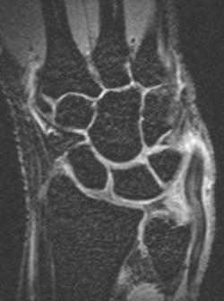
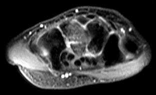
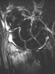
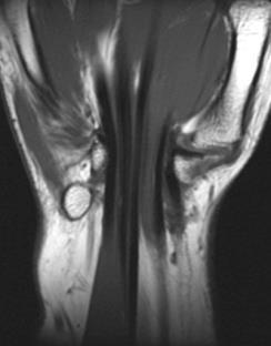
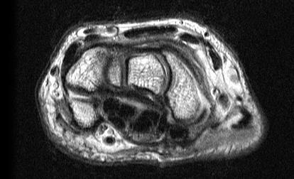
1 MR IMAGING OF THE WRIST Wrist Instability Dissociative Pattern apparent on routine radiographs Non-dissociative Stress / positional radiographs Dynamic fluoroscopy during stress Arthrography MRI / MR arthrography Look for intact carpal arcs Joints Should be Uniform in width Osseous Congruency Disruption of Carpal Arcs Acute scaphoid fracture with perilunate dislocation Widening of scapholunate interval 1


2 Make sure hand is straight Lateral view: Evaluate -Lunate tilt -capitate-lunate angle -scapholunate angle Lateral view: -Lunate tilt -capitate-lunate angle -scapholunate angle Transscaphoid perilunate dislocation Lateral view: -Lunate tilt -capitate-lunate angle -scapholunate angle 0 30 degrees Lunate tilt results in pie-shaped configuration Lateral view: -Lunate tilt -capitate-lunate angle -scapholunate angle degrees Positional Radiographs Can help demonstrate carpal malalignment, intercarpal widening Radial / ulnar deviation, lateral flexion / extension 2


 vs.")
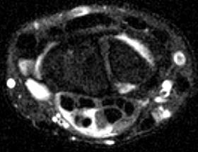
3 - Radial deviation Joint spaces maintained Scaphoid palmarflexes, rounded on AP - Ulnar deviation Joint spaces maintained Scaphoid elongates on AP - Lateral flexion / extension Imaging under stress Closed fist view Places distractive stress on carpal ring Can accentuate scapholunate widening Video fluoroscopy Monitor during motion under applied stress Visualize carpal instability patterns dynamically Capitate tilts with lunate Scapholunate widening more prominent Arthrography Inject contrast into joint Visualize contrast passing between compartments Documents ligament tear Single compartment (radiocarpal) vs. triple compartment injection (may increase sensitivity) 3


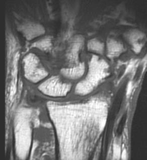
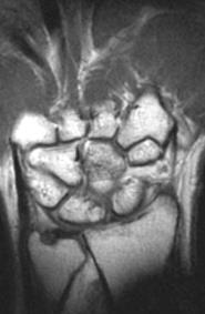
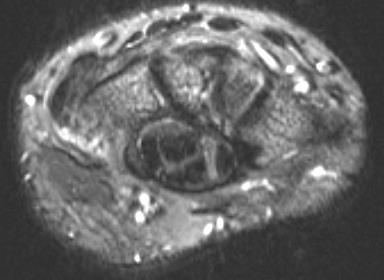
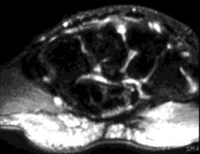
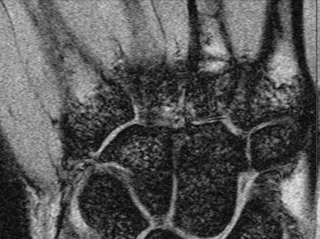
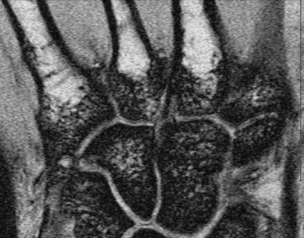
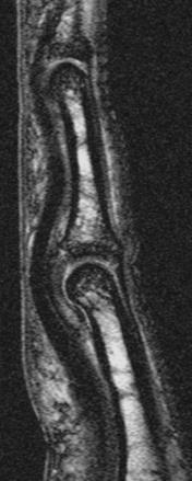
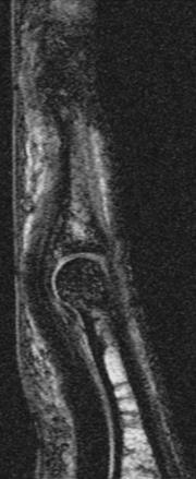
4 Early contrast extending through Lunatotriquetral ligament Radioscaphoid injection Late - contrast in midcarpal joint Scapholunate and Lunatotriquetral Ligaments NORMAL ANATOMY T2 FSE PD fat sat Scapholunate and Lunatotriquetral Ligaments DORSAL AND VOLAR BANDS Large tear SL ligament These bands are more mechanically important than central membrane Disruption of dorsal and volar bundles 4


 tear")


 DISI")


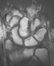
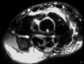
5 Scapholunate Ligament Tear Direct MR arthrogram scapholunate tear Tear on multiple slices Widening of joint / malalignment Intervening cartilage damage Malalignment MAY BE visible on MRI ***WARNING*** Dorsal tilt of lunate Scapholunate advanced collapse (SLAC wrist) Scapholunate tear Palmarflexion of scaphoid Dorsal intercalated segment instability (DISI) DISI deformity Proximal migration of capitate Carpal osteoarthritis Radiographic progression Of SLAC SLAC wrist Late Early -radioscaphoid joint narrowing Intermediate 5


")
")


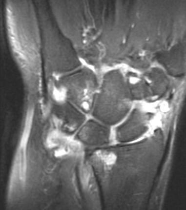
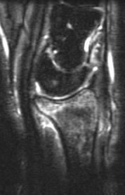
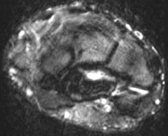
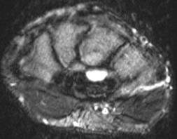
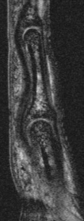
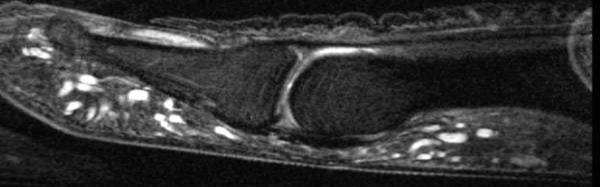
6 SLAC secondary to rheumatoid arthritis Inflammatory arthropathies can cause intrinsic ligament tears Lunatotriquetral Ligament Tear Extensive synovitis Marrow edema Lunatotriquetral ligament tear TFCC Leak or Perforation Lunate may tilt in palmar direction along with scaphoid Volar intercalated segment instability (VISI) Perforation -Contrast / fluid through ligament -Seen on only 1 slice ( ask a friend rule ) -Small diameter (1mm) Perforations may not be clinically significant TFCC Tear TFCC IMPACTION Tear -Contrast / fluid through ligament -Multiple slices / >1mm -Malalignment -Cartilage damage Tears are more likely to be symptomatic 6


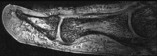
7 Ulnar Impaction AKA Ulno-lunate Abutment Direct Arthrogram Indirect Arthrogram tear of central TFC with ulnar-lunate abutment Large communication Underlying thinning Cartilage damage TFCC: Peripheral attachments Central TFCC tear It s not all about MRI! CT arthrography can also be useful Styloid Foveal Peripheral TFCC Tear Combo LT Tear (or leak?) A trickle or a FLOOD? MRI can help 7


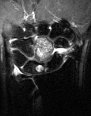
8 Flood!! Massive Peripheral TFCC Tear Peripheral TFCC Tear Extensor Carpi Ulnaris Tenosynovitis Gd Contrast injected into radioscaphoid joint Rheumatoid Arthritis Masslike synovial proliferation Rheumatoid Arthritis MRI can monitor activity, response to Tx Erosions -Capitate fracture -Distal radial fracture 8

 Progression:")


")


9 -Occult scaphoid Fracture NBA player Avascular necrosis -Lunate (negative ulnar variance) -Scaphoid (fracture) Progression: density, fracture, collapse, OA -Keinbock s disease Replacement of fat signal c/w AVN -Scaphoid fracture with AVN of the proximal pole Arthritis -Scapholunate Advanced Collapse (SLAC) -Osteoarthritis -Subchondral cysts cartilage loss, spurs -Distribution depends on etiology -Trauma, instability, predisposing factors -Inflammatory arthropathies -Classic: rheumatoid arthritis -Carpus, MCPs -Diffuse involvement -Synovitis, erosions 9


10 -Type 2 lunate with secondary OA Rheumatoid Arthritis Masslike synovial proliferation Lunate articulates with hamate Rheumatoid Arthritis Tenosynovitis in multiple sheaths suggests an Inflammatory arthropathy Septic Arthritis / Tenosynovitis DeQuervain s Tenosynovitis 1 st EXTENSOR COMPARTMENT Inflammation at distal forearm at crossing point of first and second extensor compartments Intersection syndrome 10


11 Complete Tear Extensor Tendon Partial Tear Flexor Carpi Radialis Dorsal Deep to tendons Adjacent to lunate/capitate joint Weak area of capsule Volar Radial aspect off radioscaphoid joint Adjacent to radial artery may be confused for vessel / aneurysm Other areas Into carpal tunnel Off tendon sheaths Ganglia: Common Locations Ganglion Cyst from Joint Extending Around Tendons Dorsal intercarpal ligament Fluid signal Rim enhancement Volar Radioscaphoid Ganglion The Angry Ganglion Carpal Tunnel Pisiform / hamate medially Carpal bones dorsal Flexor retinaculum volar Median nerve deep to retinaculum Flexor tendons Flexor carpi radialis: outside the carpal tunnel 11


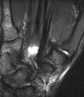
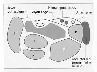
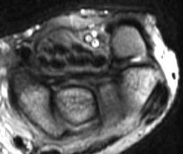
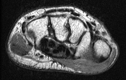
12 Carpal tunnel syndrome -Flexor tenosynovitis -Separation of tendons by synovial tissue -Mass effect from muscle in carpal tunnel Median Nerve: Proximal Enlargement and Fasciculation -Volar ganglion cyst in carpal tunnel Fasciculation: Looks like dots inside nerve proximal distal Guyon s Canal PROXIMAL GUYON S CANAL DISTAL 12


13 -Ganglion cyst with Ulnar nerve impingement Guyon s canal Summary Size matters! Larger communications (tear) are more likely to be symptomatic / mechanically significant than a leak Is the band together? Evaluate dorsal and volar bands of SL and LT Malalignment / separation Chondrosis / arthritis Routine MRI wrist: -Tendon pathology -Carpal tunnel syndrome -Ganglion cyst -Acute trauma -Osteoarthritis -AVN MRI wrist with IV contrast: -Mass -Infection -Inflammatory arthropathy MR arthrogram: -Ligament tear MRI Protocol T2 FSE PD fat sat THE FUTURE RESOLUTION T2 FSE fat sat 13

14 GRE 50 micron resolution Problem is: you miss all those 40 micron leaks. It s all relative! william.morrison@jefferson.edu Questions? 14
Introduction. The wrist contains eight small carpal bones, which as a group act as a flexible spacer between the forearm and hand.
 Wrist Introduction The wrist contains eight small carpal bones, which as a group act as a flexible spacer between the forearm and hand. Distal forearm Distal forearm 4 Distal end of the radius A. anterior
3. Ulno lunate, Ulno triquetral ligament. Poirier: Between RSC &LRL. 5. Dorsal intercarpal ligament
 CARPAL INSTABILITY Ligaments Intrinsic Scapho lunate ligament: Dorsal component stronger than volar ligament Luno triquetral ligament: Volar component stronger than dorsal ligament Extrinsic Palmar 1 Radio
Sean Walsh Orthopaedic Surgeon Dorset County Hospital
 Sean Walsh Orthopaedic Surgeon Dorset County Hospital Shapes and orientation of articular surfaces Ligaments Oblique positioning of scaphoid Tendons surrounding the joints Other soft tissues Peripheral
Acute Wrist Injuries OUCH!
 Acute Wrist Injuries OUCH! Case the athlete FOOSH from sporting event 2 days ago C/O wrist swelling, pain, worse with movement Hmmm Wrist pain Exam of the wrist - basics Appearance Swelling, bruising,
SCAHPO-LUNATE DISSOCIATION
 SCAHPO-LUNATE DISSOCIATION Introduction Scapho-lunate dissociation is the most common significant ligamentous injury of the wrist. The condition is also sometimes referred to as rotary subluxation of the
Carpal Instability: Clarification of the Most Common Etiologies and Imaging Findings
 Carpal Instability: Clarification of the Most Common Etiologies and Imaging Findings Corey Matthews DO, Nicholas Strle DO, Donald von Borstel DO Oklahoma State University Medical Center, Department of
8/25/2014. Radiocarpal Joint. Midcarpal Joint. Osteology of the Wrist
 Structure and Function of the Wrist 2 joints and 10 different bones Combine to create wrist motion Anatomical Terms: Wrist/Hand Palmar = anterior aspect of the wrist and hand Dorsal = posterior aspect
Forearm and Wrist Regions Neumann Chapter 7
 Forearm and Wrist Regions Neumann Chapter 7 REVIEW AND HIGHLIGHTS OF OSTEOLOGY & ARTHROLOGY Radius dorsal radial tubercle radial styloid process Ulna ulnar styloid process ulnar head Carpals Proximal Row
Hand and wrist emergencies
 Chapter1 Hand and wrist emergencies Carl A. Germann Distal radius and ulnar injuries PEARL: Fractures of the distal radius and ulna are the most common type of fractures in patients younger than 75 years.
Kristin Kelley, DPT, OCS, FAAOMPT Orthopaedic Manual Physical Therapy Series Charlottesville Trauma/Fractures
 WRIST/HAND PATHOLOGY Kristin Kelley, DPT, OCS, FAAOMPT Orthopaedic Manual Physical Therapy Series Charlottesville 2017-2018 Trauma/Fractures Hook of Hamate Fractures Triangular Fibrocartilage Complex (TFCC)
Trauma/Fractures WRIST/HAND PATHOLOGY. TFCC Injury. Hook of Hamate Fracture. Property of VOMPTI, LLC
 WRIST/HAND PATHOLOGY Kristin Kelley, DPT, OCS, FAAOMPT Orthopaedic Manual Physical Therapy Series Charlottesville 2017-2018 Trauma/Fractures Hook of Hamate Fractures Triangular Fibrocartilage Complex (TFCC)
Abstract Submission Form
 Abstract Submission Form All abstracts must be submitted to the AOCR by September 15 th. All information included must be the original work of the author(s) and be in typed form. Incomplete or handwritten
Index. radiologic.theclinics.com. Note: Page numbers of article titles are in boldface type.
 Index Note: Page numbers of article titles are in boldface type. A Acromioclavicular joint injuries in football players, 318, 319 ALPSA. See Anterior labroligamentous periosteal sleeve avulsion. Anterior
Scapholunate Ligament Lesions Imaging Which and when?
 Scapholunate Ligament Lesions Imaging Which and when? Kolo Frank Lesions to scapholunate ligament(sl) Most frequent cause of carpal instability Traumatic tears of SL ligament = most common ligament injury
Wrist & Hand Assessment and General View
 Wrist & Hand Assessment and General View Done by; Mshari S. Alghadier BSc Physical Therapy RHPT 366 m.alghadier@sau.edu.sa http://faculty.sau.edu.sa/m.alghadier/ Functional anatomy The hand can be divided
10/15/2014. Wrist. Clarification of Terms. Clarification of Terms cont
 Wrist Clarification of Terms Palmar is synonymous with anterior aspect of the wrist and hand Ventral is also synonymous with anterior aspect of the wrist and hand Dorsal refers to the posterior aspect
COMMON CARPAL INJURIES IN ATHLETES Nicholas A. Bontempo, MD Orthopedic Associates of Hartford I HAVE NO CONFLICTS OR DISCLOSURES TO REPORT OUTLINE
 COMMON CARPAL INJURIES IN ATHLETES Nicholas A. Bontempo, MD Orthopedic Associates of Hartford I HAVE NO CONFLICTS OR DISCLOSURES TO REPORT OUTLINE The carpus Scaphoid fracture Scapholunate ligament tear
EXAMINATION OF THE WRIST BEYOND THE BASICS OMA SPORT MED Janice Harvey MD CCFP CFFP Dip. Sp Med.
 EXAMINATION OF THE WRIST BEYOND THE BASICS OMA SPORT MED 2019 Janice Harvey MD CCFP CFFP Dip. Sp Med. CFPC CoI Templates: Slide 1 used in Faculty presentation only. FACULTY/PRESENTER DISCLOSURE Faculty:
Trapezium is by the thumb, Trapezoid is inside
 Trapezium is by the thumb, Trapezoid is inside Intercarpal Jt Radiocarpal Jt Distal Middle Proximal DIP PIP Interphalangeal Jts Metacarpalphalangeal (MCP) Jt Metacarpal Carpometacarpal (CMC) Jt Trapezium
Exam of the Injured Hand and Wrist. Christina M. Ward, MD Regions Hospital TRIA Woodbury
 Exam of the Injured Hand and Wrist Christina M. Ward, MD Regions Hospital TRIA Woodbury Disclosures We have no disclosures that are pertinent to this presentation Terminology Ring Long Index Small Thumb
Wrist & Hand Ultrasonography 대구가톨릭대학교병원재활의학과 권동락
 Wrist & Hand Ultrasonography 대구가톨릭대학교병원재활의학과 권동락 Dorsal Wrist Evaluation (1 st Compartment) EPB APL Transverse View APL, abductor pollicis longus; EPB, extensor pollicis brevis Dorsal Wrist Evaluation
Union rate: Union: Stable 94% All fracture 90% Union after surgery for nonunion with surgery 80% OA in healed scaphoid: 9%
 Complications Incidence of Non-union 1 cm displacement of fracture caused 55% Non-union It takes 5-20 yrs to develop SNAC. SNAC appears to be more common with waist fracture than a proximal pole. However
Hand & Wrist Injuries. DR MA Manjra
 Hand & Wrist Injuries DR MA Manjra 1 Background Up to 25% of all athletic injuries General population Sport people Sport specific Position specific Multifaceted Time of season Level of athlete Parents
Wrist movements, apart from the distal radioulnar joint, take place in two planes:
 The wrist consists of eight bones in two rows: the proximal and distal. The proximal row includes (starting from the radial bone): the scaphoid bone, the lunate bone, the triangular bone and the postulnar
SCAPHOID FRACTURE. Relevant antomy
 SCAPHOID FRACTURE Relevant antomy The proximal row consists of the scaphoid, the lunate, and the triquetrum. The proximal carpal row is regarded as an intercalated segment The keystone in the coordination
Alvin S. Chen, Harvard Medical School Year III Gillian Lieberman, MD Radiology Core Clerkship
 Alvin S. Chen, Harvard Medical School Year III Gillian Lieberman, MD Radiology Core Clerkship Overview Wrist: Normal Anatomy & Biomechanics Approach to Wrist Imaging: Menu of Tests & Efficacious Use Index
MCQWeek2. All arise from the common flexor origin. The posterior aspect of the medial epicondyle is the common flexor origin.
 MCQWeek2. 1. Regarding superficial muscles of anterior compartment of the forearm: All arise from the common flexor origin. The posterior aspect of the medial epicondyle is the common flexor origin. Flexor
Introduction to Ultrasound Examination of the Hand and upper
 Introduction to Ultrasound Examination of the Hand and upper Emil Dionysian, M.D. Ultrasound of upper ext. Upside Convenient Opens another exam dimension Can be like a stethoscope Helps 3-D D visualization
Wrist and Hand Anatomy/Biomechanics
 Wrist and Hand Anatomy/Biomechanics Kristin Kelley, DPT, OCS, FAAOMPT Orthopaedic Manual Physical Therapy Series Charlottesville 2017-2018 Orthopaedic Manual Physical Therapy Series 2017-2018 Anatomy -
Anatomy - Hand. Wrist and Hand Anatomy/Biomechanics. Osteology. Carpal Arch. Property of VOMPTI, LLC
 Wrist and Hand Anatomy/Biomechanics Kristin Kelley, DPT, OCS, FAAOMPT The wrist The metacarpals The Phalanges Digit 1 thumb Digit 5 digiti minimi Anatomy - Hand Orthopaedic Manual Physical Therapy Series
SPORTS INJURIES IN HAND
 Grundkurs SGSM-SSMS Sion 2015 SPORTS INJURIES IN HAND Dr S. KŠmpfen EPIDEMIOLOGY Incidence of hand, finger and wrist injuries in sports : 3% Ð 9 % RADIAL-SIDED WRIST PAIN 1)! Distal Radius Fractures 2)!
Interesting Case Series. Perilunate Dislocation
 Interesting Case Series Perilunate Dislocation Tom Reisler, BSc (Hons), MB ChB, MRCS (Ed), Paul J. Therattil, MD, and Edward S. Lee, MD Division of Plastic and Reconstructive Surgery, Department of Surgery,
Ligaments of Elbow hinge: sagittal plane so need lateral and medial ligaments
 Ligaments of Elbow hinge: sagittal plane so need lateral and medial ligaments Ulnar Collateral ligament on medial side; arising from medial epicondyle and stops excess valgus movement (lateral movement)
Physical therapy of the wrist and hand
 Physical therapy of the wrist and hand Functional anatomy wrist and hand The wrist includes distal radius, scaphoid, lunate, triquetrum, pisiform, trapezium, trapezoid, capitate, and hamate. The hand includes
CLINICAL PRESENTATION AND RADIOLOGY QUIZ QUESTION
 Donald L. Renfrew, MD Radiology Associates of the Fox Valley, 333 N. Commercial Street, Suite 100, Neenah, WI 54956 10/13/2012 Radiology Quiz of the Week # 94 Page 1 CLINICAL PRESENTATION AND RADIOLOGY
Scaphoid Fractures. Mohammed Alasmari. Orthopaedic Surgery Demonstrator Majmaah University
 Scaphoid Fractures Mohammed Alasmari Orthopaedic Surgery Demonstrator Majmaah University 1 2 Scaphoid Fractures Introduction Anatomy History Clinical examination Radiographic evaluation Classification
Hand Anatomy A Patient's Guide to Hand Anatomy
 Hand Anatomy A Patient's Guide to Hand Anatomy Introduction Few structures of the human anatomy are as unique as the hand. The hand needs to be mobile in order to position the fingers and thumb. Adequate
Mayo Clinic Disorders of the Wrist
 Mayo Clinic Disorders of the Wrist Thursday, May 19, 2016 Pre-Conference Laboratory Workshop Anatomy of the Wrist & Wrist Arthroscopy 6:30 a.m. Registration and Breakfast 7:30 a.m. Welcome and Introduction
Wrist and Hand Anatomy
 Wrist and Hand Anatomy Bone Anatomy Scapoid Lunate Triquetrium Pisiform Trapeziod Trapezium Capitate Hamate Wrist Articulations Radiocarpal Joint Proximal portion Distal portion Most surface contact found
Chapter 51 Wrist and Forearm Episode Overview
 Chapter 51 Wrist and Forearm Episode Overview 1) Describe normal radiographic relationships: a. Radial length measurement b. Radial inclination c. Volar Tilt d. Scapholunate angle e. Capitolunate angle
Surgical Technique. DISCLOSURE: This device is not approved for sale in the U.S.A. Customer Service:
 DISCLOSURE: This device is not approved for sale in the U.S.A. INDICATIONS FOR USE The KinematX Modular Wrist Arthroplasty System is indicated for the replacement of a wrist joints disabled by pain, deformity,
Arhtroscopy of the wrist joint: Setup, instrumentation, anatomy & indications
 Arhtroscopy of the wrist joint: Setup, instrumentation, anatomy & indications Andreas Panagopoulos, MD, PhD Upper Limb and Sports Medicine Surgeon Assistant Professor in Orthopaedics Patras University
Wrist and Hand Complaints
 Wrist and Hand Complaints Charles S. Day, M.D., M.B.A. Chief, Hand & Upper Extremity Surgery St. Elizabeth s Medical Center Tufts University School of Medicine Primary Care Internal Medicine 2018 Outline
PREVIEW ONLY 27/10/2014. Instabilities in the Wrist
 Be sure to convert to your own time zone at Andrew Ellis BSc (Ex. Sci), M. Phty Instabilities in the Wrist Presented by: Ben Cunningham Be sure to convert to your own time zone at Ben Cunningham Member
Clinical Orthopaedic Rehabilitation Volume 1 and 2
 Clinical Orthopaedic Rehabilitation Volume 1 and 2 COURSE DESCRIPTION This program is a practical, clinical guide that provides guidance on the evaluation, differential diagnosis, treatment, and rehabilitation
Ultrasonography of the wrist - a step-by-step approach to study protocols and normal findings
 Ultrasonography of the wrist - a step-by-step approach to study protocols and normal findings Poster No.: C-1779 Congress: ECR 2016 Type: Educational Exhibit Authors: R. R. Domingues Madaleno, A. P. Pissarra,
WRIST MRI. TFCC (SIMILAR TO MENISCUS): best seen on coronal (except for RUL, use sag)
: best seen on coronal (except for RUL, use sag)") WRIST MRI LIGAMENT (thin slice coronal GRE): low to intermediate signal on GRE; consider abnormal if high signal (equal to fluid) or discontinuity/thinning/elongation or increased intercarpal space INTRINSIC
WRIST MRI LIGAMENT (thin slice coronal GRE): low to intermediate signal on GRE; consider abnormal if high signal (equal to fluid) or discontinuity/thinning/elongation or increased intercarpal space INTRINSIC
SURGICAL/APPLIED ANATOMY
 Página 1 de 11 Copyright 2001 Lippincott Williams & Wilkins Bucholz, Robert W., Heckman, James D. Rockwood & Green's Fractures in Adults, 5th Edition SURGICAL/APPLIED ANATOMY Part of "19 - FRACTURES AND
Carpal rows injuries!
 Carpal rows injuries! Michael Papaloïzos! Center for Hand Surgery and Therapy Geneva, Switzerland no conflict of interest to declare Fractures of carpal bones! The fractured scaphoid! Fracture-dislocations
The Painful Elbow, Wrist, and Hand. Jennifer R Marks, MD
 The Painful Elbow, Wrist, and Hand Jennifer R Marks, MD The Painful Elbow A 44 yo M presents to clinic complaining of a sore elbow What further questions do you have for this patient? What is on your differential
Clinical Examination of the Hand and Wrist
 Clinical Examination of the Hand and Wrist OBJECTIVES Review the clinical anatomy and physical exam of the wrist and hand Formulate a pathoanatomic diagnosis in the clinical setting Discuss common clinical
Kinesiology of The Wrist and Hand. Cuneyt Mirzanli Istanbul Gelisim University
 Kinesiology of The Wrist and Hand Cuneyt Mirzanli Istanbul Gelisim University Bones The wrist and hand contain 29 bones including the radius and ulna. There are eight carpal bones in two rows of four to
Difference Between Angle You Can Bend Your Left Wrist Back vs Your Right Wrist Jenna Priest Science Department Altoona High School January 25, 2017
 Difference Between Angle You Can Bend Your Left Wrist Back vs Your Right Wrist Jenna Priest Science Department Altoona High School January 25, 2017 Background 1- The wrist joint (also known as the radiocarpal
Scapholunate Advanced Collapse and Scaphoid Nonunion Advanced Collapse: MDCT Arthrography Features
 Musculoskeletal Imaging Pictorial Essay Crema et al. MDCT rthrography of SLC and SNC Musculoskeletal Imaging Pictorial Essay Michel D. Crema 1,2 Joachim Zentner 3 li Guermazi 1 Nabil Jomaah 4 Monica D.
(i) Examination of the wrist surface anatomy of the carpal bones
 Examination of the wrist surface anatomy of the carpal bones") Current Orthopaedics (2005) 19, 171 179 www.elsevier.com/locate/cuor MINI-SYMPOSIUM: THE WRIST (i) Examination of the wrist surface anatomy of the carpal bones R. Srinivas Reddy, J. Compson Upper Limb
Current Orthopaedics (2005) 19, 171 179 www.elsevier.com/locate/cuor MINI-SYMPOSIUM: THE WRIST (i) Examination of the wrist surface anatomy of the carpal bones R. Srinivas Reddy, J. Compson Upper Limb
Index. Note: Page numbers of article titles are in boldface type. Hand Clin 21 (2005)
") Hand Clin 21 (2005) 501 505 Index Note: Page numbers of article titles are in boldface type. A Antibiotics, following distal radius fracture treatment, 295, 296 Arthritis, following malunion of distal
Hand Clin 21 (2005) 501 505 Index Note: Page numbers of article titles are in boldface type. A Antibiotics, following distal radius fracture treatment, 295, 296 Arthritis, following malunion of distal
Upper Limb- Sports Medicine II
 Upper Limb- Sports Medicine II I. Palpation A. With patient sitting, supine, & prone, palpate for pain, specific tenderness, swelling, effusion, local hyperthermia B. Bony Palpation 1. Carpal Bones (8)
Neglected trans-scaphoid trans-styloid volar dislocation of the lunate
 CASE REPORT Neglected trans-scaphoid trans-styloid volar dislocation of the lunate LATE RESULT FOLLOWING OPEN REDUCTION AND K-WIRE FIXATION P. Givissis, A. Christodoulou, B. Chalidis, J. Pournaras From
THE WRIST JOINT: ATHLETIC INJURIES
 THE WRIST JOINT: ATHLETIC INJURIES Gianni Rigoni FMH Handsurgery SSMS Wrist unity The wrist links the hand to the forearm 1 Anatomy Bone V IV III II T H C Tid T I P L S U R Anatomy Intrinsic ligament 2
I-A-1) Non-specific thickening of synovial membrane
 Non-specific thickening of synovial membrane") I-A-1) Non-specific thickening of synovial membrane Grayscale Metatarsal Power Doppler Dorsal aspect of metatarsophalangeal joint in right 1 st toe, longitudinal view Asterisks indicate non-specific thickening
I-A-1) Non-specific thickening of synovial membrane Grayscale Metatarsal Power Doppler Dorsal aspect of metatarsophalangeal joint in right 1 st toe, longitudinal view Asterisks indicate non-specific thickening
Ultrasonography of Peripheral Nerve -upper extremity
 Ultrasonography of Peripheral Nerve -upper extremity Department of Physical Medicine and Rehabilitation Korea University Guro Hospital Korea University College of Medicine Yoon Joon Shik Normal median
ORTHOPAEDIC INJECTION AND ASPIRATION TECHNIQUES
 ORTHOPAEDIC INJECTION AND ASPIRATION TECHNIQUES OAAPN October 20, 2016 David H. Sohn, JD MD Chief, Shoulder and Sports Medicine University of Toledo Medical Center When to aspirate? To rule out infection
MR: Finger and Thumb Injuries
 MR: Finger and Thumb Injuries Laura W. Bancroft, M.D. Professor of Radiology University of Central Florida Florida State University Outline Normal anatomy of the fingers and thumb MR imaging protocols
Main Menu. Wrist and Hand Joints click here. The Power is in Your Hands
 1 The Wrist and Hand Joints click here Main Menu K.5 http://www.handsonlineeducation.com/classes/k5/k5entry.htm[3/23/18, 1:40:40 PM] Bones 29 bones, including radius and ulna 8 carpal bones in 2 rows of
Wrist Arthritis & Partial Wrist Fusion
 Wrist Arthritis & Partial Wrist Fusion Mr Jason N Harvey MB.BS. FRACS (Orth) Hand,Wrist & Elbow Surgeon Clinical Symptoms Outline Physical Examination Diagnosis Differential Diagnosis Outline Non-operative
TRIQUETRUM FRACTURE. The triquetrum bone is one of the small bones that make up the carpus.
 TRIQUETRUM FRACTURE Introduction The triquetrum bone is one of the small bones that make up the carpus. It is also known as the triquetral bone, (and in the past the pyramidal or triangular bone) Triquetrum
Columbia/NYOH Department of Orthopaedics Hand Service Competency Requirements
 Revised 2/8/10 Columbia/NYOH Department of Orthopaedics Hand Service Competency Requirements Patient Care Faculty will evaluate the resident s ability to obtain an H&P and appropriate radiographs and formulate
Surgical Technique. SLIC Screw System
 Surgical Technique SLIC Screw System Acumed is a global leader of innovative orthopaedic and medical solutions. We are dedicated to developing products, service methods, and approaches that improve patient
Hand & Wrist Casey G. Batten MD Assistant Clinical Professor UCSF Sports Medicine
 Hand & Wrist Casey G. Batten MD Assistant Clinical Professor UCSF Sports Medicine Topics: Scaphoid Fracture Scapholunate Separation TFCC Injury Thumb Ulnar Collateral Lig (UCL) Injury Extensor Injury /
Chapter 7. Anatomy of the Triangular Fibrocartilage Complex: Current Concepts. Introduction. Anatomy. Histology
 Chapter 7 Anatomy of the Triangular Fibrocartilage Complex: Current Concepts Introduction The triangular fibrocartilage complex (TFCC) is one of the intrinsic ligaments of the wrist. It is often injured
The Elbow and the cubital fossa. Prof Oluwadiya Kehinde
 The Elbow and the cubital fossa Prof Oluwadiya Kehinde www.oluwadiya.com Elbow and Forearm Anatomy The elbow joint is formed by the humerus, radius, and the ulna Bony anatomy of the elbow Distal Humerus
triquetrum in rheumatoid arthritis
 Ann. rheum. Dis. (1976), 35, 46 Early abnormalities of pisiform and triquetrum in rheumatoid arthritis DONALD RESNICK From the Department of Radiology, Veterans Administration Hospital, San Diego, and
Vascular Pedicle Pisiform Bone Grafting for Kienbocks Disease : A Case Report
 Case Report Vascular Pedicle Pisiform Bone Grafting for Kienbocks Disease : A Case Report Nagamuneendrudu K 1, Valya B 2, Vishnu Vardhan M 3 1 Associate Professor Department of Orthopaedics Osmania Medical
Peripheral Nerve Ultrasound
 Peripheral Nerve Ultrasound Jon A. Jacobson, M.D. Professor of Radiology Director, Division of Musculoskeletal Radiology University of Michigan Normal Peripheral Nerve Ultrasound appearance: Hypoechoic
[[Sally Leaning Towards Peter To Take Cold Hand]]
![[[Sally Leaning Towards Peter To Take Cold Hand]]](/thumbs/84/91174469.jpg "[[Sally Leaning Towards Peter To Take Cold Hand]]") In this lecture we will talk about the bones of the hand, and the muscles and contents of the forearm. *The hand bones are: - Carpal bones. -Metacarpals. -Phalanges. *The carpal bones (wrist bones): They
In this lecture we will talk about the bones of the hand, and the muscles and contents of the forearm. *The hand bones are: - Carpal bones. -Metacarpals. -Phalanges. *The carpal bones (wrist bones): They
Anatomy Workshop Upper Extremity David Ebaugh, PT, PhD Workshop Leader. Lab Leaders: STATION I BRACHIAL PLEXUS
 Anatomy Workshop Upper Extremity David Ebaugh, PT, PhD Workshop Leader Lab Leaders: STATION I BRACHIAL PLEXUS A. Posterior cervical triangle and axilla B. Formation of plexus 1. Ventral rami C5-T1 2. Trunks
Vascularised pisiform bone graft Indications, technique and long-term results
 Vascularised pisiform bone graft Indications, technique and long-term results J. Norbert KUHLMANN, Cédric KRON, André BOABIGHI, Serge BAUX, Maurice MIMOU The authors report their experience with the use
Integra. Spider and Mini Spider Limited Wrist Fusion System SURGICAL TECHNIQUE
 Integra Spider and Mini Spider Limited Wrist Fusion System SURGICAL TECHNIQUE Table of contents Description... 02 Indications... 02 Contraindications... 02 Surgical Technique... 03 Spider Introduction-Four
Technique. Disclosure. Approach to Ultrasound of the Wrist. Objectives. Outline. Technique 14/09/2015
 Approach to Ultrasound of the Wrist Disclosure I have no commercial or financial interests related to the subject matter of this presentation Linda robyn, MD, FRCC MSK Radiologist Objectives At the end
31yo M with chronic basilar thumb and wrist pain that started after cross-country bicycle ride 5 yrs ago.
 31yo M with chronic basilar thumb and wrist pain that started after cross-country bicycle ride 5 yrs ago. EPL EPB APL Full-thickness tear involving the dorsal deltoid ligament of the first carpometacarpal
SYMPOSIUM ON ADVANCES IN THE MANAGEMENT OF SCAPHOID PROBLEMS Scaphoid malunion
 Hong HKJOS Kong Journal of Orthopaedic Surgery 2002;6(2):104-108. SYMPOSIUM ON ADVANCES IN THE MANAGEMENT OF SCAPHOID PROBLEMS Scaphoid malunion Department of Orthopaedics and Traumatology, Prince of Wales
The Kienböck disease and scaphoid fractures. Mariusz Bonczar
 The Kienböck disease and scaphoid fractures Mariusz Bonczar The Kienböck disease and scaphoid fractures Mariusz Bonczar Kienböck disease personal experience My special interest for almost 25 years Thesis
Carpal instability in rheumatoid arthritis and
 Annals of the Rheumatic Diseases, 1977, 36, 311-318 Carpal instability in rheumatoid arthritis and calcium pyrophosphate deposition disease Pathogenesis and roentgen appearance D. RESNICK AND G. NIWAYAMA
Common. Common Hand Problems in Elite Athletes
 Common Hand Problems in Elite Athletes Fred Corley M.D. Dept. of Orthopaedic Surgery UTHSCSA I have no disclosures concerning this talk. The University of Texas Health Science Center @ San Antonio - Orthopaedics
Radiographic Evaluation and Classification of Distal Radius Fractures
 Radiographic Evaluation and Classification of Distal Radius Fractures Robert J Medoff, MD Introduction X-rays are essential to the treatment of distal radius fractures. When combined with the age and baseline
Chapter 13. Arthroscopic Lunotriquetral Arthrodesis and Head of the Hamate Resection. Introduction. Operative Technique (Fontes) Midcarpal Exploration
 Midcarpal Exploration") Chapter 13 Arthroscopic Lunotriquetral Arthrodesis and Head of the Hamate Resection Introduction Lunotriquetral arthrodesis is a controversial procedure but is sometimes proposed as a last resort for lunotriquetral
Chapter 13 Arthroscopic Lunotriquetral Arthrodesis and Head of the Hamate Resection Introduction Lunotriquetral arthrodesis is a controversial procedure but is sometimes proposed as a last resort for lunotriquetral
American College of Radiology ACR Appropriateness Criteria Chronic Wrist Pain
 Revised 2017 American College of Radiology ACR Appropriateness Criteria Chronic Wrist Pain Variant 1: Chronic wrist pain. With or without prior injury. Best initial study. X-ray wrist Usually Appropriate
The Forearm 2. Extensor & lateral Compartments of the Forearm
 The Forearm 2 Extensor & lateral Compartments of the Forearm 1-Lateral Fascial Compartment (at the lateral side of the forearm ) *Some books mention the lateral compartment contain just the Brachioradialis
ARM Brachium Musculature
 ARM Brachium Musculature Coracobrachialis coracoid process of the scapula medial shaft of the humerus at about its middle 1. flexes the humerus 2. assists to adduct the humerus Blood: muscular branches
Clinical examination of the wrist, thumb and hand
 Clinical examination of the wrist, thumb and hand 20 CHAPTER CONTENTS Referred pain 319 History 319 Inspection 320 Functional examination 320 The distal radioulnar joint.............. 320 The wrist.......................
Netter's Anatomy Flash Cards Section 6 List 4 th Edition
 Netter's Anatomy Flash Cards Section 6 List 4 th Edition https://www.memrise.com/course/1577581/ Section 6 Upper Limb (66 cards) Plate 6-1 Humerus and Scapula: Anterior View 1.1 Acromion 1.2 Greater tubercle
CHAPTER 6: THE UPPER EXTREMITY: THE ELBOW, FOREARM, WRIST, AND HAND
 CHAPTER 6: THE UPPER EXTREMITY: THE ELBOW, FOREARM, WRIST, AND HAND KINESIOLOGY Scientific Basis of Human Motion, 12 th edition Hamilton, Weimar & Luttgens Presentation Created by TK Koesterer, Ph.D.,
Episode 52 Commonly Missed Uncommon Orthopedic Injuries. Lisfranc Injuries. Drs. Ivy Cheng & Hossein Medhian. Prepared by Dr. Keerat Grewal, Oct 2014
 Prepared by Dr. Keerat Grewal, Oct 2014 Episode 52 Commonly Missed Uncommon Orthopedic Injuries Drs. Ivy Cheng & Hossein Medhian Lisfranc Injuries Q: What is a Lisfranc injury? Lisfranc injuries are a
TFCC Tears and Repair. Jeffrey Yao, M.D. Associate Professor Department of Orthopaedic Surgery Stanford University Medical Center
 TFCC Tears and Repair Jeffrey Yao, M.D. Associate Professor Department of Orthopaedic Surgery Stanford University Medical Center Disclosures The following relationships exist: 1. Grants American Foundation
ELENI ANDIPA General Hospital of Athens G. Gennimatas
 ELENI ANDIPA General Hospital of Athens G. Gennimatas Technological advances over the last years have caused a dramatic improvement in ultrasound quality and resolution An established imaging modality
Index. Springer International Publishing Switzerland 2016 J.N. Lawton (ed.), Distal Radius Fractures, DOI /
, Distal Radius Fractures, DOI /") Index A AAOS. See American Academy of Orthopaedic Surgeons (AAOS) Abductor pollicis longus (APL) tendon, 34, 73 Acute carpal tunnel syndrome, 93 American Academy of Orthopaedic Surgeons (AAOS), 66, 238
Index A AAOS. See American Academy of Orthopaedic Surgeons (AAOS) Abductor pollicis longus (APL) tendon, 34, 73 Acute carpal tunnel syndrome, 93 American Academy of Orthopaedic Surgeons (AAOS), 66, 238
13 13/3/2012. Adel Muhanna
 13 13/3/2012 Adel Muhanna بسم هللا الرحمن الرحيم The Hand Extensor retinaculum: Deep fascia of anterior compartment of the wrist is thickened to form flexor retinaculum : a bridge that have 6 structures
RADIOGRAPHY OF THE WRIST
 RADIOGRAPHY OF THE WRIST Patient Position: WRIST PA Projection, elbow in same plane Part Position: Hand ; fingers centered to IR Central Ray: Structures Shown: NOTE: Optional AP projection best demonstrates
Nerves of Upper limb. Dr. Brijendra Singh Professor & Head Department of Anatomy AIIMS Rishikesh
 Nerves of Upper limb Dr. Brijendra Singh Professor & Head Department of Anatomy AIIMS Rishikesh 1 Objectives Origin, course & relation of median & ulnar nerves. Motor & sensory distribution Carpal tunnel
Viorel Nacu. The clinical anatomy of the Hand
 Viorel Nacu The clinical anatomy of the Hand The distal part of the upper limb is divided in to three regions: 1. The wrist (carpus) 2. The hand (metacarpus) 3. The digits (fingers) The landmarks of this