Wrist & Hand Assessment and General View
|
|
|
- Cora Lindsey
- 5 years ago
- Views:
Transcription

1 Wrist & Hand Assessment and General View Done by; Mshari S. Alghadier BSc Physical Therapy RHPT 366
2 Functional anatomy The hand can be divided into two major parts: The wrist. Five digits. Composed of eight small bones. It can accommodate movement in three planes. The greatest degree of freedom is in; Flexion extension plane. Ulnar radial deviation. Rotation about the long axis of the forearm. 2
3 Functional anatomy 3
4 Functional anatomy Because of its vascular supply, fracture of the scpahoid can lead to avascular necrosis and collapse of the proximal half of that bone. This damage leads to impairment of wrist function and progressive osteoarthritis of the wrist joint. The carpal tunnel, which contains the median nerve together with the flexor tendons of the digits, and the tunnel of Guyon, which contains the ulnar nerve. 4
5 Functional anatomy 5
6 Functional anatomy Compression injury of the ulnar nerve will affect the medial aspect of the hand, with the ulnar intrinsic muscles of the hand. This muscular compromise will lead to classic posturing of the digits called the benediction hand, referring to the appearance of a priest s hand when giving a blessing. 6
7 Functional anatomy 7

8 Functional anatomy The benediction hand deformity results from damage to the ulnar nerve. There is wasting of the interosseous muscles, the hypothenar muscles, and the two medial lumbrical muscles. 8
9 Functional anatomy 9
10 Observation The examination should begin in the waiting room. Is the arm relaxed at the side or is the patient cradling it for protection? Note whether the wrist or hand is edematous. Note the shape of the hand and if there are any changes in contour. Any deformity. 10


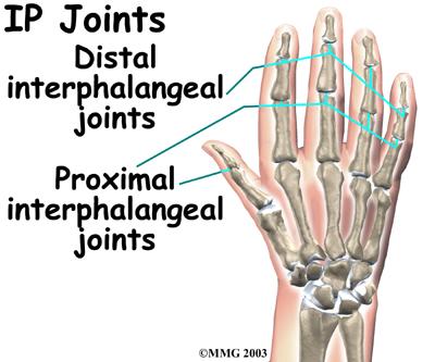
11 Observation The patient may have a swan neck, boutonni`ere deformity, or claw fingers 11
12 Observation Compare one hand to the other, remembering that the dominant hand may be larger in the normal individual. Will he or she allow you to shake their hand? Is the movement effortless and coordinated or stiff and uncoordinated? Watch the patient if he/she push ups and use his hands to stand from sitting. 12


13 Observation Clubbing and cyanosis of the nails may be secondary to pulmonary disease. 13
14 Observation Posture and standing position. Note the height of the shoulders and their relative positions. Arm swing can be limited by either loss of motion, pain, or neurological damage. Observe for symmetry of bony structures. Observe for areas of muscle wasting that may be secondary to peripheral nerve lesions. 14
15 Subjective Examination The wrist and hand are extremely active structures and complicated. Non-weightbearing, problems are; Overuse syndromes, inflammation, and trauma. Nature and behavior, location and onset of pain. Functional limitation and what is the dominant hand. Does the patient regularly participate in any vigorous sport activity that would stress the wrist or hand? 15
16 Subjective Examination What is the patient s occupation computer?? If there is a trauma? What was the mechanism of the injury? The direction of the force. The position of the upper extremity. The activity the patient was participating in at the time of the injury. Previous history of the same injury? The most common nerve roots that refer pain are C6, C7, C8, and T1. 16
17 Gentle palpation The palpatory examination is started with the patient in the sitting position. You should first examine for areas of; Localized effusion, discoloration, birthmarks, open sinuses or drainage, incisions, bony contours, muscle girth and symmetry, and skinfolds. Use firm and gentle pressure to allocate the malposition or deformities. If you harm the Pt in this part of examination the Pt will be afraid and you ll lose his confidence. 17
18 Gentle palpation The sitting position with the extremity supported on a table is preferred for ease of examination of the wrist and hand. For palpation, the hand should be in the anatomical position. 18
19 Gentle palpation A. Anterior (palmar) aspect; 1. Boney Structures; Thick skin and fascia covered the palm. 2. Soft-Tissue Structures; Many sweat glands but free of hair. Medial (Ulnar) Compartment; Flexor Carpi Ulnaris. Ulnar Artery. Ulnar Nerve. Hypothenar Eminence. 19
20 Gentle palpation Middle compartment; Palmaris Longus. Flexor Digitorum Profundus and Superficialis. Carpal Tunnel. Palmar Aponeurosis. Lateral (Radial) Compartment; Flexor Carpi Radialis. Radial Artery. Thenar Eminence. 20
21 Gentle palpation B. Medial (Ulnar) Aspect; 1. Bony Structures; Ulna Styloid Process. Triquetrum. Pisiform. Hamate. 2. Soft-Tissue Structures; Triangular Fibrocartilaginous Complex. 21
22 Gentle palpation C. Lateral (Radial) Aspect; 1. Bony Structures; Radial Styloid Process. Scaphoid (Navicular). Trapezium and Trapezoid (Greater and Lesser Multangular). First Metacarpal. 2. Soft-Tissue Structures; Anatomical Snuffbox. 22
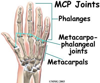
23 Gentle palpation D. Posterior (Dorsal) Aspect; 1. Bony Structures; Dorsal Tubercle of the Radius (Lister s Tubercle). Lunate. Capitate. Metacarpals. Metacarpophalangeal Joints. Phalanges and Interphalangeal Joints. Nails. 23
24 Gentle palpation 2. Soft-Tissue Structures; Extensor Retinaculum. Compartment I. Compartment II. Compartment III. Compartment IV. Compartment V. Compartment VI. 24
25 Special Tests A. Tests for ligament, capsule & joint instability; Watson s (Scaphoid shift) Test; This test is used to diagnose abnormal separation of the lunate and scaphoid bones. The examiner takes the patient wrist into ulnar deviation and slight extension with one hand. The examiner presses the thumb of the other hand against the scaphoid to prevent it from moving toward the palm. With other hand, examiner radially deviates and slightly flexes the patient s hand while maintain the pressure on scaphoid. 25

26 The Watson s Test 26
27 Special Tests B. Tests Tendons and Muscles; 1) Bunnel Littler Test (Intrinsic Muscles Versus Contracture); Tests the structures around the matacarpophalangeal joint. Meatacarpophalangeal joint held slightly extended while the examiner moves the proximal interphalangeal joint into flexion. Positive; inability to flex the proximal interphalangeal joint, due to tight intrinsic muscle or contracture of the joint capsule. If the metacarpophalangeal joints are slightly flexed, the proximal interphalageal joints flexes fully if the intrinsic muscle are tight. But it dose not flex fully if the capsule is tight. 27
28 Special Tests 2) Finkelstein s Test (de Quervain s or Hoffmann s Syndrome); This test is used to diagnose tenosynovitis of the first dorsal compartment of the wrist, which contains the tendons of the abductor pollicis longus and extensor pollicis brevis muscles. Having the patient place the thumb inside the closed fist. Take the patient s hand and deviate the hand and wrist in the ulnar direction to stretch the tendons of the first extensor compartment. Pain over the radial styloid process is pathognomonic of de Quervain s syndrome. 28
29 Special Tests C. Tests for neurological dysfunction; 1) Tinel s Sign (at the wrist); The examiner taps over the carpal tunnel over the wrist, positive test cause tingling or paresthesia into the thumb and index finger, and middle and lateral half of the ring finger. Its indicative for carpal tunnel syndrome. It should be distally to the point being pressed. 29
30 Special Tests D. Tests for circulation and swelling; Allen s Test; Used to check the patency of the radial and ulnar arteries at the level of the wrist. v=oycrz1vaehi&feature=related 30

31 Special Tests 31
32 Want to do! 1. Tennis elbow. 2. Golfer elbow. 3. Carpal Tunnel syndrome. 4. Major hand deformity. 5. De Quervain s Syndrome 6. Medial & Lateral Epicondlytis. 32
33 Thank you 33
34 References, Musculoskeletal Examination, 3rd Edition Jeffrey M. Gross, chapter 10. Orthopedic Physical Assessment, 5th edition, David J. Magee, chapter 7. 34
Kinesiology of The Wrist and Hand. Cuneyt Mirzanli Istanbul Gelisim University
 Kinesiology of The Wrist and Hand Cuneyt Mirzanli Istanbul Gelisim University Bones The wrist and hand contain 29 bones including the radius and ulna. There are eight carpal bones in two rows of four to
Main Menu. Wrist and Hand Joints click here. The Power is in Your Hands
 1 The Wrist and Hand Joints click here Main Menu K.5 http://www.handsonlineeducation.com/classes/k5/k5entry.htm[3/23/18, 1:40:40 PM] Bones 29 bones, including radius and ulna 8 carpal bones in 2 rows of
MCQWeek2. All arise from the common flexor origin. The posterior aspect of the medial epicondyle is the common flexor origin.
 MCQWeek2. 1. Regarding superficial muscles of anterior compartment of the forearm: All arise from the common flexor origin. The posterior aspect of the medial epicondyle is the common flexor origin. Flexor
Trapezium is by the thumb, Trapezoid is inside
 Trapezium is by the thumb, Trapezoid is inside Intercarpal Jt Radiocarpal Jt Distal Middle Proximal DIP PIP Interphalangeal Jts Metacarpalphalangeal (MCP) Jt Metacarpal Carpometacarpal (CMC) Jt Trapezium
Physical therapy of the wrist and hand
 Physical therapy of the wrist and hand Functional anatomy wrist and hand The wrist includes distal radius, scaphoid, lunate, triquetrum, pisiform, trapezium, trapezoid, capitate, and hamate. The hand includes
Forearm and Wrist Regions Neumann Chapter 7
 Forearm and Wrist Regions Neumann Chapter 7 REVIEW AND HIGHLIGHTS OF OSTEOLOGY & ARTHROLOGY Radius dorsal radial tubercle radial styloid process Ulna ulnar styloid process ulnar head Carpals Proximal Row
Nerves of Upper limb. Dr. Brijendra Singh Professor & Head Department of Anatomy AIIMS Rishikesh
 Nerves of Upper limb Dr. Brijendra Singh Professor & Head Department of Anatomy AIIMS Rishikesh 1 Objectives Origin, course & relation of median & ulnar nerves. Motor & sensory distribution Carpal tunnel
Elbow, Wrist & Hand Evaluation.
 Elbow, Wrist & Hand Evaluation www.fisiokinesiterapia.biz Common Injuries to the Elbow, Wrist, Hand & Fingers Lateral epicondylitis tennis elbow Medial epicondylitis golfer s s elbow, little league elbow
Upper Limb- Sports Medicine II
 Upper Limb- Sports Medicine II I. Palpation A. With patient sitting, supine, & prone, palpate for pain, specific tenderness, swelling, effusion, local hyperthermia B. Bony Palpation 1. Carpal Bones (8)
Hand and Wrist Editing file. Color Code Important Doctors Notes Notes/Extra explanation
 Hand and Wrist Editing file Color Code Important Doctors Notes Notes/Extra explanation Objectives Describe the anatomy of the deep fascia of the wrist & hand (flexor & extensor retinacula & palmar aponeurosis).
10/10/2014. Structure and Function of the Hand. The Hand. Osteology of the Hand
 Structure and Function of the Hand 19 bones and 19 joints are necessary to produce all the motions of the hand The Hand Dorsal aspect Palmar aspect The digits are numbered 1-5 Thumb = #1 Little finger
8/25/2014. Radiocarpal Joint. Midcarpal Joint. Osteology of the Wrist
 Structure and Function of the Wrist 2 joints and 10 different bones Combine to create wrist motion Anatomical Terms: Wrist/Hand Palmar = anterior aspect of the wrist and hand Dorsal = posterior aspect
[[Sally Leaning Towards Peter To Take Cold Hand]]
![[[Sally Leaning Towards Peter To Take Cold Hand]]](/thumbs/84/91174469.jpg "[[Sally Leaning Towards Peter To Take Cold Hand]]") In this lecture we will talk about the bones of the hand, and the muscles and contents of the forearm. *The hand bones are: - Carpal bones. -Metacarpals. -Phalanges. *The carpal bones (wrist bones): They
In this lecture we will talk about the bones of the hand, and the muscles and contents of the forearm. *The hand bones are: - Carpal bones. -Metacarpals. -Phalanges. *The carpal bones (wrist bones): They
Wrist and Hand Anatomy/Biomechanics
 Wrist and Hand Anatomy/Biomechanics Kristin Kelley, DPT, OCS, FAAOMPT Orthopaedic Manual Physical Therapy Series Charlottesville 2017-2018 Orthopaedic Manual Physical Therapy Series 2017-2018 Anatomy -
Anatomy - Hand. Wrist and Hand Anatomy/Biomechanics. Osteology. Carpal Arch. Property of VOMPTI, LLC
 Wrist and Hand Anatomy/Biomechanics Kristin Kelley, DPT, OCS, FAAOMPT The wrist The metacarpals The Phalanges Digit 1 thumb Digit 5 digiti minimi Anatomy - Hand Orthopaedic Manual Physical Therapy Series
divided by the bones ( redius and ulna ) and interosseous membrane into :
 and interosseous membrane into :") fossa Cubital Has: * floor. * roof : - Skin - superficial fasica - deep fascia ( include bicipital aponeurosis ) Structures within the roof : -cephalic and basilic veins -and between them median cubital
fossa Cubital Has: * floor. * roof : - Skin - superficial fasica - deep fascia ( include bicipital aponeurosis ) Structures within the roof : -cephalic and basilic veins -and between them median cubital
ARM Brachium Musculature
 ARM Brachium Musculature Coracobrachialis coracoid process of the scapula medial shaft of the humerus at about its middle 1. flexes the humerus 2. assists to adduct the humerus Blood: muscular branches
Muscular Nomenclature and Kinesiology - One
 Chapter 16 Muscular Nomenclature and Kinesiology - One Lessons 1-3 (with lesson 4) 1 Introduction 122 major muscles covered in this chapter Chapter divided into nine lessons Kinesiology study of human
The Muscular System. Chapter 10 Part C. PowerPoint Lecture Slides prepared by Karen Dunbar Kareiva Ivy Tech Community College
 Chapter 10 Part C The Muscular System Annie Leibovitz/Contact Press Images PowerPoint Lecture Slides prepared by Karen Dunbar Kareiva Ivy Tech Community College Table 10.9: Muscles Crossing the Shoulder
Clinical examination of the wrist, thumb and hand
 Clinical examination of the wrist, thumb and hand 20 CHAPTER CONTENTS Referred pain 319 History 319 Inspection 320 Functional examination 320 The distal radioulnar joint.............. 320 The wrist.......................
Muscles of the hand Prof. Abdulameer Al-Nuaimi
 Muscles of the hand Prof. Abdulameer Al-Nuaimi a.alnuaimi@sheffield.ac.uk abdulameerh@yahoo.com Thenar Muscles Thenar muscles are three short muscles located at base of the thumb. All are innervated by
LECTURE 8 HANDS: BONES AND MUSCLES
 LECTURE 8 HANDS: BONES AND MUSCLES WRIST AND HAND - Human hand can do power grip and precision grip - Thumb is 90 to the rest of the hand can do fine actions - Often able to do power actions o Take tools
13 13/3/2012. Adel Muhanna
 13 13/3/2012 Adel Muhanna بسم هللا الرحمن الرحيم The Hand Extensor retinaculum: Deep fascia of anterior compartment of the wrist is thickened to form flexor retinaculum : a bridge that have 6 structures
Wrist and Hand Anatomy
 Wrist and Hand Anatomy Bone Anatomy Scapoid Lunate Triquetrium Pisiform Trapeziod Trapezium Capitate Hamate Wrist Articulations Radiocarpal Joint Proximal portion Distal portion Most surface contact found
Wrist & Hand Ultrasonography 대구가톨릭대학교병원재활의학과 권동락
 Wrist & Hand Ultrasonography 대구가톨릭대학교병원재활의학과 권동락 Dorsal Wrist Evaluation (1 st Compartment) EPB APL Transverse View APL, abductor pollicis longus; EPB, extensor pollicis brevis Dorsal Wrist Evaluation
Lab Activity 11: Group II
 Lab Activity 11: Group II Muscles Martini Chapter 11 Portland Community College BI 231 Origin and Insertion Origin: The place where the fixed end attaches to a bone, cartilage, or connective tissue. Insertion:
Netter's Anatomy Flash Cards Section 6 List 4 th Edition
 Netter's Anatomy Flash Cards Section 6 List 4 th Edition https://www.memrise.com/course/1577581/ Section 6 Upper Limb (66 cards) Plate 6-1 Humerus and Scapula: Anterior View 1.1 Acromion 1.2 Greater tubercle
Viorel Nacu. The clinical anatomy of the Hand
 Viorel Nacu The clinical anatomy of the Hand The distal part of the upper limb is divided in to three regions: 1. The wrist (carpus) 2. The hand (metacarpus) 3. The digits (fingers) The landmarks of this
Supplied in part by the musculocutaneous nerve. Forms the axis of rotation in movements of pronation and supination
 Anatomy: Upper limb (15 questions) 1. Latissimus Dorsi: Is innervated by the dorsal scapular nerve Lies above feres major muscle Medially rotates the humerus All of the above 2. Supinator muscle is: Deep
The hand is full with sweat glands, activated at times of stress. In Slide #2 there was a mistake where the doctor mentioned lateral septum twice.
 We should only know: Name, action & nerve supply Layers - Skin - Superficial fascia - Deep fascia The hand is full with sweat glands, activated at times of stress. Deep fascia In Slide #2 there was a mistake
10/15/2014. Wrist. Clarification of Terms. Clarification of Terms cont
 Wrist Clarification of Terms Palmar is synonymous with anterior aspect of the wrist and hand Ventral is also synonymous with anterior aspect of the wrist and hand Dorsal refers to the posterior aspect
Lecture 9: Forearm bones and muscles
 Lecture 9: Forearm bones and muscles Remember, the region between the shoulder and the elbow = brachium/arm, between elbow and wrist = antebrachium/forearm. Forearm bones : Humerus (distal ends) Radius
Wrist and Hand Complaints
 Wrist and Hand Complaints Charles S. Day, M.D., M.B.A. Chief, Hand & Upper Extremity Surgery St. Elizabeth s Medical Center Tufts University School of Medicine Primary Care Internal Medicine 2018 Outline
Acute Wrist Injuries OUCH!
 Acute Wrist Injuries OUCH! Case the athlete FOOSH from sporting event 2 days ago C/O wrist swelling, pain, worse with movement Hmmm Wrist pain Exam of the wrist - basics Appearance Swelling, bruising,
Biceps Brachii. Muscles of the Arm and Hand 4/4/2017 MR. S. KELLY
 Muscles of the Arm and Hand PSK 4U MR. S. KELLY NORTH GRENVILLE DHS Biceps Brachii Origin: scapula Insertion: radius, fascia of forearm (bicipital aponeurosis) Action: supination and elbow flexion Innervation:
The Forearm 2. Extensor & lateral Compartments of the Forearm
 The Forearm 2 Extensor & lateral Compartments of the Forearm 1-Lateral Fascial Compartment (at the lateral side of the forearm ) *Some books mention the lateral compartment contain just the Brachioradialis
Introduction. The wrist contains eight small carpal bones, which as a group act as a flexible spacer between the forearm and hand.
 Wrist Introduction The wrist contains eight small carpal bones, which as a group act as a flexible spacer between the forearm and hand. Distal forearm Distal forearm 4 Distal end of the radius A. anterior
Key Points for Success:
 SELF WRIST & HAND 1 2 All of the stretches described in this chapter are detailed to stretch the right side. Key Points for Success: Sit comfortably in a position where you can straighten or fully extend
Nerves of the upper limb Prof. Abdulameer Al-Nuaimi. E. mail:
 Nerves of the upper limb Prof. Abdulameer Al-Nuaimi E-mail: a.al-nuaimi@sheffield.ac.uk E. mail: abdulameerh@yahoo.com Brachial plexus Median nerve After originating from the brachial plexus in the axilla,
Nerve Injury. 1) Upper Lesions of the Brachial Plexus called Erb- Duchene Palsy or syndrome.
 Upper Lesions of the Brachial Plexus called Erb- Duchene Palsy or syndrome.") Nerve Injury - Every nerve goes to muscle or skin so if the nerve is injured this will cause paralysis in the muscle supplied from that nerve (paralysis means loss of function) then other muscles and other
Nerve Injury - Every nerve goes to muscle or skin so if the nerve is injured this will cause paralysis in the muscle supplied from that nerve (paralysis means loss of function) then other muscles and other
Anatomy and Physiology II. Review Shoulder Girdle New Material Upper Extremities - Bones
 Anatomy and Physiology II Review Shoulder Girdle New Material Upper Extremities - Bones Anatomy and Physiology II Shoulder Girdle Review Questions From Last Lecture Can you identify the following muscles?
Clinical Examination of the Hand and Wrist
 Clinical Examination of the Hand and Wrist OBJECTIVES Review the clinical anatomy and physical exam of the wrist and hand Formulate a pathoanatomic diagnosis in the clinical setting Discuss common clinical
medial half of clavicle; Sternum; upper six costal cartilages External surfaces of ribs 3-5
 MUSCLE ORIGIN INSERTION ACTION NERVE Pectoralis Major medial half of clavicle; Sternum; upper six costal cartilages Lateral lip of intertubercular groove of horizontal adduction Medial and lateral pectoral
In the name of Allah, Most gracious, Most merciful
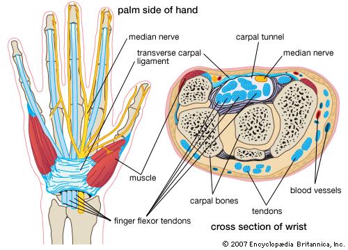
 In the name of Allah, Most gracious, Most merciful This lecture includes the following: The Palmer Oponeurosis. The Carpel tunnel. The palmaris brevis muscle. The anatomical snuffbox. The Fibrous flexor
forearm posterior compartment
 Quick revision: The anterior compartment of the forearm contains of 8 muscles... -4 superficial -1 intermediate -3 deep *All supplied by median nerve except 1 and 1/2 muscle (by ulnar N.) forearm posterior
Ligaments of Elbow hinge: sagittal plane so need lateral and medial ligaments
 Ligaments of Elbow hinge: sagittal plane so need lateral and medial ligaments Ulnar Collateral ligament on medial side; arising from medial epicondyle and stops excess valgus movement (lateral movement)
Difference Between Angle You Can Bend Your Left Wrist Back vs Your Right Wrist Jenna Priest Science Department Altoona High School January 25, 2017
 Difference Between Angle You Can Bend Your Left Wrist Back vs Your Right Wrist Jenna Priest Science Department Altoona High School January 25, 2017 Background 1- The wrist joint (also known as the radiocarpal
Manual therapy approach to the Patient with Carpal Tunnel Syndrome.
 Manual therapy approach to the Patient with Carpal Tunnel Syndrome www.fisiokinesiterapia.biz Symptoms and Signs Thumb, index, middle, and radial aspect of ring finger Hand Pain Paresthesia Numbness Pins
Shoulder joint Assessment and General View
 Shoulder joint Assessment and General View Done by; Mshari S. Alghadier BSc Physical Therapy RHPT 366 m.alghadier@sau.edu.sa http://faculty.sau.edu.sa/m.alghadier/ Functional anatomy The shoulder contains
# Anatomy. Upper Extremities Muscles and anatomy of axilla. Tiba Al-Ani 9/10/2015 Nabil. Page 0 of 16
 #10 25 Anatomy Upper Extremities Muscles and anatomy of axilla Tiba Al-Ani 9/10/2015 Nabil Page 0 of 16 Salam AWN Today s lecture is divided into two parts, the first part is the continuation of the upper
EXAMINATION OF THE WRIST BEYOND THE BASICS OMA SPORT MED Janice Harvey MD CCFP CFFP Dip. Sp Med.
 EXAMINATION OF THE WRIST BEYOND THE BASICS OMA SPORT MED 2019 Janice Harvey MD CCFP CFFP Dip. Sp Med. CFPC CoI Templates: Slide 1 used in Faculty presentation only. FACULTY/PRESENTER DISCLOSURE Faculty:
Muscles of the Upper Limb
 Muscles of the Upper Limb anterior surface of ribs 3 5 coracoid process Pectoralis minor pectoral nerves protracts / depresses scapula Serratus anterior Subclavius ribs 1-8 long thoracic nerve rib 1 ----------------
SUSPECTS THE UNUSUAL. Often-Overlooked Muscles. Sternohyoid, longus colli, and longus capitis. Flexor pollicis longus.
 THE UNUSUAL SUSPECTS Often-Overlooked Muscles By Joseph E. Muscolino, DC Sternohyoid, longus colli, and longus capitis Flexor pollicis longus Quadratus femoris 52 massage & bodywork november/december 2016
compartments of the forearm
 " forearm posterior compartment " compartments of the forearm Posterior Fascial compartment Muscles: ** The superficial group 1. Extensor carpi radialis brevis 2. Ex. digitorum 3. Ex. digiti minimi 4.
Anatomy Workshop Upper Extremity David Ebaugh, PT, PhD Workshop Leader. Lab Leaders: STATION I BRACHIAL PLEXUS
 Anatomy Workshop Upper Extremity David Ebaugh, PT, PhD Workshop Leader Lab Leaders: STATION I BRACHIAL PLEXUS A. Posterior cervical triangle and axilla B. Formation of plexus 1. Ventral rami C5-T1 2. Trunks
The Elbow and the cubital fossa. Prof Oluwadiya Kehinde
 The Elbow and the cubital fossa Prof Oluwadiya Kehinde www.oluwadiya.com Elbow and Forearm Anatomy The elbow joint is formed by the humerus, radius, and the ulna Bony anatomy of the elbow Distal Humerus
MLT Muscle(s) Patient Position Therapist position Stabilization Limb Position Picture Put biceps on slack by bending elbow.
 Patient Position Therapist position Stabilization Limb Position Picture Put biceps on slack by bending elbow.") MLT Muscle(s) Patient Position Therapist position Stabilization Limb Position Picture Put biceps on slack by bending elbow. Pectoralis Minor Supine, arm at side, elbows extended, supinated Head of Table
MLT Muscle(s) Patient Position Therapist position Stabilization Limb Position Picture Put biceps on slack by bending elbow. Pectoralis Minor Supine, arm at side, elbows extended, supinated Head of Table
The Painful Elbow, Wrist, and Hand. Jennifer R Marks, MD
 The Painful Elbow, Wrist, and Hand Jennifer R Marks, MD The Painful Elbow A 44 yo M presents to clinic complaining of a sore elbow What further questions do you have for this patient? What is on your differential
STRUCTURAL BASIS OF MEDICAL PRACTICE EXAMINATION 5. September 30, 2011
 STRUCTURAL BASIS OF MEDICAL PRACTICE EXAMINATION 5 September 30, 2011 PART l. Answer in the space provided. (12 pts) 1. Identify the structures. (2 pts) EXAM NUMBER A. Suprascapular nerve B. Axillary nerve
Ultrasonography of the wrist - a step-by-step approach to study protocols and normal findings
 Ultrasonography of the wrist - a step-by-step approach to study protocols and normal findings Poster No.: C-1779 Congress: ECR 2016 Type: Educational Exhibit Authors: R. R. Domingues Madaleno, A. P. Pissarra,
Chapter 8. The Pectoral Girdle & Upper Limb
 Chapter 8 The Pectoral Girdle & Upper Limb Pectoral Girdle pectoral girdle (shoulder girdle) supports the arm consists of two on each side of the body // clavicle (collarbone) and scapula (shoulder blade)
Clinical Orthopaedic Rehabilitation Volume 1 and 2
 Clinical Orthopaedic Rehabilitation Volume 1 and 2 COURSE DESCRIPTION This program is a practical, clinical guide that provides guidance on the evaluation, differential diagnosis, treatment, and rehabilitation
Classification of Established Volkmann s Ischemic Contracture and the Program for Its Treatment
 10 Classification of Established Volkmann s Ischemic Contracture and the Program for Its Treatment In spite of the advances made in preventive treatment of muscular ischemia at the forearm and hand, there
Dr Nabil khouri MD. MSc. Ph.D
 Dr Nabil khouri MD. MSc. Ph.D Foot Anatomy The foot consists of 26 bones: 14 phalangeal, 5 metatarsal, and 7 tarsal. Toes are used to balance the body. Metatarsal Bones gives elasticity to the foot in
SPORTS INJURIES IN HAND
 Grundkurs SGSM-SSMS Sion 2015 SPORTS INJURIES IN HAND Dr S. KŠmpfen EPIDEMIOLOGY Incidence of hand, finger and wrist injuries in sports : 3% Ð 9 % RADIAL-SIDED WRIST PAIN 1)! Distal Radius Fractures 2)!
Anatomy of the Upper Limb
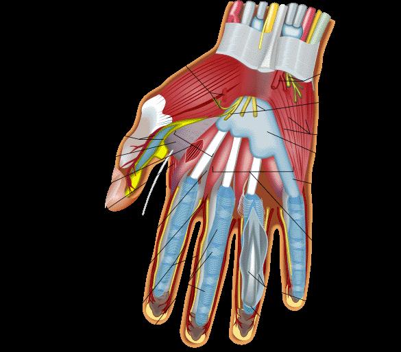
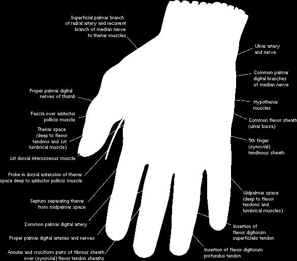
 Anatomy of the Upper Limb Figure 53: The thenar & midpalmar spaces. The synovial (tendon) sheaths of the long flexors [Figure.54] These sheaths surround the tendons of the long flexors; flexor digitorum
STRUCTURAL BASIS OF MEDICAL PRACTICE EXAMINATION 5 October 6, 2006
 STRUCTURAL BASIS OF MEDICAL PRACTICE EXAMINATION 5 October 6, 2006 PART l. Answer in the space provided. (8 pts) 1. Identify the structures. (2 pts) B C A. _pisiform B. _ulnar artery A C. _flexor carpi
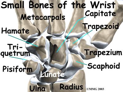
Hand Anatomy A Patient's Guide to Hand Anatomy
 Hand Anatomy A Patient's Guide to Hand Anatomy Introduction Few structures of the human anatomy are as unique as the hand. The hand needs to be mobile in order to position the fingers and thumb. Adequate
Practical 2 Worksheet
 Practical 2 Worksheet Upper Extremity BONES 1. Which end of the clavicle is on the lateral side (acromial or sternal)? 2. Describe the difference in the appearance of the acromial and sternal ends of the
Key Relationships in the Upper Limb
 Key Relationships in the Upper Limb This list contains some of the key relationships that will help you identify structures in the lab. They are organized by dissection assignment as defined in the syllabus.
This presentation is the intellectual property of the author. Contact them for permission to reprint and/or distribute.
 The Stiff Hand: Manual Therapy Sylvia Dávila, PT, CHT San Antonio, Texas Orthopedic Manual Therapy Common Applications Passive stretch Tensile force to tissue to increase extensibility of length & ROM
The hand. it's the most important subject of the upper limb because it has a clinical importance. the palm of the hand**
 Today at 12:48 AM The hand it's the most important subject of the upper limb because it has a clinical importance. the palm of the hand** -the palmar aponeurosis located in the palm of the hand which is
Dr. Mahir Alhadidi Anatomy Lecture #9 Feb,28 th 2012
 Quick Revision: Upper arm is divided into two compartments: 1. Anterior Compartment: Contains three muscles (Biceps brachii, Coracobrachialis, Brachialis). Innervated by Musculocutaneous nerve. 2. Posterior
(i) Examination of the wrist surface anatomy of the carpal bones
 Examination of the wrist surface anatomy of the carpal bones") Current Orthopaedics (2005) 19, 171 179 www.elsevier.com/locate/cuor MINI-SYMPOSIUM: THE WRIST (i) Examination of the wrist surface anatomy of the carpal bones R. Srinivas Reddy, J. Compson Upper Limb
Current Orthopaedics (2005) 19, 171 179 www.elsevier.com/locate/cuor MINI-SYMPOSIUM: THE WRIST (i) Examination of the wrist surface anatomy of the carpal bones R. Srinivas Reddy, J. Compson Upper Limb
REFERENCE DIAGRAMS OF UPPER LIMB MUSCLES: NAMES, LOCATIONS, ATTACHMENTS, FUNCTIONS MUSCLES CONNECTING THE UPPER LIMB TO THE AXIAL SKELETON
 REFERENCE DIAGRAMS OF UPPER LIMB MUSCLES: NAMES, LOCATIONS, ATTACHMENTS, FUNCTIONS MUSCLES CONNECTING THE UPPER LIMB TO THE AXIAL SKELETON A25LAB EXERCISES: UPPER LIMB MUSCLES Page 1 MUSCLES CONNECTING
Abduction of arm until your hand rich your head. Flexion of forearm at elbow joint. Extension of arm at elbow joint. Flexion of fingers 10.
 Num. answer 1. Medialy With the manubrium ( sternum ), and laterally with the acromion of the scapula 2. 1. Trapezius 2. Levator scapulae 3. Rhomboids 3. 1. Pectoralis major 2. Pectoralis minor 3. Latissiumus
Hand & Wrist Injuries. DR MA Manjra
 Hand & Wrist Injuries DR MA Manjra 1 Background Up to 25% of all athletic injuries General population Sport people Sport specific Position specific Multifaceted Time of season Level of athlete Parents
Elbow. Chapter 2 LISTEN. Mechanism of Injury (If Applicable) Pain
 Pain") Chapter 2 Elbow LISTEN Mechanism of Injury (If Applicable) Patient usually remembers their position at the time of injury Certain mechanisms of injury result in characteristic patterns Fall on outstretched
Chapter 2 Elbow LISTEN Mechanism of Injury (If Applicable) Patient usually remembers their position at the time of injury Certain mechanisms of injury result in characteristic patterns Fall on outstretched
SMALL GROUP SESSION 16 January 8 th or 10 th Shoulder pain case/ Touch workshop/ Upper and Lower Extremity Examination
 SMALL GROUP SESSION 16 January 8 th or 10 th Shoulder pain case/ Touch workshop/ Upper and Lower Extremity Examination Suggested Readings: Opatrny L. The Healing Touch. Ann Int Med 2002; 137:1003. http://www.annals.org/cgi/reprint/137/12/1003.pdf
BRACHIAL PLEXUS. DORSAL SCAPULAR NERVE (C5) supraclavicular branch innervates rhomboids (major and minor) and levator scapulae
 supraclavicular branch innervates rhomboids (major and minor) and levator scapulae") THE BRACHIAL PLEXUS DORSAL SCAPULAR NERVE (C5) supraclavicular branch innervates rhomboids (major and minor) and levator scapulae SCHEMA OF THE BRACHIAL PLEXUS THE BRACHIAL PLEXUS PHRENIC NERVE supraclavicular
THE BRACHIAL PLEXUS DORSAL SCAPULAR NERVE (C5) supraclavicular branch innervates rhomboids (major and minor) and levator scapulae SCHEMA OF THE BRACHIAL PLEXUS THE BRACHIAL PLEXUS PHRENIC NERVE supraclavicular
LIST OF STRUCTURES TO BE IDENTIFIED IN LAB: UPPER EXTREMITY REVIEW 2016
 LIST OF STRUCTURES TO BE IDENTIFIED IN LAB: UPPER EXTREMITY REVIEW 2016 BONES Ribs, sternum, clavicle Humerus: Head, greater tubercle, lesser tubercle, intertubercular sulcus, surgical neck, anatomical
Al-Balqa Applied University
 Al-Balqa Applied University Faculty Of Medicine *You can use this checklist as a guide to you for the lab. the items on this checklist represent the main features of the models that you have to know for
Functional Anatomy of the Elbow
 Functional Anatomy of the Elbow Orthopedic Institute Daryl C. Osbahr, M.D. Chief of Sports Medicine, Orlando Health Chief Medical Officer, Orlando City Soccer Club Orthopedic Consultant, Washington Nationals
Wrist movements, apart from the distal radioulnar joint, take place in two planes:
 The wrist consists of eight bones in two rows: the proximal and distal. The proximal row includes (starting from the radial bone): the scaphoid bone, the lunate bone, the triangular bone and the postulnar
MR IMAGING OF THE WRIST
 MR IMAGING OF THE WRIST Wrist Instability Dissociative Pattern apparent on routine radiographs Non-dissociative Stress / positional radiographs Dynamic fluoroscopy during stress Arthrography MRI / MR arthrography
Introduction to Ultrasound Examination of the Hand and upper
 Introduction to Ultrasound Examination of the Hand and upper Emil Dionysian, M.D. Ultrasound of upper ext. Upside Convenient Opens another exam dimension Can be like a stethoscope Helps 3-D D visualization
Cubital fossa and forearm
 Cubital fossa and forearm Cubital fossa is the triangular space in front of elbow joint. - The Cubital fossa has boundaries: apex, base, roof and floor and it has contents. The base: an imaginary horizontal
Hand and wrist emergencies
 Chapter1 Hand and wrist emergencies Carl A. Germann Distal radius and ulnar injuries PEARL: Fractures of the distal radius and ulna are the most common type of fractures in patients younger than 75 years.
RADIOGRAPHY OF THE WRIST
 RADIOGRAPHY OF THE WRIST Patient Position: WRIST PA Projection, elbow in same plane Part Position: Hand ; fingers centered to IR Central Ray: Structures Shown: NOTE: Optional AP projection best demonstrates
Human Anatomy Biology 351
 1 Human Anatomy Biology 351 Upper Limb Exam Please place your name on the back of the last page of this exam. You must answer all questions on this exam. Because statistics demonstrate that, on average,
Module 7 - The Muscular System Muscles of the Arm and Trunk
 Module 7 - The Muscular System Muscles of the Arm and Trunk This Module will cover the muscle anatomy of the arms and trunk. We have already seen the muscles that move the humerus, so this module will
I-A-1) Non-specific thickening of synovial membrane
 Non-specific thickening of synovial membrane") I-A-1) Non-specific thickening of synovial membrane Grayscale Metatarsal Power Doppler Dorsal aspect of metatarsophalangeal joint in right 1 st toe, longitudinal view Asterisks indicate non-specific thickening
I-A-1) Non-specific thickening of synovial membrane Grayscale Metatarsal Power Doppler Dorsal aspect of metatarsophalangeal joint in right 1 st toe, longitudinal view Asterisks indicate non-specific thickening
Small muscles of the hand
 By the name of Allah Small muscles of the hand Revision: The palmar aponeurosis is triangular in shape with apex and base. It is divided into 4 bands that radiate to the medial four fingers. Dupuytren
Levels of the anatomical cuts of the upper extremity RADIUS AND ULNA right
 11 CHAPTER 2 Levels of the anatomical cuts of the upper extremity AND right CUT 1 CUT 4 1 2 3 4 5 6 Isolated fixation of the radius is difficult at this level because of the anterolateral vessels and the
Kristin Kelley, DPT, OCS, FAAOMPT Orthopaedic Manual Physical Therapy Series Charlottesville Trauma/Fractures
 WRIST/HAND PATHOLOGY Kristin Kelley, DPT, OCS, FAAOMPT Orthopaedic Manual Physical Therapy Series Charlottesville 2017-2018 Trauma/Fractures Hook of Hamate Fractures Triangular Fibrocartilage Complex (TFCC)
Trauma/Fractures WRIST/HAND PATHOLOGY. TFCC Injury. Hook of Hamate Fracture. Property of VOMPTI, LLC
 WRIST/HAND PATHOLOGY Kristin Kelley, DPT, OCS, FAAOMPT Orthopaedic Manual Physical Therapy Series Charlottesville 2017-2018 Trauma/Fractures Hook of Hamate Fractures Triangular Fibrocartilage Complex (TFCC)
Figure 1: Bones of the upper limb
 BONES OF THE APPENDICULAR SKELETON The appendicular skeleton is composed of the 126 bones of the appendages and the pectoral and pelvic girdles, which attach the limbs to the axial skeleton. Although the
Structure and Function of the Hand
 Structure and Function of the Hand Some say it takes a village to raise a child, but it takes 19 bones and 19 joints in the hand for it to function smoothly. The Hand Dorsal aspect 2 3 4 The digits are
Sick Call Screener Course
 Sick Call Screener Course Musculoskeletal System Upper Extremities (2.7) 2.7-2-1 Enabling Objectives 1.46 Utilize the knowledge of musculoskeletal system anatomy while assessing a patient with a musculoskeletal