Superficial Lumps and Bumps: Ultrasound Assessment
|
|
|
- Berenice York
- 6 years ago
- Views:
Transcription


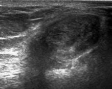
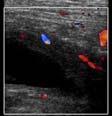
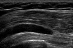
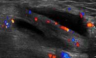
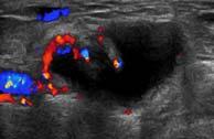
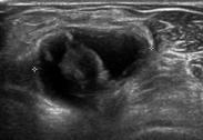
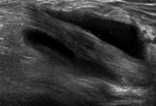
1 Posterior knee Superficial Lumps and Bumps: Ultrasound Assessment Walter Mak, MD Department of Medical Imaging St. Michael s Hospital SM SM MGas MGas MGas MGas Synovial lined Synovial cyst: extrusion of joint fluid Bursa: synovial lined potential space between osseous surfaces, ligaments or tendons SM: Semimembranosus MGas: Medial head of gastrocnemius Posterior knee Posterior knee SM MGas SM MGas MGas SM: Semimembranosus MGas: Medial head of gastrocnemius Septations, internal debris, peripheral Doppler flow SM: Semimembranosus MGas: Medial head of gastrocnemius 1


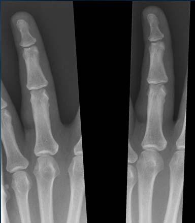
2 Anterior hip Anterior shoulder IPs IPs BT BT Peripheral Doppler flow - synovitis BT: Biceps tendon IPs: Iliopsoas Antecubital fossa Antecubital fossa BT BT BT BT BT BT Rad BT: Biceps tendon Rad: Radius Peripheral/intratendinous Doppler flow BT: Biceps tendon Finger, near proximal phalanx * * 2


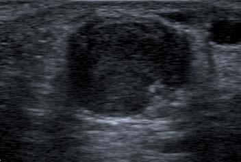
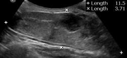
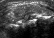
3 Mucoid/myxomatous degeneration of periarticular connective tissue dominant cyst Result of repetitive microtrauma Common in hand/wrist, foot/ankle Close association with joint or tendon sheath Dorsal radial wrist Dorsal radial wrist S L Contiguous with scapholunate ligament S: Scaphoid L: Lunate S Scapholunate ligament tear Peripheral Doppler flow SA Teefeyet al., AJR 2008; 191: L S L S: Scaphoid L: Lunate Volar radial wrist Dorsal radial wrist L Contiguous with scapholunate ligament Slightly complex, collapsed S: Scaphoid L: Lunate S Non-compressible Visible neck in only 25-35% SA Teefeyet al., AJR 2008; 191: G Wang et al., J Ultrasound Med 2007; 26: Internal Doppler flow? 3


4 FCR RA FCR RA Pitfall: artifactual flow from adjacent radial artery RA: radial artery FCR: flexor carpi radialis Mature adipocytes and uniform nuclei identical to normal adult (white) fat Arise within subcutaneous tissue 80% <5 cm; 99% 10 cm MJ Kransdorf et al., Radiology 2002;224: MD Murphey et al., RadioGraphics 2004; 24: Periscapular region Parallel to skin surface Elliptical Linear echogenic striations No internal Doppler flow <5 cm Anterior knee Supraclavicular region May be hyperechoic, isoechoic, or hypoechoic Uniform echogenicity Parallel to skin surface Elliptical Linear echogenic striations 4


")


5 Radial aspect elbow Shoulder w/o compression w/ compression Compressible Parallel to skin surface Parallel to skin surface Linear echogenic striations Slightly heterogenous Deep (intramuscular) Needs additional imaging Shoulder Linear echogenic striations Variable echogenicity Deep (intramuscular) Needs additional imaging T1 T2FS Medial anterior thigh Lower back Heterogeneous Deep (intramuscular) Needs additional imaging Heterogeneous >10 cm Exuberant Doppler flow Needs additional imaging T1 T2FS T1FS+C 5


6 Calf T1 T2FS T1 T2FS Upper back Epithelial lined, keratincontaining cysts Causes: Squamous metaplasia Deep growth after obstruction of hair follicle Post traumatic implantation into dermis HK Kim et al., Skeletal Radiol (2011) 40: Ovoid, sharply marginated Curvilinear echogenic keratin Hypoechoic clefts May scallop overlying skin Tip of finger Hypoechoic No internal Doppler flow Cortical disruption 6


7 Proximal lateral thigh Degloving injury of proximal thigh Separation of fascia from subcutaneous tissue and vascular/lymphatic disruption Collection containing blood, lymph, fat Proximal lateral thigh Proximal lateral thigh Chronic: fusiform, hypoechoic Globules of echogenic fat No internal Doppler flow May be complex Globules of echogenic fat No internal Doppler flow Proximal lateral thigh Proximal calf, near knee w/o compression w/ compression Morel-Lavallée also observed about the knee SG Tejwani et al., Am J Sports Med 2007; 35: Globules of echogenic fat Compressible Acute: may be lobulated, heterogeneous Globules of echogenic fat 7


8 Proximal forearm Congenital vascular lesion resulting from aberrant vessel angiogenesis High flow: arterial component Low flow: venous malformation hemangioma Bluish discolouration of overlying skin Dorsal to metacarpal Lateral Knee Hypoechoic vascular channels Echogenic phleboliths Doppler flow w/o compression w/ compression Hypoechoic vascular channels Doppler flow Compressible Deep, intramuscular lesions MRI Dorsal wrist ED ED Pigmented villonodular tenosynovitis Localized synovial proliferation ED ED: Extensor digitorum 8


 Dupuytren")


9 Volar Thumb Volar finger Hypoechoic Peripheral flow Longitudinal contact with tendon Does not move with digital flexion/extension Hypoechoic Longitudinal contact with tendon Circumferential contact with tendon Sole of foot PF PF PF: Plantar fascia ABH Ledderhose disease (feet) Dupuytren s contracture (hands) Benign proliferation of fibrous tissue Right plantar midfoot same patient Left plantar midfoot Fusiform, hypoechoic Continuous with plantar fascia No internal Doppler flow May be bilateral 9


10 Plantar midfoot Volar ring finger May be multiple Fusiform, hypoechoic Palpable Superficial to flexor tendon Volar distal palmar crease near 4 th MCP joint Volar forearm Irregular, hypoechoic plaque Superficial to flexor tendon Neoplasms of Schwann cell origin contained within nerve epineurium Malignant lesions: >5cm Ill defined margins Central necrosis 10


11 Lateral hindfoot Volar ulnar wrist SN SN FCU FCU UA UN UN Hypoechoic fusiform mass continuous with nerve Solid masses may show no internal Doppler flow SN: sural nerve MH Lee et al., Skeletal Radiol (2010) 39: P Belli et al, J Ultrasound Med 19: , 2000 Focal fusiform nerve thickening Anechoic, posterior acoustic enhancement No internal Doppler flow UN: ulnar nerve UA: ulnar artery FCU: flexor carpi ulnaris UA FCU Posterior thigh Proximal calf Heterogeneous Large, >5cm Central necrosis PN PN MR for complete assessment Histology for Benign vs. malignant PN PN Communication with tibio-fibular joint Compresses peroneal nerve PN: peroneal nerve Proximal ulnar aspect of forearm Along patellar tendon UN UN Anechoic No internal flow Extends along ulnar nerve UN: ulnar nerve UN Proximal long axis Distal short axis Difficult distinction from peripheral nerve sheath tumour Distal long axis Distal short axis 11


")


12 Monosodium urate crystal deposition in joints, cartilage, soft tissues Erosion, synovitis, effusion, tophus Suprapatellar knee Distal quadriceps Amorphous, hyperechoic tophus Posterior shadowing Calcified, shadowing tophus Courtesy Dr. Nabil Hussain Proximal patellar tendon (same patient) Dorsal midfoot Heterogeneous tophus, hypoechoic halo Suprapatellar region Intra-articular tophus Hyperechoic, shadowing tophus Hypoechoic halo Achilles tendon 12


 13:146 153 P P")


13 MCP joint Pubic region P P: Pubis Periarticular erosion Urate icing RG Thiele, Curr Rheumatol Rep (2011) 13: P P Several months later P: Pubis Heterotopic ossification in muscle Thigh, buttocks, elbow, calf, shoulder Time course: Early/active: <2 4 wks Subacute/intermediate: 4 wks 6 mos Mature: >6 mos Anterior elbow Intramuscular Hypoechoic, centrally hyperechoic Uninterrupted muscle fibers 13


14 Peripheral flow in early myositis ossificans M Abate et al., J Clin Ultrasound VOL. 39, NO. 3, MARCH/APRIL 2011 Ultrasound findings predate radiographic findings P Tyler et al., Semin Musculoskelet Radiol 2010;14: Two months later Anterior thigh Medial distal thigh Intramuscular Hypoechoic, centrally hyperechoic Uninterrupted muscle fibers Dense peripheral calcification Posterior acoustic shadowing Anterior ankle - synovial sarcoma Distal forearm - schwannoma 14


15 Finger - angiomyoma Thigh - lymphoma Posterior acoustic enhancement does not imply cyst! MH Lee et al., Skeletal Radiol : Finger - venous malformation Inguinal region - lymphadenopathy Gray scale appearance often nonspecific Lesion location is helpful Relation of lesion to joint, bursa, tendon, or nerve is helpful Entirely superficial Parallel to skin surface Linear echogenic striations Uniform echogenicity <10 cm May show no internal colour/power Doppler flow May show posterior acoustic enhancement Predicting histology based on ultrasound difficult MRI Large, infiltrative lesions Deep solid lesions CT/Plain Film Calcification/ossification Bone erosion 15
16 A Baker s cyst extends between which two structures? A) Semimembranosus and medial head of gastrocnemius B) Semitendinosus and medial head of gastrocnemius C) Semitendinosus and sartorius D) Semitendinosus and gracilis E) Sartorius and gracilis A Baker s cyst is located between which two tendons? A) Semimembranosus and medial head of gastrocnemius B) Semitendinosus and medial head of gastrocnemius C) Semitendinosus and sartorius D) Semitendinosus and gracilis E) Sartorius and gracilis Which of the following is LEAST helpful in ultrasound assessment of a focal mass lesion? A) Relationship to joint, nerve, or tendon B) Presence of internal flow on colour/power Doppler C) Posterior acoustic shadowing D) Posterior acoustic enhancement E) Lesion location Which of the following is LEAST helpful in ultrasound assessment of a focal mass lesion? A) Relationship to joint, nerve, or tendon B) Presence of internal flow on colour/power Doppler C) Posterior acoustic shadowing D) Posterior acoustic enhancement E) Lesion location Thank you. 16
Ultrasound Evaluation of Masses
 Ultrasound Evaluation of Masses Jon A. Jacobson, M.D. Professor of Radiology Director, Division of Musculoskeletal Radiology University of Michigan Disclosures: Consultant: Bioclinica Advisory Panel: GE,
ELENI ANDIPA General Hospital of Athens G. Gennimatas
 ELENI ANDIPA General Hospital of Athens G. Gennimatas Technological advances over the last years have caused a dramatic improvement in ultrasound quality and resolution An established imaging modality
Why? Ultrasound of the Foot. Ultrasound of the Foot. General Rules. Plantar Fascia. Plantar Fasciitis 18/09/2018
 Ultrasound of the Foot Why? Ultrasound of the Foot Plantar fasciitis Plantar fascia fibromatosis Morton s neuroma Intermetatarsal bursitis Adventitial bursitis Plantar plate tears MTP joint synovitis Ganglia
Ultrasound of the Knee
 Ultrasound of the Knee Jon A. Jacobson, M.D. Professor of Radiology Director, Division of Musculoskeletal Radiology University of Michigan Disclosures: Consultant: Bioclinica Book Royalties: Elsevier Advisory
Urgent Cases and Foreign Bodies
 Urgent Cases and Foreign Bodies Catherine J. Brandon, MD, MS University of Michigan Ann Arbor, MI, USA Introduction: Patients added on to the schedule from the emergency department or as urgent add-on
Ultrasound of soft-tissue vascular anomalies
 Ultrasound of soft-tissue vascular anomalies Oscar M. Navarro Associate Professor, University of Toronto Dept. of Diagnostic Imaging, The Hospital for Sick Children Toronto, Canada Declaration of Disclosure
I-A-1) Non-specific thickening of synovial membrane
 Non-specific thickening of synovial membrane") I-A-1) Non-specific thickening of synovial membrane Grayscale Metatarsal Power Doppler Dorsal aspect of metatarsophalangeal joint in right 1 st toe, longitudinal view Asterisks indicate non-specific thickening
I-A-1) Non-specific thickening of synovial membrane Grayscale Metatarsal Power Doppler Dorsal aspect of metatarsophalangeal joint in right 1 st toe, longitudinal view Asterisks indicate non-specific thickening
Introduction to Ultrasound Examination of the Hand and upper
 Introduction to Ultrasound Examination of the Hand and upper Emil Dionysian, M.D. Ultrasound of upper ext. Upside Convenient Opens another exam dimension Can be like a stethoscope Helps 3-D D visualization
MCQWeek2. All arise from the common flexor origin. The posterior aspect of the medial epicondyle is the common flexor origin.
 MCQWeek2. 1. Regarding superficial muscles of anterior compartment of the forearm: All arise from the common flexor origin. The posterior aspect of the medial epicondyle is the common flexor origin. Flexor
Sonographic appearances of benign soft tissue lumps and bumps. Hints and tips for differential diagnosis.
 Sonographic appearances of benign soft tissue lumps and bumps. Hints and tips for differential diagnosis. Award: Certificate of Merit Poster No.: C-0244 Congress: ECR 2016 Type: Educational Exhibit Authors:
Lecture 09. Popliteal Fossa. BY Dr Farooq Khan Aurakzai
 Lecture 09 Popliteal Fossa BY Dr Farooq Khan Aurakzai Dated: 14.02.2018 What is popliteus? Introduction Anything relating to, or near the part of the leg behind the knee. From New Latin popliteus the muscle
Rotator Cuff and Biceps Pathology
 Rotator Cuff and Biceps Pathology Jon A. Jacobson, M.D. Professor of Radiology Director, Division of Musculoskeletal Radiology University of Michigan Disclosures: Consultant: Bioclinica Advisory Board:
Sonography of Knee and Calf Pain: the differential considerations
 Sonography of Knee and Calf Pain: the differential considerations Dr. Lisa L. S.Wong Consultant Radiologist St Paul s Hospital Outline Ultrasound techniques Common pathologies in calf and posterior knee
Pragmatic ultrasound in the diagnosis of soft tissue rheumatic pain. Plamen Todorov
 Pragmatic ultrasound in the diagnosis of soft tissue rheumatic pain Plamen Todorov INTRODUCTION Soft tissue rheumatism: nonsystemic, focal pathological syndromes involving the periarticular structures.
In which arm muscle are intramuscular injections most often given? (not in text)
") AP1 Lab 9 - Muscles of the Arms and Legs Locate the following muscles on the models and on yourself. Recall anatomical position. Directional terms such as anterior, posterior, lateral, etc. all assume
AP1 Lab 9 - Muscles of the Arms and Legs Locate the following muscles on the models and on yourself. Recall anatomical position. Directional terms such as anterior, posterior, lateral, etc. all assume
Ultrasound in Rheumatology
 Arthritis Research UK Primary Care Centre Winner of a Queen s Anniversary Prize For Higher and Further Education 2009 Ultrasound in Rheumatology Alison Hall Consultant MSK Sonographer/Research Fellow Primary
Year 2004 Paper one: Questions supplied by Megan
 QUESTION 47 A 58yo man is noted to have a right foot drop three days following a right total hip replacement. On examination there is weakness of right ankle dorsiflexion and toe extension (grade 4/5).
Table of contents. Foreword. Preface. 1 Introduction Historical Perspective 00
 Table of contents Foreword Preface 1 Introduction 00 1.1 Historical Perspective 00 2 Fundamentals of musculoskeletal ultrasound 00 2.1 Frequency and wavelength 00 2.2 Generating ultrasound waves 00 2.3
HUMAN BODY COURSE LOWER LIMB NERVES AND VESSELS
 HUMAN BODY COURSE LOWER LIMB NERVES AND VESSELS October 22, 2010 D. LOWER LIMB MUSCLES 2. Lower limb compartments ANTERIOR THIGH COMPARTMENT General lfunction: Hip flexion, knee extension, other motions
Contents of the Posterior Fascial Compartment of the Thigh
 Contents of the Posterior Fascial Compartment of the Thigh 1-Muscles: B i c e p s f e m o r i s S e m i t e n d i n o s u s S e m i m e m b r a n o s u s a small part of the adductor magnus (h a m s t
Ultrasonography of Peripheral Nerve -upper extremity
 Ultrasonography of Peripheral Nerve -upper extremity Department of Physical Medicine and Rehabilitation Korea University Guro Hospital Korea University College of Medicine Yoon Joon Shik Normal median
Peripheral Nerve Ultrasound
 Peripheral Nerve Ultrasound Jon A. Jacobson, M.D. Professor of Radiology Director, Division of Musculoskeletal Radiology University of Michigan Normal Peripheral Nerve Ultrasound appearance: Hypoechoic
Ultrasound of the Hip: Anatomy, Pathology, and Procedures
 Ultrasound of the Hip: Anatomy, Pathology, and Procedures Jon A. Jacobson, M.D. Professor of Radiology Director, Division of Musculoskeletal Radiology University of Michigan Outline Hip Joint Native hip
MR IMAGING OF THE WRIST
 MR IMAGING OF THE WRIST Wrist Instability Dissociative Pattern apparent on routine radiographs Non-dissociative Stress / positional radiographs Dynamic fluoroscopy during stress Arthrography MRI / MR arthrography
The Elbow 3/5/2015. The Elbow Scanning Sequence. * Anterior Joint (The anterior Pyramid ) * Lateral Epicondyle * Medial Epicondyle * Posterior Joint
 * Lateral Epicondyle * Medial Epicondyle * Posterior Joint") Scanning Sequence * Anterior Joint (The anterior Pyramid ) * Lateral Epicondyle * Medial Epicondyle * Posterior Joint Anterior Elbow Pyramid Courtesy of Jay Smith, MD. Vice chair PMR Mayo Clinic Rochester,
Scanning Sequence * Anterior Joint (The anterior Pyramid ) * Lateral Epicondyle * Medial Epicondyle * Posterior Joint Anterior Elbow Pyramid Courtesy of Jay Smith, MD. Vice chair PMR Mayo Clinic Rochester,
Musculoskeletal Ultrasound Fundamentals
 Fundamentals Benjamin D. Levine, M.D. Associate Professor of Radiology Musculoskeletal Imaging Dept. of Radiological Sciences UCLA Health System I. Image Optimization II. Image Interpretation Artifacts
1-Muscles: 2-Blood supply: Branches of the profunda femoris artery. 3-Nerve supply: Sciatic nerve
 1-Muscles: B i c e p s f e m o r i s S e m i t e n d i n o s u s S e m i m e m b r a n o s u s a small part of the adductor magnus (h a m s t r i n g p a r t o r i s c h i a l p a r t ) 2-Blood supply:
Anatomy of the Musculoskeletal System
 Anatomy of the Musculoskeletal System Kyle E. Rarey, Ph.D. Department of Anatomy & Cell Biology and Otolaryngology University of Florida College of Medicine Outline of Presentation Vertebral Column Upper
Dupuytren's Contracture Assessment
 Dupuytren's Contracture Assessment Link to guidance: http://www.enhertsccg.nhs.uk/ bedfordshire-and-hertfordshire-priorities-forum Dupuytren's contracture - clinical presentation for patients History Examination
Compartment Syndrome
 Compartment Syndrome Chapter 34 Compartment Syndrome Introduction Compartment syndrome may occur with an injury to any fascial compartment. The fascial defect caused by the injury may not be adequate to
ARM Brachium Musculature
 ARM Brachium Musculature Coracobrachialis coracoid process of the scapula medial shaft of the humerus at about its middle 1. flexes the humerus 2. assists to adduct the humerus Blood: muscular branches
Ligaments of Elbow hinge: sagittal plane so need lateral and medial ligaments
 Ligaments of Elbow hinge: sagittal plane so need lateral and medial ligaments Ulnar Collateral ligament on medial side; arising from medial epicondyle and stops excess valgus movement (lateral movement)
Common Applications for Sonography and Guided Intervention: Shoulder
 Common Applications for Sonography and Guided Intervention: Shoulder Jon A. Jacobson, M.D. Professor of Radiology Director, Division of Musculoskeletal Radiology University of Michigan Disclosures: Consultant:
Knee, Ankle, and Foot: Normal and Abnormal Features with MRI and Ultrasound Correlation. Disclosures. Outline. Joint Effusion. Suprapatellar recess
 Knee, Ankle, and Foot: Normal and Abnormal Features with MRI and Ultrasound Correlation Jon A. Jacobson, M.D. Professor of Radiology Director, Division of Musculoskeletal Radiology University of Michigan
MSK Imaging Conference. 07/22/2016 Eman Alqahtani, MD, MPH R3/PGY4 UCSD Radiology
 MSK Imaging Conference 07/22/2016 Eman Alqahtani, MD, MPH R3/PGY4 UCSD Radiology A 51 years old female with chronic thumb pain, and inability to actively flex the thumb interphalyngeal joint Possible trigger
The Hip (Iliofemoral) Joint. Presented by: Rob, Rachel, Alina and Lisa
 Joint. Presented by: Rob, Rachel, Alina and Lisa") The Hip (Iliofemoral) Joint Presented by: Rob, Rachel, Alina and Lisa Surface Anatomy: Posterior Surface Anatomy: Anterior Bones: Os Coxae Consists of 3 Portions: Ilium Ischium Pubis Bones: Pubis Portion
The Hip (Iliofemoral) Joint Presented by: Rob, Rachel, Alina and Lisa Surface Anatomy: Posterior Surface Anatomy: Anterior Bones: Os Coxae Consists of 3 Portions: Ilium Ischium Pubis Bones: Pubis Portion
Anatomy MCQs Week 13
 Anatomy MCQs Week 13 1. Posterior to the medial malleolus of the ankle: The neurovascular bundle lies between Tibialis Posterior and Flexor Digitorum Longus The tendon of Tibialis Posterior inserts into
Ultrasound Evaluation of Posteromedial Ankle Pathology. Andrew C Cordle, M.D., Ph.D. 9/21/2018
 Ultrasound Evaluation of Posteromedial Ankle Pathology Andrew C Cordle, M.D., Ph.D. 9/21/2018 Overview: Pathology of the Posteromedial Ankle Flexor Tendon Pathology Accessory Navicular Bone Pathology Tarsal
Leg. Dr. Heba Kalbouneh Associate Professor of Anatomy and Histology
 Leg Dr. Heba Kalbouneh Associate Professor of Anatomy and Histology Skin of the Leg Cutaneous Nerves Medially: The saphenous nerve, a branch of the femoral nerve supplies the skin on the medial surface
Index. Note: Page numbers of article titles are in boldface type.
 Note: Page numbers of article titles are in boldface type. A ACJ. See Acromioclavicular joint (ACJ) Acromioclavicular joint (ACJ) procedures of, 557 559 Ankle and foot procedures of, 649 671 (See also
Where should you palpate the pulse of different arteries in the lower limb?
 Where should you palpate the pulse of different arteries in the lower limb? The femoral artery In the femoral triangle, its pulse is easily felt just inferior to the inguinal ligament midway between the
10/15/2014. Wrist. Clarification of Terms. Clarification of Terms cont
 Wrist Clarification of Terms Palmar is synonymous with anterior aspect of the wrist and hand Ventral is also synonymous with anterior aspect of the wrist and hand Dorsal refers to the posterior aspect
MUSCULOSKELETAL LOWER LIMB
 MUSCULOSKELETAL LOWER LIMB Spinal Cord Lumbar and Sacral Regions Spinal cord Dorsal root ganglion Conus medullaris Cauda equina Dorsal root ganglion of the fifth lumbar nerve End of subarachnoid space
Wrist & Hand Assessment and General View
 Wrist & Hand Assessment and General View Done by; Mshari S. Alghadier BSc Physical Therapy RHPT 366 m.alghadier@sau.edu.sa http://faculty.sau.edu.sa/m.alghadier/ Functional anatomy The hand can be divided
Ultrasound imaging of vascular anomalies: pearls and pitfalls
 Ultrasound imaging of vascular anomalies: pearls and pitfalls Oscar Navarro, MD Dept. of Medical Imaging, University of Toronto Dept. of Diagnostic Imaging, The Hospital for Sick Children Declaration of
13 13/3/2012. Adel Muhanna
 13 13/3/2012 Adel Muhanna بسم هللا الرحمن الرحيم The Hand Extensor retinaculum: Deep fascia of anterior compartment of the wrist is thickened to form flexor retinaculum : a bridge that have 6 structures
Imaging of Ankle and Foot pain
 Imaging of Ankle and Foot pain Pramot Tanutit, M.D. Department of Radiology Faculty of Medicine, Prince of Songkla University 1 Outlines Plain film: anatomy Common causes of ankle and foot pain Exclude:
The Forearm 2. Extensor & lateral Compartments of the Forearm
 The Forearm 2 Extensor & lateral Compartments of the Forearm 1-Lateral Fascial Compartment (at the lateral side of the forearm ) *Some books mention the lateral compartment contain just the Brachioradialis
Lecture 9: Forearm bones and muscles
 Lecture 9: Forearm bones and muscles Remember, the region between the shoulder and the elbow = brachium/arm, between elbow and wrist = antebrachium/forearm. Forearm bones : Humerus (distal ends) Radius
MR: Finger and Thumb Injuries
 MR: Finger and Thumb Injuries Laura W. Bancroft, M.D. Professor of Radiology University of Central Florida Florida State University Outline Normal anatomy of the fingers and thumb MR imaging protocols
Clinical Orthopaedic Rehabilitation Volume 1 and 2
 Clinical Orthopaedic Rehabilitation Volume 1 and 2 COURSE DESCRIPTION This program is a practical, clinical guide that provides guidance on the evaluation, differential diagnosis, treatment, and rehabilitation
Ultrasound of Mid and Hindfoot Pathology
 Ultrasound of Mid and Hindfoot Pathology Levon N. Nazarian, M.D. Professor of Radiology Thomas Jefferson University Hospital Disclosures None relevant to this presentation Educational Objective Following
MUSCULOSKELETAL DISORDERS: THE BIGGEST JOB SAFETY PROBLEM. What Are Musculoskeletal Disorders
 MUSCULOSKELETAL DISORDERS: THE BIGGEST JOB SAFETY PROBLEM What Are Musculoskeletal Disorders Every year more than 1.8 million workers in the United States suffer painful back and repetitive strain injuries,
Pediatric Musculoskeletal Ultrasound: Cases reviewed and lessons learned
 Pediatric Musculoskeletal Ultrasound: Cases reviewed and lessons learned Jessica Leschied, MD Sections of Pediatric and Musculoskeletal Radiology C.S. Mott Children s Hospital University of Michigan Ann
Lateral Elbow Pathology
 Lateral Elbow Pathology Jon A. Jacobson, M.D. Professor of adiology Director, Division of Musculoskeletal adiology University of Michigan Disclosures: Consultant: Bioclinica Advisory Board: GE, Philips
Gout. Crystal deposition disease: Imaging perspectives. Crystal associated arthropathies. Clinical Stages of Gout 07/06/60
 Crystal associated arthropathies Crystal deposition disease: Imaging perspectives Warapat Virayavanich, MD Ramathibodi hospital, Mahidol University Commonly seen arthropathy MSU (gout) CPPD HADD Uncommon
Sonography of soft-tissue vascular lesions
 Sonography of soft-tissue vascular lesions Oscar M. Navarro Associate Professor, University of Toronto Dept. of Diagnostic Imaging, The Hospital for Sick Children Toronto, Canada Declaration of Disclosure
Ultrasound of the Knee Joint. Jun Sung Park,M.D. Bundang General Hospital Dept. of Rehabilitation Medicine
 Ultrasound of the Knee Joint Jun Sung Park,M.D. Bundang General Hospital Dept. of Rehabilitation Medicine Clinical History and P/E Chronic or Acute Symptoms Chronic Sx. : possible of systemic articular
Accessory Muscles. Anatomy, Symptomatology, and Imaging. Melanie Chang February 16, 2017
 Accessory Muscles Anatomy, Symptomatology, and Imaging Melanie Chang February 16, 2017 Objectives Review anatomy of common accessory muscles Discuss potential role in symptom causation Describe characteristic
Shoulder Elbow Wrist/Hand
 Shoulder Elbow Wrist/Hand Randy E. Moore DC RDMS RMSK General Musculoskeletal Imaging, Inc. 1 Shoulder Tendinosis : 3 key Ultrasound Findings 1. Increased cellularity thickened and ACR inhomogeneous CLV
DISSECTION SCHEDULE. Session I - Hip (Front) & Thigh (Superficial)
 & Thigh (Superficial)") DISSECTION SCHEDULE Session I - Hip (Front) & Thigh (Superficial) Surface anatomy Inguinal region Gluteal region Thigh Leg Foot bones Hip bone Femur Superficial fascia Great saphenous vein Superficial
DISSECTION SCHEDULE Session I - Hip (Front) & Thigh (Superficial) Surface anatomy Inguinal region Gluteal region Thigh Leg Foot bones Hip bone Femur Superficial fascia Great saphenous vein Superficial
Calcific periarthritis: A love for the shoulder...yet fondness for other joints too! - a multimodality review
 Calcific periarthritis: A love for the shoulder...yet fondness for other joints too! - a multimodality review Poster No.: C-2469 Congress: ECR 2012 Type: Educational Exhibit Authors: S. Basu, W. Bhatti
Mohammad Ashraf. Abdulrahman Al-Hanbali. Ahmad Salman. 1 P a g e
 - 7 Mohammad Ashraf Abdulrahman Al-Hanbali Ahmad Salman 1 P a g e Structures under the cover of Gluteus Maximus: 1-Bones: Ileum, Femur (Head, greater trochanter and gluteal tuberosity), Ischium (ischial
Reporting Ultrasound Findings and Diagnosis
 Reporting Ultrasound Findings and Diagnosis Rodina Nestorova MD Rheumatology Centre St. Irina, Sofia Bulgarian MSUS Society Basic MSU Course 14-16 Jan 2016 Plovdiv, Bulgaria ULTRASOUND REPORT COLLECTION
Case 8 Soft tissue swelling
 Case 8 Soft tissue swelling 26-year-old female presented with a swelling on the back of the left knee joint since the last 6 months and chronic pain in the calf and foot since the last 2 months. Pain in
Ultrasound Guided Lower Extremity Blocks
 Ultrasound Guided Lower Extremity Blocks CONTENTS: 1. Femoral Nerve Block 2. Popliteal Nerve Block Updated December 2017 1 1. Femoral Nerve Block Indications Surgery involving the knee, anterior thigh,
Ultrasound in Rheumatology
 Ultrasound in Rheumatology Alison Hall Consultant MSK Sonographer Research Institute for Primary Care & Health Sciences, Keele University Department of Rheumatology, Cannock Hospital, Royal Wolverhampton
Contents. Basic Ultrasound Principles and Terminology. Ultrasound Nodule Characteristics
 Contents Basic Ultrasound Principles and Terminology Basic Ultrasound Principles... 1 Ultrasound System... 2 Linear Transducer for Superficial Images and Ultrasound-Guided FNA... 3 Scanning Planes... 4
Calcifying Aponeurotic Fibroma of the Knee: a Case Report with Radiographic and MRI Finding
 pissn 2384-1095 eissn 2384-1109 imri 2017;21:259-263 Calcifying Aponeurotic Fibroma of the Knee: a Case Report with Radiographic and MRI Finding Seung Hyun Lee 1,2, In Sook Lee 1,2, You Seon Song 1,2,
Technique. Disclosure. Approach to Ultrasound of the Wrist. Objectives. Outline. Technique 14/09/2015
 Approach to Ultrasound of the Wrist Disclosure I have no commercial or financial interests related to the subject matter of this presentation Linda robyn, MD, FRCC MSK Radiologist Objectives At the end
Hand Anatomy A Patient's Guide to Hand Anatomy
 Hand Anatomy A Patient's Guide to Hand Anatomy Introduction Few structures of the human anatomy are as unique as the hand. The hand needs to be mobile in order to position the fingers and thumb. Adequate
When Pads of Fat are a Welcome Sight: Fat Pads in Acute Musculoskeletal Imaging
 When Pads of Fat are a Welcome Sight: Fat Pads in Acute Musculoskeletal Imaging Poster No.: C-2444 Congress: ECR 2013 Type: Authors: Keywords: DOI: Educational Exhibit M. Zakhary 1, M. Adix 2, C. Yablon
LECTURE 8 HANDS: BONES AND MUSCLES
 LECTURE 8 HANDS: BONES AND MUSCLES WRIST AND HAND - Human hand can do power grip and precision grip - Thumb is 90 to the rest of the hand can do fine actions - Often able to do power actions o Take tools
Index. radiologic.theclinics.com. Note: Page numbers of article titles are in boldface type.
 Index Note: Page numbers of article titles are in boldface type. A Acromioclavicular joint injuries in football players, 318, 319 ALPSA. See Anterior labroligamentous periosteal sleeve avulsion. Anterior
Netter's Anatomy Flash Cards Section 6 List 4 th Edition
 Netter's Anatomy Flash Cards Section 6 List 4 th Edition https://www.memrise.com/course/1577581/ Section 6 Upper Limb (66 cards) Plate 6-1 Humerus and Scapula: Anterior View 1.1 Acromion 1.2 Greater tubercle
Ultrasound Guided Injections
 Ultrasound Guided Injection Technique More accurate injections Better Results! 1 Benefits: Increased Level of Certainty ie : really know how accurate PRP/Prolotherapy Avoid damage to articular cartilage
Wrist & Hand Ultrasonography 대구가톨릭대학교병원재활의학과 권동락
 Wrist & Hand Ultrasonography 대구가톨릭대학교병원재활의학과 권동락 Dorsal Wrist Evaluation (1 st Compartment) EPB APL Transverse View APL, abductor pollicis longus; EPB, extensor pollicis brevis Dorsal Wrist Evaluation
Skeletal System. Bones & Joints
 Skeletal System Bones & Joints Vertebral Column Upper Limb Lower Limb OUTLINE Clinical Related Features Arrangements Features of the Joints Vertebral Column (Overview) Costal Element Regional Features
Main Menu. Wrist and Hand Joints click here. The Power is in Your Hands
 1 The Wrist and Hand Joints click here Main Menu K.5 http://www.handsonlineeducation.com/classes/k5/k5entry.htm[3/23/18, 1:40:40 PM] Bones 29 bones, including radius and ulna 8 carpal bones in 2 rows of
Joints of the upper limb II
 Joints of the upper limb II Prof. Abdulameer Al-Nuaimi E-mail: a.al-nuaimi@sheffield.ac.uk E. mail: abdulameerh@yahoo.com Elbow joint The elbow joint is connecting the upper arm to the forearm. It is classed
MUSCLES OF THE LOWER LIMBS
 MUSCLES OF THE LOWER LIMBS Naming, location and general function Dr. Nabil khouri ROLES THAT SHOULD NOT BE FORGOTTEN Most anterior compartment muscles of the hip and thigh Flexor of the femur at the hip
Due in Lab weeks because of Thanksgiving Prelab #10. Homework #8. Both sides! Both sides!
 Lab 8 MUSCLES Due in Lab 10 2 weeks because of Thanksgiving Prelab #10 Both sides! Homework #8 Both sides! Refer to Muscles 22-23 Naming of muscles Origin Site of muscle attachment that doesn t move during
Hand and Wrist Editing file. Color Code Important Doctors Notes Notes/Extra explanation
 Hand and Wrist Editing file Color Code Important Doctors Notes Notes/Extra explanation Objectives Describe the anatomy of the deep fascia of the wrist & hand (flexor & extensor retinacula & palmar aponeurosis).
Hand and wrist emergencies
 Chapter1 Hand and wrist emergencies Carl A. Germann Distal radius and ulnar injuries PEARL: Fractures of the distal radius and ulna are the most common type of fractures in patients younger than 75 years.
11/15/2018. Temporalis Elevates & retracts mandible. Masseter = Prime mover of jaw closure. Levator scapulae Supraspinatus Clavicle.
 Due in Lab 10 Lab 8 MUSCLES 2 weeks because of Thanksgiving Prelab #10 Both sides! Homework #8 Both sides! Refer to Muscles 22-23 Examples of Origin & Insertion Naming of muscles Origin Site of muscle
ORTHOPAEDIC INJECTION AND ASPIRATION TECHNIQUES
 ORTHOPAEDIC INJECTION AND ASPIRATION TECHNIQUES OAAPN October 20, 2016 David H. Sohn, JD MD Chief, Shoulder and Sports Medicine University of Toledo Medical Center When to aspirate? To rule out infection
The Essentials Tissue Characterization and Knobology
 The Essentials Tissue Characterization and Knobology Randy E. Moore, DC, RDMS RMSK No relevant financial relationships Ultrasound The New Standard of Care Musculoskeletal sonography has become the standard
divided by the bones ( redius and ulna ) and interosseous membrane into :
 and interosseous membrane into :") fossa Cubital Has: * floor. * roof : - Skin - superficial fasica - deep fascia ( include bicipital aponeurosis ) Structures within the roof : -cephalic and basilic veins -and between them median cubital
fossa Cubital Has: * floor. * roof : - Skin - superficial fasica - deep fascia ( include bicipital aponeurosis ) Structures within the roof : -cephalic and basilic veins -and between them median cubital
Sonographic assessment of adult and juvenile rheumatoid arthritis
 Sonographic assessment of adult and juvenile rheumatoid arthritis Poster No.: C-1485 Congress: ECR 2013 Type: Educational Exhibit Authors: C. A. S. Ruano, P. L. Pegado, J. M. G. Lourenco, P. Alves, L.
Muscles of the lower extremities. Dr. Nabil khouri MD, MSc, Ph.D
 Muscles of the lower extremities Dr. Nabil khouri MD, MSc, Ph.D Posterior leg Popliteal fossa Boundaries Biceps femoris (superior-lateral) Semitendinosis and semimembranosis (superior-medial) Gastrocnemius
31yo M with chronic basilar thumb and wrist pain that started after cross-country bicycle ride 5 yrs ago.
 31yo M with chronic basilar thumb and wrist pain that started after cross-country bicycle ride 5 yrs ago. EPL EPB APL Full-thickness tear involving the dorsal deltoid ligament of the first carpometacarpal
A 24 year old male patient presented with a swelling on the dorsal aspect of left foot since 3 years. He was operated thrice before, outside, for
 A 24 year old male patient presented with a swelling on the dorsal aspect of left foot since 3 years. He was operated thrice before, outside, for same. Came to us with recurrence since last one year with
Synovial hemangioma of the suprapatellar bursa
 Synovial hemangioma of the suprapatellar bursa Poster No.: P-0040 Congress: ESSR 2013 Type: Authors: Keywords: DOI: Scientific Exhibit A. YESILDAG, S. Keskin, H. Kalkan, S. Kucuksen, U. Kerimoglu; Konya/TR
Biceps Brachii. Muscles of the Arm and Hand 4/4/2017 MR. S. KELLY
 Muscles of the Arm and Hand PSK 4U MR. S. KELLY NORTH GRENVILLE DHS Biceps Brachii Origin: scapula Insertion: radius, fascia of forearm (bicipital aponeurosis) Action: supination and elbow flexion Innervation:
Lower Limb Nerves. Clinical Anatomy
 Lower Limb Nerves Clinical Anatomy Lumbar Plexus Ventral rami L1 L4 Supplies: Abdominal wall External genitalia Anteromedial thigh Major nerves.. Lumbar Plexus Nerves relation to psoas m. : Obturator n.
Dr Nabil khouri MD. MSc. Ph.D
 Dr Nabil khouri MD. MSc. Ph.D Foot Anatomy The foot consists of 26 bones: 14 phalangeal, 5 metatarsal, and 7 tarsal. Toes are used to balance the body. Metatarsal Bones gives elasticity to the foot in
Human Anatomy Lab #7: Muscles of the Cadaver
 Human Anatomy Lab #7: Muscles of the Cadaver Table of Contents: Expected Learning Outcomes.... 1 Introduction...... 1 Identifying Muscles on Yourself.... 2 Muscles of the Anterior Trunk and Arm.. 2 Muscles
Pictorial review of gouty arthropathy
 Pictorial review of gouty arthropathy Poster No.: C-0574 Congress: ECR 2014 Type: Educational Exhibit Authors: S. F. Low, R. SRIDHARAN ; Cheras, KL/MY, KUALA LUMPUR/ MY Keywords: Education and training,
Case Studies. A. Kent Allen, DVM LAMENESS AND IMAGING IN THE SPORT HORSE
 Case Studies A. Kent Allen, DVM Author s address: Virginia Equine Imaging, 2716 Landmark School Road, The Plains, VA 20198; e-mail: vaequine@aol.com. 2007 AAEP. 1. Case Study #1: Medial Collateral Desmitis
WHAT CAN ULTRASOUND SEE IN THE CARPAL TUNNEL REGION?
 WHAT CAN ULTRASOUND SEE IN THE CARPAL TUNNEL REGION? Jay Smith, M.D. CMO, Sonex Health LLC June 2017 Modern day ultrasound (US) machines provide a powerful combination of submillimeter resolution and dynamic