Proceedings of the 12th International Congress of the World Equine Veterinary Association WEVA
|
|
|
- Cameron Carter
- 5 years ago
- Views:
Transcription


1 Proceedings of the 12th International Congress of the World Equine Veterinary Association WEVA November 2-5, 2011 Hyderabad, India Reprinted in IVIS with the Permission of WEVA Organizers
2 OCD in warmblood horses, a radiographic overview and their clinical importance. Dr. Hans Wilderjans, Dipl ECVS Dierenkliniek De Bosdreef Spelonckvaart Moerbeke-Waas Belgium hans.wilderjans@bosdreef.be Osteochondral fragments are often identified as a coincidental finding on routine radiographic screenings and prepurchase examinations, carried out at a young age (2-5 year old) in warmblood horses. The clinical relevance of these joint fragments is not always clear. Some fragments are considered relatively benign, whereas others are seen as potential hazards, and preventive arthroscopic removal is advocated. With the routine use of arthroscopy, and in the hands of experienced orthopaedic surgeons, the risk and complication rate related to arthroscopic surgery is very low. This accounts for the success of preventative arthroscopic surgery, often performed on young animals before any clinical symptoms are present. However the outcome varies from excellent to poor depending on the joint, the location of the fragment within the joint, and the presence of osteoarthritis at the time of removal of the fragment. The key question that needs to be answered in sport horses is Do osteochondral fragments (OCF) present in a joint, cause joint damage and subsequent lameness?. The answer is yes they can. However not all OCF cause cartilage fibrillation and secondary degenerative joint disease. Much will depend on the size of the fragments, how strongly they are attached to the parent bone, where they are located in the joint, the level of work, the type of work, and other external factors like abnormal strain on the joint that may dislodge the OCF. In young warmblood horses, OCF in the fetlock joint are often accompanied by cartilage fibrillation, wear lines and cartilage erosions without any clinical signs. In Thoroughbreds these changes quickly cause clinical signs affecting racing results. In warm bloods these signs are often only visible at later age when the horse is performing at higher level.
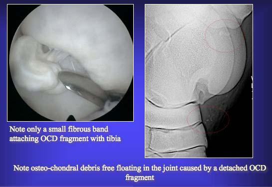
3 The OCD found in hock (tarsocrural) and stifle (femoropatellar), are the result of a disturbance of endochondral ossification. In the fetlock joint, proximal P1 fragments, synovial plica fragments and plantar proximal P1 fragments are not necessarily OCD fragments, but can be the result of delayed ossification, trauma, avulsion fragments or a separate centre of ossification. Hock OCD (intermediate ridge tibia, lateral talus ridge, medial malleolus) will mainly result in joint distension with no lameness. Lameness will only be present if the OCD fragments become detached and are free floating. This is often caused by an abnormal movement (hyperflexion of the joint), which breaks the fibrous attachments between the fragment and bone. Stifle OCD often causes lameness after several years of competing and arthroscopic surgery is strongly advised in sport horses. The ideal age for stifle surgery is between 12 and the 18 months. At this age the healing potential of bone and cartilage is a lot more important and the quality of repair seems superior, than at a later age. Postponing surgery till a later age can result in important secondary lesions in the joint such as generalised cartilage fibrillation, cartilage erosion on the patella and free floating osteochondral debris in the femoro-patellar joint. Presently the relationship between the presence of intra-articular OCF and early development of degenerative joint disease is unclear, and we can not accurately predict which fragments are benign or not. We do know that some fragments can seriously affect the athletic career of sport horses (i.e. stifle OCD, large proximal P1 fragments in the fetlock, large extensor process fragments in the DIP joint) whereas others are very unlikely to cause lameness (i.e. proximoplantar P1 fragments in the fetlock). There is a large grey area where nobody can predict the significance and impact of those fragments on an athletic career. Answering the question Is preventive removal of osteochondral fragments before starting an athletic career recommended? is not easy, and is certainly based on personal subjective experience rather than hard scientific evidence. However there is one thing we have to remember. If we wait to remove the OCF until the horse shows clinical signs (joint distension, stiffness, lameness, poor performance), we are often too late to save the joint. Unlike racing Thoroughbreds, show jumpers and dressage horses will often continue working with cartilage damage, without showing a lot of clinical signs. Sometimes clinical symptoms willnotbe seen until they reached the point of no return. At this stage a well developed degenerative joint disease
4 is often present. The waiting attitude and replacing preventive arthroscopic surgery by curative surgery is dangerous, because of the extensive secondary changes that might already be present in the joint caused by the OCF. In many joints the advantages of a preventive arthroscopic surgery outweigh the possible disadvantages of leaving the fragment in place. During this lecture several types of OCF in the fetlock, stifle, DIP joint and hock joint will be shown, and their clinical relevance will be discussed.
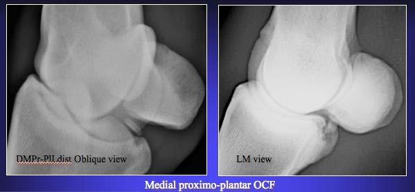
 - Sagittal ridge OCD - Proximo-plantar P1 fragments - Apical sesamoid bone")
5 Commonly encountered intra-articular OCF in the fetlock joint: - Proximo-dorsal P1 fragments - OCF in the synovial plica of the fetlock joint (dorso-proximal in the fetlock joint) - Sagittal ridge OCD - Proximo-plantar P1 fragments - Apical sesamoid bone fragments


6


7


8


9 Commonly encountered intra-articular OCF in the DIP joint: - Extensor process fragments - Palmar DIP fragments


10 Commonly encountered intra-articular OCF in the hock joint:


11 - OCD of the intermediate ridge of the distal tibia - OCD of the distal 1/3 of the lateral talus ridge - OCD of the medial malleolus

12

13

14 Commonly encountered intra-articular fragments in the stifle joint: - OCD of the lateral femur ridge - OCD of the medial femur ridge - OCF cranial or caudal in femorotibial joint


15

16
Proceedings of the 10th International Congress of World Equine Veterinary Association
 www.ivis.org Proceedings of the 10th International Congress of World Equine Veterinary Association Jan. 28 Feb. 1, 2008 - Moscow, Russia Next Congress: Reprinted in IVIS with the permission of the Conference
Proceedings of the American Association of Equine Practitioners - Focus Meeting. First Year of Life Austin, Texas, USA 2008
 www.ivis.org Proceedings of the American Association of Equine Practitioners - Focus Meeting First Year of Life Austin, Texas, USA 2008 Next AAEP Focus Meeting : Focus on the Foot Jul. 19-21, 2009 Columbus,
Proceedings of the 12th International Congress of the World Equine Veterinary Association WEVA
 www.ivis.org Proceedings of the 12th International Congress of the World Equine Veterinary Association WEVA November 2-5, 2011 Hyderabad, India Reprinted in IVIS with the Permission of WEVA Organizers
HOW DO WE DIAGNOSE LAMENESS IN YOUR HORSE?
 HOW DO WE DIAGNOSE LAMENESS IN YOUR HORSE? To help horse owners better understand the tools we routinely use at VetweRx to evaluate their horse s soundness, the following section of this website reviews
EQUINE APPROACHES TO THE DISTENDED DIGITAL FLEXOR TENDON SHEATH ORTHOPAEDICS
 Dr Hans Wilderjans, Dipl ECVS Dierenkliniek De Bosdreef Spelonckvaart 46 9180 Moerbeke-Waas Belgium www.bosdreef.be info@bosdreef.be APPROACHES TO THE DISTENDED DIGITAL FLEXOR TENDON SHEATH During this
Indications: - Anatomical Deformities:
 Indications: - Anatomical Deformities: - The ligaments present an echogenic aspect, however there are some who have helicoidal fibers whose image can give anechogenic areas that should not be confused
Ultrasonographic Examination of Joints in Horses
 Ultrasonographic Examination of Joints in Horses J.-M. Denoix, DVM, PhD, Agrégé; F. Audigié, DVM, PhD The diagnosis of lesions can be made with ultrasonography in many painful joints with no abnormal radiographic
Proceedings of the 55th Annual Convention of the American Association of Equine Practitioners
 Close this window to return to IVIS www.ivis.org Proceedings of the 55th Annual Convention of the American Association of Equine Practitioners December 5 9, 2009, Las Vegas, Nevada Program Chair : Nathaniel
Proceedings of the 12th International Congress of the World Equine Veterinary Association WEVA
 www.ivis.org Proceedings of the 12th International Congress of the World Equine Veterinary Association WEVA November 2-5, 2011 Hyderabad, India Reprinted in IVIS with the Permission of WEVA Organizers
Dr. Chris Bell BSc, DVM, MVetSc, Dip ACVS* *Elders Equine Veterinary Service, Winnipeg, MB, Canada
 Use of a nutraceutical supplement (EXCEED 6 WAY ) Med Vet Pharmaceuticals (MVP) for daily joint support in horses with osteoarthritis Minimal risk field trial and survey. Introduction: Dr. Chris Bell BSc,
Current Surgical Versus Conservative Management in the Treatment of Osteochondritis Dissecans
 Current Surgical Versus Conservative Management in the Treatment of Osteochondritis Dissecans Reproduced with permission from McIlwraith CW. Surgical versus conservative management of osteochondrosis.
Equine Skeletal System
 Equine Skeletal System EQS 110 Table of Contents Click on the different sections of the table of contents to jump through this document Functions of the Skeletal System... 3 Skeletal Strength... 3 Bone
How to Interpret Radiographs of the Stifle Joint of the Young Performance Horse
 How to Interpret Radiographs of the Stifle Joint of the Young Horse Elizabeth M. Santschi, DVM, Diplomate ACVS Author s address: Department of Veterinary Clinical Sciences, The Ohio State University, 601
Proceedings of the 56th Annual Convention of the American Association of Equine Practitioners - AAEP -
 http://www.ivis.org Proceedings of the 56th Annual Convention of the American Association of Equine Practitioners - AAEP - December 4-8, 2010 Baltimore, Maryland, USA Next Meeting : Nov. 18-22, 2011 -
Equine Skeletal System
 Equine Skeletal System EQS 110 Table of Contents Click on the different sections of the table of contents to jump through this document Functions of the Skeletal System... 3 Skeletal Strength... 3 Bone
Proceedings of the 11th Annual Resort Symposium of the American Association of Equine Practitioners AAEP
 www.ivis.org Proceedings of the 11th Annual Resort Symposium of the American Association of Equine Practitioners AAEP January 25-28, 2009 - Gold Coast, Australia ACKNOWLEDGMENTS Dr. Stephen M. Reed, Educational
Osteochondritis Dissecans
 Osteochondritis Dissecans Introduction Osteochondritis dissecans (OCD) is a problem that affects the knee, mostly at the end of the big bone of the thigh (the femur). A joint surface damaged by OCD doesn't
International Cartilage Repair Society
 OsteoArthritis and Cartilage (2005) 13, 1029e1036 ª 2005 OsteoArthritis Research Society International. Published by Elsevier Ltd. All rights reserved. doi:10.1016/j.joca.2005.07.004 Brief report Second-look
Osteochondritis Dissecans (OCD) Defective Cartilage in Young Dogs
 Defective Cartilage in Young Dogs") Osteochondritis Dissecans (OCD) Defective Cartilage in Young Dogs This morning as I was driving home from my own acupuncture appointment for an orthopedic issue, I realized it s been some time since I
Osteochondritis Dissecans (OCD) Defective Cartilage in Young Dogs This morning as I was driving home from my own acupuncture appointment for an orthopedic issue, I realized it s been some time since I
The mare was injected 7000 MBq Technetium 99 MDP and the bone phase examination was performed about three hours later.
 12 year old female used as Show Jumper History The mare was referred for scintigraphy of the front distal limbs. The referring veterinarian s complaint was chronic lameness located on the left front limb.
A Patient s Guide to Osteochondritis Dissecans of the Knee
 A Patient s Guide to Osteochondritis Dissecans of the Knee 2350 Royal Boulevard Suite 200 Elgin, IL 60123 Phone: 847.931.5300 Fax: 847.931.9072 DISCLAIMER: The information in this booklet is compiled from
Focus on Hindlimb Lameness Oklahoma City, OK, USA 2012
 www.ivis.org Proceedings of the American Association of Equine Practitioners - Focus Meeting Focus on Hindlimb Lameness Oklahoma City, OK, USA 2012 Next Focus Meetings: August 4-6, 2013 - Focus on Dentistry
As for the forelimb, treatment of condition of the hindlimb may be treated by both localised therapy, applying the laser
 MLS Master Class - Veterinary Imaging Presented by CelticSMR Ltd Free Phone (UK): 0800 279 9050 International: +44 (0) 1646 603150 AUTHOR DETAILS Carl Gorman BVSc MRCVS PUBLISHER DETAILS Mike Howe B Vet
Management of Chronic Elbow Pain
 Mr. Nashat Siddiqui Consultant Upper Limb Orthopaedic Surgeon Management of Chronic Elbow Pain Patients presenting with elbow pain can pose a diagnostic challenge, especially if there is no obvious recent
The Role of Select Imaging Studies in the Lameness Examination
 The Role of Select Imaging Studies in the Lameness Examination M. J. Martinelli, DVM, PhD and Norm Rantanen, DVM, MS The physical examination and gait analysis are the most important aspects of a lameness
PREVALENCE OF RADIOGRAPHIC CHANGES IN SOUTH AFRICAN THOROUGHBRED RACEHORSES AT THE YEARLING SALES, YOLANDI SMIT
 PREVALENCE OF RADIOGRAPHIC CHANGES IN SOUTH AFRICAN THOROUGHBRED RACEHORSES AT THE YEARLING SALES, 2008-2010 By YOLANDI SMIT Submitted in partial fulfilment of the requirements for the degree of Master
Equine Lameness & Imaging Techniques
 Equine Lameness & Imaging Techniques Peter Heidmann DVM MPH Specialist in Equine Internal Medicine Montana Equine Medical & Surgical Center www.montanaequine.com 406-285-0123 Types of lameness Skeletal
Proceedings of the 55th Annual Convention of the American Association of Equine Practitioners
 www.ivis.org Proceedings of the 55th Annual Convention of the American Association of Equine Practitioners December 5 9, 2009, Las Vegas, Nevada Program Chair : Nathaniel A. White ACKNOWLEDGMENTS Dr. David
Examining Elbow Dysplasia Prepared by the Orthopedic Foundation for Animals Orthopedic Foundation for Animals, Columbia, MO
 Examining Elbow Dysplasia Prepared by the Orthopedic Foundation for Animals Orthopedic Foundation for Animals, Columbia, MO Elbow dysplasia has been found in 78 breeds evaluated by the Orthopedic Foundation
Some Carpal Lesions in the Non-racehorse
 Published in IVIS with the permission of the AAEP Close this window to return to IVIS Some Carpal Lesions in the Non-racehorse Sue Dyson, VetMB, PhD Author s address: Centre for Equine Studies, Animal
Proceedings of the World Small Animal Veterinary Association Sydney, Australia 2007
 Proceedings of the World Small Animal Sydney, Australia 2007 Hosted by: Next WSAVA Congress CRANIAL CRUCIATE LIGAMENT INJURIES SURGICAL MANAGEMENT Warrick J. Bruce BVSc(dist), MVM, DSAS(orthopaedics),
Ankle Arthroscopy.
 Ankle Arthroscopy Key words: Ankle pain, ankle arthroscopy, ankle sprain, ankle stiffness, day case surgery, articular cartilage, chondral injury, chondral defect, anti-inflammatory medication Our understanding
ACROPODIAL DISEASES IN HORSES DIAGNOSED RADIOGRAPHICALLY: RETROSPECTIVE STUDY OF 7 CASES
 Abstract Scientific Works. Series C. Veterinary Medicine. Vol. LIX (1) ISSN 2065-1295, ISSN CD-ROM 2343-9394, ISSN Online 2067-3663, ISSN-L 2065-1295 ACROPODIAL DISEASES IN HORSES DIAGNOSED RADIOGRAPHICALLY:
 Mmmmmm Mmmmmm mmmmmmmmmmmmm mmmmmmm mmmmmmm MRI of Equine Stifle Injuries: A Review of the first 100 clinical cases Martin Waselau, Dr.med.vet., MS Diplomate ACVS, Diplomate ECVS Equine Hospital Aschheim,
Pre-operative evaluation
 Pre-operative evaluation Andrea Meyer-Lindenberg Clinic of Small Animal Surgery and eproduction Ludwig-Maximilians-University Munich Importance of pre-operative planning Evaluate patient before selecting
Diagnostic Stifle Joint Arthroscopy Using a Needle Arthroscope in Standing Horses
 Using a Needle Arthroscope in Standing Horses David D. Frisbie, DVM, PhD, Diplomate ACVS & ACVSMR, Myra F. Barrett, DVM, MS, Diplomate ACVR, C. Wayne McIlwraith, BVSc, PhD, DSc, FRCVS, Diplomate ACVS &
Clinical Case Series Adipose Derived Stem and Regenerative Cells for the Treatment of Equine Joint Injuries
 Clinical Case Series Adipose Derived Stem and Regenerative Cells for the Treatment of Equine Joint Injuries Vet-Stem, Inc., 12860 Danielson Court, Suite B, Poway, CA 92064 Developmental bone disease, osteochondritis
Cranial cruciate ligament rupture in Dogs
 Clinical sheet - Surgery Cranial cruciate ligament rupture in Dogs Cranial cruciate ligament rupture is one of the most common orthopedic conditions in dogs. Rupture of the cranial cruciate ligament is
May 2011, Issue 31. In addition to our regular ER hours, AMVS is providing emergency and critical care services to your patients: Fridays, all day
 Page 1 of 5 Having Trouble Viewing this Email? Click Here You're receiving this email because of your relationship with Aspen Meadow Veterinary Specialists. Please confirm your continued interest in receiving
198 EQUINE VETERINARY EDUCATION Equine vet. Educ. (2001) 13 (4)
 13 (4)") 198 EQUINE VETERINARY EDUCATION Equine vet. Educ. (2001) 13 (4) 198-204 Tutorial Article Use of ultrasonography in the evaluation of joint disease in horses. Part 1: Indications, technique and examination
198 EQUINE VETERINARY EDUCATION Equine vet. Educ. (2001) 13 (4) 198-204 Tutorial Article Use of ultrasonography in the evaluation of joint disease in horses. Part 1: Indications, technique and examination
Proceeding of the SEVC Southern European Veterinary Conference
 www.ivis.org Proceeding of the SEVC Southern European Veterinary Conference Oct. 17-19, 2008 Barcelona, Spain http://www.sevc.info Reprinted in the IVIS website with the permission of the SEVC www.ivis.org
Fracture and Dislocation of Metacarpal Bones, Metacarpophalangeal Joints, Phalanges, and Interphalangeal Joints ( 1-Jan-1985 )
") In: Textbook of Small Animal Orthopaedics, C. D. Newton and D. M. Nunamaker (Eds.) Publisher: International Veterinary Information Service (www.ivis.org), Ithaca, New York, USA. Fracture and Dislocation
In: Textbook of Small Animal Orthopaedics, C. D. Newton and D. M. Nunamaker (Eds.) Publisher: International Veterinary Information Service (www.ivis.org), Ithaca, New York, USA. Fracture and Dislocation
Cranial Cruciate disease
 Cranial Cruciate disease Anatomy The Cranial cruciate ligament is located in the stifle joint (or knee). It is a thick fibrous band that runs from the distal femur to the proximal tibia. It is designed
Lameness & Non- Surgical Therapies of the Equine Athlete
 Lameness & Non- Surgical Therapies of the Equine Athlete Mark T. Reilly, DVM, Dipl. ABVP (Equine) Linda J. Cimetti, DVM South Shore Equine Clinic & Diagnostic Center Lameness & Non-Surgical Therapy of
Equine Lameness. Kevin Kersh, D.V.M. Equine Surgery Resident Iowa State University
 Equine Lameness Kevin Kersh, D.V.M. Equine Surgery Resident Iowa State University Lameness Lameness associated pain can be localized by physical exam, palpation, response to hoof testers, response to flexion
Patellar Luxation. Anatomy, Function, and Dysfunction
 6910 Carpenter Fire Station Road, Cary NC 27519 Phone (919) 545-1001 Patellar Luxation This information is provided to help you understand the condition that has been diagnosed in your pet. We find that
REPORTING SERVICE: XR
 REPORTING SERVICE: XR Report number: VETCT-70603 Report date: 04/04/2017 Referring Veterinarian: xxxxx Referring Practice: xxxxx Email address: xxxxx Owner: xxxx Patient: xxxx Species: Equine Breed: Belgian
Lameness in the Rodeo Horse
 Lameness in the Rodeo Horse Robert D. Lewis, DVM Author s address: Elgin Veterinary Hospital, Inc., P.O. Box 629, Elgin, Texas 78621. 2001 AAEP. The ever-increasing popularity of the rodeo has created
Application of optical coherence tomography enhances reproducibility of arthroscopic evaluation of equine joints
 Niemelä et al. Acta Veterinaria Scandinavica 2014, 56:3 RESEARCH Application of optical coherence tomography enhances reproducibility of arthroscopic evaluation of equine joints Open Access Tytti Niemelä
Equine Appendicular Skeleton
 Equine ppendicular Skeleton Tony Pease, DVM, MS, DCV ssistant Professor of adiology North Carolina State University bjectives cquisition of radiographs adiographic anatomy adiographic patterns of disease
Medical Practice for Sports Injuries and Disorders of the Knee
 Sports-Related Injuries and Disorders Medical Practice for Sports Injuries and Disorders of the Knee JMAJ 48(1): 20 24, 2005 Hirotsugu MURATSU*, Masahiro KUROSAKA**, Tetsuji YAMAMOTO***, and Shinichi YOSHIDA****
Zoran Lončar. CONGRESS AMVAC/RoSAVA September, 2014
 Zoran Lončar Veterinary Clinic www.vetnovak.com loncarzor@yahoo.co.uk Belgrade, Serbia CONGRESS AMVAC/RoSAVA 11-13 September, 2014 ALL ABOUT THE KNEEE To become familiar with the EXAM To recognize most
Performance Lameness in Reining Horses
 Performance Lameness in Reining Horses QuickTime and a TIFF (Uncompressed) decompressor are needed to see this picture. MPH Peter Heidmann DVM Specialist in Equine Internal Medicine Montana Equine Medical
ANATOMY AND PHYSIOLOGY
 WHY? ANATOMY AND PHYSIOLOGY VETERINARY SCIENCE PROGRAM RECOGNIZE AND UNDERSTAND BASIC DIRECTIONAL AND ANATOMICAL TERMS UNDERSTAND AND SPEAK THE LANGUAGE OF ANATOMY EXPECTED TO BE ABLE TO COMMUNICATE INTELLIGENTLY
Prevalence of osteochondral lesions in the fetlock and hock joints of Standardbred horses that survived bacterial infection before 6 months of age
 Hendrickson et al. BMC Veterinary Research (2018) 14:390 https://doi.org/10.1186/s12917-018-1726-3 RESEARCH ARTICLE Open Access Prevalence of osteochondral lesions in the fetlock and hock joints of Standardbred
Proceedings of the 10th International Congress of World Equine Veterinary Association
 www.ivis.org Proceedings of the 10th International Congress of World Equine Veterinary Association Jan. 28 Feb. 1, 2008 - Moscow, Russia Next Congress: Reprinted in IVIS with the permission of the Conference
Patellar Ligament Disease.
 Patellar Ligament Disease. The patellar ligament disease is a condition of the stifle where the cartilage keeping the patella in place over knee joint is weakened or damaged. The patella is held in place
Osteochondritis Dissecans
 Osteochondritis Dissecans Carrie Lane, MA, SAMP October 14, 2011 Table of Contents Introduction What is Osteochondritis Dissecans? (OCD) The Nature of the Condition Common Treatment Approaches Rehabilitation
Proceedings of the 59th Annual Convention of the American Association of Equine Practitioners - AAEP -
 http://www.ivis.org Proceedings of the 59th Annual Convention of the American Association of Equine Practitioners - AAEP - December 7-11, 2013 Nashville, TN, USA Next Meeting : Dec. 6-10, 2014 - Salt Lake
Sequalae of Ankle Sprains: Peri Articular Fractures of the Ankle in Sports Medicine.
 Sequalae of Ankle Sprains: Peri Articular Fractures of the Ankle in Sports Medicine www.fisiokinesiterapia.biz Chronic Ankle Pain The most common cause of chronic pain following an ankle sprain is a missed
BIOMECHANICAL EVALUATION OF MEDIAL FEMORAL CONDYLAR SUBCHONDRAL CYSTIC LESIONS AND THE EFFECTS OF TREATMENT WITH INTERNAL FIXATION
 BIOMECHANICAL EVALUATION OF MEDIAL FEMORAL CONDYLAR SUBCHONDRAL CYSTIC LESIONS AND THE EFFECTS OF TREATMENT WITH INTERNAL FIXATION Abstract Purpose of the study: Subchondral cystic lesions (SCLs) in the
Welcome to the: Orthopaedic Opinion Online Website The website for the answer to all your Orthopaedic Questions
 Welcome to the: Orthopaedic Opinion Online Website The website for the answer to all your Orthopaedic Questions Orthopaedic Opinion Online is a website designed to provide information to patients who have
Patellofemoral Instability
 Disclaimer This movie is an educational resource only and should not be used to manage Patellofemoral Instability. All decisions about the management of Patellofemoral Instability must be made in conjunction
MRI KNEE WHAT TO SEE. Dr. SHEKHAR SRIVASTAV. Sr.Consultant KNEE & SHOULDER ARTHROSCOPY
 MRI KNEE WHAT TO SEE Dr. SHEKHAR SRIVASTAV Sr.Consultant KNEE & SHOULDER ARTHROSCOPY MRI KNEE - WHAT TO SEE MRI is the most accurate and frequently used diagnostic tool for evaluation of internal derangement
EMERGENCY PITFALLS IN ORTHOPAEDIC TRAUMA. Thierry E. Benaroch, MD, FRCS MCH Trauma Rounds February 9, 2009
 EMERGENCY PITFALLS IN ORTHOPAEDIC TRAUMA Thierry E. Benaroch, MD, FRCS MCH Trauma Rounds February 9, 2009 MORAL OF THE STORY Fracture distal radius and intact ulna W/O radius fracture will most likely
Treatment of meniscal lesions and isolated lesions of the anterior cruciate ligament of the knee in adults
 QUICK REFERENCE GUIDE Treatment of meniscal s and isolated s of the anterior cruciate ligament of the knee in adults June 2008 AIM OF THE GUIDELINES To encourage good practices in the areas of meniscal
Anterior Cruciate Ligament (ACL)
") Anterior Cruciate Ligament (ACL) The anterior cruciate ligament (ACL) is one of the 4 major ligament stabilizers of the knee. ACL tears are among the most common major knee injuries in active people of
Anterior Cruciate Ligament (ACL) The anterior cruciate ligament (ACL) is one of the 4 major ligament stabilizers of the knee. ACL tears are among the most common major knee injuries in active people of
A Patient s Guide to Bipartite Patella
 A Patient s Guide to Bipartite Patella 651 Old Country Road Plainview, NY 11803 Phone: 5166818822 Fax: 5166813332 p.lettieri@aol.com DISCLAIMER: The information in this booklet is compiled from a variety
Osseous Trauma in the Fetlock Region of Mature Sports Horses
 Osseous Trauma in the Fetlock Region of Mature Sports Horses Sue J. Dyson, VetMB, PhD; and Rachel Murray, VetMB, PhD Osseous trauma of the fetlock is a potentially important cause of lameness in mature
TTA. Common Tangent Method
 TTA Common Tangent Method This document is derived from a presentation by Dr. Randy Boudrieau DVM, Dipl. ACVS, ECVS, Prof. of Surgery, Cummings School of Veterinary Medicine, Tufts University IVET DESIG
Luxation of the Patella
 Luxation of the Patella The patella or knee cap is the small bone that connects the thigh muscles to the bones that comprise the shin, namely the tibia and fibula. The term luxation refers to the dislocation
Arthroscopy Of the Ankle.
 Arthroscopy Of the Ankle www.fisiokinesiterapia.biz Ankle Arthroscopy Anatomy Patient setup Portal placement Procedures Complications Anatomy Portals Anterior Anteromedial Anterolateral Anterocentral Posterior
where in the how doctors Anatomy Femoral Condyles surface be seen on to as the:
 Osteochondritis Dissecans of the Knee Introduction Welcome to BodyZone Physiotherapy's patient resource about Osteochondritis Dissecans of the Knee. Osteochondritis dissecans (OCD) is a problem that affects
BASELINE QUESTIONNAIRE (SURGEON)
") SECTION A: STUDY INFORMATION Subject ID: - - Study Visit: Baseline Site Number: Date: / / Surgeon ID: SECTION B: INITIAL SURGEON HISTORY B1. Previous Knee Surgery: Yes No Not recorded B2. Number of Previous
SECTION A: STUDY INFORMATION Subject ID: - - Study Visit: Baseline Site Number: Date: / / Surgeon ID: SECTION B: INITIAL SURGEON HISTORY B1. Previous Knee Surgery: Yes No Not recorded B2. Number of Previous
Arthroscopy. Turnberg Building Orthopaedics
 Arthroscopy Turnberg Building Orthopaedics 0161 206 4898 All Rights Reserved 2017. Document for issue as handout. Introduction An arthroscopy is a type of keyhole surgery used both to diagnose and treat
Courtney James and Jessica Gladney
 EFFECTS OF BILAYER GELATIN/BETA TRICALCIUM PHOSPHATE SPONGES LOADED WITH MESENCHYMAL STEM CELLS, CHONDROCYTES, BMP-2, AND PRP ON OSTEOCHONDRAL DEFECTS OF THE TALUS IN HORSES Courtney James and Jessica
2017 SAFSA CONGRESS PROGRAMME
 2017 SAFSA CONGRESS PROGRAMME THURSDAY, MAY 25 07h45 07h55: WELCOME & INTRODUCTIONS Forefoot I: Hallux Valgus and Lesser Toes (08h00-10h00 Lectures) 08h00 08h30: Surgical Management of Hallux Valgus Rippstein,
GASTROCNEMIUS TENDON REPAIR VETLIG USING THE STIF CAT 30 SOFT TISSUE INTERNAL FIXATION VETLIG
 VETLIG SOFT TISSUE INTERNAL FIXATION GASTROCNEMIUS TENDON REPAIR USING THE STIF CAT 30 VETLIG A R T I F I C I A L L I G A M E N T S F O R V E T E R I N A R Y U S E VETLIG MANAGEMENT OF CHRONIC GASTROCNEMIUS
How to Take Radiographs of the Metacarpophalangeal/Metatarsophalangeal Joint (Fetlock Joint)
") How to Take Radiographs of the Metacarpophalangeal/Metatarsophalangeal Joint (Fetlock Joint) Joseph W. Morgan, DVM, Diplomate ACVS Author s address: 5366 Leestown Road, Lexington, KY 40511 8904; e-mail:
How to Take Radiographs of the Metacarpophalangeal/Metatarsophalangeal Joint (Fetlock Joint) Joseph W. Morgan, DVM, Diplomate ACVS Author s address: 5366 Leestown Road, Lexington, KY 40511 8904; e-mail:
40 th Annual Symposium on Sports Medicine. Knee Injuries In The Pediatric Athlete. Disclosure
 40 th Annual Symposium on Sports Medicine Travis Murray, MD Assistant Professor University of Texas Health Science Center San Antonio Knee Injuries In The Pediatric Athlete Disclosure Dr. Travis Murray
Ankle Arthritis PATIENT INFORMATION. The ankle joint. What is ankle arthritis?
 PATIENT INFORMATION Ankle Arthritis The ankle joint The ankle is a very complex joint. It is actually made up of two joints: the true ankle joint and the subtalar ankle joint. The ankle joint consists
Common Knee Injuries
 Common Knee Injuries In 2010, there were roughly 10.4 million patient visits to doctors' offices because of common knee injuries such as fractures, dislocations, sprains, and ligament tears. Knee injury
Module 2 Objectives. Module 2 Current Research in Regenerative Therapy - Orthopedics - Rabbit Osteochondral Defect Repair Model
 Module 2 Current Research in Regenerative Therapy - Orthopedics - Module 2 Objectives Review of current research Review of equine orthopedics clinical data Review of canine orthopedics clinical data Review
This presentation is the intellectual property of the author. Contact them at for permission to reprint and/or distribute.
 B F Morrey, MD Professor of Orthopedic Surgery, UTHSCSA Financial Disclosure Dr. Bernard Morrey has disclosed that he is the Medical director of Tenex Health. OUTLINE Muscles/tendons Ligaments Articulation
Diagnosing Forelimb Lameness in Canine Patients
 OCTOBER 2018 Diagnosing Forelimb Lameness in Canine Patients DR. SEVIMA AKTAY, VMD, DACVS Diagnosing and treating forelimb lameness in dogs can often be challenging. Our patients rarely demonstrate overt
Proceedings of the 11th Annual Resort Symposium of the American Association of Equine Practitioners AAEP
 www.ivis.org Proceedings of the 11th Annual Resort Symposium of the American Association of Equine Practitioners AAEP January 25-28, 2009 - Gold Coast, Australia ACKNOWLEDGMENTS Dr. Stephen M. Reed, Educational
EPIPHYSEAL PLATE IN FEMUR
 Reviewing: Epiphyseal Plates (younger skeletons) eventually will disappear. Bones grow lengthwise up and down from each plate, and in a circular collar like fashion around the diaphysis. These plates will
MRI of the Knee: Part 4 - normal variants that may simulate disease. Mark Anderson, M.D. University of Virginia
 MRI of the Knee: Part 4 - normal variants that may simulate disease Mark Anderson, M.D. University of Virginia discuss the most common normal variants in the pediatric knee that may simulate pathology
SECTION 1 ANATOMY L A TERI A M TED H G PYRI CO
 COPYRIGHTED MATERIAL A veterinary technician comes in for work in the morning to discover that Fluffy has come in overnight after having been hit by a car. The chart note indicates that there is a cut
HISTORY AND INDICATIONS OF LATERAL TENODESIS IN ATHLETES
 HISTORY AND INDICATIONS OF LATERAL TENODESIS IN ATHLETES Written by Philippe Landreau, Qatar The treatment of anterior cruciate ligament injuries remains challenging in young athletic populations. A residual
These conditions can be differentiated by high quality craniocaudal and lateral radiographic views of the elbow joint.
 ELBOW DYSPLASIA Daniel D. Lewis, DVM, Diplomate ACVS Professor Small Animal Surgery Department of Small Animal Clinical Sciences University of Florida Gainesville, Florida The term elbow dysplasia has
Basic Principles of Fractures & Easily Missed Fractures. Mr Irfan Merchant Trauma & Orthopaedic Registrar Bedford Hospital, East of England
 Basic Principles of Fractures & Easily Missed Fractures Mr Irfan Merchant Trauma & Orthopaedic Registrar Bedford Hospital, East of England Objectives Types Fracture Patterns Fracture Healing Assessing
Proceeding of the NAVC North American Veterinary Conference Jan. 8-12, 2005, Orlando, Florida
 Proceeding of the NAVC North American Veterinary Conference Jan. 8-12, 2005, Orlando, Florida Reprinted in the IVIS website with the permission of the NAVC http:/// The North American Veterinary Conference
Case Studies. A. Kent Allen, DVM LAMENESS AND IMAGING IN THE SPORT HORSE
 Case Studies A. Kent Allen, DVM Author s address: Virginia Equine Imaging, 2716 Landmark School Road, The Plains, VA 20198; e-mail: vaequine@aol.com. 2007 AAEP. 1. Case Study #1: Medial Collateral Desmitis
Anterior knee pain.
 Anterior knee pain What are the symptoms? Anterior knee pain is very common amongst active adolescents and athletes participating in contact sports. It is one of the most common problems/injuries seen
Dukes Vet Practice
 www.dukesvets.com Getting the best out of your horse Jim Dukes, BVM&S, MRCVS Response to treatment for lameness What do we know about lameness in horses? Our approach to treatment and effectiveness What
A Patient s Guide to Knee Arthroscopy
 A Patient s Guide to Knee Arthroscopy 2350 Royal Boulevard Suite 200 Elgin, IL 60123 Phone: 847.931.5300 Fax: 847.931.9072 DISCLAIMER: The information in this booklet is compiled from a variety of sources.
KNEE ARTHROSCOPY PATIENT INFORMATION SHEET
 KNEE ARTHROSCOPY PATIENT INFORMATION SHEET Introduction It has been recommended that you undergo an arthroscopy of your knee. This information sheet is designed to explain what is involved in an arthroscopy,
ACL Athletic Career. ACL Rupture - Warning Features Intensive pain Immediate swelling Locking Feel a Pop Dead leg Cannot continue to play
 FIMS Ambassador Tour to Eastern Europe, 2004 Belgrade, Serbia Montenegro Acute Knee Injuries - Controversies and Challenges Professor KM Chan OBE, JP President of FIMS Belgrade ACL Athletic Career ACL