Is it really honeycombing? Limitations and pitfalls in radiological diagnosis of honeycombing.
|
|
|
- Jennifer Owen
- 5 years ago
- Views:
Transcription
1 Is it really honeycombing? Limitations and pitfalls in radiological diagnosis of honeycombing. J. Arenas-Jiménez, E. García-Garrigós, M. Sirera-Matilla; M.C. Planells-Alduvin, F.I. Aranda* Hospital General Universitario de Alicante. Department of Radiology and *Pathology Alicante. Spain.
2 All authors have no relevant financial disclosures
3 The honeycombing quiz. Half of the images in this slide correspond to honeycombing and the other half are entities mimicking it. In this education exhibit clues for a correct diagnosis and limitations in the radiological definition of honeycombing will be reviewed.
 have shown a relatively low interobserver agreement in the diagnosis of honeycombing, at different levels of radiologists experience.")

4 Introduction Diagnosis of radiological honeycombing is crucial for establishing an usual interstitial pneumonia pattern, for that reason a confident diagnosis is desirable. However, several studies (1,2) have shown a relatively low interobserver agreement in the diagnosis of honeycombing, at different levels of radiologists experience. There is a typical appearance (figures) of clustered peripheral thick wall air cysts that cause few problems for the vast majority of readers, but the daily radiological spectrum can be more complicated. We have focused this presentation on potential pitfalls that radiologist have to keep in mind for avoiding misdiagnosis of honeycombing, and have divided in four groups: small cysts, big cysts, bronchiectasis and centrilobular emphysema under other pulmonary conditions, mainly infection.


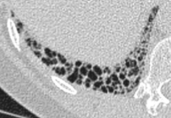
5 Small cysts Radiologists must be aware that a microscopic honeycombing under CT scans resolution frequently exists. Very small cysts in regions with other signs of fibrosis can correspond to this microscopic honeycombing. Sometimes there is a gap between what pathologist and radiologists call honeycombing. Other signs such as traction bronchiectasis and bronchiolectasis are the clue to suggest the diagnosis. CT and pathologic correlation in a 43 year-old man with a pathologic UIP pattern showing small sized honeycombing (yellow arrowheads) at HRCT.


6 Which of these should you certainly call honeycombing? A C B D


7 Which of these should you certainly call honeycombing? As you can see, A B and C-D correspond to the same patients, respectively. Probably you can have reasonable doubts in A and D due to the small cysts, but feel more comfortable making the diagnosis of honeycombing in B and C. B A C D

8 Big cysts Honeycombing in areas with emphysema and fibrosis can be difficult to differentiate from each other 64-year-old smoker with rheumatoid arthritis, pulmonary fibrosis with UIP pattern and left pleural effusion. Cystic lesions in right lower lung suggest honeycombing with big cysts in this setting.


9 Big cysts Relatively big honeycombing cysts (1-2 cm) are frequent in combined pulmonary fibrosis and emphysema syndrome (3). Thick-wall cystic lesions as shown in the image below are also frequently found in those patients.
.")

10 Bronchiectasis Sometimes, appearance and distribution of ectatic bronchi can mimic honeycombing. This is not surprising, since part of the honeycombing cysts correspond histologically to bronchiolectasis (4). Review of contiguous slices and multiplanar reformatting can be useful. When a bronchiectasis ends and a honeycombing cyst begins can be difficult to ascertain such as in the figure. There is an increasing trend to consider traction bronchiectasis and honeycombing as part of the same spectrum of remodeling of the lung due to fibrosis (5, 6).

11 Pulmonary fibrosis in a patient with scleroderma. Multiple variable sized cysts are seen. Also note dilated esophagus. In this individual image, diagnosis can be difficult. Next slide can help.

.")
12 Pulmonary fibrosis in a patient with scleroderma. Reformatted coronal CT show how at least part of cystic lesions in previous slide correspond to bronchiectasis (red arrows). Right middle lobe clearly depicts the bronchiectasis centrally.
13 Centrilobular emphysema under other pulmonary conditions Infection, inflammatory conditions and edema in patients with centrilobular emphysema can closely resemble the appearance of honeycombing. Distribution and visualization of centrilobular arteries are the clue to avoid a false diagnosis. A follow-up CT scan should solve the doubt.


14 Centrilobular emphysema under other pulmonary conditions Infection, inflammatory conditions and edema in patients with centrilobular emphysema can closely resemble the appearance of honeycombing. Distribution and visualization of centrilobular arteries are the clue to avoid a false diagnosis. A follow-up CT scan should solve the doubt.


15 Centrilobular emphysema under other pulmonary conditions Same patient 1-month follow-up
16 These figures resemble each other, but can you see the differences?


17 Emphysema Clue: You can see the centrilobular arteries and cyst are not subpleural in some cases. Honeycombing Clue: Traction bronchiectasis are seen next to the cysts.
18 The honeycombing quiz. Half of the images in this slide correspond to honeycombing and the other half are entities mimicking it. In this education exhibit clues for a correct diagnosis and limitations in the radiological definition of honeycombing have been reviewed.


 and")
 are the")
19 The honeycombing quiz. These images ARE NOT honeycombing. Bronchiectasis (above) and emphysema (bottom line) are the causes of cystic peripheral lesions.
20 The honeycombing quiz. These images ARE honeycombing.
21 References 1. Watadani et al. Radiology 2013; 266: Walsh et al. Thorax 2016; 75: Inomata et al. BMC Pulmonary Medicine 2014; 14: Staats et al. Pathol Res Pract 2015; 211:55 5. Piciucchi et al. BMC Pulmonary Medicine 2016;16:87 6. Gruden et al. AJR 2016; 206: 495
22 Other suggested readings 1. Arakawa & Honma. AJR 2011; 196: Johkoh et al. Eur J Radiol 2014; 83: 27
23 Contact information Juan Arenas-Jiménez Department of Radiology. Hospital General Universitario de Alicante. Alicante. Spain
Imaging: how to recognise idiopathic pulmonary fibrosis
 REVIEW IDIOPATHIC PULMONARY FIBROSIS Imaging: how to recognise idiopathic pulmonary fibrosis Anand Devaraj Affiliations: Dept of Radiology, St George s Hospital, London, UK. Correspondence: Anand Devaraj,
Difficulties Diagnosing Idiopathic Pulmonary Fibrosis
 1. er Encuentro Entre Neumólogos y Radiólogos, Madrid, Spain, 2016, October 14th Difficulties Diagnosing Idiopathic Pulmonary Fibrosis Simon Walsh King s College Hospital Foundation Trust London, United
The radiological differential diagnosis of the UIP pattern
 5th International Conference on Idiopathic Pulmonary Fibrosis, Modena, 2015, June 12th The radiological differential diagnosis of the UIP pattern Simon Walsh King s College Hospital Foundation Trust London,
Financial disclosure COMMON DIAGNOSES IN HRCT. High Res Chest HRCT. HRCT Pre test. I have no financial relationships to disclose. Anatomy Nomenclature
 Financial disclosure I have no financial relationships to disclose. Douglas Johnson D.O. Cardiothoracic Imaging Gaston Radiology COMMON DIAGNOSES IN HRCT High Res Chest Anatomy Nomenclature HRCT Sampling
Thoracic lung involvement in rheumatoid arthritis: Findings on HRCT
 Thoracic lung involvement in rheumatoid arthritis: Findings on HRCT Poster No.: C-2488 Congress: ECR 2015 Type: Educational Exhibit Authors: R. E. Correa Soto, M. J. Martín Sánchez, J. M. Fernandez 1 1
CT in Idiopathic Pulmonary Fibrosis: Diagnosis and Beyond
 Cardiopulmonary Imaging Review Gruden CT of Idiopathic Pulmonary Fibrosis Cardiopulmonary Imaging Review James F. Gruden 1 Gruden JF FOCUS ON: Keywords: CT, diagnosis, high-resolution CT, idiopathic pulmonary
11/10/2014. Multi-disciplinary Approach to Diffuse Lung Disease: The Imager s Perspective. Radiology
 Multi-disciplinary Approach to Diffuse Lung Disease: The Imager s Perspective Radiology Pathology Clinical 1 Role of HRCT Diagnosis Fibrosis vs. inflammation Next step in management Response to treatment
Progress in Idiopathic Pulmonary Fibrosis
 Progress in Idiopathic Pulmonary Fibrosis David A. Lynch, MB Disclosures Progress in Idiopathic Pulmonary Fibrosis David A Lynch, MB Consultant: t Research support: Perceptive Imaging Boehringer Ingelheim
Usual Interstitial pneumonia and Nonspecific Interstitial Pneumonia. Nitra and the Gangs.
 Usual Interstitial pneumonia and Nonspecific Interstitial Pneumonia Nitra and the Gangs. บทน ำและบทท ๓, ๑๐, ๑๒, ๑๓, ๑๔, ๑๕, ๑๗ Usual Interstitial Pneumonia (UIP) Most common & basic pathologic pattern
Liebow and Carrington's original classification of IIP
 Liebow and Carrington's original classification of IIP-- 1969 Eric J. Stern MD University of Washington UIP Usual interstitial pneumonia DIP Desquamative interstitial pneumonia BIP Bronchiolitis obliterans
NONE OVERVIEW FINANCIAL DISCLOSURES UPDATE ON IDIOPATHIC PULMONARY FIBROSIS/IPF (UIP) FOR PATHOLOGISTS. IPF = Idiopathic UIP Radiologic UIP Path UIP
 FOR PATHOLOGISTS. IPF = Idiopathic UIP Radiologic UIP Path UIP") UPDATE ON IDIOPATHIC PULMONARY FIBROSIS/IPF () FOR PATHOLOGISTS Thomas V. Colby, M.D. Professor of Pathology (Emeritus) Mayo Clinic Arizona FINANCIAL DISCLOSURES NONE OVERVIEW IPF Radiologic Dx Pathologic
UPDATE ON IDIOPATHIC PULMONARY FIBROSIS/IPF () FOR PATHOLOGISTS Thomas V. Colby, M.D. Professor of Pathology (Emeritus) Mayo Clinic Arizona FINANCIAL DISCLOSURES NONE OVERVIEW IPF Radiologic Dx Pathologic
5/9/2015. Multi-disciplinary Approach to Diffuse Lung Disease: The Imager s Perspective. No, I am not a pulmonologist! Radiology
 Multi-disciplinary Approach to Diffuse Lung Disease: The Imager s Perspective No, I am not a pulmonologist! Radiology Pathology Clinical 1 Everyone needs a CT Confidence in diagnosis Definitive HRCT +
Radiologic-pathologic correlation of pulmonary diseases
 The 1578 th Chest Conference/ 3 rd Biennial Clinical- Radiologic-Pathologic Correlation Radiologic-pathologic correlation of pulmonary diseases Harumi Itoh, M.D. University of Fukui, Japan Centriacinar
CTD-related Lung Disease
 13 th Cambridge Chest Meeting King s College, Cambridge April 2015 Imaging of CTD-related Lung Disease Dr Sujal R Desai King s College Hospital, London Disclosure Statement No Disclosures / Conflicts of
Outline Definition of Terms: Lexicon. Traction Bronchiectasis
 HRCT OF IDIOPATHIC INTERSTITIAL PNEUMONIAS Disclosures Genentech, Inc. Speakers Bureau Tadashi Allen, MD University of Minnesota Assistant Professor Diagnostic Radiology 10/29/2016 Outline Definition of
T he diagnostic evaluation of a patient with
 546 REVIEW SERIES Challenges in pulmonary fibrosis? 1: Use of high resolution CT scanning of the lung for the evaluation of patients with idiopathic interstitial pneumonias Michael B Gotway, Michelle M
Differential diagnosis
 Differential diagnosis Idiopathic pulmonary fibrosis (IPF) is part of a large family of idiopathic interstitial pneumonias (IIP), one of four subgroups of interstitial lung disease (ILD). Differential
Asbestosis and other pulmonary fibrosis in asbestos-exposed workers: high-resolution CT features with pathological correlations
 Eur Radiol (2016) 26:1485 1492 DOI 10.1007/s00330-015-3973-z CHEST Asbestosis and other pulmonary fibrosis in asbestos-exposed workers: high-resolution CT features with pathological correlations Hiroaki
Emphysema association in a prospective series with patients suffering from Idiopathic Pulmonary Fibrosis.
 Emphysema association in a prospective series with patients suffering from Idiopathic Pulmonary Fibrosis. David Jiménez-Restrepo, Mª Luisa Domingo Montañana, Claudia Fernandez Ruiz, Andrea Martínez Deltoro*,
Diffuse Interstitial Lung Diseases: Is There Really Anything New?
 : Is There Really Anything New? Sujal R. Desai, MBBS, MD ESTI SPEAKER SUNDAY Society of Thoracic Radiology San Antonio, Texas March 2014 Diffuse Interstitial Lung Disease The State of Play DILDs Is There
Daria Manos RSNA 2016 RC 401. https://medicine.dal.ca/departments/depar tment-sites/radiology/contact/faculty/dariamanos.html
 Daria Manos RSNA 2016 RC 401 https://medicine.dal.ca/departments/depar tment-sites/radiology/contact/faculty/dariamanos.html STEP1: Is this fibrotic lung disease? STEP 2: Is this a UIP pattern? If yes:
Dr. Daria Manos: Boehringer Ingelheim CSL Behring HIT Global. Dr. Horatiu Muller: No conflicts of interest
 Dr. Daria Manos: Boehringer Ingelheim CSL Behring HIT Global Dr. Horatiu Muller: No conflicts of interest Honeycombing Centrilobular emphysema (CLE) Paraseptal emphysema (PSE) Panlobular emphysema Cystic
World Journal of Radiology. Pulmonary fibrosis and emphysema: Is the emphysema type associated with the pattern of fibrosis?
 W J R World Journal of Radiology Submit a Manuscript: http://www.wjgnet.com/esps/ Help Desk: http://www.wjgnet.com/esps/helpdesk.aspx DOI: 10.4329/wjr.v7.i9.294 World J Radiol 2015 September 28; 7(9):
Patient with IPF and no honeycombing on HRCT. Case 1 Demosthenes Bouros, Vasilios Tzilas University of Athens
 Patient with IPF and no honeycombing on HRCT Case 1 Demosthenes Bouros, Vasilios Tzilas University of Athens CASE OVERVIEW A 76-year-old male patient presented with progressive exertional dyspnoea refractory
Criteria for confident HRCT diagnosis of usual interstitial pneumonia (UIP)
") Criteria for confident HRCT diagnosis of usual interstitial pneumonia (UIP) Assem El Essawy (1) & Amr A. Nassef (٢) Abstract Identification of interstitial pneumonia (IP) was mainly based on histological
Criteria for confident HRCT diagnosis of usual interstitial pneumonia (UIP) Assem El Essawy (1) & Amr A. Nassef (٢) Abstract Identification of interstitial pneumonia (IP) was mainly based on histological
Cryptogenic Organizing Pneumonia Diagnosis Approach Based on a Clinical-Radiologic-Pathologic Consensus
 Cryptogenic Organizing Pneumonia Diagnosis Approach Based on a Clinical-Radiologic-Pathologic Consensus Poster No.: C-1622 Congress: ECR 2012 Type: Scientific Exhibit Authors: C. Cordero Lares, E. Zorita
Pulmonary Manifestations Of Skeletal Disorders
 Pulmonary Manifestations Of Skeletal Disorders U. A. Saeed, MBBS FCPS, J. Nair, MBBS MD, R. Khosla, MD FRCR, K. Sayegh, MD FRCPC, J. Kosiuk, MD FRCPC, J. Taylor, MD FRCPC; Department of Radiology, McGill
HRCT V/S MDCT: IN DETECTION OF BRONCHIECTASIS Sowmya M 1, Shilpa Patel 2, Pravan Kumar Reddy 3
 HRCT V/S MDCT: IN DETECTION OF BRONCHIECTASIS Sowmya M 1, Shilpa Patel 2, Pravan Kumar Reddy 3 HOW TO CITE THIS ARTICLE: Sowmya M, Shilpa Patel, Pravan Kumar Reddy. HRCT v/s MDCT: In Detection of Bronchiectasis.
American Thoracic Society European Respiratory Society Classification of the Idiopathic Interstitial Pneumonias: Advances in Knowledge since 20021
 This copy is for personal use only. To order printed copies, contact reprints@rsna.org American Thoracic Society European Respiratory Society Classification of the Idiopathic Interstitial Pneumonias: Advances
Key words: CT scanners; interstitial lung diseases; polymyositis-dermatomyositis; x-ray
 Nonspecific Interstitial Pneumonia Associated With Polymyositis and Dermatomyositis* Serial High-Resolution CT Findings and Functional Correlation Hiroaki Arakawa, MD; Hidehiro Yamada, MD; Yasuyuki Kurihara,
Acute and Chronic Lung Disease
 KATHOLIEKE UNIVERSITEIT LEUVEN Faculty of Medicine Acute and Chronic Lung Disease W De Wever, JA Verschakelen Department of Radiology, University Hospitals Leuven, Belgium Clinical utility of HRCT To detect
Spectrum of Cystic Lung Disease and its Mimics. Kathleen Jacobs MD and Elizabeth Weihe MD UC San Diego Medical Center, Department of Radiology
 Spectrum of Cystic Lung Disease and its Mimics Kathleen Jacobs MD and Elizabeth Weihe MD UC San Diego Medical Center, Department of Radiology No Financial Disclosures Learning Objectives 1. Review the
IPF: Epidemiologia e stato dell arte
 IPF: Epidemiologia e stato dell arte Clinical Classification Diffuse parenchimal lung diseases Exposure-related: - occupational - environmental - medication Desquamative interstitial pneumonia Idiopathic
Imaging findings in Hypersensitivity Pneumonitis - a pictorical review.
 Imaging findings in Hypersensitivity Pneumonitis - a pictorical review. Poster No.: C-1655 Congress: ECR 2014 Type: Educational Exhibit Authors: B. M. Araujo, A. F. S. Simões, M. S. C. Rodrigues, J. Pereira;
Pulmonary manifestations of Rheumatoid Arthritis: what is there waiting to be found?
 Pulmonary manifestations of Rheumatoid Arthritis: what is there waiting to be found? Poster No.: C-1795 Congress: ECR 2015 Type: Educational Exhibit Authors: M. S. C. Rodrigues, R. Correia, A. Carvalho,
Cryptogenic Organizing Pneumonia: Serial High-Resolution CT Findings in 22 Patients
 Cardiopulmonary Imaging Original Research Lee et al. High-Resolution CT of Cryptogenic Organizing Pneumonia Cardiopulmonary Imaging Original Research Ju Won Lee 1 Kyung Soo Lee 1 Ho Yun Lee 1 Man Pyo Chung
The Egyptian Journal of Hospital Medicine (July 2017) Vol.68 (2), Page
 Vol.68 (2), Page") The Egyptian Journal of Hospital Medicine (July 2017) Vol.68 (2), Page 1135-1140 Role of High Resolution Computed Tomography in Diagnosis of Interstitial Lung Diseases in Patients with Collagen Diseases
The Egyptian Journal of Hospital Medicine (July 2017) Vol.68 (2), Page 1135-1140 Role of High Resolution Computed Tomography in Diagnosis of Interstitial Lung Diseases in Patients with Collagen Diseases
Combined pulmonary fibrosis and emphysema; prevalence and follow up among health-care personnel
 Combined pulmonary fibrosis and emphysema; prevalence and follow up among health-care personnel Poster No.: C-0698 Congress: ECR 2013 Type: Scientific Exhibit Authors: K. Chae, G. Jin, S. Chon, Y. Lee;
INVITED REVIEW SERIES: PULMONARY FIBROSIS SERIES EDITORS: MARTIN KOLB AND GERARD COX
 INVITED REVIEW SERIES: PULMONARY FIBROSIS SERIES EDITORS: MARTIN KOLB AND GERARD COX Diagnosing fibrotic lung disease: When is high-resolution computed tomography sufficient to make a diagnosis of idiopathic
Medical Policy An independent licensee of the Blue Cross Blue Shield Association
 Idiopathic Pulmonary Fibrosis Page 1 of 10 Medical Policy An independent licensee of the Blue Cross Blue Shield Association Title: Idiopathic Pulmonary Fibrosis (Esbriet /pirfenidone, Ofev /nintedanib)
tomography Assessment of bronchiectasis by computed Reid' into three types-cystic, varicose, andcylindrical.
 Thorax 1985;40:920-924 Assessment of bronchiectasis by computed tomography IM MOOTOOSAMY, RH REZNEK, J OSMAN, RSO REES, MALCOLM GREEN From the Departments of Diagnostic Radiology and Chest Medicine, St
ARTICLE IN PRESS. Ahuva Grubstein a, Daniele Bendayan b, Ithak Schactman c, Maya Cohen a, David Shitrit b, Mordechai R. Kramer b,
 Respiratory Medicine (2005) 99, 948 954 Concomitant upper-lobe bullous emphysema, lower-lobe interstitial fibrosis and pulmonary hypertension in heavy smokers: report of eight cases and review of the literature
Connective Tissue Disorder- Associated Interstitial Lung Disease (CTD-ILD) and Updates
 and Updates") Connective Tissue Disorder- Associated Interstitial Lung Disease (CTD-ILD) and Updates Maria Elena Vega, M.D Assistant Professor of Medicine Lewis Katz School of Medicine at Temple University Nothing to
Connective Tissue Disorder- Associated Interstitial Lung Disease (CTD-ILD) and Updates Maria Elena Vega, M.D Assistant Professor of Medicine Lewis Katz School of Medicine at Temple University Nothing to
A case of a patient with IPF treated with nintedanib. Prof. Kreuter and Prof. Heussel
 A case of a patient with IPF treated with nintedanib Prof. Kreuter and Prof. Heussel Case Overview This case describes the history of a patient with IPF who, at the time of diagnosis, had symptoms typical
Radiolucent Pulmonary Lesions: An Update And Diagnostic Problems
 Radiolucent Pulmonary Lesions: An Update And Diagnostic Problems Poster No.: C-2566 Congress: ECR 2013 Type: Educational Exhibit Authors: A. B. Veas-Lopez, A. Sánchez González, M. L. Rodriguez Rodriguez,
Conventional High-Resolution CT Versus Helical High- Resolution MDCT in the Detection of Bronchiectasis
 High- Resolution CT Versus MDCT in Detecting Bronchiectas is Chest Imaging Original Research A C M E D E N T U R I C A L I M A G I N G AJR 2006; 187:414 420 0361 803X/06/1872 414 American Roentgen Ray
USEFULNESS OF HRCT IN DIAGNOSIS AND FOLLOW UP OF PULMONARY INVOLVEMENT IN SYSTEMIC SCLEROSIS
 USEFULNESS OF HRCT IN DIAGNOSIS AND FOLLOW UP OF PULMONARY INVOLVEMENT IN SYSTEMIC SCLEROSIS Brestas P., Vergadis V., Emmanouil E., Malagari K. 2 nd Dept of Radiology, University of Athens, Greece ABSTRACT
Hypersensitivity Pneumonitis: Spectrum of High-Resolution CT and Pathologic Findings
 CT of Hypersensitivity Pneumonitis Chest Imaging Pictorial Essay C. Isabela S. Silva 1 ndrew Churg 2 Nestor L. Müller 1 Silva CIS, Churg, Müller NL Keywords: high-resolution CT, hypersensitivity pneumonitis,
IPF - Inquadramento clinico
 IPF - Inquadramento clinico Sergio Harari Unità Operativa di Pneumologia UTIR Servizio di Fisiopat. Resp. e Emodinamica Polmonare Ospedale S. Giuseppe, Milano Clinical Classification Diffuse parenchimal
Pulmonary fibrosis on the lateral chest radiograph: Kerley D lines revisited
 Insights Imaging (2017) 8:483 489 DOI 10.1007/s13244-017-0565-2 PICTORIAL REVIEW Pulmonary fibrosis on the lateral chest radiograph: Kerley D lines revisited Daniel B. Green 1 & Alan C. Legasto 1 & Ian
Manish Powari Regional Training Day 10/12/2014
 Manish Powari Regional Training Day 10/12/2014 Large number of different types of Interstitial Lung Disease (ILD). Most are very rare Most patients present with one of a smaller number of commoner diseases
Pediatric High-Resolution Chest CT
 Pediatric High-Resolution Chest CT Alan S. Brody, MD Professor of Radiology and Pediatrics Chief, Thoracic Imaging Cincinnati Children s s Hospital Cincinnati, Ohio, USA Pediatric High-Resolution CT Short
Serial computed tomographic evaluation in desquamative interstitial pneumonia
 Thorax 1997;52:333 337 333 Serial computed tomographic evaluation in desquamative interstitial pneumonia Masanori Akira, Satoru Yamamoto, Hideki Hara, Mitsunori Sakatani, Einosuke Ueda Abstract a better
Radiologists toolbox to differentiate alveolar versus interstitial lung diseases
 Radiologists toolbox to differentiate alveolar versus interstitial lung diseases Dr Sumer Shikhare, Dr Trishna Shimpi, Dr Ashish Chawla Khoo Teck Puat Hospital Singapore. Relevant financial disclosures
Radiologic Approach to Smoking Related Interstitial Lung Disease
 Radiologic Approach to Smoking Related Interstitial Lung Disease Poster No.: C-1854 Congress: ECR 2013 Type: Educational Exhibit Authors: K.-N. Lee, J.-Y. Han, E.-J. Kang, J. Kang; Busan/KR Keywords: Toxicity,
CT Findings in the Elderly Lung
 CT Findings in the Elderly Lung Poster No.: C-2498 Congress: ECR 2015 Type: Educational Exhibit Authors: P. Ananias, R. Coelho, H. M. R. Marques, O. Fernandes, M. Simões, L. Figueiredo; Lisbon/PT Keywords:
Eun-Young Kang, M.D., Jae Wook Lee, M.D., Ji Yung Choo, M.D., Hwan Seok Yong, M.D., Ki Yeol Lee, M.D., Yu-Whan Oh, M.D.
 Eun-Young Kang, M.D., Jae Wook Lee, M.D., Ji Yung Choo, M.D., Hwan Seok Yong, M.D., Ki Yeol Lee, M.D., Yu-Whan Oh, M.D. Department of Radiology, Korea University Guro Hospital, College of Medicine, Korea
Pulmonary manifestations of CTDs Diagnosis, differential diagnosis and treatment
 Prague, June 2014 Pulmonary manifestations of CTDs Diagnosis, differential diagnosis and treatment Katerina M. Antoniou, MD, PhD As. Professor in Thoracic Medicine ERS ILD Group Secretary Medical School,
Supplementary Appendix
 Supplementary Appendix This appendix has been provided by the authors to give readers additional information about their work. Supplement to: Hunninghake GM, Hatabu H, Okajima Y, et al. MUC5B promoter
HRCT in Diffuse Interstitial Lung Disease Steps in High Resolution CT Diagnosis. Where are the lymphatics? Anatomic distribution
 Steps in High Resolution CT Diagnosis Pattern of abnormality Distribution of disease Associated findings Clinical history Tomás Franquet MD What is the diagnosis? Hospital de Sant Pau. Barcelona Secondary
INTERSTITIAL LUNG DISEASE. Radhika Reddy MD Pulmonary/Critical Care Long Beach VA Medical Center January 5, 2018
 INTERSTITIAL LUNG DISEASE Radhika Reddy MD Pulmonary/Critical Care Long Beach VA Medical Center January 5, 2018 Interstitial Lung Disease Interstitial Lung Disease Prevalence by Diagnosis: Idiopathic Interstitial
Case 4 History. 58 yo man presented with prox IP joint swelling 2 months later pain and swelling in multiple joints Chest radiograph: bi-basilar
 Case 4 History 58 yo man presented with prox IP joint swelling 2 months later pain and swelling in multiple joints Chest radiograph: bi-basilar basilar infiltrates suggestive of pulmonary fibrosis Open
Idiopathic interstitial pneumonias (IIPs) are a group of
 are a group of") SYMPOSIA C. Isabela S. Silva, MD, PhD and Nestor L. Müller, MD, PhD Abstract: The idiopathic interstitial pneumonias (IIPs) are a group of diffuse parenchymal lung diseases of unknown etiology characterized
SYMPOSIA C. Isabela S. Silva, MD, PhD and Nestor L. Müller, MD, PhD Abstract: The idiopathic interstitial pneumonias (IIPs) are a group of diffuse parenchymal lung diseases of unknown etiology characterized
ARDS - a must know. Page 1 of 14
 ARDS - a must know Poster No.: C-1683 Congress: ECR 2016 Type: Authors: Keywords: DOI: Educational Exhibit M. Cristian; Turda/RO Education and training, Edema, Acute, Localisation, Education, Digital radiography,
Role of Computed Tomography in Diagnosis of Diffuse Lung Diseases Chauhan Jayant 1*, Panchal Pankaj 2, Faruqui Tehzeeb 3
 ORIGINAL ARTICLE Role of Computed Tomography in Diagnosis of Diffuse Lung Diseases Chauhan Jayant 1*, Panchal Pankaj 2, Faruqui Tehzeeb 3 1 MD,DTCD,Additional Professor& HOD, 2,3 MBBS, 3 rd year resident
Smoking-related Interstitial Lung Diseases: High-Resolution CT Findings
 Smoking-related Interstitial Lung Diseases: High-Resolution CT Findings Poster No.: C-2358 Congress: ECR 2013 Type: Educational Exhibit Authors: V. Cuartero Revilla, M. Nogueras Carrasco, P. Olmedilla
Diagnostic criteria for Idiopathic Pulmonary Fibrosis: a Fleischner Society White Paper
 Diagnostic criteria for Idiopathic Pulmonary Fibrosis: a Fleischner Society White Paper David A Lynch, MB Nicola Sverzellati, MD William D Travis, MD Kevin K Brown, MD Thomas V Colby, MD Jeffrey R Galvin,
Case Presentations in ILD. Harold R. Collard, MD Department of Medicine University of California San Francisco
 Case Presentations in ILD Harold R. Collard, MD Department of Medicine University of California San Francisco Outline Overview of diagnosis in ILD Definition/Classification High-resolution CT scan Multidisciplinary
Usual interstitial pneumonia: typical, possible, and inconsistent patterns
 J Bras Pneumol. 2017;43(5):393-398 http://dx.doi.org/10.1590/s1806-37562016000000368 PICTORIAL ESSAY Usual interstitial pneumonia: typical, possible, and inconsistent patterns Pedro Paulo Teixeira e Silva
Cystic Lung Disease: a Comparison of Cystic Size, as Seen on Expiratory and Inspiratory HRCT Scans
 Cystic Lung Disease: a Comparison of Cystic Size, as Seen on Expiratory and Inspiratory HRCT Scans Ki-Nam Lee, MD 1 Seong-Kuk Yoon, MD 1 Seok Jin Choi, MD 2 Jin Mo Goo, MD 3 Kyung-Jin Nam, MD 1 Index words:
How to identify interstitial pneumonias.
 How to identify interstitial pneumonias. Poster No.: C-0804 Congress: ECR 2014 Type: Educational Exhibit Authors: S. claret loaiza, M. C. Cañete Moslero, R. Carreño Gonzalez, C. de la Torre; Malaga/ES
Lung Allograft Dysfunction
 Lung Allograft Dysfunction Carlos S. Restrepo M.D. Ameya Baxi M.D. Department of Radiology University of Texas Health San Antonio Disclaimer: We do not have any conflict of interest or financial gain to
Combined Unclassifiable Interstitial Pneumonia and Emphysema: A Report of Two Cases
 CASE REPORT Combined Unclassifiable Interstitial Pneumonia and Emphysema: A Report of Two Cases Nobuhiko Nagata 1, Kentaro Watanabe 2, Michihiro Yoshimi 3, Hiroshi Okabayashi 4, Katsuo Sueishi 5, Kentaro
Monday 10 September Interstitial lung disease 15:10 15:35. The uncommon interstitial lung diseases (ILD)
") Interstitial lung disease 15:10 15:35 The uncommon interstitial lung diseases (ILD) Dr Grant Griffiths, Cwm Taf University Health Board, Cardiff Be familiar with the Diagnostic criteria for idiopathic
Interstitial lung disease 15:10 15:35 The uncommon interstitial lung diseases (ILD) Dr Grant Griffiths, Cwm Taf University Health Board, Cardiff Be familiar with the Diagnostic criteria for idiopathic
Hypothesis on the Evolution of Cavitary Lesions in Nontuberculous Mycobacterial Pulmonary Infection: Thin-Section CT and Histopathologic Correlation
 CT of Nontuberculous Mycobacterial Pulmonary Infection Tae Sung Kim 1 Won-Jung Koh 2 Joungho Han 3 Myung Jin Chung 1 Ju Hyun Lee 1 Kyung Soo Lee 1 O Jung Kwon 2 Kim TS, Koh W-J, Han J, et al. Received
10/17/2016. Nuts and Bolts of Thoracic Radiology. Objectives. Techniques
 Nuts and Bolts of Thoracic Radiology October 20, 2016 Carleen Risaliti Objectives Understand the basics of chest radiograph Develop a system for interpreting chest radiographs Correctly identify thoracic
Bronchiectasis: An Imaging Approach
 Bronchiectasis: An Imaging Approach Travis S Henry, MD Associate Professor of Clinical Radiology Cardiac and Pulmonary Imaging Section University of California, San Francisco Large Middle Small 1 Bronchiectasis
A Review of Interstitial Lung Diseases. Paul J. Wolters, MD Associate Professor Department of Medicine University of California San Francisco
 A Review of Interstitial Lung Diseases Paul J. Wolters, MD Associate Professor Department of Medicine University of California San Francisco Outline Overview of diagnosis in ILD Why it is important Definition/Classification
Radiologic pathologic discordance in biopsy-proven usual interstitial pneumonia
 ERJ Express. Published on February 25, 2016 as doi: 10.1183/13993003.01680-2015 ORIGINAL ARTICLE IN PRESS CORRECTED PROOF Radiologic pathologic discordance in biopsy-proven usual interstitial pneumonia
UIP Possibile e Probabile
 UIP Possibile e Probabile Sergio Harari U.O. di Pneumologia e UTIR Servizio di Emodinamica e Fisiopatologia Respiratoria Ospedale San Giuseppe - Milano Current definition of IPF IPF is a distinct type
Neuroendocrine Cell Hyperplasia of Infancy: Diagnosis With High- Resolution CT
 Pediatric Imaging Original Research Brody et al. CT of Neuroendocrine Cell Hyperplasia of Infancy Pediatric Imaging Original Research Alan S. Brody 1 R. Paul Guillerman 2 Thomas C. Hay 3 Brandie D. Wagner
Bronkhorst colloquium Interstitiële longziekten. Katrien Grünberg, klinisch patholoog
 Bronkhorst colloquium 2013-2014 Interstitiële longziekten De pathologie achter de CT Katrien Grünberg, klinisch patholoog K.grunberg@vumc.nl Preparing: introduction and 3 cases The introduction on microscopic
Challenges in the Diagnosis of Interstitial Lung Disease
 Challenges in the Diagnosis of Interstitial Lung Disease Kirk D. Jones, MD UCSF Dept. of Pathology kirk.jones@ucsf.edu Overview New Classification of IIP Prior classification Modifications for new classification
Eosinophilic lung diseases - what the radiologist needs to know
 Eosinophilic lung diseases - what the radiologist needs to know Poster No.: C-0803 Congress: ECR 2014 Type: Authors: Keywords: DOI: Educational Exhibit E.-M. Heursen, R. Reina Cubero, F. Japon Sola; Cádiz/ES
DIFFERENTIAL DIAGNOSIS IN THE PATHOLOGY OF ASBESTOSIS
 DIFFERENTIAL DIAGNOSIS IN THE PATHOLOGY OF ASBESTOSIS J. Gough Welsh National School of Medicine, Cardiff, Wales, Great Britain The differential diagnosis will be considered under three main headings :
ORIGINAL PAPER. Department of Radiology, Nagoya University Graduate School of Medicine, Nagoya, Japan 2
 Nagoya J. Med. Sci. 81. 41 53, 2019 doi:10.18999/nagjms.81.1.41 ORIGINAL PAPER Quantitative Follow-Up Assessment of Patients with Interstitial Lung Disease by 3D-Curved High-Resolution CT Imaging Parallel
Case 1 : Question. 1.1 What is the intralobular distribution? 1. Centrilobular 2. Perilymphatic 3. Random
 Interesting case Case 1 Case 1 : Question 1.1 What is the intralobular distribution? 1. Centrilobular 2. Perilymphatic 3. Random Case 1: Answer 1.1 What is the intralobular distribution? 1. Centrilobular
Assignable revenue codes: Explanation of services:
 computed tomography Chest/Cardiac Assignable revenue codes: Explanation of services: 0350 CT Scan General Classification 0351 CT Scan Head Scan 0352 CT Scan Body Scan 0359 CT Scan Other CT Scans Known
Imaging Small Airways Diseases: Not Just Air trapping. Eric J. Stern MD University of Washington
 Imaging Small Airways Diseases: Not Just Air trapping Eric J. Stern MD University of Washington What we are discussing SAD classification SAD imaging with MDCT emphasis What is a small airway? Airway with
Case 1: Question. 1.1 What is the main pattern of this HRCT? 1. Intralobular line 2. Groundglass opacity 3. Perilymphatic nodule
 HRCT WORK SHOP Case 1 Case 1: Question 1.1 What is the main pattern of this HRCT? 1. Intralobular line 2. Groundglass opacity 3. Perilymphatic nodule Case 1: Question 1.2 What is the diagnosis? 1. Hypersensitivity
4/17/2010 C ini n ca c l a Ev E a v l a ua u t a ion o n of o ILD U dat a e t e i n I LDs
 Update in ILDs Diagnosis 101: Clinical Evaluation April 17, 2010 Jay H. Ryu, MD Mayo Clinic, Rochester MN Clinical Evaluation of ILD Outline General aspects of ILDs Classification of ILDs Clinical evaluation
False Positive Imaging in PET-CT: When CT makes the difference Case-based
 False Positive Imaging in PET-CT: When CT makes the difference Case-based L. Lepore (1), MD; S. Rossi (1, 2), MD; MV. Ramos (1), MD; V. Rubio (1), MD; V. Soroa (1), MD; M. Volpacchio (1), MD (1) Centro
DIAGNOSTIC NOTE TEMPLATE
 DIAGNOSTIC NOTE TEMPLATE SOAP NOTE TEMPLATE WHEN CONSIDERING A DIAGNOSIS OF IDIOPATHIC PULMONARY FIBROSIS (IPF) CHIEF COMPLAINT HISTORY OF PRESENT ILLNESS Consider IPF as possible diagnosis if any of the
Epidemiology and classification of smoking related interstitial lung diseases
 Epidemiology and classification of smoking related interstitial lung diseases Šterclová M. Department of Respiratory Diseases, Thomayer Hospital, Prague, Czech Republic Supported by an IGA Grant No G 1207
Jingcai Xing 1,2,, Xiji Huang 1,, Lijuan Yang 3, Yuewei Liu 1, Hai Zhang 1 and Weihong Chen 1. Journal of Occupational Health
 Jingcai XING, et al.: Comparison of HRCT and FSR in Coal Workers Pneumoconiosis J Occup Health 2014; 56: 301 308 301 Journal of Occupational Health Comparison of High-resolution Computerized Tomography
Combined Pulmonary Fibrosis and Emphysema - A Case Series
 IOSR Journal of Dental and Medical Sciences (IOSR-JDMS) e-issn: 2279-0853, p-issn: 2279-0861.Volume 16, Issue 1 Ver. III (January. 2017), PP 15-19 www.iosrjournals.org Combined Pulmonary Fibrosis and Emphysema
Role of KL-6 in evaluating the disease severity of rheumatoid lung disease: comparison with HRCT
 Respiratory Medicine (2004) 98, 1131 1137 Role of KL-6 in evaluating the disease severity of rheumatoid lung disease: comparison with HRCT Fumiko Kinoshita a, *, Hidefumi Hamano a, Hiromi Harada a, Toshibumi
Pediatric Lung Ultrasound (PLUS) In Diagnosis of Community Acquired Pneumonia (CAP)
 In Diagnosis of Community Acquired Pneumonia (CAP)") Pediatric Lung Ultrasound (PLUS) In Diagnosis of Community Acquired Pneumonia (CAP) Dr Neetu Talwar Senior Consultant, Pediatric Pulmonology Fortis Memorial Research Institute, Gurugram Study To compare
Pediatric Lung Ultrasound (PLUS) In Diagnosis of Community Acquired Pneumonia (CAP) Dr Neetu Talwar Senior Consultant, Pediatric Pulmonology Fortis Memorial Research Institute, Gurugram Study To compare