Radiologic-pathologic correlation of pulmonary diseases
|
|
|
- Elizabeth Carroll
- 5 years ago
- Views:
Transcription
1 The 1578 th Chest Conference/ 3 rd Biennial Clinical- Radiologic-Pathologic Correlation Radiologic-pathologic correlation of pulmonary diseases Harumi Itoh, M.D. University of Fukui, Japan


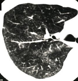
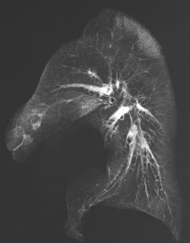
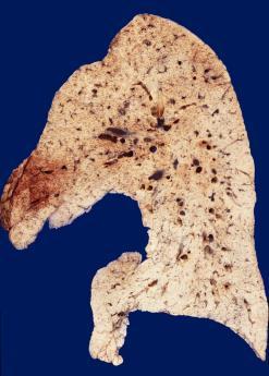
2 Centriacinar emphysema 1 Centriacinar emphysema 2 Centriacinar emphysema 3 ILS Paraseptal normal lung Interlobular septum Centrilobular normal lung Paraseptal normal lung with polygonal shape onchopneumonia 4 onchopneumonia (centrilobular nodule) 5 Pulmonary tuberculosis R 6 Cavity MB Small nodules Respiratory bronchiole Multiple nodules 1 mm 1

 7")


 11 (tree")

3 Pulmonary tuberculosis (centrilobular loose nodules) 7 Pulmonary tuberculosis 8 Centrilobular cavity Pulmonary tuberculosis (centrilobular compact nodules) 9 3 mm ILS onchiole 2 mm Pulmonary tuberculosis (centrilobular nodules) 10 Pulmonary tuberculosis (centrilobular nodules) 11 Pulmonary tuberculosis (tree in bud) mm 3 mm.... slice3 2


 15")
 16")
")

4 Pulmonary tuberculosis (tree in bud) 13 Silicosis (centrilobular nodules) Interlobular septum 14 Silicosis (centrilobular compact nodule) 15 5mm 3mm Silicosis (centrilobular small nodule) 16 Silicosis (paraseptal nodule) 17 Silicosis (paraseptal nodule) 18 Centrilobular nodule 1mm 5mm 1mm 3


")


5 Silicosis 19 Silicosis Calcified nodules 20 Silicosis (centrilobular nodule) 21 Left upper lobe Centrilobular Focal emphysema onchiole Paraseptal Pleura Interlobular septum 5mm Centrilobular nodule Interlobular septum 1mm Silicosis (centrilobular + paraseptal nodules) 22 Langerhans cell histiocytosis 23 Langerhans cell histiocytosis 24 Centrilobular nodules 5 mm Centrilobular cysts 4


 Courtesy")


 29")


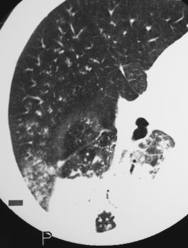
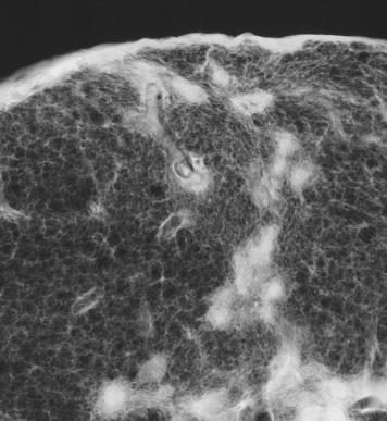
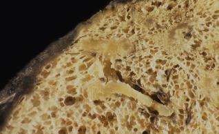
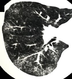
6 IPF/UIP Courtesy of Dr. T. Takemura, Japan 25 IPF/UIP mm f-nsip 27 RB ILS RB * MB Subpleural small opacities Perilobular, periacinar fibrosis (fibrosis of alveoli along pulmonary venules) Courtesy of Dr. T. Ogura, Japan mm Courtesy by Dr. T. Takemura, Japan SSc R Polygonal opacities 28 IPF/UIP (honeycomb lung) 29 Honeycomb lung (IPF/UIP) 30 ILS 2mm MB 10mm N Traction bronchiectasis Courtesy by Dr. T. Ogura, Japan 2 cm 2 cm 10mm Courtesy by Dr. T. Takemura, Japan 5
 31")


7 Honeycomb lung(ipf/uip) 31 Pulmonary fibrosis combined with emphysema Courtesy of Dr. T. Ogura, Japan 32 Pulmonary fibrosis combined with emphysema 33 Cystic change with destruction of respiratory bronchioles and alveoli A A MB * Perilobular fibrosis along and pleura V * V * * in emphysematous space A years later 160 Courtesy of Dr. T. Takemura Pulmonary edema (left heart failure) 34 Pulmonary edema 35 Pulmonary edema 36 Short septal lines in lung base 1cm thickened interlobular septum and pleura Short septal lines 3cm 6


")

8 Centilobular nodules Pulmonary ossification 37 Thickened long septa Normal long septa 38 Lymphangiosis carcinomatosa 39 ILS Coronal CT Thickened pleura Thickened bronchoarterial bundle ILS Thickened interlobular septum 3mm 3mm Left upper lobe Lymphangiosis carcinomatosa 40 Lymphangiosis carcinomatosa 41 Lymphangiosis carcinomatosa 42 Thickened bronchovascular bundles (central and periphery) Thickened bronchoarterial bundle Thickened interlobular septum 2mm 7


9 Lymphoproliferative disorder 43 Lymphoproliferative disorder 44 Pulmonary sarcoidosis 45 onchovascular bundle onchovascular bundle Thickening of centrilobular bronchovascular bundles Y.S Pulmonary sarcoidosis 46 Pulmonary sarcoidosis 47 Pulmonary sarcoidosis 48 2mm 8


10 Pulmonary sarcoidosis 49 Pulmonary sarcoidosis 50 Pulmonary thromboembolism 51 RB Pulmonary thromboembolism 52 Metastasis of hepatic cancer 53 Metastasis of hepatic cancer 54 Pulmonary artery Pulmonary arteries onchiole 2 5 mm Pulmonary vein 9


11 Metastasis of hepatic cancer 55 Cystic metastasis of bladder cancer 56 Metastasis of bladder cancer 57 Pulmonary artery: filled by tumor and thrombus onchiole Pulmonary artery: dilated with tumors 1 mm 1 Thickened blood vessels Courtesy of Dr. S. Imokawa 2 66 Perivascular tumor 1mm Courtesy of Dr. S. Imokawa Org. DAD 58 Org. DAD 59 Adenocarcinoma 60 Traction bronchiectsis Ground glass shadow Thickened back to back alveoli AD AD 1 mm Thickened back to back alveoli intervened by contrasted air space K.T 10


12 Adenocarcinoma Adenocarcinoma with lepidic growth 61 GGA Thickened back-to-back alveoli Normal back-to-back alveoli 11
HRCT in Diffuse Interstitial Lung Disease Steps in High Resolution CT Diagnosis. Where are the lymphatics? Anatomic distribution
 Steps in High Resolution CT Diagnosis Pattern of abnormality Distribution of disease Associated findings Clinical history Tomás Franquet MD What is the diagnosis? Hospital de Sant Pau. Barcelona Secondary
Usual Interstitial pneumonia and Nonspecific Interstitial Pneumonia. Nitra and the Gangs.
 Usual Interstitial pneumonia and Nonspecific Interstitial Pneumonia Nitra and the Gangs. บทน ำและบทท ๓, ๑๐, ๑๒, ๑๓, ๑๔, ๑๕, ๑๗ Usual Interstitial Pneumonia (UIP) Most common & basic pathologic pattern
Case 1 : Question. 1.1 What is the intralobular distribution? 1. Centrilobular 2. Perilymphatic 3. Random
 Interesting case Case 1 Case 1 : Question 1.1 What is the intralobular distribution? 1. Centrilobular 2. Perilymphatic 3. Random Case 1: Answer 1.1 What is the intralobular distribution? 1. Centrilobular
An Image Repository for Chest CT
 An Image Repository for Chest CT Francesco Frajoli for the Chest CT in Antibody Deficiency Group An Image Repository for Chest CT he Chest CT in Antibody Deficiency Group is an international and interdisciplinary
Acute and Chronic Lung Disease
 KATHOLIEKE UNIVERSITEIT LEUVEN Faculty of Medicine Acute and Chronic Lung Disease W De Wever, JA Verschakelen Department of Radiology, University Hospitals Leuven, Belgium Clinical utility of HRCT To detect
Case 1: Question. 1.1 What is the main pattern of this HRCT? 1. Intralobular line 2. Groundglass opacity 3. Perilymphatic nodule
 HRCT WORK SHOP Case 1 Case 1: Question 1.1 What is the main pattern of this HRCT? 1. Intralobular line 2. Groundglass opacity 3. Perilymphatic nodule Case 1: Question 1.2 What is the diagnosis? 1. Hypersensitivity
Manish Powari Regional Training Day 10/12/2014
 Manish Powari Regional Training Day 10/12/2014 Large number of different types of Interstitial Lung Disease (ILD). Most are very rare Most patients present with one of a smaller number of commoner diseases
Imaging of the Lung. István Battyány
 Imaging of the Lung István Battyány Anatomy of airways: (asszimetric dichotomy) trachea Main bronchus Lobar bronchus }lung, lobe 1. segmental bronchus subsegmental bronchus bronchus lobularis bronchiolus
Financial disclosure COMMON DIAGNOSES IN HRCT. High Res Chest HRCT. HRCT Pre test. I have no financial relationships to disclose. Anatomy Nomenclature
 Financial disclosure I have no financial relationships to disclose. Douglas Johnson D.O. Cardiothoracic Imaging Gaston Radiology COMMON DIAGNOSES IN HRCT High Res Chest Anatomy Nomenclature HRCT Sampling
Micronodular Lung Disease an algorithm
 Micronodular Lung Disease an algorithm H. Page McAdams, MD Department of Radiology Duke University Medical Center Durham, NC USA page.mcadams@duke.edu Question Which of the following lung diseases is MOST
Outline Definition of Terms: Lexicon. Traction Bronchiectasis
 HRCT OF IDIOPATHIC INTERSTITIAL PNEUMONIAS Disclosures Genentech, Inc. Speakers Bureau Tadashi Allen, MD University of Minnesota Assistant Professor Diagnostic Radiology 10/29/2016 Outline Definition of
Daria Manos RSNA 2016 RC 401. https://medicine.dal.ca/departments/depar tment-sites/radiology/contact/faculty/dariamanos.html
 Daria Manos RSNA 2016 RC 401 https://medicine.dal.ca/departments/depar tment-sites/radiology/contact/faculty/dariamanos.html STEP1: Is this fibrotic lung disease? STEP 2: Is this a UIP pattern? If yes:
Cystic Lung Disease. Cristopher A. Meyer, MD
 Cystic Lung Disease Cristopher A. Meyer, MD Air filled structure with definable wall typically less than 1 mm thick Cris A. Meyer, M.D. Professor of Radiology University of Wisconsin School of Medicine
Radiologic Approach to Smoking Related Interstitial Lung Disease
 Radiologic Approach to Smoking Related Interstitial Lung Disease Poster No.: C-1854 Congress: ECR 2013 Type: Educational Exhibit Authors: K.-N. Lee, J.-Y. Han, E.-J. Kang, J. Kang; Busan/KR Keywords: Toxicity,
Radiologists toolbox to differentiate alveolar versus interstitial lung diseases
 Radiologists toolbox to differentiate alveolar versus interstitial lung diseases Dr Sumer Shikhare, Dr Trishna Shimpi, Dr Ashish Chawla Khoo Teck Puat Hospital Singapore. Relevant financial disclosures
I don t need you. Disclosure Statement. Pathology Approach to ILD 11/5/2016. Kirk D. Jones, MD UCSF Dept of Pathology
 Pathology Approach to ILD Disclosure Statement Relevant financial relationships with a commercial interest: Boeringer Ingleheim, speaker Kirk D. Jones, MD UCSF Dept of Pathology kirk.jones@ucsf.edu I don
Systemic lupus erythematosus (SLE): Pleuropulmonary Manifestations
: Pleuropulmonary Manifestations") 08/30/10 09/26/10 Systemic lupus erythematosus (SLE): Pleuropulmonary Manifestations Camila Downey S. Universidad de Chile, School of Medicine, Year VII Harvard University, School of Medicine Sept 17,
08/30/10 09/26/10 Systemic lupus erythematosus (SLE): Pleuropulmonary Manifestations Camila Downey S. Universidad de Chile, School of Medicine, Year VII Harvard University, School of Medicine Sept 17,
Web Chapter 3. Image Gallery: Lesion detection on low dose chest CT
 Web Chapter 3 Image Gallery: Lesion detection on low dose chest CT Sarabjeet Singh, MD Mannudeep K. Kalra, MD *Eugene J. Mark, MD *James Stone, MD James H. Thrall, MD Department of Radiology and *Department
Low-dose CT Lung Cancer Screening Guidelines for Pulmonary Nodules Management Version 2
 Low-dose CT Lung Cancer Screening Guidelines for Pulmonary Nodules Management Version 2 The Committee for Management of CT-screening-detected Pulmonary Nodules 2009-2011 The Japanese Society of CT Screening
The radiological differential diagnosis of the UIP pattern
 5th International Conference on Idiopathic Pulmonary Fibrosis, Modena, 2015, June 12th The radiological differential diagnosis of the UIP pattern Simon Walsh King s College Hospital Foundation Trust London,
11/10/2014. Multi-disciplinary Approach to Diffuse Lung Disease: The Imager s Perspective. Radiology
 Multi-disciplinary Approach to Diffuse Lung Disease: The Imager s Perspective Radiology Pathology Clinical 1 Role of HRCT Diagnosis Fibrosis vs. inflammation Next step in management Response to treatment
Micronodular lung pattern - Differential diagnosis
 Micronodular lung pattern - Differential diagnosis Poster No.: P-0074 Congress: ESTI 2015 Type: Educational Poster Authors: P. Ninitas, F. Marinho, P. Campos, I. Távora ; Lisbon/PT, 1 2 2 3 1 1 3 Funchal/PT,
Mimics in chest disease: interstitial opacities
 Insights Imaging (2013) 4:9 27 DOI 10.1007/s13244-012-0207-7 PICTORIAL REVIEW Mimics in chest disease: interstitial opacities Anastasia Oikonomou & Panos Prassopoulos Received: 19 June 2012 / Revised:
ARTICLE IN PRESS. Ahuva Grubstein a, Daniele Bendayan b, Ithak Schactman c, Maya Cohen a, David Shitrit b, Mordechai R. Kramer b,
 Respiratory Medicine (2005) 99, 948 954 Concomitant upper-lobe bullous emphysema, lower-lobe interstitial fibrosis and pulmonary hypertension in heavy smokers: report of eight cases and review of the literature
Bronkhorst colloquium Interstitiële longziekten. Katrien Grünberg, klinisch patholoog
 Bronkhorst colloquium 2013-2014 Interstitiële longziekten De pathologie achter de CT Katrien Grünberg, klinisch patholoog K.grunberg@vumc.nl Preparing: introduction and 3 cases The introduction on microscopic
Interstitial Syndrome Ground glass attenuation miliary and nodular images Linear images
 Interstitial Syndrome Ground glass attenuation miliary and nodular images Linear images Dr Etienne Leroy-Terquem Centre hospitalier de Meulan les Mureaux. France French-cambodian association for pneumology
Role of Computed Tomography in Diagnosis of Diffuse Lung Diseases Chauhan Jayant 1*, Panchal Pankaj 2, Faruqui Tehzeeb 3
 ORIGINAL ARTICLE Role of Computed Tomography in Diagnosis of Diffuse Lung Diseases Chauhan Jayant 1*, Panchal Pankaj 2, Faruqui Tehzeeb 3 1 MD,DTCD,Additional Professor& HOD, 2,3 MBBS, 3 rd year resident
Workshop Cyst & Lucency. How to Approach
 Workshop Cyst & Lucency How to Approach To Approach Cystic Lung Disease True cysts? Cavitary disease Cystic bronchiectasis Mosaic attenuation Subpleural cysts Bullae Paraseptal emphysema Honeycombing Birt
I have no relevant conflicts of interest to disclose
 I have no relevant conflicts of interest to disclose Diffuse parenchymal lung disease (DPLD) and its associations Secondary lobular anatomy DPLD History, clinical findings, temporal evolution, and exposures
Typical and atypical findings of pulmonary sarcoidosis at high resolution CT
 Typical and atypical findings of pulmonary sarcoidosis at high resolution CT Poster No.: C-0169 Congress: ECR 2013 Type: Educational Exhibit Authors: L. Raposo Rodríguez, C. Mejía, B. Escobar Mallada,
American Thoracic Society European Respiratory Society Classification of the Idiopathic Interstitial Pneumonias: Advances in Knowledge since 20021
 This copy is for personal use only. To order printed copies, contact reprints@rsna.org American Thoracic Society European Respiratory Society Classification of the Idiopathic Interstitial Pneumonias: Advances
How to identify interstitial pneumonias.
 How to identify interstitial pneumonias. Poster No.: C-0804 Congress: ECR 2014 Type: Educational Exhibit Authors: S. claret loaiza, M. C. Cañete Moslero, R. Carreño Gonzalez, C. de la Torre; Malaga/ES
Spectrum of Cystic Lung Disease and its Mimics. Kathleen Jacobs MD and Elizabeth Weihe MD UC San Diego Medical Center, Department of Radiology
 Spectrum of Cystic Lung Disease and its Mimics Kathleen Jacobs MD and Elizabeth Weihe MD UC San Diego Medical Center, Department of Radiology No Financial Disclosures Learning Objectives 1. Review the
Role of High Resolution Computed Tomography in Evaluation of Pulmonary Diseases
 International J. of Healthcare & Biomedical Research, Volume:, Issue:, April, Pages 9-96 Role of High Resolution Computed Tomography in Evaluation of Pulmonary Diseases Dr. Abhijeet D. Nagapurkar*, Dr.
10/17/2016. Nuts and Bolts of Thoracic Radiology. Objectives. Techniques
 Nuts and Bolts of Thoracic Radiology October 20, 2016 Carleen Risaliti Objectives Understand the basics of chest radiograph Develop a system for interpreting chest radiographs Correctly identify thoracic
Smoking-related Interstitial Lung Diseases: High-Resolution CT Findings
 Smoking-related Interstitial Lung Diseases: High-Resolution CT Findings Poster No.: C-2358 Congress: ECR 2013 Type: Educational Exhibit Authors: V. Cuartero Revilla, M. Nogueras Carrasco, P. Olmedilla
Epidemiology and classification of smoking related interstitial lung diseases
 Epidemiology and classification of smoking related interstitial lung diseases Šterclová M. Department of Respiratory Diseases, Thomayer Hospital, Prague, Czech Republic Supported by an IGA Grant No G 1207
Is it really honeycombing? Limitations and pitfalls in radiological diagnosis of honeycombing.
 Is it really honeycombing? Limitations and pitfalls in radiological diagnosis of honeycombing. J. Arenas-Jiménez, E. García-Garrigós, M. Sirera-Matilla; M.C. Planells-Alduvin, F.I. Aranda* Hospital General
Histopathologic Approach to Interstitial Lung Disease
 Histopathologic Approach to Interstitial Lung Disease Kirk D. Jones, MD UCSF Dept of Pathology kirk.jones@ucsf.edu Disclosures I have nothing to disclose 1 Why? Much of interstitial lung disease biopsies
The Imaging Analysis of Pulmonary Sarcodiosis
 www.cancercellresearch.org ISSN: 2161-2609 Article The Imaging Analysis of Pulmonary Sarcodiosis Xin He, Chuanyu Zhang* Department of Radiology, Affiliated Hospital of Qingdao University, Qingdao, China
The gamut of cystic lung disease: a practical approach to differential diagnosis
 The gamut of cystic lung disease: a practical approach to differential diagnosis Award: Cum Laude Poster No.: P-0100 Congress: ESTI 2014 Type: Educational Poster Authors: C. Leal, R. Santos, J. P. A. Lopes,
Difficulties Diagnosing Idiopathic Pulmonary Fibrosis
 1. er Encuentro Entre Neumólogos y Radiólogos, Madrid, Spain, 2016, October 14th Difficulties Diagnosing Idiopathic Pulmonary Fibrosis Simon Walsh King s College Hospital Foundation Trust London, United
COPD in Radiology, with a Focus on Bronchiectasis and Emphysema
 November, 2002 COPD in Radiology, with a Focus on Bronchiectasis and Emphysema Evan Lyon, Harvard Medical School, Year IV Course Director Why is COPD important? Its common: 30 million Americans living
ARDS - a must know. Page 1 of 14
 ARDS - a must know Poster No.: C-1683 Congress: ECR 2016 Type: Authors: Keywords: DOI: Educational Exhibit M. Cristian; Turda/RO Education and training, Edema, Acute, Localisation, Education, Digital radiography,
Resident Case Review CHEST. Daria Manos CAR 2016
 Resident Case Review CHEST CAR 2016 Daria Manos Disclosure Speakers bureau, Roche CAR 2016 Daria Manos 1. Recognize common and critical chest radiograph and computed tomography signs and use these clues
PULMONARY TUBERCULOSIS RADIOLOGY
 PULMONARY TUBERCULOSIS RADIOLOGY RADIOLOGICAL MODALITIES Medical radiophotography Radiography Fluoroscopy Linear (conventional) tomography Computed tomography Pulmonary angiography, bronchography Ultrasonography,
T he diagnostic evaluation of a patient with
 546 REVIEW SERIES Challenges in pulmonary fibrosis? 1: Use of high resolution CT scanning of the lung for the evaluation of patients with idiopathic interstitial pneumonias Michael B Gotway, Michelle M
Vascular Lung Diseases
 Vascular Lung Diseases SESSION SPECIFIC OBJECTIVES List the major types of vascular lung disease Recognize and describe the pathology of vascular lung disease: Pulmonary embolism, thrombosis, hypertension,
Pediatric High-Resolution Chest CT
 Pediatric High-Resolution Chest CT Alan S. Brody, MD Professor of Radiology and Pediatrics Chief, Thoracic Imaging Cincinnati Children s s Hospital Cincinnati, Ohio, USA Pediatric High-Resolution CT Short
The crazy-paving pattern: A radiological-pathological correlated and illustrated overview
 The crazy-paving pattern: A radiological-pathological correlated and illustrated overview Poster No.: C-0827 Congress: ECR 2010 Type: Educational Exhibit Topic: Chest Authors: W. F. M. De Wever, J. Coolen,
Glossary of terms in thoracic imaging and clinicalradiological correlation. What every radiologist should
 Glossary of terms in thoracic imaging and clinicalradiological correlation. What every radiologist should know. Poster No.: C-1565 Congress: ECR 2012 Type: Educational Exhibit Authors: A. Moreno Pastor,
Radiologic findings of drug-induced lung disease
 Radiologic findings of drug-induced lung disease Poster No.: P-0115 Congress: ESTI 2015 Type: Educational Poster Authors: A. I. C. Santos, A. F. Roque, R. Mamede, L. Oliveira, T. Saldanha; Lisbon/PT Keywords:
Emphysema association in a prospective series with patients suffering from Idiopathic Pulmonary Fibrosis.
 Emphysema association in a prospective series with patients suffering from Idiopathic Pulmonary Fibrosis. David Jiménez-Restrepo, Mª Luisa Domingo Montañana, Claudia Fernandez Ruiz, Andrea Martínez Deltoro*,
Thoracic CT pattern in lung cancer: correlation of CT and pathologic diagnosis
 19 th Congress of APSR PG of Lung Cancer (ESAP): Update of Lung Cancer Thoracic CT pattern in lung cancer: correlation of CT and pathologic diagnosis Kazuma Kishi, M.D. Department of Respiratory Medicine,
Liebow and Carrington's original classification of IIP
 Liebow and Carrington's original classification of IIP-- 1969 Eric J. Stern MD University of Washington UIP Usual interstitial pneumonia DIP Desquamative interstitial pneumonia BIP Bronchiolitis obliterans
How to Analyse Difficult Chest CT
 How to Analyse Difficult Chest CT Complex diseases are:- - Large lesion - Unusual or atypical pattern - Multiple discordant findings Diffuse diseases are:- - Numerous findings in both sides 3 basic steps
Bronchiectasis: An Imaging Approach
 Bronchiectasis: An Imaging Approach Travis S Henry, MD Associate Professor of Clinical Radiology Cardiac and Pulmonary Imaging Section University of California, San Francisco Large Middle Small 1 Bronchiectasis
Cystic Lung Disease: a Comparison of Cystic Size, as Seen on Expiratory and Inspiratory HRCT Scans
 Cystic Lung Disease: a Comparison of Cystic Size, as Seen on Expiratory and Inspiratory HRCT Scans Ki-Nam Lee, MD 1 Seong-Kuk Yoon, MD 1 Seok Jin Choi, MD 2 Jin Mo Goo, MD 3 Kyung-Jin Nam, MD 1 Index words:
Interesting Cases. Pulmonary
 Interesting Cases Pulmonary 54M with prior history of COPD, hep B/C, and possible history of TB presented with acute on chronic dyspnea, and productive cough Hazy opacity overlying the left hemithorax
4/17/2010 C ini n ca c l a Ev E a v l a ua u t a ion o n of o ILD U dat a e t e i n I LDs
 Update in ILDs Diagnosis 101: Clinical Evaluation April 17, 2010 Jay H. Ryu, MD Mayo Clinic, Rochester MN Clinical Evaluation of ILD Outline General aspects of ILDs Classification of ILDs Clinical evaluation
Diagnosis of TB: Radiology David Finlay, MD
 TB Intensive Tyler, Texas June 2-4, 2010 Diagnosis of TB: Radiology David Finlay, MD June 3, 2010 2stages stages- Tuberculosis 1. primary infection 2. reactivation, or post primary disease 2 1 Primary
Progress in Idiopathic Pulmonary Fibrosis
 Progress in Idiopathic Pulmonary Fibrosis David A. Lynch, MB Disclosures Progress in Idiopathic Pulmonary Fibrosis David A Lynch, MB Consultant: t Research support: Perceptive Imaging Boehringer Ingelheim
Radiological Imaging of Drug-Induced Pulmonary Lesions
 Review Article imedpub Journals www.imedpub.com Journal of Clinical Radiology and Case Reports Radiological Imaging of Drug-Induced Pulmonary Lesions D souza M *, Rajiah P, Khan A and Irion K Department
Pulmonary Alveolar Microlithiasis: CT and pathologic findings in 10 patients
 Monaldi Arch Chest Dis 2005; 63: 1, 59-64 CASE REPORT Pulmonary Alveolar Microlithiasis: CT and pathologic findings in 10 patients H. Sumikawa 1,T. Johkoh 1, 2, N. Tomiyama 1, S. Hamada 1, M. Koyama 1,
Interstitial syndrome
 Interstitial syndrome Ground-glass attenuation Miliary and nodular images linear images Etienne Leroy Terquem Pierre L Her SPI / ISP Soutien Pneumologique International / International Support for Pulmonology
Lung CT: Part 2, The Interstitial Pneumonias Clinical, Histologic, and CT Manifestations
 Integrative Imaging Review Ferguson and Berkowitz CT of Interstitial Pneumonia Integrative Imaging Review CME SAM Lung CT FOCUS ON: Emma C. Ferguson 1 Eugene A. Berkowitz 2 Ferguson EC, Berkowitz EA Keywords:
NONE OVERVIEW FINANCIAL DISCLOSURES UPDATE ON IDIOPATHIC PULMONARY FIBROSIS/IPF (UIP) FOR PATHOLOGISTS. IPF = Idiopathic UIP Radiologic UIP Path UIP
 FOR PATHOLOGISTS. IPF = Idiopathic UIP Radiologic UIP Path UIP") UPDATE ON IDIOPATHIC PULMONARY FIBROSIS/IPF () FOR PATHOLOGISTS Thomas V. Colby, M.D. Professor of Pathology (Emeritus) Mayo Clinic Arizona FINANCIAL DISCLOSURES NONE OVERVIEW IPF Radiologic Dx Pathologic
UPDATE ON IDIOPATHIC PULMONARY FIBROSIS/IPF () FOR PATHOLOGISTS Thomas V. Colby, M.D. Professor of Pathology (Emeritus) Mayo Clinic Arizona FINANCIAL DISCLOSURES NONE OVERVIEW IPF Radiologic Dx Pathologic
Differential diagnosis
 Differential diagnosis Idiopathic pulmonary fibrosis (IPF) is part of a large family of idiopathic interstitial pneumonias (IIP), one of four subgroups of interstitial lung disease (ILD). Differential
Pulmonary Sarcoidosis - Radiological Evaluation
 Original Research Article Pulmonary Sarcoidosis - Radiological Evaluation Jayesh Shah 1, Darshan Shah 2*, C. Raychaudhuri 3 1 Associate Professor, 2 1 st Year Resident, 3 Professor and HOD Radiology Department,
Chest imaging III: Nodular pulmonary disease. Ádám Domonkos Tárnoki, MD, PhD Assistant professor Department of Radiology, Semmelweis University 1
 Chest imaging III: Nodular pulmonary disease Ádám Domonkos Tárnoki, MD, PhD Assistant professor Department of Radiology, Semmelweis University 1 Pattern 2 Nodular pattern Several round opacity, typically
Diagnostic Imaging of Diffuse Infiltrative Disease of the Lung
 Thematic Review Series Respiration 2004;71:4 19 DOI: 10.1159/000075642 Diagnostic Imaging of Diffuse Infiltrative Disease of the Lung Maurizio Zompatori a Claudio Bnà a Venerino Poletti c Enrica Spaggiari
Thinking Through Pathology
 Thinking Through Pathology Pathology Radiology Alessandra Cancellieri Alberto Cavazza Giorgia Dalpiaz Introduction and anatomy Secondary lobule Page 28 Elementary lesions Defi ning lesions: neoplasm Defi
Pulmonary Manifestations of Systemic Lupus Erythematosus 1
 Pulmonary Manifestations of Systemic Lupus Erythematosus 1 Kee Hyuk Yang, M.D., Yo Won Choi, M.D., Seok Chol Jeon, M.D., Choong Ki Park, M.D., Kyung in Joo, M.D., Chang Kok Hahm, M.D., Seung Ro Lee, M.D.
Cryptogenic Organizing Pneumonia Diagnosis Approach Based on a Clinical-Radiologic-Pathologic Consensus
 Cryptogenic Organizing Pneumonia Diagnosis Approach Based on a Clinical-Radiologic-Pathologic Consensus Poster No.: C-1622 Congress: ECR 2012 Type: Scientific Exhibit Authors: C. Cordero Lares, E. Zorita
Idiopathic interstitial pneumonias (IIPs) are a group of
 are a group of") SYMPOSIA C. Isabela S. Silva, MD, PhD and Nestor L. Müller, MD, PhD Abstract: The idiopathic interstitial pneumonias (IIPs) are a group of diffuse parenchymal lung diseases of unknown etiology characterized
SYMPOSIA C. Isabela S. Silva, MD, PhD and Nestor L. Müller, MD, PhD Abstract: The idiopathic interstitial pneumonias (IIPs) are a group of diffuse parenchymal lung diseases of unknown etiology characterized
Pulmonary CT Findings of Visceral Larva Migrans due to Ascaris suum
 Pulmonary CT Findings of Visceral Larva Migrans due to Ascaris suum Poster No.: E-0038 Congress: ESTI 2012 Type: Scientific Exhibit Authors: K. Honda, F. Okada, Y. Ando, A. Ono, S. Matsumoto, H. Mori;
Key words: CT scanners; interstitial lung diseases; polymyositis-dermatomyositis; x-ray
 Nonspecific Interstitial Pneumonia Associated With Polymyositis and Dermatomyositis* Serial High-Resolution CT Findings and Functional Correlation Hiroaki Arakawa, MD; Hidehiro Yamada, MD; Yasuyuki Kurihara,
Computerized Medical Imaging and Graphics
 Computerized Medical Imaging and Graphics 36 (2012) 72 84 Contents lists available at ScienceDirect Computerized Medical Imaging and Graphics journa l h o me pa g e: www.elsevier.com/locate/compmedimag
Interstitial Lung Disease in Infants and Children
 Interstitial Lung Disease in Infants and Children David A. Mong, MD SUNDAY Andrew Mong MD Beyond the interstitium (path includes airways/airspace) Radiographic diffuse disease Adult Interstitial Lung Disease
DISEASES OF THE RESPIRATORY SYSTEM 2018 DR HEYAM AWAD LECTURE 2: ATELECTASIS AND EMPHYSEMA
 DISEASES OF THE RESPIRATORY SYSTEM 2018 DR HEYAM AWAD LECTURE 2: ATELECTASIS AND EMPHYSEMA INTRODUCTION In this lecture we will discuss atelectasis which is a complication of several medical and surgical
CT findings in multifocal or diffuse non-mucinous bronchioloalveolar carcinoma (BAC)
") CT findings in multifocal or diffuse non-mucinous bronchioloalveolar carcinoma (BAC) Poster No.: C-2192 Congress: ECR 2014 Type: Educational Exhibit Authors: I. Sandu, A. R. Popita, I.-A. Brumboiu; Cluj-Napoca/RO
CT findings in multifocal or diffuse non-mucinous bronchioloalveolar carcinoma (BAC) Poster No.: C-2192 Congress: ECR 2014 Type: Educational Exhibit Authors: I. Sandu, A. R. Popita, I.-A. Brumboiu; Cluj-Napoca/RO
5/9/2015. Multi-disciplinary Approach to Diffuse Lung Disease: The Imager s Perspective. No, I am not a pulmonologist! Radiology
 Multi-disciplinary Approach to Diffuse Lung Disease: The Imager s Perspective No, I am not a pulmonologist! Radiology Pathology Clinical 1 Everyone needs a CT Confidence in diagnosis Definitive HRCT +
Thoracic Sarcoidosis Imaging Updated: Jul 19, 2013
 Thoracic Sarcoidosis Imaging Updated: Jul 19, 2013 Overview Radiography Computed Tomography Magnetic Resonance Imaging Nuclear Imaging Show All Multimedia Library References Overview For patients with
INTERSTITIAL LUNG DISEASE. Radhika Reddy MD Pulmonary/Critical Care Long Beach VA Medical Center January 5, 2018
 INTERSTITIAL LUNG DISEASE Radhika Reddy MD Pulmonary/Critical Care Long Beach VA Medical Center January 5, 2018 Interstitial Lung Disease Interstitial Lung Disease Prevalence by Diagnosis: Idiopathic Interstitial
September 2014 Imaging Case of the Month. Michael B. Gotway, MD. Department of Radiology Mayo Clinic Arizona Scottsdale, AZ
 September 2014 Imaging Case of the Month Michael B. Gotway, MD Department of Radiology Mayo Clinic Arizona Scottsdale, AZ Clinical History: A 57-year-old non-smoking woman presented to her physician as
Criteria for confident HRCT diagnosis of usual interstitial pneumonia (UIP)
") Criteria for confident HRCT diagnosis of usual interstitial pneumonia (UIP) Assem El Essawy (1) & Amr A. Nassef (٢) Abstract Identification of interstitial pneumonia (IP) was mainly based on histological
Criteria for confident HRCT diagnosis of usual interstitial pneumonia (UIP) Assem El Essawy (1) & Amr A. Nassef (٢) Abstract Identification of interstitial pneumonia (IP) was mainly based on histological
Eun-Young Kang, M.D., Jae Wook Lee, M.D., Ji Yung Choo, M.D., Hwan Seok Yong, M.D., Ki Yeol Lee, M.D., Yu-Whan Oh, M.D.
 Eun-Young Kang, M.D., Jae Wook Lee, M.D., Ji Yung Choo, M.D., Hwan Seok Yong, M.D., Ki Yeol Lee, M.D., Yu-Whan Oh, M.D. Department of Radiology, Korea University Guro Hospital, College of Medicine, Korea
Thoracic lung involvement in rheumatoid arthritis: Findings on HRCT
 Thoracic lung involvement in rheumatoid arthritis: Findings on HRCT Poster No.: C-2488 Congress: ECR 2015 Type: Educational Exhibit Authors: R. E. Correa Soto, M. J. Martín Sánchez, J. M. Fernandez 1 1
CT findings of high-attenuation pulmonary abnormalities
 Insights Imaging (2010) 1:287 292 DOI 10.1007/s13244-010-0039-2 PICTORIAL REVIEW CT findings of high-attenuation pulmonary abnormalities Naim Ceylan & Selen Bayraktaroglu & Recep Savaş & Hudaver Alper
Noninfectious Inflammatory Lung Disease: Imaging Considerations and Clues to Differential Diagnosis
 Cardiopulmonary Imaging Review Nemec et al. Noninfectious Inflammatory Lung Disease Cardiopulmonary Imaging Review Stefan Franz Nemec 1 Ronald L. Eisenberg Alexander A. Bankier Nemec SF, Eisenberg RL,
Lung Allograft Dysfunction
 Lung Allograft Dysfunction Carlos S. Restrepo M.D. Ameya Baxi M.D. Department of Radiology University of Texas Health San Antonio Disclaimer: We do not have any conflict of interest or financial gain to
OFF THE BEATEN TRACK: A Pictorial Review of Atypical Features of Pulmonary Metastases
 OFF THE BEATEN TRACK: A Pictorial Review of Atypical Features of Pulmonary Metastases Megan Hora, MD Chi Wan Koo, MD Christian Cox, MD Department of Diagnostic Radiology Thoracic Imaging Section Mayo Clinic
Multiple Parenchymal Abnormalities on Chest CT scan: Don't Panic, Let's Be Pragmatic: The Eight Questions Rule
 Multiple Parenchymal Abnormalities on Chest CT scan: Don't Panic, Let's Be Pragmatic: The Eight Questions Rule Poster No.: C-1064 Congress: ECR 2013 Type: Educational Exhibit Authors: M. Baque-Juston,
Department of Radiology, Korea University Anam Hospital, Korea University College of Medicine, Seoul, Korea 2
 Case Report pissn 1738-2637 / eissn 2288-2928 J Korean Soc Radiol 2018;79(5):276-281 https://doi.org/10.3348/jksr.2018.79.5.276 Sequential CT Findings in Two Cases of Immunoglobulin G4-Related Lung Disease:
Small airway disease: semiological and radiological evaluation. A pictorial review.
 Small airway disease: semiological and radiological evaluation. A pictorial review. Award: Magna Cum Laude Poster No.: C-3028 Congress: ECR 2018 Type: Educational Exhibit Authors: K. N. Nieto, A. Cerpa,
Anomalous pulmonary air spaces. Management radiological
 Anomalous pulmonary air spaces. Management radiological Poster No.: C-2608 Congress: ECR 2015 Type: Educational Exhibit Authors: J. C. Quintero Rivera, J. E. Millán Suárez, R. A. Corral Rivadulla, M. F.