Spectrum of Cystic Lung Disease and its Mimics. Kathleen Jacobs MD and Elizabeth Weihe MD UC San Diego Medical Center, Department of Radiology
|
|
|
- Neil Blair
- 6 years ago
- Views:
Transcription


1 Spectrum of Cystic Lung Disease and its Mimics Kathleen Jacobs MD and Elizabeth Weihe MD UC San Diego Medical Center, Department of Radiology
2 No Financial Disclosures
3 Learning Objectives 1. Review the pathologic and radiologic definition of a lung cyst 2. Illustrate classic and distinguishing HRCT features of cystic lung disease 3. Review common mimics of cystic lung disease 4. Discuss the underlying pathology of each entity
. Hansell et al. Radiology 2008; 246: 697-722.")
4 Definition of a Lung Cyst Pathology: Any round, circumscribed space surrounded by an epithelial or fibrous wall of variable thickness Radiography or CT: Round parenchymal lucency or low attenuating area with a well-defined interface with normal lung. Cysts are usually thin-walled but can have variable wall thickness (<2 mm). Hansell et al. Radiology 2008; 246:
5 Diffuse Cystic Lung Disease Syndromic Neurofibromatosis Tuberous Sclerosis (TS)/ Lymphangioleiomyomatosis (LAM) Birt Hogg Dube Smoking-Related Pulmonary Langerhans Histiocytosis Desquamative Interstitial Pneumonia Immune-Mediated Chronic Hypersensitivity Pneumonitis Lymphocytic Interstitial Pneumonia Post-Infectious Pneumocystis Pneumonia
6 Neurofibromatosis Related Diffuse Lung Disease (NF-DLD) Specific to Neurofibromatosis type-1 Ground glass (50%) Basilar reticulation/fibrosis (37%) Cysts (25%); upper lobe predominant Emphysema (25%) Other thoracic findings: cutaneous/subcutaneous neurofibromas, ribbon deformity of ribs, focal thoracic scoliosis, posterior vertebral scalloping NF-1 has increased sensitivity to cigarette smoke with early development of emphysema-like changes which may be a risk factor for NF-ILD Pathology: Lymphoplasmocytic inflammation and fibrosis in the alveolar septa, similar to NSIP Zamora et al. European Respiratory Journal 2007; 29 (1):

7 Cutaneous neurofibromas Small cysts Subpleural, basilar reticulation
8 Sporadic Lymphangioleiomyomatosis (S-LAM) Almost exclusively in women of childbearing years Diffuse thin wall ovoid cysts surrounded by normal lung Cysts measure 2-5 mm (up to mm) Uniform cyst distribution Interlobular septal thickening (interstitial edema from lymphatic obstruction) Associated with chylous effusion, recurrent pneumothorax, lymphadenopathy Pathology: Smooth muscle proliferation in the pulmonary interstitium (affects vessels, airways, lymphatics, alveolar septa, pleura) Hohman et al. European Journal of Internal Medicine 2008;18: Pallisa et al. Radiographics 2002; 22: S


9 Note: --Uniform distribution of cysts --Predominantly small cysts Bilateral apical pneumothoraces
10 Tuberous Sclerosis Complex (TSC) Associated LAM TSC is autosomal dominant, characterized by hamartomas, seizures, mental retardation 26% of women with TSC have LAM Usually less severe than S-LAM Thin wall cystic lesions 2-5 mm in size (up 25-30mm) Associated with Cortical tubers/subependymal nodules Renal angiomyolipmas Facial angiofibroma Pathology: Same as S-LAM Avila et al. Radiology 2007; 242: Costello et al. Mayo Clinic Proceedings 2000; 75:


11 Note: --Same morphology as S-LAM, but less severe --Normal intervening lung parenchyma

12 Birt-Hogg-Dube Syndrome Rare, autosomal dominant syndromes characterized by skin hamartomas, renal tumors (chromophobe RCC), pulmonary cysts (80-90%) Thin walled cysts Vary widely in size Enlongated/oval, multiseptated Subpleural Basal predominant Associated bullous emphysema Increased risk of PTX Pathology: Folliculin gene mutation leads to alveolar de-arrangement Agarwal et al. AJR 2011; 196: Tobino et al. European Journal of Radiology 2011;77:
 --Many cysts are subpleural --Basal")
13 Note: --Variable-sized cysts, some with septated appearance (arrow) --Many cysts are subpleural --Basal predominance
14 Pulmonary Langerhans Histiocystosis +Smoking history (PLCH) Symmetric, upper lobe predominant cysts Irregular small cysts (<10mm), some with thicker walls Intervening parenchyma with nodules, architectural distortion, reticulation Temporal heterogeneity Pathology: Reactive polyclonal process induced by antigens in cigarette smoke peribronchiolar infiltration of specific histiocytes called Langerhans cells (Birbeck granules) stellate interstitial nodules cavitation thick/thin wall bizarre cysts Treatment: Smoking cessation and steroids Atilli et al. RadioGraphics 2008; 28: Vassallo et al. NEJM 2002;346:

 --Upper")
15 Note: --Variable cyst morphology, but majority small cysts --Some cysts with thicker walls (arrow) --Upper lobe predominant

16 Nodules in addition to cysts Upper lobe predominant
 Basilar and peripheral distribution Pathology: Pigmented macrophages fill the alveolar spaces Treatment: Worse prognosis than RB-ILD; ~66% will stabilize or improve with steroid")
17 Desquamative Interstitial Pneumonitis (DIP) Strong association with smoking Small cysts (< 2cm) represent focal bronchiolectasis and dilated alveolar ducts Associated with patchy ground glass opacities (80%) Basilar and peripheral distribution Pathology: Pigmented macrophages fill the alveolar spaces Treatment: Worse prognosis than RB-ILD; ~66% will stabilize or improve with steroid therapy Akira et al. Thorax 1997; 52: Atilli et al. RadioGraphics 2008; 28: Seaman et al. AJR 2011; 196:


18 Note: --Background of ground glass opacities --Basilar predominance
19 Chronic Hypersensitivity Pneumonitis More common in nonsmokers Thin-walled cysts, few in number: ~10% in subacute stage and ~40% in chronic stage More common to see ground glass opacities, centrilobular nodules, air trapping (head-cheese sign) Mid/upper lobe predominant fibrosis (no or minimal honeycombing as opposed to UIP) Type III/Type IV hypersensitivity response Pathologic hallmark: Chronic interstitial infiltrates Lymphocytes, plasma cells, occasional multinucleated giant cell and histiocytes Treatment: Exposure removal and steroids Franquet et al. J Comput Assist Tomogr 2003; 27: Silva et al. Radiology 2008; 246:


20 Scattered, small cysts Air trapping Note: --Background of subpleural reticulation/fibrosis
21 Lymphocytic Interstitial Pneumonia Associated with Sjogrens, AIDS, Lupus, DIP Cystic airspaces in 68%, variable size 1-30mm, thin-walled Lower lobe predominant Fewer cysts than LAM/PLCH Associated with LAD, bronchovascular thickening, poorly defined centrilobular and subpleural nodules Absence of pleural effusion Pathology: Diffuse interstitial proliferation with polyclonal small lymphocytes/plasma cells Johkoh et al. Radiology 1999; 212:
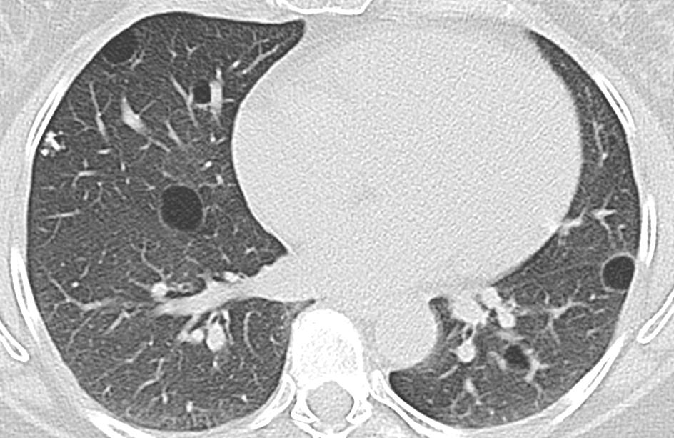
22 Note: --Scattered, lower lobe cysts
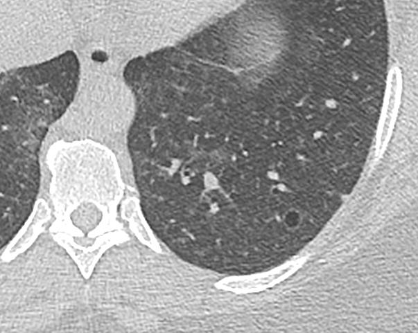
23 Pneumocystis Pneumonia Associated with T-call immunodeficiency Ground glass opacities characteristic (+/- crazy paving) Cysts/pneumatoceles in 10-34% Small, thin walled Can coalesce into bizarre shapes LAD (< 10%) Pleural effusions rare Spares the periphery Pathology: Peri-bronchiolar inflammation results in obstruction with cyst formation via a ball-valve effect Treatment: Steroid and antibiotics (Bactrim) Improve or resolve within 5 months of treatment Boddu et al. Pathology Research International 2017; Kanne at el. AJR 2012;198:W Lee et al. J of Computer Assisted Tomography 2002;26:5-12.


24 Note: --Upper lobe cysts and ground glass opacities --Relative subpleural sparing
 Quigley at el.")
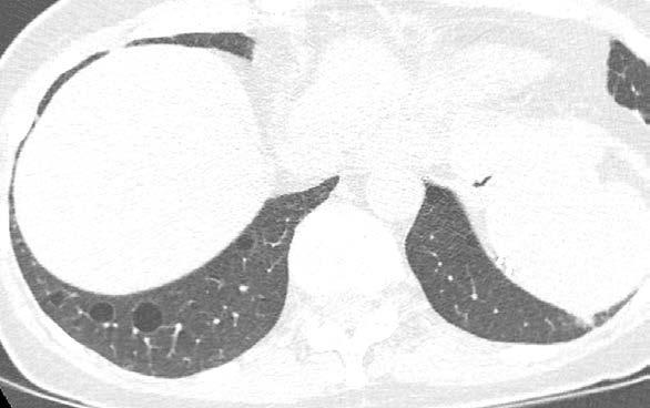
25 Focal Cystic Disease: Pneumatocele Post-infectious pneumatocele associated with Staph Aureus (most common) and Strep Pneumo Usually transient but can last for years Pathology: Combination of parenchymal necrosis and ball-valve mechanism (airway obstruction by inflammatory exudates) Quigley at el. AJR 1988;150:


26 Bronchiectasis Large pneumatocele Extensive basilar consolidation with areas of cavitation
27 Focal Cystic Disease: Congenital Pulmonary Airway Malformation Mixed solid/cystic mass Type 1 large cysts Type 2 -numerous small cysts ( cm) Type 3 microcysts Type 4 large cysts with mass effect No systemic vascular supply (as opposed to pulmonary sequestration) Distinguish from other congenital focal cystic disease: congenital lobar emphysema (involved lung is expanded) and pulmonary sequestration. Pathology: Bronchiolar proliferation and columnar/epithelium lined micro- and macrocysts Boddu et al. Pathology Research International 2017; Daltro et al. AJR 2004;183:


28 Increased lucency of the right lower lobe with multiple small cysts
29 Mimics of Cystic Lung Disease Emphysema/Bullae Cavitary Nodule/Consolidation Honeycombing Bronchiectasis


30 Cyst versus Bullae/Cavity Bullae (>1cm) and Blebs (<1 cm): cystic air spaces contiguous with the pleura and often associated with bullous emphysema Cavity: a gas-filled space, seen as a lucency or low attenuation area, within pulmonary consolidation, mass, or nodule. Usually thicker walled (>4mm) Tracheobronchopapillomatosis with cavitating nodules (arrow)

31 Cavitary/Cystic Metastatic Disease Cavitary mets rare <5% Usually epithelial origin (squamous) Peripheral Feeding vessel sign Mesenchymal (sarcomas) less frequent Can produce thin-walled cysts difficult to distinguish from LAM Stain for HMB45 and CD34 in mets Lee et al. J of Computer Assisted Tomography 2002;26:5-12. Seaman et al. AJR 2011; 196:


32 Emphysema - imperceptible wall - polygonal shaped lucency Honeycombing - stacked lucencies - architectural distortion


33 e.g: Cystic Fibrosis -Lucencies contiguous with airways (arrows) -adjacent pulmonary artery Bronchiectasis
34 Conclusion Differential diagnosis for cystic lung disease can be narrowed by considering the following: Cyst or Mimic? Distribution (diffuse vs focal, lobar predominance) Size/shape of cysts Patient sex/age/history (young female?, smoker?) Secondary imaging findings (ground glass opacities?, skin nodules?)
35 Author Contact Kathleen Jacobs:
Cystic Lung Disease. Cristopher A. Meyer, MD
 Cystic Lung Disease Cristopher A. Meyer, MD Air filled structure with definable wall typically less than 1 mm thick Cris A. Meyer, M.D. Professor of Radiology University of Wisconsin School of Medicine
Workshop Cyst & Lucency. How to Approach
 Workshop Cyst & Lucency How to Approach To Approach Cystic Lung Disease True cysts? Cavitary disease Cystic bronchiectasis Mosaic attenuation Subpleural cysts Bullae Paraseptal emphysema Honeycombing Birt
HRCT in Diffuse Interstitial Lung Disease Steps in High Resolution CT Diagnosis. Where are the lymphatics? Anatomic distribution
 Steps in High Resolution CT Diagnosis Pattern of abnormality Distribution of disease Associated findings Clinical history Tomás Franquet MD What is the diagnosis? Hospital de Sant Pau. Barcelona Secondary
Financial disclosure COMMON DIAGNOSES IN HRCT. High Res Chest HRCT. HRCT Pre test. I have no financial relationships to disclose. Anatomy Nomenclature
 Financial disclosure I have no financial relationships to disclose. Douglas Johnson D.O. Cardiothoracic Imaging Gaston Radiology COMMON DIAGNOSES IN HRCT High Res Chest Anatomy Nomenclature HRCT Sampling
Radiologic Approach to Smoking Related Interstitial Lung Disease
 Radiologic Approach to Smoking Related Interstitial Lung Disease Poster No.: C-1854 Congress: ECR 2013 Type: Educational Exhibit Authors: K.-N. Lee, J.-Y. Han, E.-J. Kang, J. Kang; Busan/KR Keywords: Toxicity,
Usual Interstitial pneumonia and Nonspecific Interstitial Pneumonia. Nitra and the Gangs.
 Usual Interstitial pneumonia and Nonspecific Interstitial Pneumonia Nitra and the Gangs. บทน ำและบทท ๓, ๑๐, ๑๒, ๑๓, ๑๔, ๑๕, ๑๗ Usual Interstitial Pneumonia (UIP) Most common & basic pathologic pattern
Acute and Chronic Lung Disease
 KATHOLIEKE UNIVERSITEIT LEUVEN Faculty of Medicine Acute and Chronic Lung Disease W De Wever, JA Verschakelen Department of Radiology, University Hospitals Leuven, Belgium Clinical utility of HRCT To detect
Liebow and Carrington's original classification of IIP
 Liebow and Carrington's original classification of IIP-- 1969 Eric J. Stern MD University of Washington UIP Usual interstitial pneumonia DIP Desquamative interstitial pneumonia BIP Bronchiolitis obliterans
11/10/2014. Multi-disciplinary Approach to Diffuse Lung Disease: The Imager s Perspective. Radiology
 Multi-disciplinary Approach to Diffuse Lung Disease: The Imager s Perspective Radiology Pathology Clinical 1 Role of HRCT Diagnosis Fibrosis vs. inflammation Next step in management Response to treatment
Daria Manos RSNA 2016 RC 401. https://medicine.dal.ca/departments/depar tment-sites/radiology/contact/faculty/dariamanos.html
 Daria Manos RSNA 2016 RC 401 https://medicine.dal.ca/departments/depar tment-sites/radiology/contact/faculty/dariamanos.html STEP1: Is this fibrotic lung disease? STEP 2: Is this a UIP pattern? If yes:
Cystic lung diseases 7/21/2017. Cystic Lung Diseases. CT definition of a lung cyst. Important clinical clues
 Cystic Lung Diseases Cystic lung diseases Aurelie Fabre Increased Awareness Spontaneous Pneumothorax High resolution imaging (HRCT) Multidisciplinary approach like interstitial lung disease CT definition
Diffuse Cystic Lung Disease at High-Resolution CT
 Cardiopulmonary Imaging Pictorial Essay Seaman et al. HRCT of Diffuse Cystic Lung Disease Cardiopulmonary Imaging Pictorial Essay Downloaded from www.ajronline.org by 37.44.193.85 on 01/05/18 from IP address
Outline Definition of Terms: Lexicon. Traction Bronchiectasis
 HRCT OF IDIOPATHIC INTERSTITIAL PNEUMONIAS Disclosures Genentech, Inc. Speakers Bureau Tadashi Allen, MD University of Minnesota Assistant Professor Diagnostic Radiology 10/29/2016 Outline Definition of
PULMONARY TUBERCULOSIS RADIOLOGY
 PULMONARY TUBERCULOSIS RADIOLOGY RADIOLOGICAL MODALITIES Medical radiophotography Radiography Fluoroscopy Linear (conventional) tomography Computed tomography Pulmonary angiography, bronchography Ultrasonography,
An Image Repository for Chest CT
 An Image Repository for Chest CT Francesco Frajoli for the Chest CT in Antibody Deficiency Group An Image Repository for Chest CT he Chest CT in Antibody Deficiency Group is an international and interdisciplinary
Smoking-related Interstitial Lung Diseases: High-Resolution CT Findings
 Smoking-related Interstitial Lung Diseases: High-Resolution CT Findings Poster No.: C-2358 Congress: ECR 2013 Type: Educational Exhibit Authors: V. Cuartero Revilla, M. Nogueras Carrasco, P. Olmedilla
Manish Powari Regional Training Day 10/12/2014
 Manish Powari Regional Training Day 10/12/2014 Large number of different types of Interstitial Lung Disease (ILD). Most are very rare Most patients present with one of a smaller number of commoner diseases
Systemic lupus erythematosus (SLE): Pleuropulmonary Manifestations
: Pleuropulmonary Manifestations") 08/30/10 09/26/10 Systemic lupus erythematosus (SLE): Pleuropulmonary Manifestations Camila Downey S. Universidad de Chile, School of Medicine, Year VII Harvard University, School of Medicine Sept 17,
08/30/10 09/26/10 Systemic lupus erythematosus (SLE): Pleuropulmonary Manifestations Camila Downey S. Universidad de Chile, School of Medicine, Year VII Harvard University, School of Medicine Sept 17,
T he diagnostic evaluation of a patient with
 546 REVIEW SERIES Challenges in pulmonary fibrosis? 1: Use of high resolution CT scanning of the lung for the evaluation of patients with idiopathic interstitial pneumonias Michael B Gotway, Michelle M
Bronkhorst colloquium Interstitiële longziekten. Katrien Grünberg, klinisch patholoog
 Bronkhorst colloquium 2013-2014 Interstitiële longziekten De pathologie achter de CT Katrien Grünberg, klinisch patholoog K.grunberg@vumc.nl Preparing: introduction and 3 cases The introduction on microscopic
HYPERSENSITIVITY PNEUMONITIS
 HYPERSENSITIVITY PNEUMONITIS A preventable fibrosis MOSAVIR ANSARIE MB., FCCP INTERSTITIAL LUNG DISEASES A heterogeneous group of non infectious, non malignant diffuse parenchymal disorders of the lower
NONE OVERVIEW FINANCIAL DISCLOSURES UPDATE ON IDIOPATHIC PULMONARY FIBROSIS/IPF (UIP) FOR PATHOLOGISTS. IPF = Idiopathic UIP Radiologic UIP Path UIP
 FOR PATHOLOGISTS. IPF = Idiopathic UIP Radiologic UIP Path UIP") UPDATE ON IDIOPATHIC PULMONARY FIBROSIS/IPF () FOR PATHOLOGISTS Thomas V. Colby, M.D. Professor of Pathology (Emeritus) Mayo Clinic Arizona FINANCIAL DISCLOSURES NONE OVERVIEW IPF Radiologic Dx Pathologic
UPDATE ON IDIOPATHIC PULMONARY FIBROSIS/IPF () FOR PATHOLOGISTS Thomas V. Colby, M.D. Professor of Pathology (Emeritus) Mayo Clinic Arizona FINANCIAL DISCLOSURES NONE OVERVIEW IPF Radiologic Dx Pathologic
I don t need you. Disclosure Statement. Pathology Approach to ILD 11/5/2016. Kirk D. Jones, MD UCSF Dept of Pathology
 Pathology Approach to ILD Disclosure Statement Relevant financial relationships with a commercial interest: Boeringer Ingleheim, speaker Kirk D. Jones, MD UCSF Dept of Pathology kirk.jones@ucsf.edu I don
Diagnosis of TB: Radiology David Finlay, MD
 TB Intensive Tyler, Texas June 2-4, 2010 Diagnosis of TB: Radiology David Finlay, MD June 3, 2010 2stages stages- Tuberculosis 1. primary infection 2. reactivation, or post primary disease 2 1 Primary
The crazy-paving pattern: A radiological-pathological correlated and illustrated overview
 The crazy-paving pattern: A radiological-pathological correlated and illustrated overview Poster No.: C-0827 Congress: ECR 2010 Type: Educational Exhibit Topic: Chest Authors: W. F. M. De Wever, J. Coolen,
Radiologic-pathologic correlation of pulmonary diseases
 The 1578 th Chest Conference/ 3 rd Biennial Clinical- Radiologic-Pathologic Correlation Radiologic-pathologic correlation of pulmonary diseases Harumi Itoh, M.D. University of Fukui, Japan Centriacinar
Neurofibromatosis-associated lung disease: a case series and literature review
 Eur Respir J 2007; 29: 210 214 DOI: 10.1183/09031936.06.00044006 CopyrightßERS Journals Ltd 2007 CASE STUDY Neurofibromatosis-associated lung disease: a case series and literature review A.C. Zamora*,#,+,
Thoracic lung involvement in rheumatoid arthritis: Findings on HRCT
 Thoracic lung involvement in rheumatoid arthritis: Findings on HRCT Poster No.: C-2488 Congress: ECR 2015 Type: Educational Exhibit Authors: R. E. Correa Soto, M. J. Martín Sánchez, J. M. Fernandez 1 1
Epidemiology and classification of smoking related interstitial lung diseases
 Epidemiology and classification of smoking related interstitial lung diseases Šterclová M. Department of Respiratory Diseases, Thomayer Hospital, Prague, Czech Republic Supported by an IGA Grant No G 1207
Case 1: Question. 1.1 What is the main pattern of this HRCT? 1. Intralobular line 2. Groundglass opacity 3. Perilymphatic nodule
 HRCT WORK SHOP Case 1 Case 1: Question 1.1 What is the main pattern of this HRCT? 1. Intralobular line 2. Groundglass opacity 3. Perilymphatic nodule Case 1: Question 1.2 What is the diagnosis? 1. Hypersensitivity
Cryptogenic Organizing Pneumonia Diagnosis Approach Based on a Clinical-Radiologic-Pathologic Consensus
 Cryptogenic Organizing Pneumonia Diagnosis Approach Based on a Clinical-Radiologic-Pathologic Consensus Poster No.: C-1622 Congress: ECR 2012 Type: Scientific Exhibit Authors: C. Cordero Lares, E. Zorita
Differential diagnosis
 Differential diagnosis Idiopathic pulmonary fibrosis (IPF) is part of a large family of idiopathic interstitial pneumonias (IIP), one of four subgroups of interstitial lung disease (ILD). Differential
ARTICLE IN PRESS. Ahuva Grubstein a, Daniele Bendayan b, Ithak Schactman c, Maya Cohen a, David Shitrit b, Mordechai R. Kramer b,
 Respiratory Medicine (2005) 99, 948 954 Concomitant upper-lobe bullous emphysema, lower-lobe interstitial fibrosis and pulmonary hypertension in heavy smokers: report of eight cases and review of the literature
Case 1 : Question. 1.1 What is the intralobular distribution? 1. Centrilobular 2. Perilymphatic 3. Random
 Interesting case Case 1 Case 1 : Question 1.1 What is the intralobular distribution? 1. Centrilobular 2. Perilymphatic 3. Random Case 1: Answer 1.1 What is the intralobular distribution? 1. Centrilobular
Interstitial Lung Disease in Infants and Children
 Interstitial Lung Disease in Infants and Children David A. Mong, MD SUNDAY Andrew Mong MD Beyond the interstitium (path includes airways/airspace) Radiographic diffuse disease Adult Interstitial Lung Disease
I have no relevant conflicts of interest to disclose
 I have no relevant conflicts of interest to disclose Diffuse parenchymal lung disease (DPLD) and its associations Secondary lobular anatomy DPLD History, clinical findings, temporal evolution, and exposures
Multiple Cystlike Lung Lesions
 Residents Section Pattern of the Month Cantin et al. Multiple Cystlike Lung Lesions in the dult Residents Section Pattern of the Month Residents inradiology Luce Cantin 1 lexander. ankier Ronald L. Eisenberg
Non-neoplastic Lung Disease II
 Pathobasic Non-neoplastic Lung Disease II Spasenija Savic Prince Pathology Program Systematic approach to surgical lung biopsies with ILD Examples (chronic ILD): Idiopathic interstitial pneumonias: UIP,
Coexistence of Lymphangioleiomyomatosis and Angiomyolipomas in a Patient of Tuberous Sclerosis Complex: a case report
 Chin J Radiol 2003; 28: 329-333 329 Coexistence of Lymphangioleiomyomatosis and Angiomyolipomas in a Patient of Tuberous Sclerosis Complex: a case report FENG-CHI HSIEH 1 KAO-LANG LIU 1 YIH-LEONG CHANG
Radiologists toolbox to differentiate alveolar versus interstitial lung diseases
 Radiologists toolbox to differentiate alveolar versus interstitial lung diseases Dr Sumer Shikhare, Dr Trishna Shimpi, Dr Ashish Chawla Khoo Teck Puat Hospital Singapore. Relevant financial disclosures
Idiopathic interstitial pneumonias (IIPs) are a group of
 are a group of") SYMPOSIA C. Isabela S. Silva, MD, PhD and Nestor L. Müller, MD, PhD Abstract: The idiopathic interstitial pneumonias (IIPs) are a group of diffuse parenchymal lung diseases of unknown etiology characterized
SYMPOSIA C. Isabela S. Silva, MD, PhD and Nestor L. Müller, MD, PhD Abstract: The idiopathic interstitial pneumonias (IIPs) are a group of diffuse parenchymal lung diseases of unknown etiology characterized
Cystic Lung Diseases: Spectrum of Radiologic Findings
 Cystic Lung Diseases: Spectrum of Radiologic Findings Poster No.: P-0012 Congress: ESTI 2014 Type: Educational Poster Authors: B. Alami, O. Addou, M. Jaffal, Y. Lamrani, M. Boubbou, I. Kamaoui, M. Maaroufi,
10/17/2016. Nuts and Bolts of Thoracic Radiology. Objectives. Techniques
 Nuts and Bolts of Thoracic Radiology October 20, 2016 Carleen Risaliti Objectives Understand the basics of chest radiograph Develop a system for interpreting chest radiographs Correctly identify thoracic
Mimics in chest disease: interstitial opacities
 Insights Imaging (2013) 4:9 27 DOI 10.1007/s13244-012-0207-7 PICTORIAL REVIEW Mimics in chest disease: interstitial opacities Anastasia Oikonomou & Panos Prassopoulos Received: 19 June 2012 / Revised:
ARDS - a must know. Page 1 of 14
 ARDS - a must know Poster No.: C-1683 Congress: ECR 2016 Type: Authors: Keywords: DOI: Educational Exhibit M. Cristian; Turda/RO Education and training, Edema, Acute, Localisation, Education, Digital radiography,
Histopathologic Approach to Interstitial Lung Disease
 Histopathologic Approach to Interstitial Lung Disease Kirk D. Jones, MD UCSF Dept of Pathology kirk.jones@ucsf.edu Disclosures I have nothing to disclose 1 Why? Much of interstitial lung disease biopsies
CTS NEWS. President s Message. Dear members and friends of the California Thoracic Society,
 January 30, 2019 CTS NEWS President s Message Dear members and friends of the California Thoracic Society, I am incredibly honored to be serving as the President of CTS for the next year during such an
August 2018 Imaging Case of the Month: Dyspnea in a 55-Year-Old Smoker. Michael B. Gotway, MD
 August 2018 Imaging Case of the Month: Dyspnea in a 55-Year-Old Smoker Michael B. Gotway, MD Department of Radiology Mayo Clinic Arizona Scottsdale, AZ USA Clinical History: A 55 year old woman presented
5/9/2015. Multi-disciplinary Approach to Diffuse Lung Disease: The Imager s Perspective. No, I am not a pulmonologist! Radiology
 Multi-disciplinary Approach to Diffuse Lung Disease: The Imager s Perspective No, I am not a pulmonologist! Radiology Pathology Clinical 1 Everyone needs a CT Confidence in diagnosis Definitive HRCT +
INTERSTITIAL LUNG DISEASE. Radhika Reddy MD Pulmonary/Critical Care Long Beach VA Medical Center January 5, 2018
 INTERSTITIAL LUNG DISEASE Radhika Reddy MD Pulmonary/Critical Care Long Beach VA Medical Center January 5, 2018 Interstitial Lung Disease Interstitial Lung Disease Prevalence by Diagnosis: Idiopathic Interstitial
An Introduction to Radiology for TB Nurses
 An Introduction to Radiology for TB Nurses Garold O. Minns, MD September 14, 2017 TB Nurse Case Management September 12 14, 2017 EXCELLENCE EXPERTISE INNOVATION Garold O. Minns, MD has the following disclosures
Lung CT: Part 2, The Interstitial Pneumonias Clinical, Histologic, and CT Manifestations
 Integrative Imaging Review Ferguson and Berkowitz CT of Interstitial Pneumonia Integrative Imaging Review CME SAM Lung CT FOCUS ON: Emma C. Ferguson 1 Eugene A. Berkowitz 2 Ferguson EC, Berkowitz EA Keywords:
TB Radiology for Nurses Garold O. Minns, MD
 TB Nurse Case Management Salina, Kansas March 31-April 1, 2010 TB Radiology for Nurses Garold O. Minns, MD April 1, 2010 TB Radiology for Nurses Highway Patrol Training Center Salina, KS April 1, 2010
Pulmonary Manifestations Of Skeletal Disorders
 Pulmonary Manifestations Of Skeletal Disorders U. A. Saeed, MBBS FCPS, J. Nair, MBBS MD, R. Khosla, MD FRCR, K. Sayegh, MD FRCPC, J. Kosiuk, MD FRCPC, J. Taylor, MD FRCPC; Department of Radiology, McGill
Review Article Pathologic and Radiologic Correlation of Adult Cystic Lung Disease: A Comprehensive Review
 Hindawi Pathology Research International Volume 2017, Article ID 3502438, 17 pages https://doi.org/10.1155/2017/3502438 Review Article Pathologic and Radiologic Correlation of Adult Cystic Lung Disease:
Eun-Young Kang, M.D., Jae Wook Lee, M.D., Ji Yung Choo, M.D., Hwan Seok Yong, M.D., Ki Yeol Lee, M.D., Yu-Whan Oh, M.D.
 Eun-Young Kang, M.D., Jae Wook Lee, M.D., Ji Yung Choo, M.D., Hwan Seok Yong, M.D., Ki Yeol Lee, M.D., Yu-Whan Oh, M.D. Department of Radiology, Korea University Guro Hospital, College of Medicine, Korea
Lung Allograft Dysfunction
 Lung Allograft Dysfunction Carlos S. Restrepo M.D. Ameya Baxi M.D. Department of Radiology University of Texas Health San Antonio Disclaimer: We do not have any conflict of interest or financial gain to
The radiological differential diagnosis of the UIP pattern
 5th International Conference on Idiopathic Pulmonary Fibrosis, Modena, 2015, June 12th The radiological differential diagnosis of the UIP pattern Simon Walsh King s College Hospital Foundation Trust London,
Is it really honeycombing? Limitations and pitfalls in radiological diagnosis of honeycombing.
 Is it really honeycombing? Limitations and pitfalls in radiological diagnosis of honeycombing. J. Arenas-Jiménez, E. García-Garrigós, M. Sirera-Matilla; M.C. Planells-Alduvin, F.I. Aranda* Hospital General
Bubbles in the lung. Page 1 of 29
 Bubbles in the lung. Poster No.: C-1048 Congress: ECR 2015 Type: Educational Exhibit Authors: A. Arango, A. Martínez de Alegría, R. García Figueiras, S. Baleato González, M. C. Ageitos Casais, M. V. Trujillo
The gamut of cystic lung disease: a practical approach to differential diagnosis
 The gamut of cystic lung disease: a practical approach to differential diagnosis Award: Cum Laude Poster No.: P-0100 Congress: ESTI 2014 Type: Educational Poster Authors: C. Leal, R. Santos, J. P. A. Lopes,
Hypersensitivity Pneumonitis: Spectrum of High-Resolution CT and Pathologic Findings
 CT of Hypersensitivity Pneumonitis Chest Imaging Pictorial Essay C. Isabela S. Silva 1 ndrew Churg 2 Nestor L. Müller 1 Silva CIS, Churg, Müller NL Keywords: high-resolution CT, hypersensitivity pneumonitis,
Imaging findings in Hypersensitivity Pneumonitis - a pictorical review.
 Imaging findings in Hypersensitivity Pneumonitis - a pictorical review. Poster No.: C-1655 Congress: ECR 2014 Type: Educational Exhibit Authors: B. M. Araujo, A. F. S. Simões, M. S. C. Rodrigues, J. Pereira;
September 2014 Imaging Case of the Month. Michael B. Gotway, MD. Department of Radiology Mayo Clinic Arizona Scottsdale, AZ
 September 2014 Imaging Case of the Month Michael B. Gotway, MD Department of Radiology Mayo Clinic Arizona Scottsdale, AZ Clinical History: A 57-year-old non-smoking woman presented to her physician as
Criteria for confident HRCT diagnosis of usual interstitial pneumonia (UIP)
") Criteria for confident HRCT diagnosis of usual interstitial pneumonia (UIP) Assem El Essawy (1) & Amr A. Nassef (٢) Abstract Identification of interstitial pneumonia (IP) was mainly based on histological
Criteria for confident HRCT diagnosis of usual interstitial pneumonia (UIP) Assem El Essawy (1) & Amr A. Nassef (٢) Abstract Identification of interstitial pneumonia (IP) was mainly based on histological
Typical and atypical findings of pulmonary sarcoidosis at high resolution CT
 Typical and atypical findings of pulmonary sarcoidosis at high resolution CT Poster No.: C-0169 Congress: ECR 2013 Type: Educational Exhibit Authors: L. Raposo Rodríguez, C. Mejía, B. Escobar Mallada,
Diagnosing Idiopathic Pulmonary Fibrosis on Evidence-Based Guidelines
 Diagnosing Idiopathic Pulmonary Fibrosis on Evidence-Based Guidelines Rebecca Keith, MD Assistant Professor, Division of Pulmonary and Critical Care Medicine National Jewish Health, Denver, CO Objectives
Diffuse Interstitial Lung Diseases: Is There Really Anything New?
 : Is There Really Anything New? Sujal R. Desai, MBBS, MD ESTI SPEAKER SUNDAY Society of Thoracic Radiology San Antonio, Texas March 2014 Diffuse Interstitial Lung Disease The State of Play DILDs Is There
Pulmonary manifestations of Rheumatoid Arthritis: what is there waiting to be found?
 Pulmonary manifestations of Rheumatoid Arthritis: what is there waiting to be found? Poster No.: C-1795 Congress: ECR 2015 Type: Educational Exhibit Authors: M. S. C. Rodrigues, R. Correia, A. Carvalho,
Progress in Idiopathic Pulmonary Fibrosis
 Progress in Idiopathic Pulmonary Fibrosis David A. Lynch, MB Disclosures Progress in Idiopathic Pulmonary Fibrosis David A Lynch, MB Consultant: t Research support: Perceptive Imaging Boehringer Ingelheim
Imaging: how to recognise idiopathic pulmonary fibrosis
 REVIEW IDIOPATHIC PULMONARY FIBROSIS Imaging: how to recognise idiopathic pulmonary fibrosis Anand Devaraj Affiliations: Dept of Radiology, St George s Hospital, London, UK. Correspondence: Anand Devaraj,
TB Intensive Houston, Texas
 TB Intensive Houston, Texas October 15-17, 17 2013 Diagnosis of TB: Radiology Rosa M Estrada-Y-Martin, MD MSc FCCP October 16, 2013 Rosa M Estrada-Y-Martin, MD MSc FCCP, has the following disclosures to
The role of high-resolution computed tomography in the follow-up of diffuse lung disease
 SERIES RADIOLOGY The role of high-resolution computed tomography in the follow-up of diffuse lung disease Brett M. Elicker, Kimberly G. Kallianos and Travis S. Henry Number 2 in the Series Radiology Edited
Cavitation in primary lung cancer:
 Cavitation in primary lung cancer: characteristic features and mechanisms Yoshie Kunihiro 1,2), Taiga Kobayashi 2), Nobuyuki Tanaka 3), Tsuneo Matsumoto 1), Naofumi Matsunaga 2) 1) National Hospital Organization,
Congenital Lung Malformations: Radiologic-Pathologic Correlation
 Acta Radiológica Portuguesa, Vol.XVIII, nº 70, pág. 51-60, Abr.-Jun., 2006 Congenital Lung Malformations: Radiologic-Pathologic Correlation Marilyn J. Siegel Mallinckrodt Institute of Radiology, Washington
CT findings in multifocal or diffuse non-mucinous bronchioloalveolar carcinoma (BAC)
") CT findings in multifocal or diffuse non-mucinous bronchioloalveolar carcinoma (BAC) Poster No.: C-2192 Congress: ECR 2014 Type: Educational Exhibit Authors: I. Sandu, A. R. Popita, I.-A. Brumboiu; Cluj-Napoca/RO
CT findings in multifocal or diffuse non-mucinous bronchioloalveolar carcinoma (BAC) Poster No.: C-2192 Congress: ECR 2014 Type: Educational Exhibit Authors: I. Sandu, A. R. Popita, I.-A. Brumboiu; Cluj-Napoca/RO
How to identify interstitial pneumonias.
 How to identify interstitial pneumonias. Poster No.: C-0804 Congress: ECR 2014 Type: Educational Exhibit Authors: S. claret loaiza, M. C. Cañete Moslero, R. Carreño Gonzalez, C. de la Torre; Malaga/ES
IPF: Epidemiologia e stato dell arte
 IPF: Epidemiologia e stato dell arte Clinical Classification Diffuse parenchimal lung diseases Exposure-related: - occupational - environmental - medication Desquamative interstitial pneumonia Idiopathic
Diagnostic Imaging of Diffuse Infiltrative Disease of the Lung
 Thematic Review Series Respiration 2004;71:4 19 DOI: 10.1159/000075642 Diagnostic Imaging of Diffuse Infiltrative Disease of the Lung Maurizio Zompatori a Claudio Bnà a Venerino Poletti c Enrica Spaggiari
A Rare Case of Lymphangioleiomyomatosis in Sri Lanka
 A Rare Case of Lymphangioleiomyomatosis in Sri Lanka Author s Details: (1) Dushantha Madegedara (2) Asela Rasika Bandara (3) Sachini Seneviratne (4) Samadara Nakandala (5) Rathnayake R.M.D.H.M - (1) (2)
Excavated pulmonary nodule: steps to diagnosis?
 Excavated pulmonary nodule: steps to diagnosis? Poster No.: C-1044 Congress: ECR 2014 Type: Authors: Keywords: DOI: Educational Exhibit W. Mnari, M. MAATOUK, A. Zrig, B. Hmida, M. GOLLI; Monastir/ TN Metastases,
Micronodular lung pattern - Differential diagnosis
 Micronodular lung pattern - Differential diagnosis Poster No.: P-0074 Congress: ESTI 2015 Type: Educational Poster Authors: P. Ninitas, F. Marinho, P. Campos, I. Távora ; Lisbon/PT, 1 2 2 3 1 1 3 Funchal/PT,
Interesting Cases. Pulmonary
 Interesting Cases Pulmonary 54M with prior history of COPD, hep B/C, and possible history of TB presented with acute on chronic dyspnea, and productive cough Hazy opacity overlying the left hemithorax
American Thoracic Society European Respiratory Society Classification of the Idiopathic Interstitial Pneumonias: Advances in Knowledge since 20021
 This copy is for personal use only. To order printed copies, contact reprints@rsna.org American Thoracic Society European Respiratory Society Classification of the Idiopathic Interstitial Pneumonias: Advances
Role of Computed Tomography in Diagnosis of Diffuse Lung Diseases Chauhan Jayant 1*, Panchal Pankaj 2, Faruqui Tehzeeb 3
 ORIGINAL ARTICLE Role of Computed Tomography in Diagnosis of Diffuse Lung Diseases Chauhan Jayant 1*, Panchal Pankaj 2, Faruqui Tehzeeb 3 1 MD,DTCD,Additional Professor& HOD, 2,3 MBBS, 3 rd year resident
Pulmonary Complications of Antineoplastic Agents : Era of Targeted Therapy
 Pulmonary Complications of Antineoplastic Agents : Era of Targeted Therapy Poster No.: C-1230 Congress: ECR 2013 Type: Educational Exhibit Authors: H. Y. Kim, J. H. Hwang, Y.-W. Chang, J. Y. Moon; Seoul/KR
David E. Griffith, MD has the following disclosures to make:
 Diagnosis of TB: Radiology David E. Griffith, MD March 13, 2015 TB for Pulmonologist March 13, 2015 Phoenix, AZ EXCELLENCE EXPERTISE INNOVATION David E. Griffith, MD has the following disclosures to make:
Radiological features of idiopathic interstitial pneumonia: a pictorial review
 Radiological features of idiopathic interstitial pneumonia: a pictorial review Poster No.: C-2012 Congress: ECR 2013 Type: Educational Exhibit Authors: M. Piccoli, F. Roccasalva, S. Palmucci, G. Cappello,
Radiolucent Pulmonary Lesions: An Update And Diagnostic Problems
 Radiolucent Pulmonary Lesions: An Update And Diagnostic Problems Poster No.: C-2566 Congress: ECR 2013 Type: Educational Exhibit Authors: A. B. Veas-Lopez, A. Sánchez González, M. L. Rodriguez Rodriguez,
Chest Radiology Interpretation: Findings of Tuberculosis
 Chest Radiology Interpretation: Findings of Tuberculosis Get out your laptops, smart phones or other devices pollev.com/chestradiology Case #1 1 Plombage Pneumonia Cancer 2 Reading the TB CXR Be systematic!
The Egyptian Journal of Hospital Medicine (July 2017) Vol.68 (2), Page
 Vol.68 (2), Page") The Egyptian Journal of Hospital Medicine (July 2017) Vol.68 (2), Page 1135-1140 Role of High Resolution Computed Tomography in Diagnosis of Interstitial Lung Diseases in Patients with Collagen Diseases
The Egyptian Journal of Hospital Medicine (July 2017) Vol.68 (2), Page 1135-1140 Role of High Resolution Computed Tomography in Diagnosis of Interstitial Lung Diseases in Patients with Collagen Diseases
Smoking-related interstitial lung disease
 Smoking-related interstitial lung disease Sergio Harari U.O. di Pneumologia UTIR Servizio di Fisiopatologia Respiratoria e Emodinamica Polmonare Ospedale S. Giuseppe MultiMedica Milano Milano, 7 Ottobre
How to Analyse Difficult Chest CT
 How to Analyse Difficult Chest CT Complex diseases are:- - Large lesion - Unusual or atypical pattern - Multiple discordant findings Diffuse diseases are:- - Numerous findings in both sides 3 basic steps
Tuberculosis: The Essentials
 Tuberculosis: The Essentials Kendra L. Fisher, MD, PhD THORACIC TUBERCULOSIS: THE BARE ESSENTIALS Kendra Fisher MD, FRCP (C) Department of Radiology Loma Linda University Medical Center TUBERCULOSIS ()
A Review of Interstitial Lung Diseases. Paul J. Wolters, MD Associate Professor Department of Medicine University of California San Francisco
 A Review of Interstitial Lung Diseases Paul J. Wolters, MD Associate Professor Department of Medicine University of California San Francisco Outline Overview of diagnosis in ILD Why it is important Definition/Classification
CT in Idiopathic Pulmonary Fibrosis: Diagnosis and Beyond
 Cardiopulmonary Imaging Review Gruden CT of Idiopathic Pulmonary Fibrosis Cardiopulmonary Imaging Review James F. Gruden 1 Gruden JF FOCUS ON: Keywords: CT, diagnosis, high-resolution CT, idiopathic pulmonary
Cystic Lung Disease: a Comparison of Cystic Size, as Seen on Expiratory and Inspiratory HRCT Scans
 Cystic Lung Disease: a Comparison of Cystic Size, as Seen on Expiratory and Inspiratory HRCT Scans Ki-Nam Lee, MD 1 Seong-Kuk Yoon, MD 1 Seok Jin Choi, MD 2 Jin Mo Goo, MD 3 Kyung-Jin Nam, MD 1 Index words: