Pediatric Lung Ultrasound (PLUS) In Diagnosis of Community Acquired Pneumonia (CAP)
|
|
|
- Francine Haynes
- 5 years ago
- Views:
Transcription
1 Pediatric Lung Ultrasound (PLUS) In Diagnosis of Community Acquired Pneumonia (CAP) Dr Neetu Talwar Senior Consultant, Pediatric Pulmonology Fortis Memorial Research Institute, Gurugram
2 Study To compare the accuracy of PLUS with CXR in hospitalized, pediatric patients of community acquired pneumonia (CAP)
3 Background

4 CAP Leading cause of morbidity & mortality worldwide Key to Success in Management Focus is upon early detection & Rx
5 Why PLUS? Pediatric Lung Ultrasound (PLUS) recently evolved from conventional role in diagnosing pulmonary edema & fluid status in PICU to a revolutionary role of imaging pulmonary parenchyma, both in & out of ICU
")

6 What about CXR? Chest Radiography (CXR) facilitates in diagnosis of, both pneumonia as well as related complications
7 However CXR has limitations.. o Ionizing radiation Especially - multiple CXRs Increased risk of cancer o High degree of intra-& interobserver variations o Hence Overestimation Increased use of unnecessary antibiotics o Lack of findings does not r/o diagnosis, esp when strong clinical suspicion
8 Limitations o Time delay ordered & when actually done o Transport o Expensive o May miss the diagnosis: Retro cardiac lesions Juxta diaphragmatic Early stage Lesions < 1 cm o False positive
9 What about CT Chest? Considered as gold standard tool in lung imaging, but.. Greater Limitations: o Higher exposure to ionizing radiation o Limited availability o Cost issues o Difficult patient cooperation, hence need for anesthesia or sedation o Lack of portability o Time Delay
 o Accurate o Clinician")
10 Again Why PLUS? Several Advantages o No Radiation o Simple o Reliable o Available as point of care ultrasound (POCUS) o Accurate o Clinician based o Learning curve is faster o Lesser cost o Saves time / Quick to perform Advantage of being available at Bedside
 Sub cm lesions o Repeatable, FU")
11 Yes, PLUS! o Portable o Diagnostic in: Specific areas which may be missed by CXR (radio occult conditions) Sub cm lesions o Repeatable, FU easy o Reduces need for repeat CXR o Immediate report o Hence. Improved care o More use in resource - limited set up
12 Materials and Methods Study Area Study Type Study Population Dept of Pediatrics, Fortis Memorial Research Institute (FMRI), Gurugram Prospective observational study:1 yr All hospitalized, clinically suspected cases of CAP [as per British Thoracic Society (BTS) guidelines]; age of 3 mths to 18 yrs; meeting inclusion criteria
13 o Informed Written Consent o Ethics Clearance taken o No conflict of interest o No Funding
14 Sample Size o Accuracy of point of care lung ultrasound found in other similar articles was found to range between 60-90%. o Therefore, assuming (p)=80% as accuracy with 10% margin of error, minimum required sample size at 5% level of significance was 62 pts o Our Sample size was 100 (n)
15 Inclusion Criteria o Clinical suspicion of CAP as per BTS o Age: 3 mths to 18 yrs o Willing to participate o Mild / uncomplicated clinical course o Imaging &PLUS within 6 hrs of each other & within 24 hrs of hospitalization
16 Exclusion Criteria o Age:< 3 mths or > 18 yrs o K/C/O congenital lung disease o Known C/O chronic / complex condition o Chronic Resp condition o Known C/O malignancy o Hemodynamically unstable o CXR done from outside or not within 6 hrs of PLUS o Mechanical ventilation o Congenital heart disease o Immunocompromised pt o Bronchial Asthma


17 Phillips IU22 Clips / Images Recorded
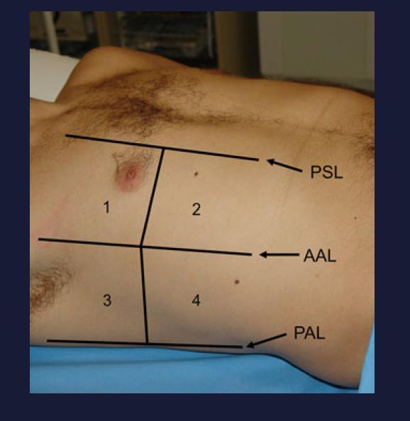
18 Lung Ultrasound o Simple & portable ultrasound machine o Curvilinear (3.5-5 megahertz) & linear probes (high resolution; megahertz) Each hemi thorax divided: 1 Anterior superior 2 Anterior inferior 3 Lateral superior 4 Lateral inferior 5 Posterior superior 6 Posterior inferior
19 Zones
20 Lung Ultrasound Definitions Consolidation Nonaerated lung Lobar Consolidation - lobe or a segment affected Peripheral Consolidation - focal area of nonaerated lung, typically abutting pleural surface
21 Air bronchogram Punctate or branching echogenicities within areas of lung consolidation Static No motion within bronchi Dynamic - move within the bronchi Mass o Focal solid lesion o Doesn t appear to be arising from lung parenchyma Doppler o +/- of color Doppler flow within an area of consolidation or mass
22 (N) Lung o Pleural Line o Sliding Sign o A Lines o Seashore Sign M Mode

23 Longitudinal Scan o Pleural line - regular echogenic line - moves continuously during respiration o Lung Sliding Sign - Pleural motion

24 Transverse Scan A lines o Pleura-lung interface o Parallel curvilinear o Regular intervals from pleura o Normal aeration pattern

25 M-mode cursor over pleural line; 2 patterns: o Motionless portion of chest above pleural line - horizontal waves, o Sliding below pleural line - granular pattern, sand Seashore Sign


26 o Absence of A lines o Hypoechogenic area; poorly defined borders o Compact underlying comet tail artifacts B o Air Bronchograms o Fluid bronchograms o Hepatization of lung o Shred Sign Pneumonia


27 B Lines o Vertical comet-tail Artifacts - pleural line o Erase A lines o Move with lung sliding o Presence is due to fluidrich subpleural interlobular septae, surrounded by air

28 Hepatization Air content further decreases (lung consolidations), acoustic window becomes completely open, & lung - directly visualized like solid parenchyma (liver/ spleen)
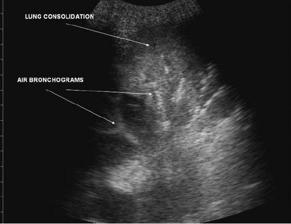
29 Air bronchograms

30 Shred Sign Deeper border of consolidated lung tissue that makes contact with aerated lung tissue is irregular & shredded
31 INTERPRETATION CODE-SHEET OF LUNG ULTRASOUND LUNG ULTRASOUND REPORT FORM PATIENT IDENTIFICATION NUMBER: DATE OF ULTRASOUND: OVERALL IMPRESSION FOR BOTH LUNGS: READER: IMPRESSION: IMPRESSION RIGHT LUNG LEFT LUNG Normal Yes / No Yes/No Pneumonia Yes/No Yes/No Pleural effusion Yes/No Yes/No Pneumothorax Yes/No Yes/No Others Yes/No Yes/No
32 Impression Lung Anterior Superior Anterior Inferior Lateral Superior Lateral Inferior Posterior Superior Posterior Inferior PLEURAL SPACE Normal Yes/No Yes/No Yes/No Yes/No Yes/No Yes/No Pneumothorax Yes/No Yes/No Yes/No Yes/No Yes/No Yes/No Effusion Yes/No Yes/No Yes/No Yes/No Yes/No Yes/No LUNG Normal Yes/No Yes/No Yes/No Yes/No Yes/No Yes/No Interstitial Disease Yes/No Yes/No Yes/No Yes/No Yes/No Yes/No Consolidation Yes/No Yes/No Yes/No Yes/No Yes/No Yes/No If Yes Solid Solid Solid Solid Solid Solid Cavitory Cavitory Cavitory Cavitory Cavitory Cavitory Air Bronchogram Yes/No Yes/No Yes/No Yes/No Yes/No Yes/No Static Static Static Static Static Static Dynamic Dynamic Dynamic Dynamic Dynamic Dynamic Mass Yes/No Yes/No Yes/No Yes/No Yes/No Yes/No Doppler (Perfused) Yes/No Yes/No Yes/No Yes/No Yes/No Yes/No
33 X-ray chest X-ray chest taken in PA view; results interpreted by 3 trained radiologists. Radiologists were blinded to patients history & examination findings
34 PATIENT IDENTIFICATION NUMBER: CHEST X-RAY REPORT FORM READER: DATE OF CHEST X-RAY: IMPRESSION: Overall impression for Both Lungs IMPRESSION RIGHT LUNG LEFT LUNG Normal Yes / No Yes/No Lobar Consolidation Yes/No Yes/No Patchy Consolidation Yes/No Yes/No Pneumothorax Yes/No Yes/No Atelectasis Yes/No Yes/No
35 Statistical Analysis o Descriptive statistics analyzed with SPSS version 17.0 software o Continuous variables presented as mean ± SD o Categorical variables expressed as frequencies & percentages o The Pearson's chi-square test or the chi-square test of association used to determine if there was a relationship between two categorical variables o p < 0.05 was considered statistically significant
36 Formula used n = =1.96*1.96*0.8* *0.1 =61.46 o o o o Where p is observed accuracy of point of care PLUS in diagnosis of CAP in children q = 1 p d is the margin of error is the ordinate of standard normal distribution at α% level of significance
37 Results o Total of 112 hospitalized children (CAP) o 100 patients met selection criteria & recruited o 56 (56%) were boys o Mean age + SD of children in yrs was 4.31 ± 4.41 o Radiological diagnosis of pneumonia - 58 (58%) patients o Lung ultrasound was abnormal in 86 (86%) patients
38
39 In CXR positive pts, PLUS positive in all (58/ 58) (100%) Whereas, in radiologically (N) pts, but clinically diagnosed CAP, lung ultrasound was abnormal in 28/42 (66.67%) patients Thus, lung ultrasound (LUS) has a high sensitivity (100%) with specificity of 33.3% (for diagnosing radiologically proven cases of CAP) PPV was 67.4%, NPV was 100% & Accuracy was 72%
40
41 o Difference in diagnosis of CAP by chest radiology & PLUS, showed the chi square statistic of , with a p value of (highly significant) o Comparison of lung Ultrasound & x ray diagnosis, showed the number of observed agreements to be 72. No of agreements expected by chance was Cohen s kappa coefficient (k) was 0.367, with SE of kappa = % confidence interval: From to o Strength of agreement is considered to be 'fair
42

43 Conclusion PLUS is a highly sensitive test in diagnosing CAP It can be used as a first use diagnostic modality in suspected cases of CAP, thus replacing CXR Significantly reduce radiation exposure in this vulnerable pt grp Towards A Radiation Free Pulmonology Practice
44 Acknowledgements - Team Pediatric Team Radiology team Statistician Parul LUS technician Staff Patients
45 Thank You
Initially for cardiac echo Subsequent studies non-cardiac applications
 No disclosures But Heavy accent Initially for cardiac echo Subsequent studies non-cardiac applications 1973: Goldberg et al in JCUS 30 mediastinal masses in pts. age 1-84 yrs. 1977: Kangarloo et al in
Lung sonography in the diagnosis of pneumothorax.
 Lung sonography in the diagnosis of pneumothorax. Poster No.: C-0526 Congress: ECR 2011 Type: Educational Exhibit Authors: K. Stefanidis, K. Vintzilaios, D. D. Cokkinos, E. Antypa, S. Dimopoulos, S. Nanas,
Chest Ultrasound: Pneumothorax
 WINFOCUS BASIC ECHO (WBE) Chest Ultrasound: Pneumothorax Mark Hamlin, MD, MS Associate Professor of Anesthesiology and Surgery University of Vermont College of Medicine Co-Director of Surgical Critical
Definitions and diagnostic implications of terms used in the chest radiograph and lung ultrasound diagnoses of pneumonia.
 Supplementary 1 Definitions and diagnostic implications of terms used in the chest radiograph and lung ultrasound diagnoses of pneumonia. Imaging finding Definition Implication CR Consolidation Interstitial
A Practical Approach to Ultrasound Assessment of Respiratory Distress
 A Practical Approach to Ultrasound Assessment of Respiratory Distress Yanick Beaulieu, MD, FRCPC Director, Bedside Ultrasound Curriculum Division of Cardiology and Critical Care Hôpital du Sacré-Coeur
This appendix was part of the submitted manuscript and has been peer reviewed. It is posted as supplied by the authors.
 This appendix was part of the submitted manuscript and has been peer reviewed. It is posted as supplied by the authors. - Figure S1: The four quadrant approach lung ultrasound at the bedside. * The anterolateral
Lung ultrasound in the critically ill patient Pleural Effusions
 Lung ultrasound in the critically ill patient Pleural Effusions Rohit Patel, MD University of Florida Health Director, Critical Care Ultrasound Surgical ICU Center for Intensive Care Gainesville, Florida
Bedside ultrasound - Lung ultrasound in the Intensive Care Unit
 Bedside ultrasound - Lung ultrasound in the Intensive Care Unit Kishore K. Pichamuthu, Professor, Department of Critical Care, Christian Medical College, Vellore. Summary In an ICU setting, ultrasonographic
OVERVIEW. Need for USG. Weaning assessment. Mechanics of USG. Pneumonia / VAP. Principles of lung USG. Prone position ventilation assessment
 OVERVIEW Need for USG Mechanics of USG Principles of lung USG BLUE protocol Alveolar syndrome Interstitial syndrome Weaning assessment Pneumonia / VAP Prone position ventilation assessment ETT positioning
Ultrasound. FAST Focused Assessment with Sonography in Trauma
 Ultrasound FAST Focused Assessment with Sonography in Trauma Rohit Patel, MD University of Florida Health Director, Critical Care Ultrasound Surgical ICU Center for Intensive Care Gainesville, Florida
NON INVASIVE LIFE SAVERS. Ultrasound in PICU
 VOL 1 NO.1 Jan March 2014 54 Table 1. Selected Applications of Point-of-Care Ultrasonography, According to Medical Specialty. Specialty Ultrasound Applications Anesthesia Cardiology Guidance for vascular
Lung ultrasound in the critically ill patient BASICS
 Lung ultrasound in the critically ill patient BASICS Rohit Patel, MD University of Florida Health Director, Critical Care Ultrasound Surgical ICU Center for Intensive Care Gainesville, Florida Introduction
Objectives. The Extended FAST Exam. Focused Assessment e With Sonography In. Trauma (FAST)
") Northern California Emergency Ultrasound Course Objectives The Extended FAST Exam Rimon Bengiamin, MD, RDMS UC SF Discuss the components of the EFAST exam Evaluate the utility of the EFAST Review how to
Northern California Emergency Ultrasound Course Objectives The Extended FAST Exam Rimon Bengiamin, MD, RDMS UC SF Discuss the components of the EFAST exam Evaluate the utility of the EFAST Review how to
Bedside Sonographic Diagnosis of Pneumothorax in Pediatric Patients: A Preliminary Report Chia-Wang Tang 1, Kai-Sheng Hsieh 1 1
 ORIGINAL ARTICLE Bedside Sonographic Diagnosis of in Pediatric Patients: A Preliminary Report Chia-Wang Tang 1, Kai-Sheng Hsieh 1 1 Division of Pediatric Pulmonology, Department of Pediatrics, Kaohsiung
The Intensive Care Unit
 Imaging of the ADRS patient: Risk of transportation and alternative to repetitive radiation exposure Jean-Jacques Rouby Pitié-Salpêtrière Hospital The Intensive Care Unit The Intensive Care Unit Multidisciplinary
Role of Transthoracic Ultrasound in Detection of Pneumonia in ICU Patients
 Med. J. Cairo Univ., Vol. 83, No. 1, June: 307-314, 2015 www.medicaljournalofcairouniversity.net Role of Transthoracic Ultrasound in Detection of Pneumonia in ICU Patients FARES AUF, M.D.; AHMED ABO-NAGLH,
Ultrasound in the ICU
 Ultrasound in the ICU Kristine E. W. Breyer, MD Assistant Professor Anesthesia & Critical Care Medicine UCSF DISCLOSURES: NONE Definition The Ultrasound Exam Types & Uses Training Clinical Examples Objectives
ASSESSMENT OF LUNG PARENCHYMAL ABNORMALITIES
 2016 by the author Thank you for viewing this presentation. We would like to remind you that this material is the property of the author. It is provided to you by the ERS for your personal use only, as
FAST Focused Assessment with Sonography in Trauma
 FAST Focused Assessment with Sonography in Trauma Wilma Rodriguez Mojica,MD,FACR Professor of Radiology UPR School of Medicine Ultrasound Section - Radiological Sciences Department OBJECTIVES Understand
TB Radiology for Nurses Garold O. Minns, MD
 TB Nurse Case Management Salina, Kansas March 31-April 1, 2010 TB Radiology for Nurses Garold O. Minns, MD April 1, 2010 TB Radiology for Nurses Highway Patrol Training Center Salina, KS April 1, 2010
Thoracic Ultrasound: Pictorial review of pneumothorax, the fastest and easiest method to diagnose.
 Thoracic Ultrasound: Pictorial review of pneumothorax, the fastest and easiest method to diagnose. Poster No.: C-1588 Congress: ECR 2014 Type: Educational Exhibit Authors: J. A. Guirola, V. Mayoral Campos,
Accuracy of Lung Ultrasonography in Diagnosis of Community Acquired Pneumonia as Compared to Chest X-Ray in Pediatric Age Group
 The Egyptian Journal of Hospital Medicine (July 2018) Vol. 72 (8), Page 4977-4983 Accuracy of Lung Ultrasonography in Diagnosis of Community Acquired Pneumonia as Compared to Chest X-Ray in Pediatric Age
EUROPEAN ASSOCIATION OF VETERINARY DIAGNOSTIC IMAGING EUROPEAN COLLEGE OF VETERINARY DIAGNOSTIC IMAGING
 EISAGOGIKO EUROPEAN ASSOCIATION OF VETERINARY DIAGNOSTIC IMAGING EUROPEAN COLLEGE OF VETERINARY DIAGNOSTIC IMAGING ARISTOTLE UNIVERSITY OF THESSALONIKI SCHOOL OF VETERINARY MEDICINE SECTION OF RADIOLOGY
RSV infection and lung ultrasound
 RSV infection and lung ultrasound Neonatology Clinic, University Hospital, Krakow Joanna Hurkała Joanna Pietras Agnieszka Ochoda-Mazur Poznań, 28.09.2018 1 Disclosure In relations to this presentation,
Point-of-care lung ultrasound
 Ultrasound Point-of-care lung ultrasound Philips tutorial Michael B. Stone, MD, RDMS Director, Division of Emergency Ultrasound Department of Emergency Medicine Brigham and Women s Hospital, Boston, MA
Extended FAST Exam. Goal of Trauma Care. Golden Hour of Trauma
 Extended FAST Exam Goal of Trauma Care Golden Hour of Trauma Best INITIAL screening modality in trauma efast 2014 LLSA Article (ACEP Policy Statement) Level B Recommendation: In hemodynamically unstable
POCUS for the Internist: Lungs & Pericardial Effusions
 POCUS for the Internist: Lungs & Pericardial Effusions Jeremy S. Boyd, MD, FACEP Asst. Professor of Emergency Medicine Vanderbilt University Medical Illustrations courtesy of Robinson Ferre, MD, FACEP
Signs in Chest Radiology
 Signs in Chest Radiology Jonathan H. Chung, MD Disclosures No pertinent disclosures Jonathan H. Chung, MD Assistant Professor Institute t of fadvanced d Biomedical Imaging National Jewish Health Denver,
Radiation Exposure in Pregnancy. John R. Mayo UNIVERSITY OF BRITISH COLUMBIA
 Radiation Exposure in Pregnancy John R. Mayo UNIVERSITY OF BRITISH COLUMBIA Illustrative Clinical Scenario 32 year old female 34 weeks pregnant with recent onset shortness of breath and central chest pain
Background Focused Assessment with Sonography in Trauma. Johann Baptist Dormagen, MD, PhD
 Focused Assessment with Sonography in Trauma Johann Baptist Dormagen, MD, PhD Unit of Abdominal and Oncologic Radiology Department of Radiology and Nuclear Medicine Oslo University Hospital, Norway 8 th
An Introduction to Radiology for TB Nurses
 An Introduction to Radiology for TB Nurses Garold O. Minns, MD September 14, 2017 TB Nurse Case Management September 12 14, 2017 EXCELLENCE EXPERTISE INNOVATION Garold O. Minns, MD has the following disclosures
Perioperative Ultrasonography Ehab Farag, MD, FRCA Hesham Elsharkawy David G. Anthony, M.D.
 Perioperative Ultrasonography Ehab Farag, MD, FRCA Hesham Elsharkawy David G. Anthony, M.D. Cleveland Clinic, Cleveland OH 1 Complications during central venous catheterization (CVC) occur 2% -15% of the
Introduction to Chest Radiography
 Introduction to Chest Radiography RSTH 366: DIAGNOSTIC TECHNIQUES Alan Alipoon BS, RCP, RRT Instructor Department of Cardiopulmonary Sciences 1 Introduction Discovered in 1895 by Wilhelm Roentgen Terminology
Dr Francis Ogaro MTRH ELDORET
 Dr Francis Ogaro MTRH ELDORET TB in children often severe, disseminated and can progress rapidly and with poor outcome TB diagnosis in children has relied on clinical, imaging, microscopy and TST findings.
Ultrasound basics Part 1
 Ultrasound basics Part 1 'Ultrasound enhanced critical care medicine' Rohit Patel, MD University of Florida Health Director, Critical Care Ultrasound Surgical ICU Center for Intensive Care Gainesville,
Chest X-ray Interpretation
 Chest X-ray Interpretation Introduction Routinely obtained Pulmonary specialist consultation Inherent physical exam limitations Chest x-ray limitations Physical exam and chest x-ray provide compliment
Interpreting thoracic x-ray of the supine immobile patient: Syllabus
 Interpreting thoracic x-ray of the supine immobile patient: Syllabus Johannes Godt Dep. of Radiology and Nuclear Medicine Oslo University Hospital Ullevål NORDTER 2017, Helsinki Content - Why bedside chest
Epidermiology Early pulmonary embolism
 Epidermiology Early pulmonary embolism Sitang Nirattisaikul Faculty of Medicine, Prince of Songkla University 3 rd most common cause of cardiovascular death in the United States, following ischemic heart
Lung ultrasound in follow-up of low birth weight with respiratory distress syndrome: clinical application and reduction of x-rays examinations
 Lung ultrasound in follow-up of low birth weight with respiratory distress syndrome: clinical application and reduction of x-rays examinations Poster No.: C-1724 Congress: ECR 2011 Type: Scientific Paper
10/17/2016. Nuts and Bolts of Thoracic Radiology. Objectives. Techniques
 Nuts and Bolts of Thoracic Radiology October 20, 2016 Carleen Risaliti Objectives Understand the basics of chest radiograph Develop a system for interpreting chest radiographs Correctly identify thoracic
Sorting the sheep from the goats
 Sorting the sheep from the goats How do we improve the diagnosis of pediatric respiratory diseases under low-resource conditions? Pediatric Grand Rounds February 27, 2015 It doesn t matter. refugee camp
and localized ground glass opacities, or bronchiolar focal or multifocal micronodules;
 E1 Chest CT scan and Pneumoniae_YE Claessens et al- Supplementary methods Level of CAP probability according to CT scan - definite CAP: systematic alveolar condensation, or alveolar condensation with peripheral
Do you want to be an excellent Radiologist? - Focus on the thoracic aorta on lateral chest image!!!
 The lateral chest radiograph: Challenging area around the thoracic aorta!!! Do you want to be an excellent Radiologist? - Focus on the thoracic aorta on lateral chest image!!! Dong Yoon Han 1, So Youn
Lung sequestration and Scimitar syndrome
 Lung sequestration and Scimitar syndrome Imaging approaches M. Mearadji International Foundation for Pediatric Imaging Aid Rotterdam, The Netherlands Pulmonary sequestration Pulmonary sequestration (PS)
Certificate in Clinician Performed Ultrasound (CCPU) Syllabus. Lung
 Syllabus. Lung") Certificate in Clinician Performed Ultrasound (CCPU) Syllabus Lung Page 1 of 8 01/17 Lung Syllabus Purpose: This unit is designed to cover the theoretical and practical curriculum for lung ultrasound in
Certificate in Clinician Performed Ultrasound (CCPU) Syllabus Lung Page 1 of 8 01/17 Lung Syllabus Purpose: This unit is designed to cover the theoretical and practical curriculum for lung ultrasound in
Background & Indications Probe Selection
 Teresa S. Wu, MD, FACEP Director, EM Ultrasound Program & Fellowship Co-Director, Simulation Based Training Program & Fellowship Associate Program Director, EM Residency Program Maricopa Medical Center
What to Do with Small Lung Nodules Hanh Vu Nghiem, MD William Beaumont Hospital Royal Oak, Michigan
 What to Do with Small Lung Nodules Hanh Vu Nghiem, MD William Beaumont Hospital Royal Oak, Michigan Small Lung Nodules What to do with small lung nodules? We biopsy them when requested What are our accuracy
Basic of Ultrasound Physics E FAST & Renal Examination. Dr Muhammad Umer Ihsan MBBS,MD, DCH CCPU,DDU1,FACEM
 Basic of Ultrasound Physics E FAST & Renal Examination Dr Muhammad Umer Ihsan MBBS,MD, DCH CCPU,DDU1,FACEM What is Sound? Sound is Mechanical pressure waves What is Ultrasound? Ultrasounds are sound waves
Radiology of the respiratory disease
 Radiology of the respiratory disease [ Color index: Important Notes Extra ] [ Editing file Feedback Share your notes Shared notes ] Resources: - 435 Slides - 434 Team - 435 Notes Done by: - Mai Alageel
Chest Radiology Interpretation: Findings of Tuberculosis
 Chest Radiology Interpretation: Findings of Tuberculosis Get out your laptops, smart phones or other devices pollev.com/chestradiology Case #1 1 Plombage Pneumonia Cancer 2 Reading the TB CXR Be systematic!
Introduction & Physics of ED Ultrasound. Objectives. What? - Limited Studies. Who? - ED Docs
 Introduction & Physics of ED Ultrasound Martine Sargent, MD Ultrasound Director, Assistant Professor UCSF Department of Emergency Medicine San Francisco General Hospital & Trauma Center Objectives Who?
Thyroid Nodules: US Risk Stratification. Alex Tessnow, MD, FACE, ECNU University of Texas Southwestern Associate Professor of Medicine Dallas, Texas
 Thyroid Nodules: US Risk Stratification Alex Tessnow, MD, FACE, ECNU University of Texas Southwestern Associate Professor of Medicine Dallas, Texas Which of the following is true? A. All echogenic foci
Chest X rays and Case Studies. No disclosures. Outline 5/31/2018. Carlo Manalo, M.D. Department of Radiology Loma Linda University Children s Hospital
 Chest X rays and Case Studies Carlo Manalo, M.D. Department of Radiology Loma Linda University Children s Hospital No disclosures. Outline Importance of history Densities delineated on radiography An approach
Ultrasound Principles cycle Frequency Wavelength Period Velocity
 ! Teresa S. Wu, MD, FACEP Director, EM Ultrasound Program & Fellowship Co-Director, Simulation Based Training Program & Fellowship Associate Program Director, EM Residency Program Maricopa Medical Center
Role of chest ultrasonography in the prognostic evaluation of patients with community acquired pneumonia: a prospective study
 International Journal of Research in Medical Sciences Rennis KD et al. Int J Res Med Sci. 2017 Jan;5(1):25-30 www.msjonline.org pissn 2320-6071 eissn 2320-6012 Original Research Article DOI: http://dx.doi.org/10.18203/2320-6012.ijrms20164510
Pulmonary Embolism. Thoracic radiologist Helena Lauri
 Pulmonary Embolism Thoracic radiologist Helena Lauri 8.5.2017 Statistics 1-2 out of 1000 adults annually are diagnosed with deep vein thrombosis (DVT) and/or pulmonary embolism (PE) About half of patients
The efficacy of bedside chest ultrasound: from accuracy to outcomes
 EUROPEAN RESPIRATORY UPDATE EFFICACY OF BEDSIDE CHEST ULTRASOUND The efficacy of bedside chest ultrasound: from accuracy to outcomes Mark Hew 1,2 and Tunn Ren Tay 1,3 Affiliations: 1 Allergy, Immunology
Case 1. A 35-year-old male presented with fever, cough, and purulent sputum for one week. This was his CXR (Fig. 1.1). What is the diagnosis?
. What is the diagnosis?") 1 Interpreting Chest X-Rays CASE 1 Fig. 1.1 Case 1. A 35-year-old male presented with fever, cough, and purulent sputum for one week. This was his CXR (Fig. 1.1). What is the diagnosis? CASE 1 Interpreting
1 Interpreting Chest X-Rays CASE 1 Fig. 1.1 Case 1. A 35-year-old male presented with fever, cough, and purulent sputum for one week. This was his CXR (Fig. 1.1). What is the diagnosis? CASE 1 Interpreting
Tuberculosis: The Essentials
 Tuberculosis: The Essentials Kendra L. Fisher, MD, PhD THORACIC TUBERCULOSIS: THE BARE ESSENTIALS Kendra Fisher MD, FRCP (C) Department of Radiology Loma Linda University Medical Center TUBERCULOSIS ()
How, why and when - Chest US guided biopsy?
 How, why and when - Chest US guided biopsy? Poster No.: P-0092 Congress: ESTI 2014 Type: Educational Poster Authors: D. Penha, E. Pinto, A. M. D. Costa ; Lisbon/PT, Amadora/PT Keywords: Thorax, Oncology,
Evaluation of the chest
 Evaluation of the chest part 1 Nagy Endre SZEGEDI TUDOMÁNYEGYETEM ÁOK, RADIOLÓGIAI KLINIKA, SZEGED Indication In case of complaints or symptoms: In suspicion of lesions, diseases or injuries of the chest
4/16/2017. Learning Objectives. Interpretation of the Chest Radiograph. Components. Production of the Radiograph. Density & Appearance
 Interpretation of the Arthur Jones, EdD, RRT Learning Objectives Identify technical defects in chest radiographs Identify common radiographic abnormalities This Presentation is Approved for 1 CRCE Credit
Shedding Light on Neonatal X-rays. Objectives. Indications for X-Rays 5/14/2018
 Shedding Light on Neonatal X-rays Barbara C. Mordue, MSN, NNP-BC Neonatal Nurse Practitioner LLUH Children s Hospital, NICU Objectives Utilize a systematic approach to neonatal x-ray interpretation Identify
We are IntechOpen, the world s leading publisher of Open Access books Built by scientists, for scientists. International authors and editors
 We are IntechOpen, the world s leading publisher of Open Access books Built by scientists, for scientists 3,800 116,000 120M Open access books available International authors and editors Downloads Our
Principles of Ultrasound. Cara C. Prideaux, M.D. University of Utah PM&R Sports Medicine Fellow March 14, 2012
 Principles of Ultrasound Cara C. Prideaux, M.D. University of Utah PM&R Sports Medicine Fellow March 14, 2012 None Disclosures Outline Introduction Benefits and Limitations of US Ultrasound (US) Physics
HOW TO IMAGE AND DESCRIBE CONGENITAL LUNG MALFORMATIONS
 HOW TO IMAGE AND DESCRIBE CONGENITAL LUNG MALFORMATIONS Paul Thacker, MD Assistant Professor Departments of Radiology and Pediatrics Medical University of South Carolina DISCLOSURES I have no relevant
Children are not small adults Children are Not Small Adults Anatomic considerations Pliable bony & cartilagenous structures - Significant thoracic inj
 PEDIATRIC CHEST TRAUMA Children are not small adults Role of imaging Spectrum of injury Children are not small adults Children are Not Small Adults Anatomic considerations Pliable bony & cartilagenous
PULMONARY TUBERCULOSIS RADIOLOGY
 PULMONARY TUBERCULOSIS RADIOLOGY RADIOLOGICAL MODALITIES Medical radiophotography Radiography Fluoroscopy Linear (conventional) tomography Computed tomography Pulmonary angiography, bronchography Ultrasonography,
ARDS - a must know. Page 1 of 14
 ARDS - a must know Poster No.: C-1683 Congress: ECR 2016 Type: Authors: Keywords: DOI: Educational Exhibit M. Cristian; Turda/RO Education and training, Edema, Acute, Localisation, Education, Digital radiography,
Eosinophilic lung diseases - what the radiologist needs to know
 Eosinophilic lung diseases - what the radiologist needs to know Poster No.: C-0803 Congress: ECR 2014 Type: Authors: Keywords: DOI: Educational Exhibit E.-M. Heursen, R. Reina Cubero, F. Japon Sola; Cádiz/ES
Abdominal Ultrasonography
 Abdominal Ultrasonography David A. Masneri, DO, FACEP, FAAEM Assistant Professor of Emergency Medicine Assistant Director, Emergency Medicine Residency Medical Director, Operational Medicine Division Center
Thyroid and Parathyroid Ultrasound Protocol
 Thyroid and Parathyroid Ultrasound Protocol Reviewed By: Anna Ellermeier, MD Last Reviewed: December 2017 Contact: (866) 761-4200, Option 1 **NOTE for all examinations: 1. If documenting possible flow
Pulmonary Embolism. Pulmonary Embolism. Pulmonary Embolism. PE - Clinical
 Pulmonary embolus - a practical approach to investigation and treatment Sam Janes Wellcome Senior Fellow and Respiratory Physician, University College London Background Diagnosis Treatment Common: 50 cases
Tests Your Pulmonologist Might Order. Center For Cardiac Fitness Pulmonary Rehab Program The Miriam Hospital
 Tests Your Pulmonologist Might Order Center For Cardiac Fitness Pulmonary Rehab Program The Miriam Hospital BASIC ANATOMY OF THE LUNGS Lobes of Lung 3 lobes on the Right lung 2 lobes on the Left Blood
Lung ultrasound: routine practice for the next generation of internists
 REVIEW Lung ultrasound: routine practice for the next generation of internists H.R.W. Touw 1,2, P.R. Tuinman* 1,3,4, H.P.M.M. Gelissen 1, E. Lust 1, P.W.G. Elbers 1,3,4 Departments of 1 Intensive Care
Terminology Tissue Appearance
 By Marc Nielsen, MD Advantages/Disadvantages Generation of Image Ultrasound Machine/Transducer selection Modes of Ultrasound Terminology Tissue Appearance Scanning Technique Real-time Portable No ionizing
Pulmonary Ultrasound in Emergency Medicine and Critical Care
 Pulmonary Ultrasound in Emergency Medicine and Critical Care www.rmgultrasound.com Author: Virginia M Stewart, MD RDMS RDCS RDMSK Dr Stewart is a practicing Emergency Physician in Eastern Virginia, USA.
Top Tips for Pleural Disease in 2012
 Top Tips for Pleural Disease in 2012 The unilateral pleural effusion on the Post Take Ward Round Pleural Effusion on CXR Bedside ultrasound + Pleural aspirate Empyema Nil evidence infection Admit IV antibiotics
Certificate in Clinician Performed Ultrasound (CCPU) Syllabus. Lung
 Syllabus. Lung") Certificate in Clinician Performed Ultrasound (CCPU) Syllabus Lung ASUM Quality CCPU Syllabi Released: 21 March 2013 Approved by: CEO Lung Purpose: This unit is designed to cover the theoretical and practical
Certificate in Clinician Performed Ultrasound (CCPU) Syllabus Lung ASUM Quality CCPU Syllabi Released: 21 March 2013 Approved by: CEO Lung Purpose: This unit is designed to cover the theoretical and practical
Introduction to Interventional Pulmonology
 Introduction to Interventional Pulmonology Alexander Chen, M.D. Director, Interventional Pulmonology Assistant Professor of Medicine and Surgery Divisions of Pulmonary and Critical Care Medicine and Cardiothoracic
Thorax Lecture 2 Thoracic cavity.
 Thorax Lecture 2 Thoracic cavity. Spring 2016 Dr. Maher Hadidi, University of Jordan 1 Enclosed by the thoracic wall. Extends between (thoracic inlet) & (thoracic outlet). Thoracic inlet At root of the
B-I-2 CARDIAC AND VASCULAR RADIOLOGY
 (YEARS 1 3) CURRICULUM FOR RADIOLOGY 13 B-I-2 CARDIAC AND VASCULAR RADIOLOGY KNOWLEDGE To describe the normal anatomy of the heart and vessels including the lymphatic system as demonstrated by radiographs,
THYROID NODULES: THE ROLE OF ULTRASOUND
 THYROID NODULES: THE ROLE OF ULTRASOUND NOVEMBER 2017 DR. DEAN DURANT DEFINITION Thyroid nodule: Focal area within the thyroid gland with echogenicity different from surrounding parenchyma. THYROID NODULES
Ultrasound-guided Aspiration of the Iatrogenic Pneumothorax Caused by Paravertebral Block
 Case Report Korean J Pain 2012 January; Vol. 25, No. 1: 33-37 pissn 2005-9159 eissn 2093-0569 http://dx.doi.org/10.3344/kjp.2012.25.1.33 Ultrasound-guided Aspiration of the Iatrogenic Pneumothorax Caused
Lecture 3. Inflammatory Processes
 Lecture 3 Inflammatory Processes Process: Increased vascular permeability Water and cellular infiltrations Results: Abscess, ulceration, cavitation Penetration, perforation and fistula formation Scarring,
CHEST Recent Advances in Chest Medicine
 CHEST Recent Advances in Chest Medicine Thoracic Ultrasonography for the Pulmonary Specialist Seth J. Koenig, MD; Mangala Narasimhan, DO, FCCP; and Paul H. Mayo, MD, FCCP Thoracic ultrasonography is a
Thyroid Nodules: US Risk Stratification and FNA Guidelines
 Thyroid Nodules: US Risk Stratification and FNA Guidelines Mark A. Lupo, MD, FACE, ECNU Thyroid & Endocrine Center of Florida Assistant Clinical Professor of Medicine Florida State University, College
The Role of the FAST exam in the EDRU
 The Role of the FAST exam in the EDRU A. Robb McLean, MD, MHCM Vice Chair of Clinical Operations, Department of Emergency Medicine Joint Trauma Conference June 20, 2017 Disclosures Goals Describe the performance,
The McMaster at night Pediatric Curriculum
 The McMaster at night Pediatric Curriculum Community Acquired Pneumonia Based on CPS Practice Point Pneumonia in healthy Canadian children and youth and the British Thoracic Society Guidelines on CAP Objectives
Evaluation of the of the sensitivity, accuracy and positive predictive value of ultrasonography in the diagnosis of Appendicitis.
 West African Journal of Ultrasound Vol 17 Number 2 (2016) Evaluation of the of the sensitivity, accuracy and positive predictive value of ultrasonography in the diagnosis of Appendicitis. 1 2 3 Oguntola
Imaging of Pleural Effusion: Comparing Ultrasound, X-Ray and CT findings
 Imaging of Pleural Effusion: Comparing Ultrasound, X-Ray and CT findings Poster No.: C-2067 Congress: ECR 2017 Type: Educational Exhibit Authors: J. M. Almeida, N. Antunes, C. Leal, L. Figueiredo ; Lisboa/PT,
Surgical indications: Non-malignant pulmonary diseases. Punnarerk Thongcharoen
 Surgical indications: Non-malignant pulmonary diseases Punnarerk Thongcharoen Non-malignant Malignant as a pathological term: Cancer Non-malignant = not cancer Malignant as an adjective: Disposed to cause
Cardiac tamponade and Pericardiocentesis Made Easy
 Cardiac tamponade and Pericardiocentesis Made Easy www.cardiconcept.com Etiology of pericardial diseases. Non Infectious cause Infectious cause European Heart Journal (2015) 36, 2921 2964 Recommendations
8/14/2017. Objective: correlate radiographic findings of common lung diseases to actual lung pathologic features
 What is that lung disease? Pulmonary Patterns & Correlated Pathology Dr. Russell Tucker, DACVR Objective: correlate radiographic findings of common lung diseases to actual lung pathologic features Improved
Pulmonary infarction semiology in CT. Revision of 80 cases.
 Pulmonary infarction semiology in CT. Revision of 80 cases. Poster No.: C-0369 Congress: ECR 2012 Type: Scientific Exhibit Authors: M. González Vázquez, D. Castellon, J. Calatayud, N. Silva 1 2 1 1 1 1
Thyroid Nodules and Ultrasound. Patrick Vos Department of Radiology St. Paul s Hospital Vancouver, BC
 Thyroid Nodules and Ultrasound Patrick Vos Department of Radiology St. Paul s Hospital Vancouver, BC No Financial Disclosures Patrick Vos Department of Radiology St. Paul s Hospital Vancouver, BC Acknowledgements
Background & Indications Probe Selection
 Teresa S. Wu, MD, FACEP Director, EM Ultrasound Program & Fellowship Co-Director, Simulation Based Training Program & Fellowship Associate Program Director, EM Residency Program Maricopa Medical Center
Hepatobiliary Ultrasound Rimon Bengiamin, MD, RDMS Assistant Clinical Professor Director of Emergency Ultrasound UCSF Fresno. Objectives. Why?
 Hepatobiliary Ultrasound Rimon Bengiamin, MD, RDMS Assistant Clinical Professor Director of Emergency Ultrasound UCSF Fresno Objectives Discuss the goals of point-of-care biliary ultrasound Review the
Chest Ultrasonography - A Quick and Accurate Diagnostic Tool in Pediatric Emergency Department and Intensive Care Unit
 Review Article Chest Ultrasonography - A Quick and Accurate Diagnostic Tool in Pediatric Emergency Department and Intensive Care Unit Dinakara Prithviraj 1, Suresh A 2 1 Associate Professor, Chief Neonatologist,