Appendix #37. Review of Research Relevant to Direct Current and Alternating Current Transmission Lines and Health. Northern Pass Project
|
|
|
- Reginald West
- 6 years ago
- Views:
Transcription

1 Appendix #37 Review of Research Relevant to Direct Current and Alternating Current Transmission Lines and Health Northern Pass Project
2 Review of Research Relevant to Direct Current and Alternating Current Transmission Lines and Health Northern Pass Project Prepared for Northern Pass, LLC 670 North Commercial Street, Suite 207 Manchester, NH Prepared by: Exponent Science Drive Suite 200 Bowie, MD October 5, 2015 Exponent, Inc.
3 Contents List of Figures List of Tables Acronyms and Abbreviations Limitations Executive Summary Alternating Current Transmission Lines Direct Current Transmission Lines iii Page vi vii viii Introduction 1 PART I THE ELECTRICAL ENVIRONMENT OF ALTERNATING CURRENT AND DIRECT CURRENT TRANSMISSION LINES 3 Overview of Electric and Magnetic Fields 5 Electromagnetic Spectrum 6 Sources and Exposure 7 Extremely low frequency electric and magnetic fields 7 Static magnetic fields 12 Static electric fields 13 Space charge 14 PART II - METHODS FOR EVALUATING SCIENTIFIC RESEARCH 18 Weight-of-Evidence Reviews 20 EMF Exposure Considerations 21 Methods of Health Research Studies 23 Epidemiologic studies 23 Experimental laboratory studies 30 PART III - EVALUATION OF ALTERNATING CURRENT ELECTRIC AND MAGNETIC FIELDS AND HEALTH 34 Guidelines for Extremely Low Frequency Electric and Magnetic Fields 35 World Health Organization Assessment of Extremely Low Frequency Electric and Magnetic Fields 37 Reviews by Other Scientific Organizations 44 National Institute of Environmental Health Sciences 44 International Agency for Research on Cancer 44 x xi xi xiii
4 International Commission on Non-Ionizing Radiation Protection 45 Scientific Committee on Emerging and Newly Emerging Health Risks 46 Current Status of Extremely Low Frequency Electric and Magnetic Field Research on Health 47 Childhood leukemia 48 Childhood brain cancer 52 Breast cancer 53 Adult leukemia and brain cancer 55 Reproductive/developmental effects 58 Neurodegenerative diseases 61 Cardiovascular disease 63 In vivo studies related to carcinogenesis 64 Research on livestock, wild animals, and plants 75 PART IV - EVALUATION OF DIRECT CURRENT ELECTRIC AND MAGNETIC FIELDS AND HEALTH 80 Guidelines for Static Electric and Magnetic Fields 81 WHO Assessment of Static Electric and Magnetic Fields 82 Reviews by Other Scientific Organizations 84 Static electric and magnetic fields 84 Air ions 87 Current Status of Static Electric and Magnetic Field Health Research 88 Static magnetic fields 88 Static electric fields 93 Relevance of static electric fields to human health 97 Space Charge 99 Human studies 102 Animal studies 108 Research on livestock, wild animals, and plants 122 PART V SUMMARY AND CONCLUSIONS 129 Alternating current 345-kV and 115-kV transmission lines 130 Direct current ±320 kv transmission line 132 References 137 Annex 1 Experimental animal studies of static electric fields Annex 2 Air ions and respiratory function outcomes: A comprehensive review iv
5 Annex 3 Annex 4 Annex 5 Annex 6 Air ions and mood outcomes: A review and meta-analysis Experimental animal studies of air ions Quantitative assessment of animal studies of air ions Magnitude of reported effects in animals as a function of ion density v
6 List of Figures Page Figure 1. Common sources of EMF in the home 8 Figure 2. Extremely low frequency magnetic- and electric-field levels in the environment. 9 Figure 3. Basic IARC method for classifying exposures based on potential carcinogenicity. 39 Figure 4. Possible explanations for the observed association between ELF magnetic fields and childhood leukemia. 41 vi
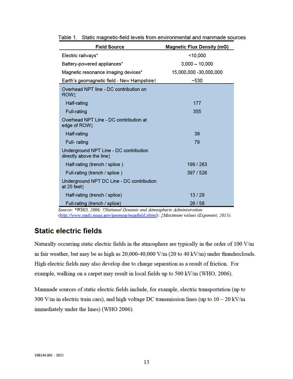
7 List of Tables Page Table 1. Static magnetic-field levels from environmental and manmade sources 13 Table 2. Comparison of static electric-field levels from the proposed project to other sources 14 Table 3. Air ion levels from a variety of background sources 16 Table 4. Measured fraction of aerosols ( micrometers [µm]) carrying charges in various locations 17 Table 5. Criteria for evaluating whether an association is causal 29 Table 6. Criteria for evaluating experimental studies 32 Table 7. Exposures corresponding to Basic Restrictions (limits) for public exposure to 60-Hz electric and magnetic fields 36 Table 8. Exposure values for static electric and magnetic fields 81 vii
8 Acronyms and Abbreviations 5HT 5-hydroxytryptamine 5HIAA 5-hydroxyindole acetic acid µm Micrometer AC Alternating current ALL Acute lymphoblastic leukemia ALS Amyotrophic lateral sclerosis AMI Acute myocardial infarction AMP Adenosine monophosphate CAT Catalase CI Confidence interval CO 2 Carbon dioxide CSC Connecticut Siting Council DC Direct current DMBA Dimethylbenz[a]anthracene ECG Electrocardiogram EEG Electroencephalogram ELF Extremely low frequency EMF Electric and magnetic fields F344 Fischer 344 FDA Food and Drug Administration FEV Forced expiratory volume G Gauss GHz Gigahertz GSH-Px Glutathione peroxidase Hz Hertz IARC International Agency for Research on Cancer ICES International Committee on Electromagnetic Safety ICNIRP International Committee on Non-Ionizing Radiation Protection IFN-γ Interferon-γ IL Interleukin viii
9 ISCO JEM khz kv/m L/min m MDA mg/kg mg MHz MPD MRI MW nm NO NPT NRPB OR OSI ROW RR SCENIHR SMD SOD T TAC TOS TWA V/m WHO WNI International Standard Classification of Occupations Job exposure matrix Kilohertz Kilovolts per meter Liters per minute Meter Malondialdehyde Milligrams per kilogram Milligauss Megahertz Mean proportional difference Magnetic resonance imaging Megawatt Nanometer Nitrous oxide Northern Pass Transmission Line National Radiological Protection Board Odds ratio Oxidative stress index Right of way Relative risk Scientific Committee on Newly Identified Health Risks Standardized mean difference Superoxide dismutase Tesla Total anti-oxidant capacity Total oxidant status Time weighted average Volts per meter World Health Organization Water-generated negative air ions ix
10 Limitations At the request of Northern Pass Transmission LLC, Exponent prepared this summary report on the status of research related to alternating current and direct current electric and magnetic fields and health. The findings presented herein are made to a reasonable degree of scientific certainty. Exponent reserves the right to supplement this report and to expand or modify opinions based on review of additional material as it becomes available, through any additional work, or review of additional work performed by others. The scope of services performed during this investigation may not adequately address the needs of other users of this report, and any re-use of this report or its findings, conclusions, or recommendations presented herein are at the sole risk of the user. The opinions and comments formulated during this assessment are based on observations and information available at the time of the investigation. No guarantee or warranty as to future life or performance of any reviewed condition is expressed or implied. x
11 Executive Summary The proposed Northern Pass Transmission (NPT) project includes transmission lines, converter terminals, and additional equipment to be installed in existing substations. These are sources of either 60-Hertz (Hz) alternating current (AC) electric and magnetic fields in the extremely-lowfrequency range, or direct current (DC) (i.e., static) electric and magnetic fields and space charge (air ions and charged aerosols). The projected levels of exposure to these aspects of the transmission line environment are presented in the companion report, Electrical Environment of the Proposed Northern Pass Transmission Project: DC Electric Field, DC Magnetic Field, Air Ion Density, AC Electric Field, AC Magnetic Field, Audible Noise, and Radio Noise (Exponent, 2015) contained in Appendix 38. The purpose of this assessment is to determine if the exposures from the NPT AC and DC transmission lines are likely to have any adverse effects on humans, livestock, wildlife, and plants in the project area. This assessment describes the methods and considerations used by scientists to systematically evaluate research in which the strength, limitations, and relevance of individual studies are a key component. Alternating Current Transmission Lines The combined operation of the proposed 345-kilovolt (kv) NPT line at full rating and the existing 115-kV and lower voltage distribution lines at peak load would produce a maximum value beneath the conductors on the right of way (ROW) of <1 milligauss (mg) to 366 mg. The AC magnetic-field levels diminish quickly with distance away from the conductors so that at the edges of the ROW these field levels are far lower between 0.1 mg and 92 mg except for an approximately 2,000-foot segment where the magnetic field on one side of the ROW is calculated to be 127 mg. These levels in turn will fall still lower to background levels outside the ROW. These levels are far below the limits on exposure of the general public, termed Basic Restrictions, recommended by the International Commission on Non-Ionizing Radiation Protection (ICNIRP) and the International Committee on Electromagnetic Safety (ICES)(12,420 mg and 9,146 mg, respectively). Similarly, the combined AC electric field from the proposed NPT 345-kV Line and the existing 115-kV lines is greatest on the ROW, with values between 1.0 kilovolts per meter (kv/m) and xi
12 5.2 kv/m, which diminish with distance at the edge of the ROW (0.0 to 2.7 kv/m). These electric-field levels are below the ICNIRP and ICES Basic Restrictions for 60-Hz electric fields (36.6 kv/m and 26.8 kv/m, respectively). The presence of trees, shrubs, fences, buildings, and other objects between the transmission lines and areas outside the ROW would reduce levels of electric field to a considerable extent. During the past 35-plus years, a large amount of research studies have been conducted to investigate potential health effects associated with EMF exposure below currently existing scientifically based exposure guidelines. These research studies include epidemiologic studies conducted in human populations, and laboratory studies conducted in live animals (in vivo studies) and in isolated cells and tissues (in vitro studies). A number of multidisciplinary expert panels on behalf of national and international agencies have evaluated the existing evidence for potential health effects by conducting health risk assessments employing the weight-of-evidence scientific process. These agencies include, among others, the World Health Organization (WHO), the International Agency for Research on Cancer, ICNIRP, the National Institute of Environmental Health Sciences, and most recently, the European Union s Scientific Committee on Emerging and Newly Identified Health Risks, which published its health risk assessment in None of these agencies concluded that the available scientific evidence confirms the existence of any adverse health effects of exposure to AC electric and magnetic fields at levels below scientifically based exposure limits. As the WHO currently states on its website, [b]ased on a recent in-depth review of the scientific literature, the WHO concluded that current evidence does not confirm the existence of any health consequences from exposure to low level electromagnetic fields. 1 The WHO calls for the adoption of scientifically-based exposure guidelines (such as those developed by ICNIRP and ICES) that are protective of all known adverse health effects and for adoption of low or no cost measures that minimize fields without jeopardizing electrical safety. A review of the relevant available scientific literature does not suggest any consistent or convincing effects of AC electric or magnetic fields on humans, livestock, wildlife, and plants. 1 xii
13 Direct Current Transmission Lines Unlike AC transmission lines, the static electric and magnetic fields from DC transmission lines do not oscillate over time and, therefore, are sources of static fields. The geomagnetic field of the earth is a static magnetic field. The intensities of the earth s static magnetic field and that from the NPT DC line are at least about 4,500 times lower than recommended limits on continuous exposure of the public (ICNIRP, 2009). No scientific or health agencies have recommended limits on static electric fields or space charge but the National Radiological Protection Board of Great Britain (NRPB, 2004a) has noted that the likelihood of annoying sensory perception of static electric fields increases above 25 kv/m. The levels of static electric and magnetic fields and space charge associated with the NPT DC line are well within the range of background exposures from natural and environmental sources. Considerable research has been conducted on the mechanism of interaction of static magnetic fields with humans and animals. This research shows that humans cannot detect these fields, except for very high fields in experimental magnetic resonance imaging devices, and provides no scientific basis to project adverse effects of the very low static magnetic fields associated with the NPT DC line. The static electric fields from the NPT DC line will be below published thresholds for human detection. Research on humans and laboratory animals exposed to static electric fields and space charge was reviewed and does not indicate that the levels of these exposures around the NPT DC line would pose any health hazard. Studies of cattle, wildlife, and crops in the environment of ±400-kV and ± 500-kV DC transmission lines have not reported adverse effects nor do experimental studies of static fields or space charge suggest adverse effects on these populations. None of the health and scientific agencies that have reviewed research relevant to the electrical environment of the proposed NPT ±320-kV DC line concluded that exposures at the projected levels would have an adverse effect on human health or the environment. xiii
14 Introduction The Northern Pass Transmission (NPT) project s infrastructure will bring 1,000 megawatts (MW), (with a potential transfer capability of up to 1,090 MW) of clean, low-cost energy from Hydro-Québec s world-class hydroelectric plants in Canada to New Hampshire and other parts of New England. The NPT project will include a new direct current (DC) transmission line from the Canadian border to Franklin, New Hampshire, where a newly built converter terminal will convert the electricity from DC to alternating current (AC). From there, a new 345-kV AC transmission line will carry electricity to an existing substation in Deerfield, New Hampshire, and into New England s power grid. Electricity is transmitted as current from generating sources via high-voltage transmission lines to substations, distribution lines, and then finally to our homes and workplaces for consumption. The vast majority of electricity worldwide and in the United States is transmitted as AC. In the transmission of AC electricity, the current flow changes direction at 60 full cycles per second (i.e., a frequency of 60 Hertz [Hz]). In the transmission of DC electricity, the current flows in only one direction (i.e., at a frequency of 0 Hz); for this reason, DC is commonly referred to as static. DC transmission lines typically are used to transmit electricity over longer distances. All electricity is associated with electric and magnetic fields (EMF). 2 Potential effects associated with AC EMF in the extremely low frequency (ELF) range, as well as with static fields, have been studied extensively by scientists over the past decades and these scientific research results have been reviewed by a number of national and international expert panels. Based on this research, scientifically-based guidelines have been developed by scientific organizations to protect the health and safety of the general public and workers from the known harmful effects of fields that may occur at high exposure levels. None of the expert reviews of EMF and static fields concluded that there is convincing or conclusive evidence to link any 2 The acronym EMF is used in this report to refer exclusively to AC EMF (or ELF EMF) in the frequency range associated with AC transmission lines; when we discuss DC transmission, we refer to these fields separately as static electric and magnetic fields or DC fields to minimize confusion of these fields of different frequency
15 adverse health effects with electric- or magnetic-field exposure at levels that could be encountered in the immediate vicinity of either AC or DC transmission lines. This report provides an overview of the nature and common sources of ELF and static electric and magnetic fields, the scientific research on potential effects, and the expert reviews that have been conducted to evaluate research results in this area. There are no national guidelines or standards in the United States to regulate exposure from either AC or static electric and magnetic fields. The World Health Organization (WHO) recommends adherence to guidelines that have been developed by the International Commission on Non-Ionizing Radiation Protection (ICNIRP) or to those developed by the International Committee for Electromagnetic Safety (ICES)
16 PART I THE ELECTRICAL ENVIRONMENT OF ALTERNATING CURRENT AND DIRECT CURRENT TRANSMISSION LINES 3
17 The NPT infrastructure will include two main components; an AC transmission line and a DC transmission line. 3 In association with these two components, the following exposures are considered and discussed in this report. Exposures associated with operation of the AC transmission line include: ELF electric fields; and ELF magnetic fields. Exposures associated with the operation of the DC transmission line include: Static electric fields; Static magnetic fields; and Space charge (air ions and charged aerosols). 3 A terminal at which DC power will be converted to AC power is part of the proposed NPT project. The exposures at the boundary of the proposed terminal site will be dominated by the AC and DC transmission lines connecting to the terminal and so the evaluation of the fields from these lines also includes the terminal. 4
18 Overview of Electric and Magnetic Fields In modern environments, people are surrounded by both natural and man-made sources of electric and magnetic fields. Among natural sources, the earth s geomagnetic field and the electric field present in the atmosphere due to weather events are common examples. The human body itself represents a source of electric fields, as well, because the function of the heart, muscles, and the brain are all driven by electrical impulses. Man-made electricity is also a source of electric and magnetic fields as they both are properties of the space near electrical sources. Forces are experienced by objects capable of interacting with these fields; electric charges are subject to a force in an electric field, and moving charges experience a force in a magnetic field. Electric fields are the result of voltages applied to electrical conductors and equipment. The electric field is expressed in measurement units of volts per meter (V/m) or kilovolts per meter (kv/m), where 1 kv/m = 1,000 V/m. Conducting objects including fences, buildings, and our own skin and muscle easily block electric fields. Therefore, certain appliances within homes and workplaces are the major source of electric fields indoors, while power lines are the major source of electric fields outdoors. Magnetic fields are produced by the flow of electric currents. Unlike electric fields, however, most materials (including the earth) do not readily block magnetic fields. The strength of a magnetic field is expressed as magnetic flux density in units of gauss (G) or milligauss (mg), where 1 G = 1,000 mg. 4 The strength of the magnetic field at any point depends on characteristics of the source, including (in the case of power lines) the arrangement of the conductors, the amount of current flow, and distance from the conductors. 4 Scientists also refer to magnetic flux density in units of microtesla (µt). Magnetic flux density in mg units can be converted to µt by dividing by 10(i.e., 1 mg = 0.1 µt). 5
19 Electromagnetic Spectrum Electricity and electromagnetic energy is characterized by frequency and wavelength. Frequency is the number of times the electric current or the voltage changes direction or completes a full cycle per second. Frequency is measured in Hz or its multiples, such as kilohertz (khz), megahertz (MHz), or gigahertz (GHz). Wavelength is inversely related to frequency; the higher the frequency, the shorter the wavelength. The wavelength and frequency are key determinants of the energy level of the fields and of the way electromagnetic energy interacts with physical objects, including the human body and other living organisms. Very-high-frequency fields, such as X-rays and gamma rays (millions of billion Hz) have high energy that can cause damage to living tissues. Frequencies in the MHz and GHz range, belonging to the radiofrequency range may, at high exposure levels, result in heating. ELF fields (typically designated between Hz, which include the 60-Hz fields associated with electric power used in North America) and static fields (0 Hz) associated with DC electricity can be found at the lower end of the electromagnetic spectrum. Neither ELF fields nor static fields associated with the proposed Project have sufficient energy to produce effects like those caused by X-rays or result in heating like microwaves. 6
20 Sources and Exposure The intensity of both electric fields and magnetic fields diminishes with increasing distance from the source. For example, higher electric- and magnetic-field levels are measured close to the conductors of distribution and transmission lines and decrease rapidly with increasing distance from the conductors. Transmission line fields generally decrease with distance from the conductors in proportion to the square of the distance, creating a bell-shaped curve of field strength. Extremely low frequency electric and magnetic fields Since electricity is such an integral part of our infrastructure (e.g., transportation systems) and our homes, schools, and businesses, people living in modern communities are surrounded by these fields every day (Figure 1). Most electricity we use in our everyday environment is AC electricity associated with ELF fields. While field levels decrease with distance from the source, any home, school, office, or other location where electricity is present, tends to have background ELF-field levels as a result of the combined effect of numerous ELF sources. In this project, the dominant sources of ELF fields are the existing 115-kV AC transmission lines and the proposed 345-kV line between Franklin and Deerfield, New Hampshire. 7
 compared to typical levels")
21 Figure 1. Common sources of EMF in the home (appliances, wiring, currents running on water pipes, and nearby distribution and transmission lines). Figure 2 outlines typical EMF levels measured in residential settings and occupational environments (all of which contribute to a person s background EMF field level) compared to typical levels measured near standard distribution and transmission line rights-of-way (ROW). The fields from underground transmission lines are not included in this figure, as they are a rare source of exposure. The magnetic field over buried conductors can be as high, or even higher, than that of an overhead line, but the magnetic field will diminish more quickly with distance. No electric field will be produced above ground by underground cables. In general, the background AC magnetic-field level as estimated from the average of measurements throughout a house away from appliances may range up to12 mg or so, while levels can be hundreds of mg in close proximity to appliances. Background levels of electric fields range up to 20 V/m, while appliances produce levels up to hundreds of V/m (NRPB, 2001). 8
22 Figure 2. Extremely low frequency magnetic- and electric-field levels in the environment. 9
23 Electric fields are easily blocked by conductive objects, building materials, and vegetation; thus, indoor exposure is dominated by indoor sources, such as domestic electric appliances in homes, or electric tools and equipment in workplaces. On the other hand, magnetic fields are not easily shielded; therefore, indoor exposure is dependent on both indoor and outdoor sources. In practice, magnetic fields are easier to measure compared to electric fields. The above factors largely contribute to the focus of ELF health research studies on magnetic fields rather than on electric fields. Experiments have yet to show which aspect of exposure to ELF magnetic fields, if any, may be relevant to biological systems. The current standard of exposure to these fields for health research is long-term, average personal exposure, which is the average of all exposures from the varied magnetic-field sources encountered in the many places we spend our days and nights. As expected, this exposure is different for every person and is difficult to approximate. Exposure assessment is a source of uncertainty in the epidemiologic studies of ELF magnetic fields and health (WHO, 2007). Some basic conclusions drawn from surveys of the general public s exposure to magnetic fields are: Residential sources of ELF magnetic-field exposure: o Residential magnetic-field levels are caused by currents carried by nearby transmission and distribution systems, internal electrical wiring in the homes, pipes or other conductive paths, and electrical appliances and devices (Zaffanella, 1993). o The highest magnetic-field levels are typically found directly next to appliances (Zaffanella, 1993). The National Institute of Environmental Health Safety (NIEHS) identified field levels at various distances from a number of common appliances in the home the highest reported measured values at 6-inches from selected appliances were: can opener, 1,500 mg; dishwasher; 200 mg; electric range, 200 mg; and washing machine, 100 mg; to name a few (NIEHS, 2002). o Several parameters affect personal magnetic-field exposures at home: residence type, residence size, type of water line, and proximity to overhead power lines. Persons living in small homes, apartments, homes with metallic piping, and homes close to 10
24 three-phase electric power distribution and transmission lines tend to have higher athome magnetic-field levels (Zaffanella and Kalton, 1998). Personal ELF magnetic-field exposure: o A survey of 1,000 randomly selected persons in the United States who wore a magnetic-field meter that recorded the magnetic field twice each second reported that the average of all measurements taken over 24-hours (i.e., their time-weighted average [TWA] exposure), was less than 2 mg for the vast majority of persons, but magnetic fields up to 1,000 mg were encountered in school, home, work, and travel environments (Zaffanella and Kalton, 1998). 5 o In general, personal magnetic-field exposure is greatest at work and when traveling (Zaffanella and Kalton, 1998). Workplace ELF magnetic-field exposure o Some occupations (e.g., electric utility workers, sewing machine operators, telecommunication workers, industrial welders) have higher exposures due to work near equipment with high ELF magnetic-field levels (NIEHS, 2002). Magnetic-field exposure from AC power lines o The ELF magnetic-field levels associated with power lines vary substantially depending on their configuration and current load, among other factors. At a distance of 300 feet and during average electricity demand, however, the magnetic-field levels from many transmission lines are often similar to the background levels found in most homes (Savitz et al., 1989). 5 TWA is the average exposure of a person to a chemical or physical agent over a given specified time period (i.e., an 8-hour workday or a 24-hour day). The average is determined by sampling the exposure of interest throughout the time period. 11
25 Static magnetic fields There are natural and artificial sources of static magnetic fields. The natural geomagnetic field of the earth originates from its metallic core and the electrical current existing in the upper layer of its crust. The strength of this field varies; it is highest at the magnetic poles (~700 mg), and lowest at the equator (~200 mg). In addition to this natural geomagnetic field, static magnetic fields are produced artificially by unvarying electric currents and by permanent magnets. Manmade sources of static magnetic fields include electric transportation (up to 10 G inside high-speed trains), industrial processes (e.g., workers during aluminum production may be exposed up to several hundred G), and medical equipment (e.g., magnetic resonance imaging [MRI] scanners may be associated with static magnetic fields up to 30,000 G or more). Artificial static magnetic fields also are produced by battery-powered toys and appliances, magnets in appliances (e.g., ear phones and telephone speakers), energy technologies, and various industries (Table 1). DC transmission lines are another source of static magnetic fields, and calculated contributions of the proposed NPT DC line to the ambient environment are referenced for comparison. Depending on the orientation of a DC transmission line with respect to the geomagnetic field of the earth, the static magnetic field from the DC transmission line can either add to or subtract from the strength of the earth s geomagnetic field. 12
26

27
28 development of charged particles. This corona activity may occur in the immediate vicinity (within about an inch) of the conductor. Suspended particles in the air (e.g., dust, water droplets, and insects) that deposit on a conductor form point sources for corona by increasing the local electric fields on the immediate surface, thus, increasing the formation of air ions. Corona is strongly affected by the environment, weather conditions (especially those that are associated with humidity, precipitation, or droplet formation), and the season of the year. Corona occurs to a lesser degree when the conductors are clean and smooth. Corona results in the generation of positive and negative air ions of the same polarity as the conductor producing corona. Since the voltage on DC conductors does not change polarity as it does on an AC transmission line, air ions of the same polarity as the conductor continuously move away from it to the opposing conductor or to the ground and are neutralized. This movement of air ions is also influenced by the wind. Experimental measurements have determined that the chemical components of air ions produced under DC lines are similar to those that are naturally occurring, but they persist for a shorter time because of the electric field from the conductors (Eisele, 1989a, 1989b). Air ions Air ions are either individual or small clusters of atoms or molecules in the air carrying a net imbalance of electric charges, resulting in either positive or negative air ions. Air ions are ubiquitous in our environment. For example, clean rural air typically contains about 500 to 2,000 small positive air ions/cm 3 and slightly fewer small negative air ions (Kotaka, 1978). Air ions have diameters of around 1 to 10 nanometers (nm). As described below, many common man-made and natural processes are sources of air ions. Typical air ion concentrations measured in several environments are listed in Table 3. 15
29 Table 3. Air ion levels from a variety of background sources Conditions Ions/cm 3 Air humidified by boiling water (e.g., from a tea kettle)* 1,000,000 10,000,000 In large towns Up to 80,000 In a candlelit room Up to 27,600 Near an open flame 200, , feet from a small waterfall 1,500 2, feet from a small waterfall 5,000-20, feet from a highway (30 vehicles/minute) 6,900 15,000 5 feet downwind of vehicle exhaust 34,500 69,000 4 feet from a negative air ion generator -26,000 Overhead NPT DC line the edge of the ROW Fair weather -15,254, +25,247 Foul weather -32,738, +32,755 Overhead NPT DC line - on the ROW Fair weather -59,524, +102,646 Foul weather -142,032, +138,651 Source: *Carlon, 1980; Johnson, 1982; Maximum median values (Exponent, 2015); Laakso et al., Negative values refer to negative ion polarity Charged aerosols When small air ions are attached to ambient particles suspended in the air, which occurs very shortly after the formation of air ions, they form charged aerosols. The most common size of charged aerosols is in the range of 20 to 200 nm. Although the concentration of aerosols is commonly measured and reported by scientists, limited data are available on the concentration of charged aerosols. In Table 4, reported ambient measurements of charged aerosols are shown. 16
30 Table 4. Measured fraction of aerosols ( micrometers [µm]) carrying charges in various locations Chicago and Environs Location Fraction charged (% ± s.d.) N. Downtown 9 ± 1 S. Downtown 11 ± 1 Wood Dale, suburb 11 ± 4 Rural 9 ± 1 Manitoba Downtown Winnipeg 9 ± 1 Upwind of ± 450 kv & ± 500 kv DC lines 8 ± 2 Downwind of ± 450 kv & ± 500 kv DC lines 8 ± 2 Source: Bailey et al.,
31 PART II - METHODS FOR EVALUATING SCIENTIFIC RESEARCH 18
32 Introduction Science is more than a collection of facts. It is a method of obtaining information and of reasoning to ensure that the information and conclusions are accurate and correctly describe physical and biological phenomena. Many misconceptions in human reasoning occur when people casually interpret their observations and experience. Therefore, scientists use systematic methods to conduct and evaluate scientific research and assess the potential impact of a specific agent on human health. This process is designed to ensure that more weight is given to those studies of better quality and studies with a given result are not selected out from all of the studies available to advocate or suppress a preconceived idea of an adverse effect. Scientists and scientific agencies and organizations use these standard methods to draw conclusions about the many exposures in our environment. 19
33 Weight-of-Evidence Reviews The scientific process entails looking at all the evidence on a particular issue in a systematic and thorough manner to evaluate if the overall data presents a logically coherent and consistent picture. This is often referred to as a weight-of-evidence review, in which all studies are considered together, giving more weight to studies of higher quality and using an established analytic framework to arrive at a conclusion about a possible causal relationship. Weight-ofevidence reviews are typically conducted within the larger framework of health risk assessments or evaluations of particular exposures or exposure circumstances that qualitatively and quantitatively define health risks. Weight-of-evidence and health risk assessment methods have been described by several agencies, including the International Agency for Research on Cancer (IARC), which routinely evaluates drugs, food, chemical and physical agents, and exposure environments for their ability to cause cancer; the WHO International Programme for Chemical Safety; and the US Environmental Protection Agency, all of which set guidance for public exposures (USEPA, 1993; WHO, 1994; USEPA, 1996; IARC, 2002). Two steps precede a weight-of-evidence evaluation: a systematic review to identify the relevant literature and an evaluation of each study to determine its strengths and weaknesses. The following sections discuss important considerations in the evaluation of human health studies of EMF in a weight-of-evidence review, including exposure considerations, study design, methods for estimating risk, bias, and the process of causal inference. The purpose of discussing these considerations here is to provide context for the weight-of-evidence evaluations by scientists and health agencies discussed in later sections of the report. 20
34 EMF Exposure Considerations Exposure assessment methods range widely in studies of ELF EMF, including: the classification of residences based on the relative capacity of nearby power lines to produce magnetic fields (i.e., wire code categories); occupational titles; calculated magnetic-field levels based on job histories (a job-exposure matrix [JEM]); residential distance from nearby power lines; spot measurements of magnetic-field levels inside or outside residences; 24-hour and 48-hour measurements of magnetic fields in a particular location in the house (e.g., a child s bedroom); calculated magnetic-field levels based on the characteristics of nearby power installations; and personal 24-hour and 48-hour magnetic-field measurements. Each of these methods has strengths and limitations (Kheifets and Oksuzyan, 2008). Since magnetic-field exposures occur virtually everywhere and vary over a lifetime as the places we frequent and the sources of EMF in those places change, making valid estimates of personal magnetic-field exposure is challenging. Furthermore, without a biological basis to define a relevant exposure metric (e.g., average, peak) and a defined critical period for exposure (e.g., in utero, shortly before diagnosis), relevant and valid assessments of exposure are problematic. Exposure misclassification is one of the most significant concerns in epidemiologic studies of ELF EMF. In general, long-term personal measurements are the metric recommended by epidemiologists to estimate exposure in their studies. Other methods are generally weaker because they may not be strong predictors of long-term exposure and do not take into account all magnetic-field sources. EMF can be estimated indirectly by assigning an estimated amount of EMF exposure to an individual based on calculations considering nearby power installations or a person s job title. For example, a relative estimate of exposure could be assigned to all machine operators based on historical information on the magnitude of the magnetic field produced by the machine. Indirect measurements are not as accurate as direct measurements because they do not contain information specific to that person or the exposure situation. In the example of machine operators, the indirect measurement may not account for how much time any one individual spends working at that machine or any potential variability in magnetic fields produced by the 21
35 machines over time, and occupational measurements do not take into account the worker s residential magnetic-field exposures. While an advance over earlier methods, JEMs still have some important limitations, as highlighted in a review by Kheifets et al. (2009) summarizing an expert panel s findings. 6 A person s occupation provides some relative indication of the overall magnitude of his or her occupational magnetic-field exposure, but it does not take into account the possible variation in exposure due to different job tasks within occupational titles, the frequency and intensity of contact to relevant exposure sources, or variation by calendar time. This was highlighted in a study of 48-hour magnetic-field measurements of 543 workers in Italy in a variety of occupational settings, including ceramics, mechanical engineering, textiles, graphics, retail, food, wood, and biomedical industries (Gobba et al., 2011). There was significant variation in this study between the measured TWA magnetic-field levels for workers in many of the International Standard Classification of Occupations (ISCO) job categories, which the authors attributed to variation within the industry task-defined ISCO categories. Since exposure to high levels of static fields cannot be expected in residential environments, epidemiologic studies of static fields have focused primarily on occupational settings, where equipment operating with large DC currents is used. Welders, workers in the aluminum industry, workers in chloralkali plants, and more recently, health care workers working with MRI scanners were included in these types of epidemiologic studies. There are, however, severe methodological limitations in these studies that affect their interpretation. Most of the epidemiologic studies of these worker populations lack proper exposure assessment. Exposure to static fields is identified and measured by job titles and by years of employment in these occupations. In addition, workers in these examined occupations are potentially exposed to other agents, including exposure at other frequencies (e.g., radiofrequency fields from MRIs) and exposure to various chemical substances (e.g., heavy metals in welding fumes). 6 Kheifets et al. (2009) reported on the conclusions of an independent panel organized by the Energy Networks Association in the United Kingdom in 2006 to review the current status of the science on occupational EMF exposure and identify the highest priority research needs. 22
36 Methods of Health Research Studies Research studies relevant for potential health effects can be broadly classified into two groups: 1) epidemiologic observations of people and 2) experimental studies of animals, humans, cells, and tissues in laboratory settings. Epidemiologic studies investigate how disease is distributed in populations and what factors influence or determine this disease distribution (Gordis, 2000). Epidemiologic studies attempt to establish causes for human disease while observing people as they go about their normal, daily lives. Such studies are designed to quantify and evaluate the associations between disease and reported exposures to environmental factors. Epidemiologic studies The most common types of epidemiologic studies in the EMF literature are case-control and cohort studies. In case-control studies, the exposures of people with and without the disease of interest are compared. Often, people are interviewed or their personal records (e.g., medical records or employment records) are reviewed in order establish the exposure history for each individual. The exposure histories of the diseased and non-diseased populations are compared to determine whether any statistically significant differences in exposure histories exist. A difference in the exposure of the case and control persons may suggest an association between the exposure and the disease. In cohort studies, on the other hand, individuals within a defined cohort of people (e.g., all persons working at a utility company) are classified as exposed or nonexposed and followed over time for the incidence of disease. Researchers then compare disease incidence in the exposed and non-exposed groups and so can directly estimate exposure-related risks. Experimental studies are designed to test specific hypotheses under controlled conditions and are vital to assessing cause-and-effect relationships. An example of a human experimental study relevant to this area of research would be a study that measures the impact of magnetic-field exposure on acute biological responses in humans, such as hormone levels. These studies are conducted in laboratories under controlled conditions. In vivo and in vitro experimental studies are also conducted under controlled conditions in laboratories. In vivo studies expose laboratory animals to very high levels of a chemical or 23
37 physical agent to determine whether exposed animals develop cancer or other effects at higher rates than unexposed animals, while attempting to control other factors that could possibly affect disease rates (e.g., diet and genetics). In vitro studies of isolated cells and tissues are also important because they can help scientists understand biological mechanisms as they relate to the same exposure in intact humans and animals. The results of experimental studies of animals, and particularly those of isolated tissues or cells, however, may not always be directly extrapolated to human populations. In these in vitro studies, the responses of cells and tissues outside the body may not reflect the response of those same cells if maintained in a living system, so their relevance cannot be assumed. Therefore, it is both necessary and desirable to explore agents that could present a potential health threat in animal and epidemiologic studies as well. Both of these approaches epidemiologic and experimental laboratory studies have been used to evaluate whether exposure to EMF has any adverse effects on human health. Epidemiologic studies are valuable because they are conducted in human populations, but they are limited by their non-experimental design and typically retrospective nature. In epidemiologic studies of EMF, for example, researchers cannot control the amount of individual exposure to EMF, the contribution from different field sources, how exposure occurs over time, or individual behaviors that could affect disease risk, such as diet or smoking. In valid risk assessments of EMF, epidemiologic studies are considered alongside experimental studies of laboratory animals, while studies of isolated tissues and cells are generally acknowledged as being supplementary. Estimating risk Epidemiologists measure the statistical association between exposures and disease in order to estimate risk. In this context, risk simply refers to an exposure that is associated with a health event and does not imply that a causal relationship has been established. 7 This brief summary of risk is included to provide a foundation for understanding and interpreting statistical associations in epidemiologic studies as risk estimates. 7 The following definition is provided of a risk factor in a dictionary of epidemiology terms: an aspect of personal behavior or lifestyle, an environmental exposure, or an inborn or inherited characteristic, that, on the basis of epidemiological evidence, is known to be associated with health-related condition(s) considered important to prevent (Last, 2001, p. 160). 24
38 Two common types of risk estimates are absolute risk and relative risk (RR). Absolute risk, also known as incidence, is the amount of new disease that occurs in a given period of time. For example, the absolute risk of invasive childhood cancer in children ages 0 19 years for 2004 was 14.8 per 100,000 children (Ries et al., 2007). RR estimates are calculated to evaluate whether a particular exposure or inherent quality (e.g., EMF, diet, genetics, race) is associated with a disease outcome. This is calculated by looking at the absolute risk in one group relative to a comparison group. For example, white children in the 0 19 year age range had an estimated absolute risk of childhood cancer of 15.4 per 100,000 in 2004, and African American children had an estimated absolute risk of 13.3 per 100,000 in the same year. By dividing the absolute risk of white children by the absolute risk of African American children, we obtain a RR estimate of This RR estimate can be interpreted to mean that white children have a risk of childhood cancer that is 16% greater than the risk of African American children. Additional statistical analysis is needed to evaluate whether this association is statistically significant, as defined in the following sub-section. It is important to understand that risk is estimated differently in cohort and case-control studies because of the way the studies are designed. Traditional cohort studies can provide a direct estimate of RR, while case-control studies can only provide indirect estimates of RR, called odds ratios (OR). The OR is calculated as the ratio of the odds of being exposed among the cases to the odds of being exposed among the controls (where the odds of being exposed are the ratio of the probability of being exposed divided by the probability of not being exposed within the specific case or control groups). ORs approximate the RRs well, but only under certain conditions (e.g., if the disease of interest is relatively rare in the population). For this reason, among others, cohort studies usually provide more reliable estimates of the risk associated with particular exposures. Case-control studies are more common than cohort studies, however, because of they are less costly and more time efficient. Thus, the association between a particular disease and exposure is measured quantitatively in an epidemiologic study as either the RR estimate (cohort studies) or OR (case-control studies). The general interpretation of a RR estimate equal to 1.0 is that the exposure is not associated with the occurrence of the disease. If the RR estimate is greater than 1.0, the inference is that the exposure is associated with an increased incidence of the disease. On the other hand, if the RR estimate is less than 1.0, the inference is that the exposure is associated with a reduced incidence 25
39 of the disease. The magnitude of the RRestimate is often referred to as its strength (i.e., strong vs. weak). Stronger associations are given more weight because they are less susceptible to the effects of bias. Statistical significance Statistical significance testing provides an idea of whether or not a statistical association is caused by chance alone, i.e., whether the association is likely to be observed this way upon repeated testing or whether it is simply a chance occurrence. The term statistically significant or statistically significant association is used in epidemiologic studies to describe the tendency of the level of exposure and the occurrence of disease to be linked, with chance as an unlikely explanation. Statistically significant associations, however, are not automatically an indication of cause-and-effect, because the interpretation of statistically significant associations depends on many other factors associated with the design and conduct of the study, including how the data were collected and the size of the study. Confidence intervals (CI) are typically reported along with RR and OR values. A CI is a range of values for an estimate of effect that has a specified probability (e.g., 95%) of including the true estimate of effect; CIs evaluate statistical significance, but do not address the role of bias, as described further below. A 95% CI indicates that, if the study were conducted a very large number of times, 95% of the measured estimates would be within the upper and lower confidence limits. The range of the CI is also important for interpreting estimated associations, including the precision and statistical significance of the association. A very wide CI indicates great uncertainty in the value of the true risk estimate. This is usually due to a small number of observations. A narrow CI provides more certainty about where the true RR estimate lies. Another way of interpreting the CI is if the 95% CI does not include 1.0, the probability of an association being due to chance alone is 5% or lower and the result is considered statistically significant, as discussed above. Statistical variation, however, while easily estimated, is just one of the sources of uncertainty in the characterization of epidemiological associations. Additional uncertainties may result from bias (e.g., participation, selection or recall biases) and confounding by alternative exposures. These additional uncertainties are not quantified by statistical testing 26
40 and the assessment of their influence on the overall interpretation requires expert evaluation of information from outside the studies themselves. Meta-analysis and pooled analysis In epidemiologic research, the results of studies with a smaller number of participants may be difficult to distinguish from normal, random variation. This is also the case for sub-group analyses where few cases are estimated to have high exposure levels, such as in case-control studies of childhood leukemia and TWA ELF magnetic-field exposure greater than 3-4 mg. Meta-analysis is an analytic technique that combines the published results from a group of studies into one summary result. A pooled analysis, on the other hand, combines the raw, individual-level data from the original studies and analyzes all of the data from the studies together. These methods are valuable because they increase the number of individuals in the analysis, which allows for a more robust and stable estimate of association. Meta- and pooled analyses are also important tools for qualitatively synthesizing the results of a large group of studies. The disadvantage of meta- and pooled analyses is that they can convey a false sense of consistency across studies if only the combined estimate of effect is considered (Rothman and Greenland, 1998). These analyses typically combine data from studies with different study populations, methods for measuring and defining exposure, and disease definitions. This is particularly true for analyses that combine data from case-control studies, which often use very different methods for the selection of cases and controls and exposure assessment. Therefore, in addition to the synthesis or combining of data, meta- and pooled analyses should be used to understand what factors cause the results of the studies to vary (e.g., publication date, study design, possibility of selection bias), and how these factors affect the associations calculated from the data of all the studies combined (Rothman and Greenland, 1998). Meta- and pooled analyses are valuable techniques in epidemiology; however, in addition to calculating a summary RR, they should follow standard techniques (Stroup et al., 2001) and analyze the factors that contribute to any heterogeneity between the studies. 27
41 Bias in epidemiologic studies One key reason that results of non-experimental epidemiologic studies cannot directly provide evidence for cause-and-effect is the presence of bias. Bias is defined as any systematic error in the design, conduct or analysis of a study that results in a mistaken estimate of an exposure s effect on the risk of disease (Gordis, 2000, p. 204). In other words, sources of bias are factors or research situations that can mask a true association or cause an association that does not truly exist. As a result, the extent of bias, as well as its types and sources, is one of the most important considerations in the interpretation of epidemiologic studies. Since it is not possible to fully control human populations, perfectly measure their exposures, or control for the effects of all other risk factors, bias will exist in some form in all epidemiologic studies of human health. Experimental studies, on the other hand, more effectively manage bias because of the tight control the researchers have over most study variables. One important source of bias occurs when a third variable, a confounder, confuses the relationship between the exposure and disease of interest because of its relationship to both. Consider an example of a researcher whose study finds that people who exercise have a lower risk of diabetes compared to people who do not exercise. It is known that people who exercise more also tend to consume healthier diets and healthier diets may lower the risk of diabetes. If the researcher does not control for the impact of diet, it is not possible to say with certainty that the lower risk of diabetes is due to exercise and not to a healthier diet. In this example, diet is the confounding variable. Cause vs. association and evaluating epidemiologic studies Epidemiologic studies can help suggest factors that may contribute to the risk of disease, but they are not used as the sole basis for drawing inferences about cause-and-effect relationships. Since epidemiologists do not have control over the many other factors to which people are exposed in their studies (e.g., chemicals, pollution, infections) and diseases can be caused by the complex interaction of many factors, the results of epidemiologic studies must be interpreted with caution. A single epidemiologic study is rarely unequivocally supportive or non-supportive of causation; rather, a weight is assigned to the study based on the validity of its methods, and all studies (epidemiologic, in vivo, and in vitro) must be considered together in a weight-of-evidence review to arrive at a conclusion about possible causality between an exposure and disease (Rothman and 28
42 Greenland, Scientific guidance for assessing the overall epidemiologic evidence for causality was formally proposed by Sir Austin Bradford Hill (Hill, 1965). Hill put forth nine criteria for use in an evaluation of causality for associations observed in epidemiologic studies. These criteria included strength of association, consistency, specificity, temporality, biological gradient, plausibility, coherence, experiment, and analogy. Hill cautioned that, while none of these criteria are sine qua non of causality, the more the epidemiologic evidence meets these guidelines, the more convincing the evidence is for a potential causal interpretation. The use of these guidelines is recommended after chance is ruled out with reasonable certainty as a potential explanation for the observed epidemiologic association. In 1964, the Surgeon General of the United States published a landmark report on smokingrelated diseases (HEW, 1964). As part of this report, nine criteria, similar to those proposed by Hill for evaluating epidemiologic studies (along with experimental data) for causality, were outlined. In a more recent version of this report, these criteria have been reorganized into seven criteria. In the earlier version, coherence, plausibility, and analogy were considered as distinct items, but are now summarized together because they have been treated in practice as essentially reflecting one concept (HHS, 2004). Table 5 provides a listing and brief description of each criterion. Table 5. Criteria for evaluating whether an association is causal Criteria Consistency Strength of the association Specificity Temporality Coherence, plausibility, and analogy Biologic gradient Experiment Description Repeated observation of an association between exposure and disease in multiple studies of adequate statistical power, in different populations, and at different times. The larger (stronger) the magnitude and statistical strength of an association between exposure and disease, the less likely such an effect is the result of chance or unmeasured confounding. The exposure is the single (or one of a few) cause of disease. The exposure occurs prior to the onset of disease. The association cannot violate known scientific principles and the association must be consistent with experimentally demonstrated biologic mechanisms. This is also known as a dose-response relationship, i.e., the observation that the stronger or greater the exposure is, the stronger or greater the effect. Supporting experimental data may strengthen a causal hypothesis. For example, when a change in disease outcome is observed in response to either an experimental or nonexperimental change in exposure patterns in a population. Source: Department of Health and Human Services,
43 The criteria were meant to be applied to statistically significant associations that have been observed in the cumulative epidemiologic literature; if no statistically significant association has been observed for an exposure, then the criteria are not relevant. Similar to Hill s criteria noted above, the criteria developed by the Department of Health and Human Services were intended to serve as a guide in evaluating associations for causal inference, rather than as a checklist. Theoretically, it is possible for an exposure to meet all seven criteria, but still not be deemed a causal factor. Also, no one criterion can provide indisputable evidence for causation; nor can any single criterion, aside from temporality, rule out causation. In summary, the judicious consideration of these criteria is useful in evaluating epidemiologic studies, but they cannot be used as the sole basis for drawing inferences about cause-and-effect relationships. In line with the criteria of coherence, plausibility, and analogy, epidemiologic studies are considered along with in vivo and in vitro studies in a comprehensive weight-ofevidence review. Epidemiologic support for causality is usually based on high-quality studies reporting consistent results across many different populations and study designs that are supported by the experimental data collected from in vivo and in vitro studies. Experimental laboratory studies Critical evaluations of experimental laboratory studies Experimental laboratory studies of humans, animals, and cells and tissues complement epidemiologic studies. While epidemiologic studies investigate people, which are the species of primary interest, human populations have large variations in genetics, diet, the exposure being studied, and other health-related exposures. In laboratories, these variables (e.g., the intensity and duration of exposure) can be controlled to provide precise information regarding biological effects on cells or animals. Very few variables can be controlled in epidemiologic studies because scientists are merely observing individuals going about their ordinary lives; it is neither ethical nor feasible to conduct a controlled experiment testing the effect of high levels of a potentially harmful exposure on humans. Taken together, epidemiologic, animal, and cellular studies provide a more complete picture of a possible disease etiology than any one of these study types alone. 30
44 A wide variety of methods are available for assessing possible harm or toxicity associated with exposures to EMF in experimental studies. The two general types of experimental studies are studies of human volunteers or whole animals (called in vivo studies), and studies of isolated cells and tissues obtained from human or animal sources (called in vitro studies). In vivo laboratory studies in which animals receive high exposures provide an important basis for evaluating the safety of environmental exposures, chemicals, and medicines. From a public health perspective, long-term (chronic) studies in which animals are exposed over most of their lifetime are of high importance in assessing potential risks of cancer and chronic disease and are widely used by health agencies to assess health risks to humans from medicines, chemicals, and physical agents (USEPA, 1993; WHO, 1994; IARC, 2002 preamble; USEPA, 2002). In vitro studies are widely used to investigate the mechanisms for effects that are observed in living organisms. The relative value of in vitro tests to human health risk assessment, however, is less than that of in vivo and epidemiologic studies. Responses of cells and tissues outside the body may not reflect the response of those same cells if maintained in a living system and so their relevance cannot be assumed (IARC, 1992). The mechanism underlying effects observed in vitro may not correspond to mechanisms underlying complex processes like nervous system activity and carcinogenesis (the progression of normal cells to cancerous cells). It may be difficult to extrapolate from simple cellular systems to complex, higher organisms to predict risks to health. In addition, the results of in vitro studies cannot be interpreted in terms of potential human health risks unless they are performed in a well-studied and validated test system. For these reasons, IARC and other agencies treat data from in vitro studies as supplementary to data obtained from epidemiologic and in vivo animal studies. In vitro studies are not used by any health agency to directly assess risks to human health. Effects that are observed in vitro may or may not be observed in intact animals or humans, and vice versa. Specific methods are used to reduce subjectivity and avoid systematic error (also called bias), in experimental scientific experiments. These are summarized in Table 6. The methods used in experiments that are designed to test hypotheses about cause-and-effect include the random assignment of subjects to control or comparison groups, the unbiased collection of information (e.g., blind to the exposure), and replication of results. Again, as with Hill s criteria, each criterion for evaluating causation in experimental studies is not met with a simple yes or no 31
45 answer; rather, they serve as guidance for weighing the evidence to reach a decision about cause and effect. The more firmly these criteria are met by the studies, the more convincing the evidence. These principles of science apply to experimental studies in the laboratory (NAS, 1977) and, with some modification, to observational epidemiologic studies in humans with regard to EMF. Similar guidelines for the review and evaluation of experimental studies and clinical trials that are not specific to electric and magnetic fields have been recommended by other agencies (AHRQ, 2002; OHAT, 2015). Table 6. Criteria for evaluating experimental studies Avoiding unwanted effects Exposure classification Sensitivity Objectivity Statistical significance Consistency Quantifiable results Appropriateness of methodologies The experimental techniques should be chosen to avoid effects of such intervening factors as microshocks, noise, corona discharges, vibrations and chemicals Extreme care should be taken to determine the effective EMF field, voltage, or current in the organism The sensitivity of the experiments should be adequate to ensure a reasonable probability that an effect would be detected if it existed The experimental and observational techniques, methods and conditions should be objective. Blind scoring (where the investigator making the observations is unaware of the experimental variable being tested) should be used whenever there is a possibility of investigator bias. Double-blind protocols (where neither the investigator making the observations nor the experimental subject are aware of the experimental variable being tested) should be used in studies of people when the experimental subjects perceptions may be unwittingly influenced by suggestions. If an effect is claimed, the result should be demonstrated at a level where chance is an unlikely explanation The results of a given experiment should be internally consistent among different ways of analyzing the data, and consistent across studies with respect to the effects of interest The results should be quantifiable and replicable. In the absence of independent confirmation, a result should not be viewed as definitive The biological and engineering methodologies should be sound and appropriate for the experiment Quality and characteristics of experimental research studies The overall weight the individual studies contribute to a comprehensive scientific assessment of the potential effect of an exposure on specific outcomes is greatly determined by the quality of the studies, which is further influenced by the characteristics of the individual studies and their design. Study subject characteristics (e.g., age, gender, health status) have substantial influence on whether the specific findings from the study can be extrapolated to the general population as a whole or just to a certain segment of the population. The number of study subjects included in the individual treatment groups influences the statistical uncertainties related to any observed effects. Studies with a small number of subjects have substantial statistical uncertainties, while studies with large populations have fewer statistical uncertainties. Randomization of study 32
46 subjects to individual treatment groups, as well as into positive and negative control groups, helps to limit, or at least reduce, the likelihood of systematic bias due to confounding by balancing the distribution of extraneous factors potentially influencing the investigated outcomes. Blinding of the study subjects (single blinding) and blinding of both the study subjects and the investigators (double blinding) as to the exposure assignment of the study subjects reduces the potential for bias due to subject or investigator anticipation of potential effects. Thorough characterization of the exposure scenarios and measurement of actual exposures as well as inclusion of multiple exposure categories contributes to the assessment of whether any observed effect is related to the actual exposure under consideration. Careful and methodological definition of the examined outcomes in a study is also a key factor in the evaluation. Biological response vs. disease in human health When interpreting research studies, it is important to distinguish between a reported biological response and an indicator of disease. This is relevant because exposure to EMF may elicit a biological response that is simply a normal response to environmental conditions. This response might not be a disease, cause a disease, or otherwise be harmful. There are many exposures or factors encountered in day-to-day life that elicit a biological response that are neither harmful, nor cause a disease. For example, when an individual walks from a dark room indoors to a sunny day outdoors, the pupils of the eye naturally constrict to limit the amount of light passing into the eye. This constriction of the pupil is a biological response to the change in light conditions. Pupil constriction, however, is neither a disease itself, nor is it known to cause disease. 33
47 PART III - EVALUATION OF ALTERNATING CURRENT ELECTRIC AND MAGNETIC FIELDS AND HEALTH
48 Guidelines for Extremely Low Frequency Electric and Magnetic Fields There are currently no federal regulations in the United States or state regulations in New Hampshire to limit exposure to ELF electric and magnetic fields. Exposure guidelines and standards are established by scientific organizations or regulatory agencies to limit human exposure to physical and chemical agents. The initial step in setting guidelines and standards is a properly conducted health risk assessment using the weight-ofevidence approach, to determine whether the exposure in question poses a potential health risk to the general public or occupationally exposed workers. As described in earlier sections of this report, a health risk assessment comprises a systematic evaluation of findings from scientific research, including epidemiologic studies of humans, in vivo laboratory studies of animals, and in vitro laboratory studies of cells and tissues. These scientific approaches provide complementary information on various aspects of potential effects for the overall risk assessment process. Multi-disciplinary expert panels that are typically assembled by health agencies or scientific organizations to conduct health risk assessments include scientists with expertise in the relevant scientific disciplines. The objective of standards and guidelines is to ensure that human exposures are kept below the level at which any established potentially adverse effect is known to occur. Following the health risk assessment, the scientists aim to identify the lowest exposure level at which research suggests that an effect could occur, that is, the lowest observable adverse effect level. Exposure guidelines are typically set well below this level, using safety factors to ensure that unrecognized limitations in the research and exposure assessment, and any potential varying sensitivity within the population are accounted for. The safety factor is the ratio of the lowest known effect level and the exposure limit set by the guideline or standard. Exposure guidelines for ELF EMF have been set by ICNIRP and ICES, among others. The exposure limits established by these organizations are shown in Table 7. The WHO has recommended the implementation of these guidelines as a protection against known adverse acute effects involving stimulation of the nervous system. 35
49 Table 7. Exposures corresponding to Basic Restrictions (limits) for public exposure to 60- Hz electric and magnetic fields Exposure Calculated to Meet Basic Restriction* Basic Restriction in tissue Magnetic Field Organization (Year) Category (V/m) (mg) ICES (2002) General public , Electric Field (kv/m) ICNIRP (2010) General public , * Exposures calculated according to Kavet et al. (2012) as producing an internal electric field corresponding to the Basic Restriction cited in the public exposure guideline. 36
50 World Health Organization Assessment of Extremely Low Frequency Electric and Magnetic Fields The WHO is a scientific organization within the United Nations system whose mandate includes providing leadership on global health matters, shaping health research agendas, and setting norms and standards. The WHO established the International EMF Project in 1996 in response to public concerns about exposures to EMF and possible adverse health outcomes. The project s membership includes 8 international organizations, 8 collaborating institutions, and over 54 national authorities. The overall purpose of the Project is to assess health and environmental effects of exposure to static and time-varying fields in the frequency range GHz. A key objective of the EMF Project is to evaluate the scientific literature and make a status report on health effects to be used as the basis for a coherent international response, including the identification of important research gaps and the development of internationally acceptable standards for EMF exposure. As part of their Environmental Health Criteria Programme, the WHO published a Monograph in 2007 summarizing health research on exposures in the ELF range. The Monograph used standard scientific procedures, as outlined in its Preamble and described above to conduct the review. The Task Group responsible for the report s overall conclusions consisted of scientists from around the world with relevant expertise in a wide range of disciplines. The Task Group relied on the conclusions of a previous weight-of-evidence review (i.e., IARC, 2002), 8 where possible, and mainly focused on evaluating new studies published after the IARC review of ELF EMF (with regard to cancer) in The WHO Task Group and IARC use specific terms to describe the strength of the evidence in support of causality between specific agents and cancer. These categories are described here 8 The term weight-of-evidence review is used in this report to denote a systematic review process by a multidisciplinary, scientific panel involving experimental and epidemiologic research to arrive at conclusions about possible health risks. The WHO Monograph on ELF EMF does not specifically describe their report as a weight-of-evidence review. Rather, they describe conducting a health risk assessment. A health risk assessment differs from a weight-of-evidence review in that it also incorporates an exposure and exposure-response assessment. 37
51 because, while they are meaningful to scientists who are familiar with the IARC process, they can create an undue level of concern with the general public. Sufficient evidence of carcinogenicity is assigned to a body of epidemiologic research if a positive association has been observed in studies in which chance, bias, and confounding can be ruled out with reasonable confidence. Limited evidence of carcinogenicity describes a body of epidemiologic research where the findings are inconsistent or there are outstanding questions about study design or other methodological issues that preclude making a conclusion. Inadequate evidence of carcinogenicity describes a body of epidemiologic research where it is unclear whether the data are supportive or unsupportive of causation because there is a lack of data or there are major quantitative or qualitative issues. A similar classification system is used for evaluating in vivo studies and mechanistic data for carcinogenicity. Summary categories are assigned by considering the conclusions of each body of evidence (epidemiologic, in vivo, and in vitro) together (Figure 3). In vitro research is not described in Figure 3 because it provides ancillary information and, therefore, is used to a lesser degree in evaluating carcinogenicity and is classified simply as strong, moderate, or weak. Categories include (from highest to lowest risk): carcinogenic to humans, probably carcinogenic to humans, possibly carcinogenic to humans, unclassifiable, and probably not carcinogenic to humans. These categories are intentionally meant to err on the side of caution, giving more weight to the possibility that the exposure is truly carcinogenic and less weight to the possibility that the exposure is not carcinogenic. The category possibly carcinogenic to humans denotes exposures for which there is limited evidence of carcinogenicity in epidemiologic studies and less than sufficient evidence of carcinogenicity in studies of experimental animals. 38

 or not")


52 Figure 3. Basic IARC method for classifying exposures based on potential carcinogenicity. The IARC has reviewed over 900 substances and exposure circumstances to evaluate their potential carcinogenicity. Over 80% of exposures fall in the categories of possibly carcinogenic to humans (29%) or not classifiable as to its carcinogenicity (52%). This occurs because it is nearly impossible to prove that something is completely safe and few exposures show a clear-cut or probable risk, so most agents will end up in either of these two categories. Throughout the history of the IARC, only one agent has been classified as probably not carcinogenic to humans, which illustrates the conservatism of the evaluations and the difficulty in proving the absence of an effect beyond all doubt. 39
53 The WHO report published in 2007 provided the following overall conclusions with regard to ELF EMF: Acute biological effects [i.e., short-term, transient health effects such as a small shock] have been established for exposure to ELF electric and magnetic fields in the frequency range up to 100 khz that may have adverse consequences on health. Therefore, exposure limits are needed. International guidelines exist that have addressed this issue. Compliance with these guidelines provides adequate protection. Consistent epidemiological evidence suggests that chronic low-intensity ELF magnetic field exposure is associated with an increased risk of childhood leukaemia. However, the evidence for a causal relationship is limited, therefore exposure limits based upon epidemiological evidence are not recommended, but some precautionary measures are warranted (WHO, 2007, p. 355). With regard to specific diseases, the WHO concluded the following: Childhood cancers. The WHO report paid particular attention to childhood leukemia because the most consistent epidemiologic association in the area of ELF EMF and health research has been reported between this disease and long-term exposure to higher than average magnetic-field levels. Two pooled analyses reported a statistical association between childhood leukemia and TWA magnetic-field exposure greater than 3 4 mg (Ahlbom et al., 2000; Greenland et al., 2000); it is this data, categorized as limited epidemiologic evidence, that resulted in the classification of ELF magnetic fields as possibly carcinogenic by the IARC in The WHO report systematically evaluated several factors that might be partially, or fully, responsible for the consistent association, including: chance, misclassification of magnetic-field exposure, confounding from hypothesized or unknown risk factors, and selection bias (Figure 4). The authors concluded that chance is an unlikely explanation since the pooled analyses had a large sample size and decreased variability due to chance. Control selection bias probably occurs to some extent in these studies and would result in an overestimate of the true association, but would not necessarily explain the entire observed association. It is less likely that confounding occurs, although the possibility that some yet-to-be identified confounder is 40
 through innovative research is currently the highest priority in the field of ELF EMF research.")

54 responsible for the association cannot be fully excluded. Finally, exposure misclassification would likely result in an underestimate of the true association, although that may not always be the case. The WHO concluded that reconciling the epidemiologic data on childhood leukemia and the negative experimental findings (i.e., no hazard or risk observed) through innovative research is currently the highest priority in the field of ELF EMF research. Given that few children are expected to have average magnetic-field exposures greater than 3-4 mg, however, the WHO stated that the public health impact of magnetic fields on childhood leukemia would likely be minimal, if the association was determined to be causal. Figure 4. Possible explanations for the observed association between ELF magnetic fields and childhood leukemia. Fewer studies have been published on magnetic fields and childhood brain cancer compared to studies of childhood leukemia. The WHO Task Group described the results of these studies as inconsistent and limited by small sample sizes and recommended a meta-analysis to clarify the research findings. Breast cancer. The WHO concluded that the more recent published studies on breast cancer and ELF EMF exposure that they evaluated were higher in quality compared with earlier studies, and for that reason, they provide strong support to previous consensus statements that magnetic-field exposure does not influence the risk of breast cancer. In summary, the WHO stated [w]ith these [recent] studies, the evidence for an association between ELF magnetic-field exposure and the 41
55 risk of female breast cancer is weakened considerably and does not support an association of this kind (WHO, 2007, p. 9). The WHO recommended no further research with respect to breast cancer and magnetic-field exposure. Adult leukemia and brain cancer. The WHO concluded, In the case of adult brain cancer and leukaemia, the new studies published after the IARC monograph do not change the conclusion that the overall evidence for an association between ELF [EMF] and the risk of these diseases remains inadequate (WHO, 2007, p. 307). The WHO panel recommended updating the existing cohorts of occupationally-exposed individuals in Europe and pooling the epidemiologic data on brain cancer and adult leukemia to confirm the absence of an association. In vivo research on carcinogenesis. The WHO concluded the following with respect to in vivo research, [t]here is no evidence that ELF exposure alone causes tumours. The evidence that ELF field exposure can enhance tumour development in combination with carcinogens is inadequate (WHO, 2007, p. 10). Recommendations for future research included the development of a rodent model for childhood acute lymphoblastic leukemia (ALL) and the continued investigation of whether magnetic fields can act as a co-carcinogen. In vitro research on carcinogenesis. The WHO concluded that magnetic-field exposure below 50,000 mg was not associated with genotoxicity in vitro. There was some evidence, however, to suggest that magnetic fields above these levels might interact with other genotoxic agents to induce damage. Evidence for an association between magnetic fields and altered apoptosis or expression of genes controlling cell cycle progression was considered inadequate. Reproductive and developmental effects. The WHO concluded that, overall, the body of research does not suggest that maternal or paternal exposures to ELF EMF cause adverse reproductive or developmental outcomes. The evidence from epidemiologic studies on miscarriage was described as inadequate and further research on this possible association was recommended, although it was designated as low priority. In vivo research on reproductive and developmental effects. The WHO Task Group concluded that the available in vivo studies were inadequate for drawing conclusions regarding the potential effects of magnetic fields on the reproductive system. Furthermore, the Task Group concluded 42
56 that studies conducted in mammalian models showed no adverse developmental effects associated with magnetic-field exposure. Neurodegenerative disease. The WHO reported that the majority of epidemiologic studies have reported associations between occupational magnetic-field exposure and mortality from Alzheimer s disease and amyotrophic lateral sclerosis (ALS), although the design and methods of these studies were relatively weak (e.g., disease status was based on death certificate data, exposure was based on incomplete occupational information from census data, and there was no control for confounding factors). The WHO concluded that there is inadequate data in support of an association between magnetic fields and Alzheimer s disease or ALS. The panel highly recommended that further studies be conducted in this area, particularly studies where the association between magnetic fields and ALS is estimated while controlling for the possible confounding effect of electric shocks. In vivo research on neurological effects. The WHO stated that various animal models were used to investigate possible field-induced effects on brain function and behavior. Few brief, transient responses had been identified. Cardiovascular disease. It has been hypothesized that magnetic-field exposure reduces heart rate variability, which in turn increases the risk for acute myocardial infarction (AMI). With one exception (Savitz et al., 1999), however, none of the studies of cardiovascular disease morbidity and mortality has shown an association with exposure. Whether a specific association exists between exposure and altered autonomic control of the heart remains speculative and the overall evidence does not support an association. Experimental studies of both short- and long-term exposure indicate that, while electric shock is an obvious health hazard, other hazardous cardiovascular effects associated with ELF EMF are unlikely to occur at exposure levels commonly encountered environmentally or occupationally. 43
57 Reviews by Other Scientific Organizations While the health risk assessment reports published by the WHO were perhaps the most comprehensive reviews of the available literature at the time, other national and international health and scientific organizations have also published reviews of the available science on potential health effects related to ELF EMF. National Institute of Environmental Health Sciences The National Institute of Environmental Health Sciences (NIEHS) of the United States, one of the research institutes of the National Institutes of Health, conducted a comprehensive review of the scientific literature on potential ELF EMF health effects in 1998, as part of the Electric and Magnetic Fields Research and Public Information Dissemination (RAPID) Program mandated by the U.S. Congress in the 1992 Energy Policy Act. The expert working group assembled by NIEHS conducted a thorough weight-of-evidence review of the literature on both cancer and non-cancer outcomes using the procedures and evaluation methods of IARC. From epidemiologic studies of humans, the NIEHS working group found only limited evidence of a statistical association from studies of residential exposure to ELF EMF and childhood leukemia and from occupational studies of ELF EMF and chronic adult leukemia. As the NIEHS working group report explains, however, limited evidence means that systematic errors, such as bias, confounding, and exposure or outcome misclassification could not be ruled out as an explanation for the observed findings. Based on this limited evidence, the NIEHS working group classified ELF EMF as possibly carcinogenic, in a decision that the NIEHS called conservative (NIEHS, 1998, p. 402). For all other cancer and non-cancer adverse health outcomes, the NIEHS expert working group found only inadequate, weak, or no evidence from human epidemiologic and laboratory animal studies. Following its own review and research, the National Toxicology Program of NIEHS has not described magnetic fields as reasonably anticipated to be [a] human carcinogen (NTP, 2014). International Agency for Research on Cancer The IARC, the cancer research agency of the WHO, and a leading scientific and health authority on cancer research and cancer causation, reviewed the literature to evaluate potential 44
58 carcinogenic effects of ELF EMF in The IARC expert working group classified ELF magnetic fields as possibly carcinogenic (Group 2B) based on limited statistical evidence from childhood leukemia epidemiologic studies. The evidence was classified as inadequate for all other childhood and adult cancers from human epidemiologic studies and for all cancers from laboratory animal studies for ELF magnetic fields. Evidence for all cancers from both epidemiologic studies and laboratory animal studies was inadequate for ELF electric fields. International Commission on Non-Ionizing Radiation Protection ICNIRP is a leading scientific organization for setting guidelines to protect the public from potential harmful effects of ELF EMF exposure. ICNIRP is also the formally recognized organization for providing guidance on standards for non-ionizing radiation exposure for the WHO. ICNIRP conducted its most recent review in It concluded that the existing ICNIRP guidelines are protective of the well-established acute effects of ELF EMF exposure, which are due to direct stimulation of nerves and muscles, induction of retinal phosphenes, and surface electric charges that may occur at field levels much higher than those the general public may encounter. In agreement with conclusions from IARC and WHO, ICNIRP also concluded that other than the limited epidemiologic evidence from studies of childhood leukemia and residential exposure to ELF magnetic fields, the evidence for other diseases are inconclusive or not in support of a potential causal association. With respect to the childhood leukemia literature, ICNIRP reached the following conclusion: [t]he currently existing scientific evidence that prolonged exposure to low frequency magnetic fields is causally related with an increased risk of childhood leukemia is too weak to form the basis for exposure guidelines (ICNIRP, 2010, p. 824). Based on the epidemiologic evidence on cancer development, ICNIRP concluded as follows: [i]n general, the initially observed associations between Hz magnetic fields and various cancers were not confirmed in studies designed to see whether the initial findings could be replicated (ICNIRP, 2010, p. 823). Based on research on laboratory animals, ICNIRP concluded: the animal cancer data, particularly those from large-scale lifetime studies, are almost universally negative (ICNIRP, 2010, p. 823) 45
59 Scientific Committee on Emerging and Newly Emerging Health Risks Scientific Committee on Emerging and Newly Emerging Health Risks (SCENIHR) is an independent group of scientific experts with the mandate to provide advice on public health and risk assessments to the Department of Health and Consumer Protection of the European Commission. SCENIHR provides opinions on emerging or newly-identified health and environmental risks and on broad, complex, or multidisciplinary issues requiring a comprehensive assessment of risks to consumer safety or public health and related issues not covered by other community risk assessment bodies. SCENIHR has conducted periodic reviews on the health literature related to EMF as well, in 2007, 2009, and SCENIHR released its most recent review and opinion on potential health effects of EMF in March 2015 (SCENIHR, 2015). The report updates and reviews relevant scientific peerreviewed literature published since the previous report issued in Overall, the conclusions are consistent with those of earlier SCENIHR opinions and IARC and WHO reviews. The SCENIHR conclusions recognize the indication of a statistical association in some of the epidemiologic literature on childhood leukemia, for which, however, chance, bias, and confounding cannot be ruled out as explanation. With respect to childhood leukemia and exposure to ELF EMF, SCENIHR reiterates that no mechanisms have been identified and no support is existing [sic] from experimental studies that could explain these findings, which, together with shortcomings of the epidemiological studies prevent a causal interpretation (SCENIHR, 2015, p. 16). The SCENIHR opinion also concludes that existing studies do not provide convincing evidence of an increased risk of neurodegenerative diseases, including dementia, related to ELF MF [magnetic field] exposure (SCENIHR, 2015, p. 186). SCENIHR also found no evidence for adverse pregnancy outcomes or effects on reproductive function in humans in relation to ELF fields. 46
60 Current Status of Extremely Low Frequency Electric and Magnetic Field Research on Health Since the publication of the 2007 WHO report, scientific interest in various aspects of a potential effect of ELF EMF on human health continued and research results have continued to be published. The subsequent relevant scientific literature published up to July 2012 was systematically reviewed in previous documents submitted by Exponent to the Connecticut Siting Council (CSC) as part of applications filed by Northeast Utilities, the parent company of the Public Service Company of New Hampshire. 9 These two reviews were judged by the CSC in its 2014 EMF Best Management Practices as consistent with the scientific consensus articulated by the WHO and other scientific organizations (CSC, 2014, p. 5). This section provides a brief summary of recent results on selected health outcomes with a particular focus on the more recent literature following New studies published in English were identified by a structured literature search using PubMed, a search engine provided by the National Library of Medicine and the National Institutes of Health, which includes over 23 million up-to-date citations from the MEDLINE database of scholarly journals, as well as ahead of print citations, and life science journals that submit full text articles to PubMed Central. A well-defined search strategy was used to identify literature published from August 1, 2012 through February 1, Overall, the results of the scientific studies published following the WHO evaluation have not provided sufficient new evidence to alter the WHO conclusion. 9 Exponent, Inc. Current Status of Research on Extremely Low Frequency Electric and Magnetic Fields and Health: Interstate Reliability Project, June 10, 2011 ; Exponent, Inc. Update of Research on Extremely Low Frequency Electric and Magnetic Fields and Health, May 1, 2011 July 31, 2012, Stamford Reliability Cable Project, August 30, 2012 (Connecticut Siting Council Dockets No. 424 and 435). 10 PubMed contains an extensive database of publications; however, some studies are indexed well after their publication date. For that reason, there may be studies included in this report that were actually published prior to August 1, 2012, but indexed after that date. While extensive efforts were made to identify relevant studies, it is possible that some studies reporting on the association between a disease and some measure of EMF exposure were missed. Many occupational and environmental case-control studies of cancer are published, some of which examine a large number of possible exposures; if no reference to EMF is made in the abstract, title, or keywords, for example, these studies may not have been identified using our search strategy. The most informative studies in this field, however, will be identified by our search strategy. 47
61 Childhood leukemia Childhood leukemia remained the main focus of ELF EMF related epidemiologic research. This was primarily motivated by the limited statistical association observed in childhood leukemia studies summarized in the 2002 IARC and the 2007 WHO health risk assessments. As a follow up to the two pooled analyses conducted by Ahlbom et al. (2000) and Greenland et al. (2000), Kheifets et al. (2010a) conducted a pooled analysis that combined data from seven childhood leukemia studies published between 2000 and They included close to 11,000 cases and 13,000 controls in their analysis. Similar to the two pooled analyses from 2000, it did not show an association between ELF EMF exposure and childhood leukemia at exposure levels below 0.3 microtesla (µt) (i.e., 3 mg). At exposure levels above 0.3 and 0.4 µt (3 and 4 mg), the authors reported a moderate risk increase, but unlike in the previous studies, it was weaker (OR 1.4) and statistically non-significant. More recently, three large case-control studies from France, Denmark, and the United Kingdom have assessed the risk of childhood leukemia in relation to residential proximity to high-voltage power lines (Sermage-Faure et al., 2013; Bunch et al., 2014; Pedersen et al., 2014). The French study used geocoded information on residential addresses and power line locations to examine the risk of childhood leukemia in association with distance to power lines between 2002 and Overall, the study included 2,779 cases of childhood leukemia and 30,000 control children (Sermage-Faure et al., 2013) and reported no statistically significant increase in leukemia risk with distance to power lines. The authors, however, noted a statistically non-significant risk increase in a sub-analysis within 50 m of kv lines, but this was based on a small number of cases (n=9). A similar study from Denmark included 1,698 cases of childhood leukemia and 3,396 healthy control children (Pedersen et al., 2014). The authors reported no risk increases for childhood leukemia with residential distance to power lines. In the third publication, Bunch et al. (2014) reported on a study that updated and extended the 2005 study conducted by Draper et al. (2005) in the United Kingdom. The update extended the study period by 13 years, included Scotland in addition to England and Wales, and included 132- kv lines in addition to 275-kV and 400-kV transmission lines. Bunch et al. is the largest study to date it included over 53,000 childhood cancer cases, diagnosed between 1962 and 2008, and over 66,000 healthy children as controls. Overall, the authors reported no association with 48
62 residential proximity to power lines with any of the voltage categories. In the overall analysis of the updated data, the statistical association that was reported in the earlier study (Draper et al., 2005) was no longer apparent. An analysis by calendar time indicated that the association was evident only in the earlier decades (1960s and 1970s) but not present in the later decades starting from the 1980s (Bunch et al., 2014). This weakens the argument that the associations observed earlier are due to magnetic-field effects. The strengths of these three studies include their large size and their population-based design that minimized the potential for selection bias. All of these studies, however, relied on distance to power lines as their main exposure metric, which is known to be a poor predictor of actual residential magnetic-field exposure. The limitations of distance as an exposure proxy also have been discussed by several observers in the scientific literature in the context of the French study (Bonnet-Belfais et al., 2013; Clavel et al., 2013). Chang et al. (2014) recently provided a detailed discussion of the limitations of exposure assessment methods based on geographical information systems. Swanson et al. (2014a) have provided additional empirical data to demonstrate the limitations of distance assessments in childhood cancer epidemiologic studies basing the exposure assessment on distance from power lines. They concluded that reliance on postcode without the exact address, which may be the case for some of the study subjects in epidemiologic studies, is probably not acceptable for accurate magnetic-field assessment in the subjects homes. A hospital-based case-control study of EMF and childhood leukemia included 79 cases and 79 matched controls in the Czech Republic (Jirik et al., 2012). Exposure was measured in the participants homes, in the vicinity of the residences, and at the participants schools. No association was reported between the measured magnetic field and leukemia risk. The study was small and provided insufficient information on the methods of case ascertainment, control selection, subject recruitment, and exposure assessment to fully assess its quality. A recent pooled analysis (Schüz et al., 2012) included magnetic-field exposure and clinical data on more than 3,000 cases of childhood leukemia from Canada, Denmark, Germany, Japan, the United Kingdom, and the United States. The authors reported no association between magneticfield exposure and overall survival or relapse of disease in children with leukemia after 49
63 diagnosis. This large study failed to confirm suggestions of an association based on a much smaller number of cases in two earlier studies (Foliart et al., 2006; Svendsen et al., 2007). Researchers also examined the association between occupational exposures of fathers and the risk of childhood leukemia in their children in the United Kingdom (Keegan et al., 2012). The study included a total of 15,785 cases of childhood leukemia diagnosed between 1962 and 2006 and a similar number of matched controls in the analyses. EMF exposure was among the 33 investigated occupational exposures. Occupational EMF exposure of the fathers was not statistically significantly related to leukemia in their children when all types of leukemia, lymphoid leukemia (the most common type), or myeloid leukemia were considered. The authors reported a statistically significant increase for leukemia classified as other types, which included but 7% of the leukemia cases. Zhao et al. (2014a) conducted a meta-analysis of nine case-control studies of EMF exposure and childhood leukemia published between 1997 and The authors reported a marginal statistically significant association between average exposure above 4 mg and all types of childhood leukemia (OR 1.57, 95% CI, ). The meta-analysis relied on published results from some of the same studies included in previous pooled analyses, and thus provided little new insight. Swanson et al. (2014b) investigated the potential role of corona ions from AC power lines in childhood cancer development in the largest-to-date epidemiologic study of childhood cancer conducted in the United Kingdom. This investigation followed up a hypothesis suggesting that charging of aerosols generated by corona activity might increase exposure to ambient airborne substances leading to increased risk of certain cancers, including childhood cancers. The authors used an improved model to predict exposure to corona ions using meteorological data on wind conditions and power line characteristics and proximity to residential address. The authors concluded that their results provided no empirical support for the corona ion hypothesis. Several methodological studies have also examined the potential role of causal and alternative, non-causal explanations for the reported epidemiologic associations. Swanson and Kheifets (2012) proposed that if the biological mechanism explaining the epidemiologic association involves free radicals then, due to the small timescale of the reactions, the effects of ELF EMF 50
64 and the earth s geomagnetic fields would be similar. Thus, to test this hypothesis the authors evaluated whether the magnitude of the earth s geomagnetic field modifies the effects reported by ELF EMF childhood leukemia studies from various parts of the world. The results were not in full support of the hypothesis. Swanson (2013) examined differences in residential mobility among residents who lived at varying distances from power lines in order to assess if these differences in mobility may explain the statistical association of leukemia with residential proximity to power lines. The study reported some variations in residential mobility, but only small ones, and not such as to support the hypothesis (Swanson, 2013, p. N9). A third study evaluated whether selection bias may play a role in the association between childhood leukemia and residential magnetic-field exposure (Slusky et al., 2014). The authors used wire code categories to assess exposure among participant and nonparticipant subjects in the Northern California Childhood Leukemia Study. While the authors reported systematic differences between participant and nonparticipant subjects in both wire code categories and socioeconomic status, these differences did not appear to influence the association between childhood leukemia and exposure estimates. The limitations of the study include the use of wire code categories to assess exposure, which is known to be a poor predictor for actual magnetic-field exposure, and that the study showed no association between magnetic fields and childhood leukemia among the participant subjects. Recent reviews continue to highlight that the observed epidemiologic association between EMF and childhood leukemia remains unexplained and there are no supportive data from laboratory animal studies or known biophysical mechanisms that could explain a carcinogenic effect (Ziegelberger et al., 2011; Teepen and van Dijck, 2012; Grellier et al., 2014). Grellier et al.(2014) estimated that, if the association was causal, approximately 1.5% to 2% of leukemia cases might be attributable to ELF EMF in Europe. They conclude that this contribution is small and is characterized by considerable uncertainty (Grellier et al., 2014, p. 61). Authors continue to emphasize that further understanding may be gained by studies of improved methodology and reduced potential for bias and by international and interdisciplinary collaborations (Teepen and van Dijck, 2012; Mezei et al., 2014). 51
65 Assessment In summary, while some of the recently published large and methodologically advanced studies showed no association (e.g., Bunch et al., 2014; Pedersen et al., 2014), the association between childhood leukemia and magnetic fields observed in some other studies remains unexplained. Thus, the results of recent studies do not change the classification of the epidemiologic data as limited, which is also the assessment by the SCENIHR (2015) panel. It should be noted that magnetic fields are just one small area in the large body of research on the possible causes of childhood leukemia. There are many other hypotheses under investigation that point to possible genetic, environmental, and infectious explanations for childhood leukemia, which have similar or stronger support in epidemiologic studies (Ries et al., 1999; McNally and Parker, 2006; Belson et al., 2007; Rossig and Juergens, 2008; Eden, 2010). Childhood brain cancer Following the WHO research recommendations, both meta- and pooled analyses were conducted summarizing the childhood brain cancer literature (Mezei et al., 2008; Kheifets et al., 2010b). Mezei et al. (2008) included published results from 13 epidemiologic in their metaanalysis. Overall, no statistically significant associations were reported regardless of the exposure assessment methods used in the studies. Kheifets et al. (2010b) included primary data from 10 studies on a total of over 8,000 children diagnosed with brain cancer. No consistent risk increase or exposure-response relationship was observed regardless of the type of exposure metrics, cutpoints, adjustment for confounders, exclusion of particular studies, and analytical methods used. The previously described case-control epidemiologic study by Bunch et al. (2014) also included cases of brain cancer (n=11,968) and other solid tumors (n=21,985) among children in the United Kingdom between 1962 and No overall associations were reported for childhood brain cancer or other childhood cancers. The results of the methodological study that investigated the accuracy of distance assessment in childhood cancer studies (Swanson et al., 2014a) are also relevant for childhood brain cancer. The study that investigated the role of corona ions in childhood cancer development, similarly to childhood leukemia, reported no consistent associations for childhood brain cancer (Swanson et al., 2014b). 52
66 Assessment The most recent publication by Bunch et al. (2014), similarly to previous studies, did not report an association between estimated magnetic-field exposure and brain tumors among children. This is in line with the previous assessment that the weight of the recent data does not support an association between magnetic-field exposures and the development of childhood brain cancer (SCENIHR, 2015). The recent data do not alter the classification of the epidemiologic data in this field as inadequate. Breast cancer Researchers in the United Kingdom published a large case-control study that investigated risk of adult breast cancer, leukemia, brain tumors, and malignant melanoma in relation to magneticfield exposure and residential distance to high-voltage power lines (Elliott et al., 2013). The study included incident cancer cases, including 29,202 female breast cancer cases, from England and Wales diagnosed between 1974 and 2008, and a total of over 79,000 controls between the age of 15 and 74 years. Location of power lines and residential addresses were identified based on data from geographical information systems. Magnetic-field exposure was calculated for each control address and for each case address for the year of and 5 years prior to diagnosis. Risk of female breast cancer showed no association with distance to power lines or with estimated magnetic fields. Following publication, the study received criticism regarding its exposure assessment, exposure categorization, and the potential for confounding (de Vocht, 2013; Philips et al., 2013; Schüz, 2013). Sorahan (2012) studied cancer incidence among more than 80,000 electricity generation and transmission workers in the United Kingdom between 1973 and Standardized registration rates were calculated among the workers compared to rates observed in the general population. No statistically significant increases were reported for breast cancer among either men or women. There was no trend for breast cancer incidence with year of hire, years of being employed, or years since leaving employment. The strengths of the study include its prospective nature and its large size. It is, however, limited in exposure assessment because risk was not calculated by magnetic-field exposure levels, and incidence rates were compared to an external reference group. 53
67 Koeman et al. (2014) investigated occupational exposure to ELF magnetic fields and cancer incidence in a cohort of about 120,000 men and women in the Netherlands Cohort study. The researchers used a case-cohort approach to analyze their data and identified 2,077 breast cancer cases among women and no breast cancer among men in the cohort. Exposure to ELF magnetic fields was assigned based on job title using a JEM. Breast cancer showed no association with the level of estimated ELF magnetic-field exposure, or the length of employment, or cumulative exposure in the exposed jobs. Li et al. (2013) conducted a nested case-cohort analysis of breast cancer incidence among more than 267,000 female textile workers in Shanghai. The researchers identified 1,687 incidence breast cancer cases in the cohort between 1989 and 2000 and compared their estimated exposure to 4,702 non-cases. Exposure was assessed based on complete work history and a JEM specifically developed for the cohort. No association was observed between cumulative exposure and risk of breast cancer regardless of age, histological type, and whether lag period was used or not. An accompanying editorial opined that this well-designed study further adds to the already large pool of data not supporting an association between ELF EMF and breast cancer (Feychting, 2013). The editorial suggests that further breast cancer studies have little new knowledge to add, following the considerable improvement in study quality over time in breast cancer epidemiologic studies, and with the evidence being consistently negative (Feychting, 2013, pp. 1046, 1049). Meta-analyses for breast cancer were conducted by Chinese investigators for both female (Chen et al., 2013; Zhao et al., 2014b) and male breast cancers (Sun et al., 2013). Chen et al. (2013) included 23 case-control studies published between 1991 and Based on all 23 studies, the authors estimated a slight, but statistically significant association between breast cancer and ELF magnetic-field exposure (OR 1.07; 95% CI ), which was slightly higher for estrogen receptor positive and premenopausal cancer (OR 1.11). The conclusion of the authors that ELF magnetic fields might be related to breast cancer is contrary to the conclusion of the WHO and other risk assessment panels, which may be due to the reliance of the meta-analysis on earlier and methodologically less advanced studies in the meta-analysis. Zhao et al. (2014b) reported the results of their meta-analysis of 16 case-control epidemiologic studies of ELF EMF and breast cancer published between 2000 and They reported a weak but statistically 54
68 significant association, which appeared to be stronger among non-menopausal women. The conclusion of the authors that ELF magnetic fields might be related to breast cancer is contrary to the conclusion of the WHO and other risk assessment panels. Similar to the previous metaanalysis, this may be due to the inclusion of earlier and methodologically less advanced studies in the meta-analysis. Sun et al (2013) conducted a meta-analysis of male breast cancer including 7 case-control and 11 cohort studies. The combined analysis showed a statistically significant association between male breast cancer and exposure to ELF EMF (OR 1.32, 95% CI, ). Methodological limitations, the small number of cases in the individual studies, and the potential for publication bias may contribute to the findings. Assessment The recent large case-control and cohort studies, which report no association with female breast cancer, add to growing support against a causal role for magnetic-field exposure in breast cancer development, both in residential and occupational settings. A recent review by SCENIHR concluded that, overall, studies on adult cancers show no consistent associations (SCENIHR, 2015, p. 158). Adult leukemia and brain cancer Leukemia and brain cancer have been the most studied diseases among adult cancer in EMF epidemiologic studies. Following the WHO research recommendations contained in the EHC report, Kheifets et al. (2008) conducted a meta-analysis that combined relevant published studies on occupational ELF EMF exposure and adult leukemia and brain cancer. The meta-analysis updated previous meta-analyses conducted by some of the same researchers on adult leukemia and brain cancer (Kheifets et al, 1995; Kheifets et al., 1997). While the new analysis showed a small statistically significant risk increase for leukemia and brain cancer, the risk increase was smaller in more recent and higher quality studies, no consistent patterns were observed within diseases subtypes, and no exposure-response pattern was observed in studies with multiple exposure categories. Based on their findings, the authors concluded that the lack of a clear pattern of EMF exposure and outcome risk does not support a hypothesis that these exposures are responsible for the observed excess risk (Kheifets et al., 2008, p. 677). 55
69 Case-control studies in Brazil and the United Kingdom investigated the association between estimated residential exposure due to high-voltage power lines and risk of adult leukemia and brain cancer (Marcilio et al., 2011; Elliott et al., 2013). Brazilian epidemiologists used death certificates in their case-control study to identify leukemia and brain cancer deaths registered in the metropolitan region of São Paulo between 2002 and 2005 (Marcilio et al., 2011). The researchers reported no association between residential proximity to high-voltage transmission lines or calculated magnetic-field exposure from high-voltage transmission lines and brain cancer mortality. For adult leukemia deaths, a statistically significant association was reported for residences within 50 m of a transmission line at the time of death, but the association was limited to lower voltage lines, which does not support the causal role of magnetic fields in the associations. Adult leukemia showed a non-significant association with calculated exposures greater than 3 mg from these transmission lines. The Brazilian study conducted no subtype analyses for either leukemia or brain cancer. The reliance on death certificates, as opposed to inclusion of newly diagnosed cases, is a major limitation of the study. Epidemiologists in the United Kingdom included newly diagnosed leukemia and brain cancer cases using the National Cancer Registry in their case-control study (Elliott et al., 2013). No risk increases or statistical trends were reported for leukemia and brain cancer either with residential distance from the nearest high-voltage power lines or with calculated magnetic fields. Several large cohort studies also have assessed occupational exposure to ELF EMF and adult leukemia and brain cancer development. Dutch epidemiologists followed a cohort of more than 120,000 men and women aged years at the time of enrollment for an average of 17 years in the Netherlands as part of the Netherlands Cohort Study (Koeman et al., 2014). Cancer incidence was examined in association with estimated occupational exposures to ELF EMF. The authors identified 233 brain cancer cases and 1,228 cases of hematopoietic malignancies among cohort members during the follow up period. For brain cancer, no statistically significant risk increase or trend was observed for cumulative ELF magnetic-field exposure either among men or women. For all leukemia combined, no overall increases in risk or trend were observed in association with cumulative exposure to ELF magnetic fields among either men or women. In some of the sub-analyses, however, statistically significant associations were noted for acute myeloid leukemia and follicular lymphoma. 56
70 Sorahan published several analyses that examined cancer incidence among more than 70,000 electricity generation and transmission workers in the United Kingdom between 1973 and 2010 (Sorahan, 2012, 2014a, 2014b). Both internal (within the cohort of workers) and external (to the general population) comparisons of cancer registration rates were conducted to assess the potential effects of EMF exposure on cancer development. The studies reported no consistent association between brain cancer risk and estimated cumulative, recent, and distant occupational exposure to ELF EMF. When compared to the general population in the United Kingdom, no increasing risk of leukemia with higher exposure to ELF EMF was observed either among men or women. Based on internal comparisons within the cohort, no overall association or trend was observed for leukemia with cumulative, recent, or distant occupational exposure to magnetic fields. In some sub-analyses, however, the author reported a statistically significant association for ALL. Rodriguez-Garcia and Ramos (2012) reported inverse correlations between acute myeloid leukemia, ALL, and the distance to thermoelectric power plants and high-density power line networks in their study of hematologic cancers in a region of Spain from 2000 to This study, however, has severe limitations due to the use of aggregated data, rudimentary methods of exposure assessment, and the lack of an adequate comparison group. Turner et al. (2014) reported results from the INTEROCC study, which is an international casecontrol study of brain cancer and occupational exposure to ELF EMF. A total of 3,761 cases of brain cancer and 5,404 controls were included from Australia, Canada, France, Germany, Israel, New Zealand, and the United Kingdom between 2000 and Exposure was assessed based on individual job history and a JEM. There was no association with lifetime cumulative exposure, average exposure, or maximum exposure for either glioma or meningioma. The authors, however, reported an association for both brain cancer types with exposure in the 1 to 4 year time-window prior to diagnosis. A statistical decrease in risk for glioma was also reported in the highest maximum exposure category. Assessment Recent studies did not provide substantial new evidence for a risk increase of adult brain cancer or leukemia risk with either occupational or residential exposure to ELF EMF. The data remain 57
71 inadequate as reported earlier (EHFRAN, 2012). As mentioned above, the most recent SCENIHR report states that, overall, studies on adult cancers show no consistent associations (SCENIHR, 2015, p. 158). Reproductive/developmental effects Early epidemiologic studies and laboratory studies of miscarriage in mammals did not provide support for an association between ELF EMF and pregnancy outcomes. According to the NIEHS expert panel, the evidence for an association was inadequate (NIEHS, 1998). According to review of the epidemiologic literature by the ICNIRP Standing Committee on Epidemiology in 2001, the existing evidence suggested that there is no association. In 2002, two studies received considerable attention because of a reported association between peak magnetic-field exposure greater than approximately 16 mg and miscarriage: a prospective cohort study of women in early pregnancy (Li et al., 2002) and a nested case-control study of women who miscarried compared to their late-pregnancy counterparts (Lee et al., 2002). Although these two studies included improved exposure assessment methods, they received criticism for other methodological shortcomings. The Li et al. study (2002), for example, was criticized by the National Radiological Protection Board of Great Britain (NRPB) inter alia because of the potential for selection bias, a low compliance rate, measurement of exposure after miscarriages, and the selection of exposure categories after inspection of the data (NRPB, 2002). Following the publication of these two studies, however, a hypothesis was put forth that the observed association may be the result of behavioral differences between women with healthy pregnancies that went to term (less physically active) and women who miscarried (more physically active) (Savitz, 2002). It was proposed that physical activity is associated with an increased opportunity for peak magnetic-field exposures, and the nausea experienced in early, healthy pregnancies and the cumbersomeness of late, healthy pregnancies would reduce physical activity levels, thereby decreasing the opportunity for exposure to peak magnetic fields. Furthermore, nearly half of women who had miscarriages reported in the cohort by Li et al. (2002) had magnetic-field measurements taken after miscarriage occurred, when changes in physical activity may have already occurred, and all measurements in Lee et al. (2002) occurred post-miscarriage. 58
72 The scientific panels that have considered the two pregnancy outcomes studies concluded that the possibility of this bias precludes making any conclusions about the effect of magnetic fields on miscarriage (NRPB, 2004; FPTRPC, 2005; WHO, 2007). The WHO concluded, [t]here is some evidence for increased risk of miscarriage associated with measured maternal magneticfield exposure, but this evidence is inadequate (WHO, 2007, p. 254) and recommended further epidemiologic research. Later methodological studies demonstrating correlation between mobility and peak EMF exposures provided support for the mobility hypothesis (Mezei et al., 2006; Savitz et al., 2006; Lewis et al., 2015). Since 2012, a small number of epidemiologic studies investigated the relationship between ELF EMF exposure and miscarriage or stillbirth, overall, providing no strong new evidence for an association. A study in China (Wang et al., 2013), identified 413 pregnant women at 8 weeks of gestation between 2010 and The researchers measured magnetic-field levels at the front door and the alley in front of the participants homes. No statistically significant association was seen with average exposure at the front door, but the authors reported an association with maximum magnetic-field values measured in the alleys in front of the homes. Magnetic-field levels measured at the front door or at the street are very poor predictors of home and personal exposure, thus the study provides a fairly limited contribution to current knowledge. A study from Iran (Shamsi Mahmoudabadi et al., 2013) reported results of a hospital-based casecontrol study that included 58 women with spontaneous abortion and 58 pregnant women. The measured magnetic-field levels were reported as statistically significantly higher among the cases than among controls. The study provides little weight to an overall assessment, however, due to limited information provided on subject recruitment, exposure assessment, type of metric used and potential confounders, and the small number of subjects. A Canadian study (Auger et al., 2012) investigated the association between stillbirth and residential proximity to power lines. The authors identified over 500,000 births and 2,033 stillbirths in Québec and determined distance between postal code at birth address and the closest power line. No consistent association or trend was reported between stillbirth and residential distance. Reliance on distance to power lines and using the postal code for address information is a major limitation of the study s exposure assessment. 59
73 Two studies examined various birth outcomes in relation to ELF EMF exposure. A study from the United Kingdom investigated birth outcomes in relation to residential proximity to power lines during pregnancy between 2004 and 2008 in northwest England (de Vocht et al., 2014). The researchers examined hospital records of over 140,000 births and distance to the nearest power lines were determined using geographical information systems. The authors reported moderately lower birth weight within 50 m of power lines, but observed no statistically significant increase in risk of any adverse clinical birth outcomes (such as preterm birth, small for gestational age, or low birth weight). The limitations of the study include its reliance on distance for exposure assessment and the potential for confounding by socioeconomic status as also discussed by the authors. A study from Iran reported no association between ELF EMF and pregnancy and developmental outcomes, such as duration of pregnancy, birth weight and length, head circumference, and congenital malformations (Mahram and Ghazavi, 2013). The study, however, provided little information on subject selection and recruitment, thus it is difficult to assess its quality. An Italian study reported that blood melatonin levels statistically significantly increased among 28 newborns 48 hours after being removed from incubators with assumed elevated ELF EMF exposure, but not among 28 control newborns who were not in incubators (Bellieni et al., 2012). Neither the before nor the after values were statistically different from each other in the two groups (incubator vs. control), thus the clinical significance of the findings, if any, is unclear. Su et al. (2014) conducted a cross-sectional study in Shanghai to examine correlations between magnetic-field exposure and embryonic development. The authors identified 149 pregnant women who were seeking induced termination of pregnancy during the first trimester. Personal 24-hour measurements were conducted for women within four weeks of the termination. Ultrasound was used to determine embryonic bud and sac length prior to the termination. The authors reported an association with maternal daily magnetic-field exposure and embryonic bud length. The study has a number of severe limitations, including the cross-sectional design, which cannot distinguish if exposure measured after termination described exposure during the first trimester and so it is impossible to assess causality; in addition, there is a lack of careful consideration of gestational age, which is a major determinant of embryonic bud length. Thus, the study provides little, if any weight in a weight-of-evidence assessment. 60
74 Assessment The recent epidemiologic studies on pregnancy and reproductive outcomes provided little new insight in this research area and do not change the classification of the data from earlier assessments as inadequate (EFHRAN, 2012). SCENIHR concluded that recent results do not show an effect of the ELF fields on the reproductive function in humans (SCENIHR, 2015, p. 185). Neurodegenerative diseases The WHO EHC concluded that the evidence was inadequate to link any of the neurodegenerative diseases to EMF exposure; however, the WHO research recommendations for ELF EMF included further scientific work in this area. Following up on these recommendations, several epidemiologic studies have been published investigating neurodegenerative diseases, primarily Alzheimer disease and ALS. A potential association between residential proximity to high-voltage transmission lines and neurodegenerative diseases has been investigated in Switzerland, Brazil, Denmark, and the Netherlands. Huss et al. (2009) evaluated mortality between 2000 and 2005 due to neurodegenerative disease in relation to distance from residence to the nearest high-voltage power lines within a cohort of the entire Swiss general population with age above 30 years. Overall, no statistically significant increase in mortality due to Alzheimer s disease was observed among those who lived within 50 m of the nearest kv transmission line. The association was stronger with longer duration of residence within 50 m, and was statistically significant for those who lived for at least 10 years within this distance. No associations were reported for Alzheimer s disease beyond 50 m or for Parkinson s disease, ALS, or multiple sclerosis at any distance. While the study was large, reliance on distance for exposure assessment (a source of potentially large exposure misclassification) and reliance on death certificates for case verification (known to be prone to underreport Alzheimer s disease) are serious limitations of the study. Brazilian epidemiologists investigated the association between magnetic-field exposure from overhead transmission lines and ALS (Marcilio et al., 2011). The study included 367 adult cases (40 years of age or older) of ALS and 308 controls. The authors estimated risk for four different 61
75 distances from power lines and found no increase in risk in any of the categories after adjusting for race, education, and marital status. A population-based case-control study (Frei et al., 2013) examined the relationship between residential distance to power lines and neurodegenerative diseases covering the entire population of Denmark between 1994 and Distance from the nearest power line to the residential address for all newly-reported cases and matched controls were determined using geographical information systems. Overall, none of the investigated diseases, including Alzheimer s disease and other types of dementia, ALS, Parkinson s disease, or multiple sclerosis was related to residential proximity to power lines. The inclusion of newly-diagnosed cases from hospital discharge records represents a significant methodological improvement over mortality studies. The study, however, was limited by the methods used for the exposure assessment. Seelen et al. (2014) conducted a population-based case-control study including 1,139 ALS cases diagnosed in the Netherlands between 2006 and 2013 and 2,864 frequency-matched controls. Case and control addresses were geocoded and the shortest distances to the nearest high-voltage power line ( kv) were determined. No statistically significant associations were reported for ALS with residential proximity to power lines with any of the included voltages. An ad hoc meta-analysis for ALS that included the current results along with two previously published studies (Marcilio et al., 2011; Frei et al., 2013) yielded an overall OR of 0.9 (95% CI, ) for living within 200 m of a high-voltage power line. Reconstruction of lifetime residential history represents a methodological improvement. The main limitation of the current study, similarly to previous power-line studies, is the use of distance to power lines as a surrogate for magnetic-field exposure. Distance to power lines is a poor predictor of actual magnetic-field levels and known to be subject to substantial measurement errors (Chang et al. 2014). Weak to no evidence of an association was presented in two recent meta-analyses of occupational exposure to ELF magnetic fields and neurodegenerative disease (Zhou et al., 2012; Vergara et al., 2013); hence, the authors concluded that potential within-study biases, evidence of publication bias, and uncertainties in the various exposure assessments greatly limit the ability to infer an association, if any, between occupational exposure to magnetic fields and 62
76 neurodegenerative disease. In sum, these recent meta-analyses provide no convincing evidence of a relationship between ELF EMF and neurodegenerative disease. It has been hypothesized that the weak and inconsistent association between ELF EMF and ALS might be explained by electric shocks. Following up on this hypothesis, several recent studies addressed the issue of the potential role of electric shocks in the development of neurodegenerative and neurological diseases, but none of them presented convincing evidence for an association (Das et al., 2012; Grell et al., 2012; Vergara et al., 2013; Huss et al., 2014; van der Mark et al., 2014).. Assessment The recent studies continue to be limited by uncertainties about the estimates of magnetic-field exposure. Further research in this area will be needed to address the limitations of research to date on neurodegenerative disease (SSM, 2010; EHFRAN, 2012). The SCENIHR report concluded that newly published studies do not provide convincing evidence of an increased risk of neurodegenerative diseases, including dementia, related to ELF MF exposure (SCENIHR, 2015, p. 7). Cardiovascular disease It has been hypothesized that magnetic-field exposure reduces heart rate variability, which in turn is considered to be a risk factor for acute cardiac death, including AMI (Sastre, 1999). In a large cohort of utility workers, Savitz et al. (1999) reported an increased risk of arrhythmiarelated deaths and deaths due to AMI. Subsequent studies that specifically followed up on this hypothesis did not report a statistically significant increase in cardiovascular disease mortality or incidence related to occupational magnetic-field exposure, and were not able to confirm the initial findings (Sahl et al., 2002; Johansen et al., 2002; Ahlbom et al., 2004). Based on these results, the WHO concluded that [o]verall, the evidence does not support an association between ELF exposure and cardiovascular disease (WHO, 2007, p. 220). Since the WHO research agenda did not include any further recommendations for research related to cardiovascular disease, there was limited interest in this area following the publication of the WHO EHC in
77 One recent study from the Netherlands evaluated the relationship between occupational exposure to ELF EMF and cardiovascular disease mortality (Koeman et al., 2013). The study identified more than 8,000 cardiovascular deaths among the more than 120,000 men and women in the Netherlands Cohort Study during a 10-year period. Occupational exposure was determined by linking occupational histories to an ELF-magnetic-field JEM. The authors reported no association between cumulative occupational ELF-magnetic-field exposure and cardiovascular mortality or death due to any of the subtypes of cardiovascular disease. The authors concluded that their results add to the combined evidence that exposure to ELF-MF [magnetic fields] does not increase the risk of death from CVD [cardiovascular disease] (Koeman et al, 2013, p. 402). Assessment The recent study (Koeman et al., 2013) reported no association between ELF magnetic fields and cardiovascular disease, thus confirming earlier conclusions of the WHO and other health agencies about the lack of an association between magnetic fields and cardiovascular disease. In vivo studies related to carcinogenesis In the field of ELF EMF research, a number of research laboratories have exposed rodents, including those with a particular genetic susceptibility to cancer, to high levels of magnetic fields over the course of the animals lifetime and performed tissue evaluations to assess the incidence of cancer in many organs. In these studies, magnetic-field exposure has been administered alone (to test for the ability of magnetic fields to act as a complete carcinogen), in combination with a known carcinogen (to test for a promotional or co-carcinogenetic effect), or in combination with a known carcinogen and a known promoter (to test for a co-promotional effect). The WHO review described four large-scale, long-term studies of rodents exposed to magnetic fields over the course of their lifetime that did not report increases in any type of cancer (Mandeville et al., 1997; Yasui et al., 1997; McCormick et al., 1999; Boorman et al., 1999a, 1999b). No directly relevant animal model for childhood ALL existed at the time of the WHO report. Some animals, however, develop a type of lymphoma similar to childhood ALL and studies exposing predisposed transgenic mice to ELF magnetic fields did not report an increased 64
78 incidence of this lymphoma type (Harris et al., 1998; McCormick et al., 1999; Sommer and Lerchl, 2004). Studies investigating whether exposure to magnetic fields can promote cancer or act as a cocarcinogen used known cancer-causing agents, such as ionizing radiation, ultraviolet radiation, or other chemicals. No effects were observed for studies on chemically-induced preneoplastic liver lesions, leukemia or lymphoma, skin tumors, or brain tumors; however, the incidence of 7,12- dimethylbenz[a]anthracene (DMBA)-induced mammary tumors was increased with magneticfield exposure in a series of experiments in Germany (Löscher et al., 1993, 1994, 1997; Mevissen et al., 1993a, 1993b, 1996a, 1996b, 1997, 1998; Baum et al., 1995; Löscher and Mevissen, 1995; Mevissen et al., 1996a, 1996b, 1998; Mandeville et al., 1997), suggesting that magnetic-field exposure increased the proliferation of mammary tumor cells. These results were not replicated in a subsequent series of experiments in a laboratory in the United States (Anderson et al., 1999; Boorman et al., 1999a; Boorman et al., 1999b), possibly due to differences in experimental protocol and the species strain. In Fedrowitz et al. (2004), exposure enhanced mammary tumor development following exposure to DMBA in one sub-strain (Fischer 344 rats), but not in another sub-strain that was obtained from the same breeder, which argues against a promotional effect of magnetic fields. 11 Some studies have reported an increase in genotoxic effects among exposed animals (e.g., DNA strand breaks in the brains of mice [Lai and Singh, 2004]), although the results have not been replicated. In summary, the WHO concluded the following with respect to in vivo research: [t]here is no evidence that ELF [EMF] exposure alone causes tumours. The evidence that ELF field exposure can enhance tumour development in combination with carcinogens is inadequate (WHO, 2007, p. 322). Recommendations for future research included the development of a rodent model for childhood ALL and the continued investigation of whether magnetic fields can act as a promoter or co-carcinogen. 11 The WHO concluded with respect to the German studies of mammary carcinogenesis, [i]nconsistent results were obtained that may be due in whole or in part to differences in experimental protocols, such as the use of specific substrains (WHO, 2007, p. 321). 65
79 No new animal bioassays of tumor development due to magnetic-field exposure alone or in combination with known cancer initiators have been identified since the study by Bernard et al. (2008), which was the first study to use an animal model of ALL, the most common leukemia type in children, and was reviewed in a previous update. Our literature search, however, did identify various in vivo studies examining potential mechanisms that could precipitate cancer development. As discussed above, some studies reported that DMBA-initiated mammary tumors were increased in a particular strain of rats exposed to magnetic fields in a single laboratory. To further investigate this phenomenon, Fedrowitz and Löscher (2012) evaluated gene expression in pooled samples of mammary tissue from both Fischer 344 (F344) rats (magnetic-field susceptible) and Lewis rats (magnetic-field insensitive) following 2 weeks of continuous exposure to 1,000 mg, 50-Hz magnetic fields. Control rats of both strains were sham exposed and analyses were conducted in a blinded manner. Based on a 2.5-fold change in gene expression as the cut-off for establishing an exposure-related response, only 22 of 31,100 gene transcripts were found to be altered with magnetic-field exposure in the two rat strains combined. Genes showing the greatest change in expression in response to magnetic-field exposure in F344 rats (with no change in gene expression observed in Lewis rats) were α-amylase (a 832-fold decrease), parotid secretory protein (a 662-fold decrease), and carbonic anhydrase 6 (a 39-fold decrease). To follow-up on these findings, Fedrowitz et al. (2012) examined α-amylase activity in mammary tissues collected from the two rat strains in previous experiments. In initial experiments using tissues collected in 2005 through 2006, magnetic-field exposure was associated with increased α-amylase activity in cranial mammary tissues, but not caudal mammary tissues, from both F344 and Lewis rats. Thus, the response did not appear to correlate with the observed rat strain susceptibility to magnetic-field exposure. In later experiments using tissues collected in 2007 through 2008, α-amylase activity in the cranial tissues was unaffected by magnetic-field exposure, but increased in the caudal tissues of F344 rats (and not the tissues of Lewis rats) in response to magnetic-field treatment. Additional experiments looked at α- amylase protein expression and its correlation with tissue differentiation following treatment with diethylstilbestrol. Overall, the findings of this study are contradictory, making interpretation difficult regarding the potential role of α-amylase expression in the observed sensitivity of F344 rats to magnetic-field exposure. 66
80 Another study investigated the therapeutic potential of high magnetic-field exposures in the treatment of tumors. El-Bialy and Rageh (2013) injected female mice with Ehrlich ascites carcinoma cells, then treated them with 3 mg/kg cisplatin on days 1, 4, and 7, or exposed them to 100,000 mg, 50-Hz magnetic fields for 14 days (1 hour per day), or both. A control group was saline-treated, but not sham exposed to magnetic fields, and analyses were not reported to have been conducted in a blinded manner. Both magnetic-field exposure and cisplatin treatment, alone or in combination, were associated with reduced tumor volume; the strongest response was observed with the combination treatment. This response appeared to be associated with reduced cell proliferation, but also increased DNA damage (as assessed using the Comet and micronucleus assays). These results suggest that magnetic-field exposure may have therapeutic applications in the treatment of tumors; however, because the field strength was relatively high, it is possible that the observed response was due to an induced electric field. Two recent animal studies examined the ability of magnetic-field exposure to cause DNA damage. Saha et al. (2014) exposed pregnant mice to one of three different magnetic field (50- Hz) exposure conditions: 1,000 mg for 2 hours on day 13.5 of gestation, 3,000 mg (continuous) for 15 hours on day 12.5 of gestation, or 3,000 mg (intermittent: 5 minutes on, 10 minutes off) for 15 hours on day 12.5 of gestation. Controls were either untreated or sham-exposed under these same conditions, but with the exposure equipment turned off. Additional animals were exposed to either 10 or 25 Gray of X-irradiation on day 13.5 of gestation; however, the amount of time for which these treatments were given is not known. Although X-irradiation was associated with increased DNA double strand breaks and cell apoptosis in the embryonic brain cells of the ventricular and subventricular zones, none of the magnetic-field conditions had a significant effect on these parameters. These analyses were not conducted in a blinded manner; however, the potential influence of the animal litter was taken into account in the statistical analysis. In a related study, Korr et al. (2014) continuously exposed mice for 8 weeks to 1,000 mg or 10,000 mg, 50-Hz magnetic fields. Controls were not sham-exposed, but maintained in the same room as the magnetic-field-exposed animals. At the end of the exposure period, the animals were injected with radiolabeled thymidine to look for DNA single-strand breaks and unscheduled DNA synthesis in the liver, kidneys, and brain using an autoradiographic method. A slight reduction in mitochondrial DNA synthesis was observed in the epithelial cells of the kidney collecting ducts at 1,000 mg, but no increase in DNA single-strand breaks was observed. 67
81 At 10,000 mg, a slight reduction in unscheduled DNA synthesis (likely related to reduced mitochondrial DNA synthesis) was observed in the epithelial cells of the choroid plexus of the brain s fourth ventricle and the kidney collecting duct, but again, there was no difference in the degree of DNA single-strand breaks observed between treated and control animals. These investigations were conducted in a blinded manner. Two studies examined the genotoxic potential of magnetic-field exposures. Miyakoshi et al. (2012) continuously exposed 3-day old rats to 100,000 mg, 50-Hz magnetic fields for 72 hours, treated them with 5 or 10 milligrams per kilogram (mg/kg) bleomycin, or both; control animals were sham exposed (with the exposure system turned off). Brain astrocytes were then examined in culture for the presence of micronuclei. In other experiments, the animals were treated as just described, but also administered tempol, an antioxidant. Magnetic-field exposure alone or in combination with 5 mg/kg bleomycin appeared to have no effect on micronuclei formation, but was reported to increase the frequency of micronuclei resulting from co-treatment with 10 mg/kg bleomycin. Tempol co-exposure was reported to reduce micronuclei formation, suggesting a role for activated oxygen species in their formation. In a study by Villarini et al. (2013), male mice were exposed to 1,000 to 20,000 mg, 50-Hz for 7 days (15 hours per day), then sacrificed immediately after exposure or 24 hours later. The striatum, hippocampus, and cerebellum were evaluated for DNA damage using the Comet assay. Control mice were sham-exposed (with the exposure system turned off), mice exposed to whole-body X-irradiation served as DNA damage positive controls, and the Comet assay data were evaluated in a blinded manner. Mice exposed to 10,000 or 20,000 mg, but not lower strength magnetic fields, showed evidence of DNA fragmentation in the brain tissues when sacrificed immediately following exposure. By 24 hours post-exposure, however, the levels of DNA fragmentation were back to baseline, indicating either that any associated DNA damage was reversible or the fragmentation was an indicator of apoptosis (as observed in Focke et al., 2010), which disappeared as the apoptotic cells were removed during the 24-hour recovery period. In other investigations in this same study, magnetic-field exposures had no effect on the expression of heat shock proteins The WHO report described the results of in vitro studies on the expression of heat shock genes as inconsistent or inconclusive (WHO, 2007, p. 347). 68
82 Oxidative stress is a condition in which oxygen free radical levels in the body are elevated and is one mechanism by which DNA damage, as well as other forms of cellular damage, may occur. Numerous recent in vivo studies have evaluated whether magnetic-field exposure may be associated with oxidative stress, with mixed results. Seifirad et al. (2014) examined the expression of various markers, including the lipid peroxidation markers malondialdehyde, conjugated dienes, and total antioxidant capacity, in the blood following exposure of rats to a 5,000 mg, 60-Hz magnetic fields for either 4 hours (acute) or 14 days (chronic). The acute exposure was associated with increased total antioxidant capacity, while the chronic exposure was associated with increased malondialdehyde levels and a reduced total antioxidant capacity. Although the controls were reportedly sham-exposed, it is not known if this was for the acute or chronic exposure condition, making interpretation difficult. Blinded analyses and control of environmental conditions also were not reported. In another study, Glinka et al. (2013) examined the expression of various antioxidant markers in the blood and liver of male rats following 30 minutes of exposure to 100,000 mg, 40-Hz magnetic fields, for 6, 10, or 14 days. The purpose of this analysis was to examine the potential role of magnetic fields in the treatment of wounds; thus, the rats were first wounded surgically prior to exposure. Controls were sham exposed, but blinded analyses were not reported. Further, no details on the preparation of liver homogenates or the methods used to analyze the various samples were reported. Differences from control in the expression of the antioxidant markers superoxide dismutase, glutathione peroxidase, and malondialdehyde were reported in either the blood or the liver on various days, but no clear pattern of expression was apparent. No differences in the expression of glutathione S-transferase was observed. It should be noted, however, that control values varied considerably across the different study days, which may be related to a confounding effect associated with the wound healing process. Hassan and Abdelkawi (2014) exposed male rats to 100,000 mg, 50-Hz magnetic fields for 1 hour per day for 30 days. Other groups of rats were treated with cadmium chloride or both cadmium chloride and magnetic-field exposure. Although it was reported that the controls were sham-exposed, based on the methods description, this does not appear to be the case; also, analyses were not conducted in a blinded manner. Both magnetic-field exposure and cadmium 69
83 treatment were reported to increase the total oxidant status and protein carbonyls present in the blood; both exposures combined results in an increased response over either single condition alone. Deng et al. (2013) conducted a similar study in which mice were exposed to 20,000 mg, 50-Hz magnetic fields for 4 hours per day, 6 days per week for 8 weeks. In this case, other treatment groups were exposed to aluminum or both magnetic fields and aluminum. Control mice were not reported to have been sham-exposed and analyses were not reported to have been conducted in a blinded manner. Both brain and serum levels of superoxide dismutase were reported to be lower in all exposure conditions compared to controls. In contrast, malondialdehyde levels were increased in all exposure groups. Other analyses looking at behavior and brain pathology were also conducted in this study, but are not reported here. Cui et al. (2012) exposed male mice (n=24/group) to 1,000 or 10,000 mg, 50-Hz for 4 hours per day for 12 weeks, after which the expression of oxidative stress markers was evaluated in the hippocampus and striatum. Control mice were sham-exposed. Although exposure to 1,000 mg was not reported to have produced any differences from control mice, 10,000 mg increased levels of malondialdehyde (MDA), a marker of lipid peroxidation, and reduced levels of the antioxidant enzymes catalase (CAT) and glutathione peroxidase (GSH-Px). The total anti-oxidant capacity (TAC) of these tissues was also reported to be reduced. In a similar study, Deng et al. (2013) exposed male mice (n=15/group) to 20,000 mg, 50-Hz for 4 hours per day (6 days per week) for 8 weeks. Other groups of mice were treated with aluminum or both aluminum and magnetic fields; control mice were not reported to have been sham-exposed. Following magnetic-field exposure, levels of the anti-oxidant enzyme superoxide dismutase (SOD) were reduced and MDA levels increased in both brain tissue and serum. Duan et al. (2013) examined the expression of oxidative stress markers in the hippocampus and serum of male mice (n=10/group) continuously exposed to a higher intensity magnetic field of 80,000 mg, 50-Hz for 28 days. Control mice were reported to have been sham-exposed. Additional groups of mice were co-exposed to lotus seedpod procyanids to evaluate their antioxidant potential; only the findings associated with magnetic-field exposure alone are discussed here. As in the above studies, MDA levels were increased and antioxidant enzymes (SOD, CAT, and GSH-Px) were reduced with exposure. Nitric oxide (NO), an important intracellular messenger molecule and free radical compound, and NO synthase were also increased with 70
84 treatment. It is interesting to note that the magnetic-field-exposed animals also weighed approximately 10 grams less than controls by the end of study. Using the rat (n=6/group), Manikonda et al. (2014) examined the effects of 90 days continuous exposure to 500 or 1,000 mg, 50-Hz on markers of oxidative stress in the hippocampus, cerebellum, and cortex. Controls were sham-exposed. Results were similar across tissues, but the cortex was a bit less responsive than the other two tissues. No significant changes were observed at 500 mg. At 1,000 mg, reactive oxygen species were increased and GSH levels reduced. In contrast with the other studies described here, however, SOD and GSH-Px levels were increased. It was also observed that the animals in both magnetic-field-exposed groups were more physically active than controls; how this increased activity may have contributed to the observed alterations, however, is not known. To examine the possible acute effects of magnetic-field exposure, Martínez-Sámano et al. (2012) exposed rats (n=8/group) that were either restrained or unrestrained to a 24,000 mg, 60-Hz magnetic field for 2 hours only. Control animals were sham-exposed (with the exposure system turned off), but analyses were not reported to have been conducted in a blinded manner. None of the examined markers of oxidative stress were affected by magnetic-field exposure in the liver. Kidney and heart tissues showed decreases in GSH levels; plasma and brain exhibited reduced SOD activity. CAT enzymes were also reduced and lipid peroxidation increased in the brain following magnetic-field exposure. No effects on brain NO, lipid content, or plasma corticosterone levels were observed. In another study, Akdag et al. (2013a) examined the effects of 1,000 and 5,000 mg, 50-Hz exposure for 2 hours per day over 10 months on the rat brain. Control rats were sham-exposed (with the magnetic-field generator turned off) and investigators were blinded as to the exposure status of the animals. Exposure to both 1,000 and 5,000 mg magnetic fields (n=10/group) was associated with increased levels of MDA and protein carbonyl, an oxidative product. In another study by the same group of investigators (Akdag et al., 2013b) and using the same exposure regimen, markers of oxidative stress were unaffected by magneticfield exposure in the testes; these markers included MDA, myeloperoxidase, CAT, TAC, total oxidant status (TOS), and the oxidative stress index (OSI). The higher exposure of 5,000 mg, however, was associated with increased levels of apoptosis compared to controls. Finally, Kiray et al. (2013) exposed rats (n=14/group) to a 30,000 mg, 50-Hz magnetic field for 4 hours per day 71
85 for 2 months. From the study report, it is not clear if control rats were sham exposed and blinded analyses were not reported. Lipid peroxidation was reported to be increased and levels of antioxidative enzymes (SOD and GSH-Px) decreased in the heart. Markers of apoptosis (programmed cell death) were increased and morphological changes in the heart were also observed. One study looked at the effects of electric-field exposures on antioxidant status in the brain and retina of rats (Akpinar et al., 2012). Rats (n=10/group) were exposed to electric fields at either 12 kv/m or 18 kv/m for 1 hour per day for 14 days. The magnetic fields associated with these exposures were not reported. From the study report, it is not clear if controls were sham exposed or if blinded analyses were conducted. Both electric-field strengths were associated with increased lipid peroxidation in the brain and retina of exposed rats. Total antioxidant status levels were reduced and TOS and OSI levels increased in both tissues; these three markers, however, are interrelated and likely represent separate measurements of the same phenomenon. Overall, it is hard to draw any firm conclusions from these studies of oxidative stress markers because the numbers of animals per group were generally small, the exposure parameters and oxidative stress markers examined varied across the studies, negative controls (i.e., unexposed animals) were not always sham-exposed, positive controls (i.e., animals treated with agents known to cause the response being investigated) were not included in the study, and only a few of the analyses were reported to have been conducted in a blinded manner. Although markers of oxidative stress were generally increased with higher rather than lower magnetic-field exposures, it is not known if this effect is reversible or even biologically relevant. Independent replication of findings in studies with greater sample sizes and blinded analyses is needed. The immune system is thought to play an important role in the immunosurveillance against cancer cells. Further, ALL, one of the cancers of concern for EMF exposures in children, arises in cells of the immune system. Thus, there is an interest in the potential effects of EMF exposures on immune function. To address this, Salehi et al. (2013) examined the effects of long-term magnetic-field exposure on the expression of various cytokines (including certain interleukins and interferon-γ [IFN-γ]), which are important factors in regulating immune function. Male rats were exposed to a 100 mg, 50-Hz magnetic field for 2 hours per day for 72
86 3 months. Control rats were sham exposed (with the exposure system turned off), but analyses were not reported to have been conducted in a blinded manner. No differences in body weight, or weights of the spleen and thymus (two important immune system organs) were noted between the two groups. Serum concentrations of interleukin (IL)-12 were reduced with exposure, but levels of IFN-γ, IL-4, and IL-6 were unaffected. Spleen and blood cells were also collected from the animals after exposure to measure in vitro cytokine production. IL-6 production, but not production of the other cytokines, was increased in both cell types in response to phytohemagglutinin stimulation. In another study, Selmaoui et al. (2011) examined the effects of both continuous and intermittent exposure to a 100 mg, 50-Hz magnetic field on interleukins in human subjects. The control subjects were sham exposed, but in a separate room from that of the exposed group. In the intermittent condition, the exposure apparatus was 1 hour on and 1 hour off, with the magnetic field switched on and off over a 15-second cycle during the on operation. No exposure-related changes were observed with continuous exposure. In the intermittent condition, IL-6 expression was increased while the expression of four other interleukins (IL-1β, IL-1RA, IL-2, and IL-2R) was unaffected. The study authors cautioned that further study was required before any firm conclusions could be drawn from these findings. A well-designed double-blind study (Kirschenlohr et al., 2012) examined gene expression in the white blood cells of 17 pairs of human subjects following exposure to a 620 mg, 50-Hz magnetic field on four different days (2 hours per day) over 2 weeks. On each exposure day, one member of each pair was exposed to the magnetic field and the other either exposed to sham conditions (with the current passing through the two coils of the exposure apparatus in opposing directions so that the magnetic field was cancelled, but the total current remained the same) or not exposed. On the next day, the exposures were reversed (the previously exposed subject was sham exposed or not exposed, and vice-versa). Blood samples were collected just prior to and following exposures, as well as at multiple times throughout the exposure period. Gene expression in one set of the collected blood samples (collected in week 1) was determined via microarray analysis with an emphasis on genes previously reported to respond to EMF exposure (i.e., immediate early genes involved in stress, inflammatory, and proliferative and apoptotic responses). The samples collected just prior to exposure were used as reference samples. Any indications of a possible positive finding were verified using the second set of collected blood 73
87 samples. Based on their analyses, the study investigators reported that no genes showed a consistent response to magnetic-field exposure. In a similarly well-conducted study, Kabacik et al. (2013) looked for changes in the expression of genes in the bone marrow of juvenile mice exposed to a 1,000 mg, 50-Hz magnetic field for 2 hours. The premise for conducting this research was that many types of leukemia are derived from cells in the bone marrow; thus, changes in gene expression in the bone marrow may relate to the development of these cancers. Control mice were sham-exposed and the experiment repeated in multiple groups of exposed and unexposed mice. In order to confirm consistent changes with exposure, gene expression in these replicate samples was analyzed in a blinded manner using multiple methods and in different laboratories. Again, no consistent changes in gene expression in response to magnetic-field exposure were found. Assessment As previously noted, no new animal bioassays of long-term magnetic-field exposure or of ELF EMF as a possible carcinogen or co-carcinogen have been identified since the publication by Bernard et al (2008). Rather, various shorter-term, mechanistic studies have been conducted to investigate potential mechanisms related to carcinogenesis, including genotoxicity, oxidative stress, alterations in gene expression, and immune-function changes. Many of these studies suffer from various methodological deficiencies, including small samples sizes, the absence of sham-exposure treatment groups, and analyses that were not conducted in a blinded manner. Further, consistency across the body of studies is commonly lacking, with some studies reporting effects and other studies showing no change. These studies do not change the WHO conclusion that the overall evidence from in vivo studies does not support a role of EMF exposures in direct genotoxic effects; however, the potential for non-genotoxic effects remains inconclusive. Two particularly well-conducted studies evaluated potential differences in gene expression resulting from magnetic-field exposure. These studies employed sham exposures, replicate samples, and blinded analyses using multiple experimental methods of measuring gene expression in multiple laboratories; they also took into consideration the potential statistical power of the studies. Neither of these studies reported consistent changes in gene expression due to magnetic-field exposure. Another study looked at the possible anti-carcinogenic therapeutic potential associated with high magnetic-field strengths, an area for which more research is still warranted to address 74
88 the influence of potential confounding variables on observed outcomes. One particularly wellconducted study on genotoxicity found no effect of magnetic-field exposure on DNA double strand breaks. This study employed positive control X-irradiation, sham exposure of negative controls, and blinded analyses. Further, the results are generally consistent with those of another recent investigation that found no influence of magnetic-field exposure on the induction of DNA single strand breaks in the brain, liver, or kidneys of exposed mice. Overall, the in vivo studies published since the last update do not alter the previous conclusion of the WHO that there is inadequate evidence of carcinogenicity due to ELF EMF exposure. Further, the limited recent investigations suggest that DNA single and double strand breaks do not occur as a result of magnetic-field exposure. Research on livestock, wild animals, and plants Although less systematic compared to research on potential human health effects, economic and environmental considerations have also resulted in a substantial amount of scientific research on the potential effects of ELF EMF on livestock, and some research on wild animals and plants. The research studies on livestock mostly focused on species with sizable economic impact, such as cattle and sheep, and concentrated on outcomes of reproduction, milk production, and growth. ELF EMF research related to wild animals and plants has been of less interest. Overall, while sporadic associations were reported for various measures from some of the studies on animals and plants, no consistent or convincing evidence of any harmful effects of ELF EMF has emerged in either animal or plant research that would have relevance to farm operations or wildlife around high-voltage transmission lines. The following sections provide a brief summary of the research that has been conducted on the possible effects of exposure to ELF EMF on livestock, wildlife, and plants. Some research in this area has been previously reviewed for ICNIRP (Sheppard, 2000). Livestock Among farm animals, cattle have been the most extensively investigated species in the ELF EMF literature. Early farm surveys and field observation of grazing cattle near 765-kV and 400-kV transmission lines in the United States and Sweden reported no consistent differences in behavior, fertility, or productivity on farms intersected by high-voltage transmission lines 75
89 compared to farms away from transmission lines (Busby et al., 1974; Ware, 1974; Amstutz and Miller, 1980; Algers et al., 1982; Hennichs, 1982; Algers and Hennichs, 1985; Algers and Hultgren, 1986, 1987). A few observational studies with severe methodological limitations examined cattle orientation in relation to the earth s geomagnetic field and EMF from power lines, but reported no consistent or independently replicated findings (Begall et al., 2008; Burda et al., 2009; Hert et al., 2011; Slaby et al., 2013). Researchers at McGill University and the Hydro-Québec Research Institute in Québec, Canada, conducted a series of controlled experiments using standardized methods to estimate potential influence of EMF exposure on various physiological parameters of dairy cows. The investigated outcomes included measures of reproductive function (e.g., oestrus cycle and gestational hormone concentrations), quantity and quality of milk production, feed intake, blood concentrations of pineal gland and thyroid hormones, and circadian rhythm (e.g., Rodriguez et al., 2002, 2003, 2004; Burchard et al., 2003, 2004, 2007). The experiments were designed to assess the potential effects of exposure to EMF that the cows would experience standing directly beneath 735-kV AC (60 Hz) transmission lines. Electric fields up to 10 kv/m and magnetic fields up to 300 mg were generated separately and in combination. While in some of the investigated parameters small differences (a few percent) were observed (e.g., feed intake, length of oestrus cycle), these differences appeared to be within physiological ranges and showed no consistent pattern. There were no consistent differences in various measures of milk yield (e.g., fat-corrected milk yield, fat yield, protein yield), hormone concentrations (e.g., melatonin, prolactin, thyroxine), or neurotransmitter concentrations in the cerebrospinal fluid. As the researchers concluded in their most recent paper, [t]he absence of abnormal clinical signs and the absolute magnitude of the significant changes detected during MF [magnetic field] exposure, make it plausible to preclude any major animal health hazard (Burchard et al., 2007, p. 471). A number of studies evaluated sheep and the potential effects of ELF EMF from high-voltage transmission lines on their hormone levels (melatonin, cortisol), weight gain, wool production, behavior, onset of puberty, and immune function (Stormshak et al., 1992; McCoy et al., 1993; Lee et al., 1993; Thompson et al., 1995; Hefeneider et al., 2001). While some variability was 76
90 reported in some of the parameters, no changes were consistently observed or replicated in these studies. Wildlife Observational studies on movement patterns of large mammals in the vicinity of high-voltage transmission lines were conducted by two research teams in the United States. One study was conducted in Montana and observed elk near a 500-kV transmission line (Picton et al., 1985). The study did not report any abnormal movements of elk. Although no effects of electric or magnetic fields were evident in the study, Picton et al. (1985) reported that during periods of high noise attributable to corona effects, elk chose to move through timbered terrain located away from the 500-kV transmission line, as opposed to moving through open grasslands near the line. While the authors attributed this movement pattern to the higher noise levels above background, alternative explanations, such as less exposure to meteorological events and lower accumulations of snow, cannot be excluded as reasons for the elk s altered movement patterns. Another study, conducted by the Bonneville Power Administration of the U.S. Department of Energy, examined the effects of another 500-kV transmission line on big game species (deer and elk) in Northern Idaho (Goodwin, 1975). The authors did not report any abnormal movement patterns within the right-of-way, where the measured electric fields were 2 7 kv/m and the calculated magnetic fields were up to 100 mg. Goodwin s observations suggested that the deer and elk were not adversely impacted by audible noise. Goodwin concluded that most wildlife is able to adapt easily to any constant or rhythmic noise provided it is below their threshold for pain (Goodwin, 1975, p. 43). For example, deer are often found grazing on the side of a noisy highway and are typically only disturbed by sudden sounds, such as the braking of a car. Rogers et al. (1980) reported that cattle grazing under a 1,100-kV transmission line were startled when the line was first energized, apparently from the quick burst of audible noise; however, the cattle resumed grazing or resting in the area underneath the line within a few minutes, suggesting that audible noise does not have long-term effects. Other research studies also did not report that audible noise caused any long-term changes in the movement of animals (Schrieber et al., 1976; Rogers et al., 1982). Several studies investigated the potential effects of transmission lines on reindeer behavior. A Norwegian study of migration, range use, and grazing patterns of wild reindeer that used aerial 77
91 surveys and ground observations concluded that transmission lines transecting reindeer habitat resulted in no aversion effects, represented no barriers for reindeer migration, and that reindeer were not displaced by power lines (Reimers et al., 2007). Another Norwegian study investigated the potential influence of high-voltage power lines on the area use and behavior of domestic reindeer kept in enclosures (Flydal et al., 2009). The authors concluded in their study that power lines were not likely to influence pasture use and the activity cycle of reindeer. They also concluded that power line load (i.e., the source of the magnetic field) has no effect on reindeer behavior. A study that assessed noise perception of reindeer from power lines reported that reindeer are able to hear noise from power lines at frequencies above 250 Hz (Flydal et al., 2003); however, based on audiogram results, the authors concluded that humans are better able to perceive noise from power lines than reindeer. Overall, based on albeit a limited literature, there is no consistent or convincing indication of any negative or harmful effects from power lines on health and behavior of reindeer. Plants Field experiments near high-voltage transmission lines investigated plant responses in a number of species. Researchers examined several farm crops (such as barley, field corn, soybeans, wheat, alfalfa, and tobacco) growing in the vicinity of an operating 765-kV transmission line and reported that the plants developed normally (Hodges et al., 1975; Hodges and Mitchell, 1984). A comprehensive study performed under a 1,100 to 1,200-kV experimental transmission system reported that barley and peas grown directly under the line were not affected by exposures from the line (Rogers et al., 1980; Lee and Clark, 1981; Warren et al., 1981; Rogers et al., 1982). Additional studies reported no effects on the growth of onion roots grown in containers near an ultra-high-voltage line in Indiana (Greene, 1983) and no effects on wheat grown outdoors in electric fields of 5 and 12 kv/m (Conti et al., 1989). Two studies reported reduced crop growths near 500-kV lines in Tennessee and Arkansas, although the reductions were only observed for one of several crops under investigation and the authors concluded that the results were not definitive (Hilson et al., 1983; Parsch and Norman 1986). Researchers at Pennsylvania State University conducted a series of laboratory experiments, in which they exposed over 70 species of plants, including food, fiber, and feed crops, weeds, native plants, and several tree species, to 60-Hz electric fields up to 50 kv/m intensities 78
92 (Bankoske et al., 1976; McKee et al., 1978; Reed and McKee, 1985). No statistically significant differences were found between control and exposed plants for responses such as seed germination, seedling emergence, seedling growth, leaf area per plant, flowering, seed production, germination of the seeds produced under the electric fields, longevity, and biomass production. Other laboratory studies on plant growth reported conflicting results, with some studies reporting reduced growth and other studies reporting increased growth among plants exposed to high levels of electric or magnetic fields (Miller et al., 1979; Marino et al., 1983; Brulfert et al., 1985; Krizaj and Valencic, 1989; Ruzic et al., 1992; Mihai et al., 1994; Smith et al., 1993; Davies, 1996; De Souza et al., 2008). The levels of EMF exposure in many of these studies, however, were much higher than the levels encountered near high-voltage transmission lines. 79
93 PART IV - EVALUATION OF DIRECT CURRENT ELECTRIC AND MAGNETIC FIELDS AND HEALTH 80
94 Guidelines for Static Electric and Magnetic Fields Several organizations have set or discussed exposure guidelines for public and occupational exposure to static electric and magnetic fields, including ICNIRP, NRPB in the United Kingdom, and the Food and Drug Administration (FDA) in the United States. Exposure values published by these organizations are shown in Table 8. Table 8. Exposure values for static electric and magnetic fields Organization (Year) Category Value Static magnetic field exposure limits FDA (2003) MRI scanners, age > 1 month 80,000 G FDA (2003) MRI scanners, age 1 month 40,000 G ICNIRP (2009) General public 4,000 G Static electric field exposure value (not a limit) NRPB (2004) General public 25 kv/m* *Annoyance reported above this value. 81
95 WHO Assessment of Static Electric and Magnetic Fields The WHO report published in 2006 provided a comprehensive review of potential health and biological effects of static fields as an Environmental Health Criteria report (WHO, 2006). The conclusions were: Short-term exposure to static magnetic fields in the tesla range [i.e., above 10,000 G] and associated field gradients revealed a number of acute effects (WHO, 2006, p. 216). With regard to static magnetic fields, the available evidence from epidemiological and laboratory studies is not sufficient to draw any conclusions about chronic and delayed effects. IARC (2002) concluded that there was inadequate evidence in humans for the carcinogenicity of static magnetic fields, and no relevant data available from experimental animals. They are therefore not at present classifiable as to their carcinogenicity to humans (WHO, 2006, p. 216). This conclusion is the same as the earlier report by IARC (2002) on potential effects of static magnetic fields, but the context for these conclusions is clearer in the WHO document. The exposure range for which the WHO identified scientific uncertainty and insufficient evidence is above 0.01 Tesla (T) (i.e., 100 G). Based on these findings, the WHO recommended additional research at higher exposure levels. The WHO further recommended cost-effective precautionary measures that would apply to high field exposures resulting from the industrial and scientific use of DC magnetic fields (WHO, 2006) In their discussion of studies on the effects of static electric field in animals, the WHO concluded [n]o evidence of adverse health effects have been noted, other than those associated with the perception of the surface electric charge (WHO, 2006, p. 5). The WHO also noted that the IARC had not identified any studies of long-term exposure to static electric fields from which any conclusions on chronic or delayed effects could be made, which rendered the evidence 82
96 insufficient to determine the potential carcinogenicity of static electric fields. On the whole, the WHO concluded that the only adverse acute health effects [related to static electric fields] are associated with direct perception of fields and discomfort from microshocks (WHO, 2006b, p. 8). 83
97 Reviews by Other Scientific Organizations Static electric and magnetic fields International Agency for Research on Cancer The IARC, which is a research agency of the WHO responsible for cancer research and a leading scientific and health authority on cancer causation, published it review of the scientific literature on potential carcinogenic effect of static electric and magnetic fields in The IARC concluded in their monograph that the evidence does not support a cause-and-effect relationship between static electric fields or static magnetic fields and cancer. The IARC Working Group classified static fields as not classifiable as to their carcinogenicity to humans (Group 3), because of inadequate evidence from both human and in vivo animal studies (IARC, 2002, p. 338). The Group 3 classification is reserved for agents where studies cannot be interpreted as showing either the presence or absence of carcinogenic effects because of major qualitative or quantitative limitations, or no data on cancer in experimental animals [in vivo] are available (IARC, 2002, p. 25). National Radiological Protection Board The NRPB of the United Kingdom published a review of scientific evidence on limiting exposure to electromagnetic fields (0-300 GHz), which also included evaluation of the scientific literature of static electric and magnetic fields. For static electric fields, avoidance of direct perception and annoyance due to microshocks were recommended for limiting exposure at 20 kv/m and 25 kv/m, respectively. No adverse health effects were identified. The following conclusions were reached by the NRPB from epidemiologic studies of static magnetic fields (NRPB, 2004, p. 22): Studies of workers exposed to static magnetic fields up to several tens of millitesla do not overall demonstrate raised health risks. However, the number of studies, their size and the information on exposure levels are generally limited. 84
98 From human experimental studies, the following conclusions were reached about static magnetic field effects (NRPB, 2004, p. 25): Vertigo, nausea, a metallic taste and phosphenes can be induced during movement in static magnetic fields larger than about 2 T [20,000 G]. In addition, flow potentials induced in a magnetic field of this value have been calculated to generate electric fields near the sino-atrial node of the heart of about 200 mv m-1 during the relative refractory period of the cardiac cycle, when cardiac excitability is relatively low. Based on laboratory animal studies, the following conclusions were reached (NRPB, 2004, p. 30): Apart from possible field dependent changes on localized blood flow in the skin, and on neuroendocrine effects associated with behavior, no consistent effects have been reported using fields below 2 T [20,000 G], although the possibility of biological effects increased with exposure to fields of 5-8 T [50,000-80,000 G] and above. There is little information regarding possible effects of chronic exposure. Advisory Group on Non-Ionizing Radiation The Advisory Group on Non-Ionizing Radiation (AGNIR) conducted a weight-of-evidence review on static magnetic fields in 2008 for the Health Protection Agency of the United Kingdom. In their review of the literature on static fields, the AGNIR concluded: Overall, the available evidence shows at levels of static magnetic field exposure above about 2 T [20,000 G], transient sensory effects occur in some individuals; these effects relate at least in part to movement in the field. No serious or permanent health effects have been found from human exposure at levels up to 8T [80,000 G], but scientific investigation has been limited. The effects of human exposure to fields above 8 T are unknown, but some cardiovascular and 85
99 sensory effects would be expected to increase with stronger fields (AGNIR, 2008, p. 3). Scientific Committee of Emerging and Newly Identified Health Risks Periodic updates on the recently published relevant scientific literature related to possible human health effects of electromagnetic fields (including radiofrequency, intermediate frequency, extremely low frequency, and static fields) were published by SCENIHR in 2007, 2009, and Regarding epidemiologic studies of static fields, the 2007 report (SCENIHR, 2007, p. 38) states: the available evidence from epidemiological studies is not sufficient to draw any conclusions about potential health effects of static magnetic field exposure. Known effects of magnetic fields are orientation of forces applied on biological molecules with magnetic properties: hemoglobin, rhodopsin (visual pigment), free radicals, nitric oxide; these effects are detectable at field levels of about 1 T [10,000 G], without known health consequences (SCENIHR, 2007, p. 38). On static magnetic fields, the 2009 update states: Although fair numbers of studies have been published since the last opinion, the conclusion drawn there stands; there is still a lack of adequate data for proper risk assessment of static magnetic fields. Short term effects have been observed primarily on sensory functions for acute exposure. However, there is no consistent evidence for sustained adverse health effects from short term exposure up to several teslas (SCENIHR, 2009, p. 10). 86
100 On static magnetic fields, the 2015 update states: Observational studies have shown that movement in strong SMF [static magnetic fields] may cause effect such as vertigo and nausea. These can be explained by established interaction mechanisms and are more likely to occur in fields above 2 T [20,000 G] (SCENIHR, 2015, p. 7). Air ions The Advisory Group on Non-Ionizing Radiation In 2004, AGNIR reviewed research results for the NRPB on potential health effects of air ions and charged aerosols in relation to the hypothesis that corona ions may increase the dose of airborne pollutants in various target tissues by attaching electric charge to these pollutants. The conclusion of AGNIR was that [t]he additional charges on particles downwind of power lines could also lead to deposition on exposed skin. However, any increase in deposition is likely to be much smaller than increases caused by wind (AGNIR, 2004, p. 47). Their conclusion identified uncertainties about the inhalation of charged particles, but stated, However, it seems unlikely that corona ions would have more than a small effect on the long-term health risks associated with particulate pollutants, even in the individuals who are most affected (AGNIR, 2004, p. 48)). This assessment has been reaffirmed by the WHO (2007). Ministry of Health of the Russian Federation Since low levels of air ions in buildings have been alleged as symptomatic of poor indoor air quality, the Ministry of Health of the Russian Federation has recommended that (+) and (-) air ion levels be maintained between a minimum level of 400 ions/cm 3 and a maximum level of 50,000 ions/cm 3 for public and industrial quarters (MHRF, 2003). The basis for the guideline was not described in this hygienic norm and so is not cited here as an exposure guideline. 87
101 Current Status of Static Electric and Magnetic Field Health Research Static magnetic fields All magnetic fields have the movement of electric charges in common, but all magnetic fields are not the same. The magnitude and direction of static magnetic fields do not vary (or vary very little) over time. In contrast, a time-varying magnetic field is produced by AC sources; these fields vary in both magnitude and direction. This distinction between static and AC magnetic fields has implications for the ways in which fields from these sources interact with objects, including biological organisms. Unlike AC magnetic fields, static magnetic fields do not induce voltages and currents in nearby stationary conductive objects. Mechanisms of interaction Static magnetic fields interact with living tissue by a number of well-established mechanisms, including those involving electrodynamic, magnetomechanical, or atomic and subatomic forces (WHO, 2006; AGNIR, 2008; Yamaguchi-Sekino et al., 2011). Electrodynamic effects involve the interaction of magnetic fields with electrolyte flows, leading to the induction of electrical potentials and currents. These have been measured in the aorta and the heart as well as specialized organs of some organisms, and currents can also be induced by movements of the body in gradient fields. Magneto-mechanical effects involve the orientation of macromolecular assemblies in homogenous fields, and the translation or torque on paramagnetic or ferromagnetic molecular species in strong gradient fields. Another type of interaction of static fields with tissue occurs at the atomic or subatomic level. These mechanisms are not applicable to very weak static-magnetic field exposure because of the very low magnetic susceptibility of tissues and the absence of significant amounts of ferromagnetic material in the body. High intensity magnetic fields (e.g., 100 1,000 G) have been reported to influence certain chemical reactions, such as free radical reactions, and these potentially could influence biological reactions as well. None of these mechanisms, however, is known to predict harm from exposures at the low intensities of static magnetic fields associated with a DC transmission line. 88
102 Research summary on static magnetic fields MRI scanners, which make use of very strong static magnetic fields, radiofrequency electromagnetic fields, and time-varying gradient magnetic fields in the visualization of the body interior for medical diagnostic purposes, have been the main impetus for research on static magnetic fields. The focus of this research is the effect of static magnetic fields on humans and animals to assess safety parameters. For this reason, reviews of research on potential static magnetic field effects by the WHO (2006) and SCENIHR (2009, 2015) are of interest to health practitioners who, along with their patients, have exposure to fields from MRI scanners. Human studies As summarized in comprehensive reviews over the past decade, there is no evidence that exposure to static magnetic fields at environmental levels poses health or safety risks (WHO, 2006; AGNIR, 2008; SCENIHR, 2009, 2015). This conclusion is supported by recent reviews of human studies that indicate exposure to static magnetic fields at the lower level found in an MRI scanner more than 15,000 times greater than the total potential static magnetic field (530 mg in New Hampshire) plus the NPT DC line ( mg on the ROW) have no direct adverse effects. 13 Heinrich et al. (2011) evaluated human studies published between 1992 and 2007 in a metaanalysis of five studies in the area of neuropsychology because of previous reports that strong magnetic fields associated with MRI devices affect cognitive functions. The static magnetic flux densities in the studies ranged from 0.7 T to 8 T [7,000 G 80,000 G]. The authors describe no significant effects on vital physiological signs (e.g., heart rate and blood pressure). On neuropsychological tests, reports of cognitive effects could not be conclusively related to the strength of the magnetic field, but on sensory tests, modest effects on sensory perception, particularly visual contrast sensitivity and dizziness were consistently reported. The review by Yamaguchi-Sekino et al. (2011) had a broader scope and included mechanisms of interaction, dosimetry, and biological and therapeutic applications of strong magnetic fields in medicine 13 Magnetic fields at the high field strengths near MRI magnets pose safety risks from the attraction of iron or steel objects to the magnet. In MRI facilities, the area around the magnet where the static magnetic field is > 5,000 mg is a controlled access area to prevent such objects from being subject to strong attractive forces (FDA, 1998) 89
103 including cancer treatment, and a review of 11 studies on brain electric activity, sensor and motor responses, cognitive function, electrocardiogram (ECG), blood pressure, heart rate, and skin temperature. No responses outside the normal range were reported in eight studies that tested effects of static magnetic fields from T (500 94,000 G) for periods of 5 minutes to 1 hour. Three studies reported short-term changes in sensation, electroencephalogram (EEG), or neuronal activity monitored by glucose utilization for exposures from 1 7 T (10,000 70,000 G) for longer time periods. Studies of subjects exposed to high fields but not included in the above reviews reported no effect of 1.5 T and 7 T (15,000 70,000 G) exposures on tests of cognitive function in 25 subjects (Schlamann et al., 2010). Increases and decreases in brain regional blood flow were reported in 14 subjects that had 0.3 T (3,000 G) commerciallyavailable permanent magnets placed against the head (Kim et al., 2010). More recent human studies are reviewed in the recently-released review by SCENIHR (2015). Animal studies To address gaps in static magnetic field research regarding exposure to strong (1 5 T [10,000 50,000 G]) and ultra-strong (> 5 T[> 50,000 G]) static magnetic fields, AGNIR made recommendations regarding high-strength static magnetic fields (AGNIR, 2008). Specific to laboratory animal research on this topic, they proposed that studies of long-term exposures be conducted; in particular, lifetime bioassays to address the carcinogenic potential of static magnetic-field exposures were considered a high priority. In addition, studies on the possible developmental effects (particularly on the brain) of static magnetic-field exposure in utero were recommended. Finally, AGNIR indicated that additional research should be conducted to address the neurobehavioral responses observed with exposure to strong magnetic fields above 7 T [70,000 G]. It was noted by AGNIR that much of the research regarding immediate and delayed neurobehavioral effects with high static magnetic-field exposures comes from a single laboratory; as such, replication and confirmation of these data in other laboratories was considered a research priority. To address all of these research needs, AGNIR recommended that studies using genetically modified animal models (transgenic and knockout animals) be considered to help delineate any observed subtle effects. Since publication of the 2008 AGNIR report, additional animal research on static magnetic-field exposure has been conducted. These include studies on neurobehavior (László et al., 2009; 90
104 Antal and László, 2009; László and Gyires, 2009; Elferchichi et al., 2011a; Houpt et al., 2010, 2011, 2012, 2013; Maaroufi et al., 2013), developmental studies in mice (László and Pórszász, 2011; Hoyer et al., 2012), studies on oxidative stress and DNA damage (Amara et al., 2009a, 2009b, 2011; Politański et al., 2010), studies on metabolic disorders (Lahbib et al., 2010; Elferchichi et al., 2010, 2011b; Ghodbane et al., 2011a, 2011b; László et al., 2011), beneficial effect of magnetic fields on healing processes (Jing et al., 2010; Jaberi et al., 2011; Aydin and Bezer, 2011; Ekici et al., 2012), studies on cardiovascular function (Gmitriv, 2010, 2013), studies on bone (Leesungbok et al., 2013; Xu et al., 2011), and studies on other minor topics (Lin et al., 2009; Monfared et al., 2009; Miryam et al., 2010; Narinyan et al., 2012; Aïda et al., 2013; 2014). These studies were conducted at high static magnetic-field exposures as discussed below. In addition, two reviews were published. Ghodbane et al. (2013) reviewed studies of static magnetic fields on indicators of oxidative stress, damage to cellular genetic material, and therapeutic enhancement of cancer therapies by static magnetic fields. Almost all of the studies regarding oxidative stress involved static magnetic fields of 128 mt (1,280 G) or greater. These studies reported changes in enzymatic activity, antioxidant status, and other indicators although not always consistently. Moderate (128 mt [1,280 G]) to strong (> 1 T [10,000 G]) static magnetic fields had little or no effect on indictors of DNA damage but showed increased apoptosis (the process by which cells shut down and are eliminated). The studies reviewed suggest that moderate to strong static magnetic fields have biological effects but did not indicate any direct adverse effects. The review noted several studies in which static magnetic fields combined with anti-cancer drugs enhanced the survival of mice with experimenter-induced tumors. Yu and Shang (2014) surveyed 39 studies on 6 mouse and rat models that had been used to study effects of static magnetic fields on cardiovascular, skeletal, endocrine, reproductive, neural, lymphatic, muscular, and digestive systems. The magnetic flux densities in these studies ranged from 2.8 mt (28 G) to 8 T (80,000 G). Few adverse effects were reported and those were at magnetic flux densities about 100,000-fold greater than the earth s geomagnetic field (or that of the NPT DC line). Where effects were reported, the effects were short-lived. The vast majority of these studies do not address the research priorities proposed by AGNIR in Four studies on animal neurobehavior (László et al., 2009; Antal and László, 2009; László and Gyires, 2009; Elferchichi et al., 2011a) were conducted at relatively low magnetic-field 91
105 strength much lower than the 7 T (70,000 G) threshold proposed for study by AGNIR. Two studies conducted by the laboratory of Houpt and colleagues (Houpt et al., 2010, 2011) report on behavioral responses to strong static magnetic fields previously addressed by this laboratory and require replication and confirmation in other laboratories. The two latest studies by Houpt (2012, 2013) did investigate effects of ultra-strong static magnetic fields (> 5 T [> 50,000 G]). Rats and mice exposed to 14.1 T (14,100 G) static magnetic fields exhibited head deviation, and circling locomotor and swimming behavior that depended upon orientation to the magnetic field consistent with stimulation of the vestibular system by a Lorenz-force mechanism. This phenomenon has also been observed in zebra fish exposed to 11.7 T (11,700 G) static magnetic fields and is believed to result from magneto hydrodynamic interactions with lymph flow in the vestibular system (Ward et al., 2014; 2015). Comprehensive studies published by a German research group investigated pre- and postconception development of mice (Hoyer et al., 2012; Zaun et al., 2013; Zahedi et al., 2013) with exposures to static magnetic fields at 1.5 T and 7 T (15,000 G and 70,000 G). No adverse effects on behavioral indicators of development fertility, pregnancy endpoints, or post-natal development were reported. Another study reported that daily magnetic-field exposure may prevent or postpone preterm birth in mice exposed to lipopolysaccharide (László and Pórszász, 2011). Other animal studies in addition to those summarized above are reviewed in the recently released review by SCENIHR (2015). Only a few of the studies addressed research topics recommended by AGNIR. Studies of neurobehavioral outcomes and reproduction and development have been published by several research groups but none of the studies examined the long-term effects of exposure or utilized genetically modified animal models. The recent SCENIHR review also contained recommendations for research on strong magnetic fields that assign priority to studies relating to occupational exposures from MRI devices (SCENIHR, 2015). Relevance of static magnetic fields to human health The earth s geomagnetic field and the static magnetic fields under the conductors of the NPT DC line are about 4,000-fold lower than the recommended maximum exposure for the general public. No comprehensive review, including the SCENIHR (2015) review of studies published 92
106 since AGNIR s 2008 review or the previous SCENIHR (2009) review, has reported that static magnetic fields at environmental levels have adverse effects on people or animals. The uncertainty identified in reviews concerns potential for effects of long-term exposure to very high levels of magnetic fields (in the several T [10,000 G] range) produced by MRI devices. The design and aims of the recent human and animal studies are not very relevant to static magnetic field levels at or close to either that of the earth s geomagnetic field or that produced by a DC transmission line. These studies do demonstrate, however, that short-term exposures to static magnetic fields even far above the ICNIRP environmental guideline for public exposure of 400 mt (4,000 G) are not capable of producing adverse biological effects. Consideration of this new research does not to change the overall weight-of-evidence regarding the potential health effects of static magnetic-field exposure. Static electric fields The conductors of the proposed NPT DC transmission line will operate at a constant voltage of ± 320 kv, i.e., the (+) pole will maintain a (+) potential of 320 kv with respect to the ground, while the (-) pole will maintain a (-) potential of 320 kv with respect to the ground. The voltage on each of the poles is a source of a static electric field, which is identical to naturally-occurring static fields. The intensity of the electric field is greatest at the conductor surface and decreases rapidly with distance away from the conductor. In addition, in some weather conditions corona on conductors will increase levels of space charge under the conductors that will increase the total electric field measured at ground level. Mechanisms of interaction The potential direct interaction of static electric fields with humans and animals is limited to the movement of surface charges on the surface of the body; the electric field does not enter the body (IARC, 2002). At sufficiently high levels, an electric field can be perceived by the movement of hair on the body. This is most likely to occur while standing in a vertical electric field in which the body will distort the field and enhance the strength of the field at the top of the head to levels above that of the rest of the body s surface. A psychophysical study of the ability of human subjects under carefully controlled conditions in a laboratory to detect a static electric 93
107 field reported a range of perception thresholds, but the average critical detection value was 40.1 kv/m. The threshold for a standing person in this study was estimated to be 20% lower. When the ion current density, which is jointly determined by the electric field and air ion concentration and mobility, was simultaneously raised to 120 nanoamperes per meter squared (1.33 x 10 5 ions/cm 3 ), the threshold was lowered to 25 kv/m (Blondin et al., 1996). Testing done outdoors under DC transmission lines indicates that most persons would not detect electric fields at levels less than about 25 kv/m (Clairmont et al., 1989; NRPB, 2004a). Under conditions in which a static field is directed to a local portion of the body such as a hand or arm, it cannot be detected at fields up to 65 kv/m (Chapman et al., 2005). While it has been reported that the perception of a static field is enhanced by simultaneous exposure to an AC field when both lines are on the same structure, the distance between the NPT DC line and adjacent AC circuits in this project is sufficient to minimize this potential interaction (Clairmont et al., 1989; Chartier, 1981). Unlike AC electric fields, static electric fields are not capable of coupling effectively to conductive objects and so the current density intercepted by a person under a DC transmission line is on the order of a few microamperes, which is below the threshold for detection of DC currents (Bracken, 1980). Even for large vehicles parked underneath a DC transmission line or long parallel fences, the charge collected is limited by leakage current to the ground so the possibility of perception is minimal; under experimental worst-case conditions, the only noticeable effect of touching a large, well-grounded vehicle would be a microshock, weaker than what a person might experience after shuffling across a carpet and touching a metal object. This finding has been confirmed for DC transmission lines operating at higher voltages (Maruvada et al., 1982) and DC test lines (EPRI, 1978; Bracken, 1980). The presence of shrubbery and trees on and off the ROW, and other electrically-conducting materials, including buildings and cars, can effectively block exposure to the electric field. Overall, independent of the ability to perceive the field by the mechanical stimulation of sensory receptors on the body (e.g., hair and skin), there is no biophysical mechanism to explain how exposure to static electric fields could directly influence biological processes. 94
108 Research summary on static electric fields Apart from studies of perception, scientists have expressed little interest in pursuing research on biological responses to static electric fields in human subjects apart from those referenced above. This is because in the presence of an electric field, the free charges in any conducting object will redistribute themselves in order to cancel (for a good conductor like copper) or significantly reduce (for a poor conductor) the electric field at the surface of the object. Therefore, no electric field (or only a minimal one for poor conductors) will enter the object itself. Substantially more research has been conducted on the responses of laboratory animals to static electric fields, particularly because static electric fields have been included in research studies of space charge (discussed in the next section) to determine whether any effects of exposure to air ions were the result of the air ions per se or just a response to the static electric field created by the presence of electric charges in the air. And, unlike the static magnetic-field studies, which are numerous and have been frequently reviewed for health and scientific agencies, static electric-field studies are fewer in number and were last reviewed by Bailey et al. (1996). A comprehensive search of the literature was performed to identify studies of static electric fields on laboratory animals published in English language scientific journals after 1982 using PubMed, a search engine provided by the National Library of Medicine and the National Institutes of Health, which includes over 23 million up-to-date citations from the MEDLINE database of scholarly journals, as well as ahead of print citations, and life science journals that submit full text articles to PubMed Central. A well-defined search strategy was used to identify relevant indexed literature. 14 Studies published before 1982 also were identified from the Minnesota Environmental Quality Board review of the literature published in The studies identified from these sources are listed in Annex 1, which summarizes the findings along with information on the species, strain sex, number of animals per group, whether the control animals were administered sham exposures, whether confounders were addressed, whether the investigators were blinded as to the exposure status of the animals during testing and analyses, and whether the animals were randomly assigned to control and exposed groups. 14 In addition, many of the early studies were identified through a search of an extensive personal database on this subject maintained by Dr. Bailey, and additional studies were identified by reviewing the reference lists in all the studies obtained through the PubMed literature search. 95
109 Six studies examined the behavior and learning of animals exposed to static electric fields. Several studies reported that exposure of rats to static electric fields affected the indicators of behavior or arousal, likely due to direct perception of the field. At low field levels (< 16 kv/m), no or minimal responses were reported (Lott and McCain, 1973; Mayyasi and Terry, 1969; Bailey and Charry, 1986; Gromyko and Krivodaeva, 1992). At higher field intensities, particularly those well above 30 kv/m, effects on both activity and performance were reported, most clearly tied to the rats avoidance of exposure to fields at levels sufficient to move the body hair (Creim et al., 1993). Movement of cat body hair by static electric fields 17 kv/m also has been shown to stimulate sensory hair receptors but no direct effects on internal nerve afferents were reported (Kato et al., 1986; listed under Other in Annex 1). The avoidance by rats of areas of a cage associated with exposures > 30 kv/ is not mediated by sensations of nausea or systemic illness, as Creim et al. (1995) demonstrated no response to 55 kv/m static electric fields of either polarity in a testing paradigm that is very sensitive to aversive ingested foods and chemicals. Two studies reported no effect of static electric fields on levels of the neurotransmitter serotonin, the rate of serotonin utilization as indexed by the ratio of 5-hydroxyindole acetic acid/ 5-5- hydroxytryptamine (5HIAA/5HT), or the levels of norepinephrine and dopamine neurotransmitters in the brain (Charry and Bailey, 1985; Bailey and Charry, 1987). Other studies examined the effect of static electric fields on susceptibility to infection or reproduction and growth of mice and rats (Krueger and Levine, 1967; Krueger et al., 1970, 1974; Marino et al, 1974a; Fam, 1981; Kellogg et al., 1985a, 1985b; Kellogg and Yost, 1986). A unique study in which two generations of rats were continuously exposed to electric fields of 340 kv/m found no effects of exposure on reproduction, growth, blood parameters, or organ histology (Fam, 1981). Several laboratories investigated biochemical responses of guinea pigs and mice exposed to static electric fields at or below 16 kv/m (Krueger and Smith, 1958a; Marino et al, 1974a, 1974b; Mitchell et al., 1978; Güler and Atlay, 1996; Güler et al., 1996, 2004, 2006). Either no or minor responses were reported. At higher electric field strengths, >25 kv/m and 200 kv/m, other minor physiological changes were reported (Cieslar et al., 2008; Arzruny et al., 1999; Harutyunyan and Artsruni, 2013). At much higher field level of 450 kv/m, it was reported that an anti-tumorigenic chemical had increased effectiveness in reducing the growth of implanted tumors in mice (Gray et al., 2000). This same laboratory reported in an unpublished (and 96
110 therefore, non-peer reviewed) study that 201 kv/m and 378 kv/m fields enhanced growth of another type of implanted tumor in mice (Gray et al., 2004). Most of the above studies have serious exposure and methodological limitations. Despite the recognition over 25 years ago that studies of static electric fields must be carefully designed and performed to avoid artifacts and confounding effects (MEQB, 1982; Charry, 1987), methods to avoid these problems have not been commonly implemented. It is unclear from the design of some of the studies whether reported effects are due to static electric field exposure per se or these artifacts. Altogether, the literature provides little evidence that static electric fields have direct biological effects of potential health significance in animals, even when the field exposures are over 100 kv/m. Relevance of static electric fields to human health The lack of direct exposure of internal tissues to static electric fields effectively precludes any significant direct biological effects. Indirect effects, including perception and spark discharges, would be weak and limited to areas on the ROW, if they occurred at all. Scientific reviews have considered both human and animal studies that involved exposures to static electric fields over a wide range of intensities. The literature is limited in its breadth and depth because of the recognition by scientists that interactions with organisms are limited by physical principles (i.e., no induction of currents and voltages in the body), and because no research has emerged to suggest important biological effects not related to overt perception of the field. Some laboratory studies of static electric fields have reported behavioral or biological responses, while others have not. It is unclear in some of the studies whether reported effects in humans or animals are due to field exposure or artifacts. The experiments that have been done with humans and animals do not indicate any consistent response to static electric-field exposures over a wide range of intensities except for perception, and provide no basis to conclude that exposure to electric fields, such as those associated with a DC transmission line, pose health risks. The reported physiological responses of laboratory animals are consistent with the observation that the sensory perception of high levels of electric fields is likely the cause of the variety of 97
111 physiological responses reported. This conclusion is consistent with assessments of the research by other reviewers (MEQB, 1982; IARC, 2002; NRPB, 2004a, b; WHO, 2006; SCENIHR, 2015). To reduce the possibility of perception of static electric fields and annoying stimulation of hair and skin, the NRPB has recommended keeping exposures of the general public below 25 kv/m (NRPB,2004a). A higher value of 42 kv/m was recommended as an upper limit (basic restriction) in an earlier Comité Européen de Normalisation Électrotechnique pre-standard (CENELEC, 1995). In fair weather, the calculated static electric field of the NPT line will be well below 25 kv/m, even underneath the conductors, and so likely will be imperceptible. In foul weather, during which persons are less likely to be on the ROW, the static electric field will be higher but still will not exceed this value. Moreover, the likelihood of field perception from the movement of hair on the head and surface of the body would be reduced because of wind and clothing worn in wet or other foul weather. The experiments that have been done with humans and animals do not indicate any reliable response to static electric-field exposures over a wide range of intensities except for perception, and provide no basis to conclude that exposures to electric fields, such as those associated with the electric field of a DC transmission line, pose health risks. 98
112 Space Charge Space charge in the form of air ions and charged aerosols is encountered at varying levels in everyday natural environments and in environments altered by human development. A DC transmission line is one of the man-made sources of space charge. When the electric field at the surface of a conductor becomes large enough to dislodge one or more electrons from the air molecules in the immediate vicinity (i.e., corona), usually within 2 to 3 centimeters of the conductor, air ions are formed. Suspended particles, dust, liquid droplets, and insects that deposit on a conductor enhance the electric field at its surface, and become sources of corona, and thus form air ions. Corona occurs to a lesser degree when transmission line conductors are clean and smooth. Corona production from DC conductors, therefore, is strongly affected by the environment, particularly weather conditions (i.e., humidity, temperature, and precipitation) and the season of the year. In fair weather, with little debris on the conductors, corona occurs to a lesser degree than in foul weather; however, all DC transmission lines in operation generally produce corona to some degree because of deposits on their surfaces. Corona on a conductor results in the generation of positive and negative ions of the same polarity as the conductor. Thus, a positive conductor in corona acts as a source of positive air ions, while a negative conductor in corona acts as a source of negative air ions. Since the voltage on the DC conductors does not change polarity as it does on an AC line, the air ions of the same polarity as the conductor continuously move away from the conductor. 15 Many of the air ions generated from DC conductors migrate away from the conductor where they are produced towards the conductor of opposite polarity; the ions are then neutralized by recombination with air ions of the opposite polarity en route or by contacting the conductor of opposite polarity. The static electric field primarily drives the remainder of the electrically charged air ions toward the conductor of the opposite polarity or toward the ground, with a few being driven upward above the conductors. Movements of air ions are also influenced by the wind. 15 When an AC line is in corona, air ions formed in the process are alternately repelled and attracted as voltage polarity changes on the conductors at 60 Hz. Therefore, there is little movement of air ions away from AC conductors that are in corona. 99
113 One process that eliminates air ions is the transfer of charge to ambient airborne aerosols. The charges on aerosols in many locations around DC transmission lines have not been extensively measured, nor have methods been perfected to calculate charged aerosol levels around DC transmission lines. Carter and Johnson (1988) measured charged aerosol concentrations at 70 m, 150 m, and 300 m downwind of a ±500-kV monopolar test line in fair weather by measuring the charge concentration within a wire cage (small air ions were excluded by a potential on the cage). The level was highest at 70 m downwind and was markedly reduced at 150 m and 300 m. To estimate an upper limit on aerosol charging, Johnson and colleagues subsequently took measurements around a test line designed to produce air ion levels two to three times greater than those of a typical DC transmission line. They found that the distribution of charges was bipolar with a slight predominance of negative charges out to 200 m downwind, with the most common number of charges (charge/aerosol) being a single charge. Johnson and Carter also took some spot measurements downwind of the Pacific Intertie ±500-kV transmission line in California, which showed similar results (GE/DOE, 1989). Overall, while a small effect of these DC sources could be measured, the fraction of aerosols with charges was similar to levels measured in other environments, although levels were sometimes slightly higher or lower. Charged particles can be attracted to and deposited on the skin and in the respiratory system by electrostatic forces. This route of interaction of air ions and charged aerosols with the body has been given less attention because ambient aerosols are already charged to some degree, and for particles up to 1 micrometer (μm) in diameter, multiple charges up to 5 Q or 10 Q (Q = the charge on a single electron) are quite likely (Kunkel, 1950). Several studies have been conducted to determine the concentrations of charged particles that may have an effect on the respiratory system. Laboratory studies by Melandri et al. (1977, 1983) have demonstrated that deposition in the respiratory tract is increased when there is a high density of charge on aerosol particles. These researchers determined the particle charge threshold that enhanced deposition in the human respiratory tract above that of uncharged particles 9 Q for 0.3 μm diameter particles and as high as 21 to 49 Q for 0.6 μm and 1.0 µm diameter particles. 16 The Melandri et al. studies were confirmed by Prodi and Mularoni (1985) 16 For comparison, dust and pollen are generally composed of particles with a diameter of 1.0 µm or larger, while fumes and smoke generally are composed of particles with a diameter less than 1.0 μm. 100
114 who reported that 29 Q was required to enhance deposition of 0.6 μm diameter particles. In a report on conditions that simulated a room ionizer operating in a closed room, Scheuch et al. (1990) found particle concentrations of 50 Q, but the deposition of particles in the human respiratory tract was increased by less than 2% for 0.5 μm diameter particles and by less than 6.4% for 1.4 μm particles. In a study on aerosol particles carried downwind of a DC transmission line, Hoppel (1980) calculated the upper bound for the median charge on particles of different sizes as a function of particle concentration and charging time. Hoppel s results suggest that few aerosol particles with a diameter less than 1.0 μm would acquire a charge greater than 10 Q, which is within the range of a Maxwell-Boltzman distribution that describes the charge on aerosols under equilibrium conditions (AGNIR, 2004). Therefore, these results indicate that a DC transmission line would not add sufficient charge to aerosols beyond that already present. Despite these studies, speculation arose based on theoretical grounds as to whether the addition of electrical charge from corona on AC transmission lines increases respiratory exposure to ambient aerosols. No experimental evidence in humans, however, has been put forth to support this specific claim (Fews et al., 1999, 2002). Several assessments have criticized this hypothesis on multiple grounds including the lack of established relevance to health (IARC, 2002; AGNIR, 2004; Jeffers, 2005; WHO, 2007). Modeling of aerosol charging by a DC transmission line conducted in 2007 confirmed that a DC transmission line could not add sufficient additional charges to aerosols to cause enhanced deposition in the respiratory tract (Jeffers, 2007). Bailey et al. (2012) measured charges on aerosols around a ±463 and a ±500-kV DC transmission line in Manitoba and in a variety of other environments. The prevalence of charged aerosols was less than approximately 12% in all environments and was similar both upwind and downwind of the DC transmission lines. Measurements of the charge on single aerosols indicated that upwind and downwind of these lines the charge on almost all aerosols was 10 Q/aerosol. This confirms the reasonableness of the modeling by Jeffers (2007) and Hoppel (1980) and indicates that corona activity on the proposed ±320 kv NPT line, which will be less than on the ±463 and a ±500-kV DC lines, would not increase exposure to ambient aerosols. 101
115 Human studies Since the early 20th century, it has been hypothesized that air molecules or small particles suspended in the air with acquired electric charges (i.e., air ions with either positive or negative charges) may exert biological and potential health effects. The primary outcomes hypothesized to be associated with air ion exposures were various psychological outcomes, such as alterations in mood states and mental well-being, and physiological outcomes related to the respiratory system. Most studies in this research area were conducted in experimental settings, while one observational study assessed the health experience of people living near a DC transmission line. A cross-sectional epidemiologic study sponsored by the Vermont Department of Public Service was conducted in the densely-populated community of Saugus, California, that is located along the ROW of the Pacific Intertie DC transmission line (Nolfi and Haupt, 1982). The study assessed the health experience of people living close to the ROW compared to a similar population living away from the transmission line. At the time of the study, the transmission line had been operating at ±400 kv for almost 12 years (in 1984 it was upgraded to ±500 kv). The study participants were divided based on distance from the transmission line corridor into near groups (within 0.14 miles) and far groups (between 0.65 to 0.85 miles). Data were collected from door-to-door interviews at 128 households, and the survey was conducted in a blinded manner. All members of the household over the age of 2 were included for a total of 438 interviews. The interviews included questions to ascertain the person s assessment of their general overall health, specific data such as number of illness days and doctor visits in a prescribed time period, and various health outcomes and symptoms such as respiratory congestion, headaches, rashes, tension, dizziness, and drowsiness. The responses from the near and far groups were compared; the researchers observed no differences for any of the endpoints measured, indicating no acute health impacts. Although the study was conducted in a blinded manner, study quality could have been improved by better exposure measurements and a higher response rate. Nevertheless, the study is consistent with the results of human experimental and clinical studies reviewed below. 102
116 Over the decades, a number of human experimental and clinical studies have looked for effects of exposure to air ions on neurobehavioral responses and the respiratory system. The quality of these studies varies greatly in several important aspects: 1) the experimental design (e.g., the number of study subjects and inclusion of randomization and blinding), 2) the method of exposure assessment and exposure characterization, and 3) the investigated outcome measures. Two comprehensive papers recently reviewed the human experimental air ion literature on neurobehavioral responses (Perez et al., 2013) and the respiratory system (Alexander et al. (2013). These two reviews are summarized below and included in their entirety in Annexes 2 and 3, respectively. Human experimental studies of mood related outcomes Perez et al. (2013) provided a comprehensive review and meta-analysis of human experimental studies of exposure to negatively or positively charged air ions and various outcomes related to mood and mental well-being. The researchers relied on the structured literature review conducted by the Minnesota Environmental Quality Board to identify relevant historical literature up to They then conducted their own structured literature search using PubMed and ProQuest search engines to identify relevant scientific literature between 1982 and 2012, reviewing peer-reviewed publications in the English language. In total, the authors identified 33 studies that met their inclusion criteria and provided sufficient detail for a qualitative assessment or quantitative meta-analysis. The included studies were grouped into four outcome categories based on the investigated parameters; these were activation, anxiety and mood; relaxation and sleep; personal comfort ratings; and depression. The studies varied greatly in sample size, ranging between 4 and 124 participants. Four studies investigated the effects of positive and negative air ions on activation, anxiety, and mood. A study of 10 healthy adults and 2 subjects with chronic neurologic conditions reported reduced alpha activity on EEGs of 10 subjects, which was considered a nonspecific response, during exposure to positive or negative air ionization, or both (Silverman and Kornbleuh, 1957). A study of 85 adults reported increased tension and irritability after exposure to positive air ions (Charry and Hawkinshire, 1981) compared to ambient exposure. A double-blind study of nine subjects with bronchial asthma reported no changes in mood scores after exposure to either positive or negative air ions (Dantzler et al., 1983). Another double-blind study of healthy 103
117 volunteers reported increased anxiety and excitement after exposure to positive air ions, with decreased level of excitement after exposure to negative air ions (Gianinni et al., 1986). Fifteen studies evaluated the effect of negative air ions only on activation, anxiety, and mood. The number of subjects in the individual studies varied between 8 and 112, and the study designs included unblinded, single-, and double-blind studies of either healthy volunteers or psychiatric patients. Overall, these studies reported mixed results; while some observed decreases in anxiety and depressive symptoms with exposure to negative air ions, others observed no change. Four studies examined the impact of both negative and positive air ionization on relaxation and sleepiness and reported conflicting results. One study (Hawkins, 1981) examined the influence of negative and positive air ionization in an office environment on personal ratings of thermal comfort, alertness, and well-being in a double-blind, crossover experiment. The author reported higher subjective ratings of alertness, atmospheric freshness, and environmental and personal warmth with negative ionization. Of the twelve studies that examined the effects of negative air ions only on relaxation and sleepiness, about half reported some increase in relaxation and alertness, while the rest reported no effect on relaxation, drowsiness, or sleep patterns. Personal comfort rating of negative and positive air ionization was examined in three studies, while three more recent studies examined the influence of negative ionization only. An early single-blind study of 10 young male adults reported more pleasant feelings with exposure to negative ionization and increased unpleasantness with positive ionization (McGurk, 1959). More recent studies by Finnegan et al. (1987) and Watanabe et al. (1997) reported no association with ionization, while Lips et al. (1987) reported increased subjective well-being and improved comfort with exposure to enhanced negative air ions. Changes in depression and depressive symptoms were examined in nine studies. In all of these studies, the relationship to negative air ion exposure was studied. Deleanu and Stamatiu (1985) reported improvement in over 50% of the 45 patients with depression. Several double-blind randomized clinical trials by Terman and Terman (1995, 2006), Terman et al. (1998), and Goel et al. (2005) with sample sizes between 21 and 124, and single-blind experiments by Goel and Etwaroo (2006) and Flory et al. (2010) observed significant improvements in depressive symptoms with high-density exposure to negative air ions. A few other studies by Dauphinais et 104
118 al. (2012) and Harmer et al. (2012) reported no significant changes. The authors of the systematic review also performed a meta-analysis combining data from five of the studies of exposure to negative air ions and depression symptoms severity scores as measured by the 29- item Structured Interview Guide for the Hamilton Depression Rating Scale, Seasonal Affective Disorders (SIGH-SAD). High-density and low-density exposure to negative air ions was associated with a statistically significant decrease on the SIGH-SAD scale of 14 points and 7 points, respectively, on average. When the dose-response relationship was examined by length of exposure, however, no statistical trend was observed. There was no statistical indication of publication bias. Overall, the authors of this systematic and comprehensive review concluded that the existing scientific evidence does not point to a consistent beneficial or detrimental effect of negative or positive air ionization on mental well-being based on studies of anxiety, mood, relaxation and sleep, and personal comfort (Perez et al., 2013). The meta-analysis of studies on depression, however, indicated a statistically significant decrease in depression scores (i.e., improvement in the subjects depressive states) in association with exposure to negative air ions. A causal basis for this association was not corroborated by the dose-response analysis, which showed no association between length of exposure and the decrease in symptoms. The authors called for cautious interpretation of the observed association and pointed out several limitations of these findings. Most of the included studies did not control for or report environmental factors (e.g., electric fields, air flow, humidity, temperature) that may influence subjective well-being. Most studies were conducted by the same research group; thus, the results are lacking truly independent confirmation. The exposure assessment methods and air ion concentration also varied greatly in the individual studies, and most studies on mood indicators had fairly low scientific quality. The authors also pointed out that the actual air ion concentrations expressed in parts per trillion in the studies are vanishingly small and well-controlled animal studies show no changes in neurotransmitter levels at these exposure levels. Human experimental studies of respiratory function outcomes Alexander et al. (2013) provided a comprehensive review of the available scientific literature on human experimental studies of exposure to air ions and respiratory function outcomes. The authors systematically reviewed the English language literature published up to They 105
119 relied on the comprehensive review of the subject area conducted for the Minnesota Environmental Quality Board to identify relevant literature published up to A systematic search using PubMed and DIALOG search engines was conducted to identify relevant publications between 1982 and Human experimental studies were included if the study subjects were exposed to negatively or positively charged small air ions and reported respiratory function outcomes (e.g., forced expiratory volume [FEV]), metabolic or other physiological measures [blood pressure], or asthmatic or subjective symptoms [wheezing]. In total, the researchers identified 23 studies, published between 1933 and 1993, that met the inclusion criteria. To quantify the associations, meta-analyses were conducted, where relevant, for similar exposure and outcome groupings. Studies varied in their included population characteristics, ion polarity evaluations, and investigated outcome measures; thus, the authors grouped and reported studies in three main categories: 1) pulmonary and ventilation measures, 2) metabolic and other physiologic measures, and 3) subjective sensation and symptom relief. The sample size in the individual studies varied between 7 and 123. Seventeen studies examined various pulmonary and ventilatory outcomes in relation to negative or positive air ion exposure. The earliest studies by Herrington (1935) and Winsor and Beckett (1958) showed no associations for respiratory rate and maximum breathing capacity among 11 adult subjects and 5 adult subjects, respectively, with exposure to either positive or negative air ions. A number of experiments that involved patients with various pulmonary conditions (mild obstructive lung disease, allergic respiratory conditions, bronchial asthma) reported no statistically significant associations between exposure to air ions for various lengths of time and different pulmonary and ventilatory measures such as FEV, histamine threshold, forced vital capacity, oxygen uptake, and maximum mid-expiratory flow rate (Lefcoe, 1963; Blumstein et al., 1964; Motley and Yanda, 1966; Jones et al., 1976; Dantzler et al., 1983; Nogrady and Furnass, 1983; Wagner et al., 1983; Kirkham et al., 1984). In other experimental studies, the authors reported varied results among different study populations for a number of investigated outcomes. Palti et al. (1966) reported that exposure to negative air ions resulted in reduced respiratory spastic attacks in 13 infants with bronchial asthma and exposure to positive air ions was associated with increased spastic attacks in 6 normal infants. Albrechtsen (1978) reported conflicting results from two separate experiments: in one experiment, the author reported increased FEV with exposure to both positive and negative air ions among 15 patients with 106
120 bronchial asthma, but in the second experiment, no obvious changes were observed. Osteballe et al. (1979) reported small, but statistically significant, improvements in lung function in 9 of 15 patients with bronchial asthma. Ben Dov et al. (1983) observed reductions in exercise-induced bronchial reactivity in 10 of 11 asthmatic children in a double-blind experiment when exposed to negative air ions, but reported no appreciable effects on resting lung function. In a randomized double-blind experiment, Lipin et al. (1984) reported a significantly greater fall in FEV following exercise, but no changes in ventilation and oxygen consumption, in 12 asthmatic children when exposed to positive air ions. Reilly and Stevenson (1993) examined the effects of negative air ions in a crossover experiment in eight healthy adult males. The authors reported reductions in mean oxygen uptake and minute ventilation during rest, but no significant differences were noted during exercise. Warner et al. (1993) evaluated the effect of air ionizers on airborne house dust mite concentrations in a double-blind, placebo-controlled crossover trial in the homes of 20 children with allergic asthma. The authors reported a significant decrease in allergen concentration, but no changes in peak expiratory flow rate, symptom scores, and treatment usage among the children. Several of the studies that investigated some measures of respiratory function also investigated other physiological measures. The investigated outcome measures included total metabolism, pulse rate, blood pressure, body temperature, and red and white blood cell count. While most studies reported no statistically significant differences in these measures, some studies reported various associations. For example, Motley and Yanda (1966) reported slightly lower pulse rate when exposed to negative versus positive air ions; however, the authors performed no statistical analyses to ascertain the reliability of the reported differences. Dantzler et al. (1983) reported a statistically significant increase in blood pressure after two hours of exposure to positive air ions in a double blind experiment with nine adults. Reilly and Stevenson (1993) reported significant decreases in resting rectal temperature, heart rate, and metabolic rate in study subjects while exposed to negative air ions, but no changes were observed during exercise. A small number of studies reported conflicting results on subjective sensations and symptomatic relief in asthmatic subjects in association with exposure to positive or negative air ions. The review authors pooled the results and conducted a meta-analysis with data from three studies that evaluated peak expiratory flow rate in association with exposure to negative air ions. The 107
121 weighted difference in group means after exposure to negative air ions was not statistically significant either in the morning (mean differences of 2 L/min, 95% CI, -16 to 19 L/min) or in the evening hours (mean differences of 6 L/min, 95%CI, -12 to 24 L/min) compared to values before exposure. Overall, the authors of this comprehensive review concluded that the available scientific evidence, in spite of its limitations and numerous differences in the individual studies, is not in support of the hypothesis that either negative or positive air ions play an appreciable role in, or have an influence on, human respiratory function. The literature does not support the beneficial role of exposure to negative air ions in symptomatic relief of asthmatic symptoms. Similarly, there is no support for detrimental effects of positive air ions on respiratory measures. Animal studies As discussed above, and in major reviews (MEQB, 1982; Bailey et al., 1996) a large number of human experimental studies have investigated the potential effects of space charge on behavior and physiology. To expand upon the scope of those studies, experimental animal studies identified in these reviews and other more recent studies identified by a search of the literature were reviewed in depth and summarized below by topic for this report. Data from experimental animal studies are important for addressing potential health risks to humans for several reasons. First, in many of these studies, laboratory animals were exposed to air ions at much higher levels and for a longer duration (particularly in terms of an animal s lifespan) than in the human studies. For exposure to aerosol particles lower than 2 µm, the inhaled dose to the tracheobronchial region of the respiratory tract of the rat is 3 4 times greater than that of a human (USEPA, 2004). In addition, a wider range of behavioral and physiological measures were examined in the animal studies than in human studies. Since there are substantial similarities between humans and other mammals in terms of how their physiological systems function, responses observed in laboratory animals are generally considered in the assessment of potential responses in humans, particularly in regard to health and safety. Studies evaluated here were identified from the latest review of experimental animal literature on the effects of positive and negative air ion exposure (Bailey et al., 1996) and from a systematic search of the literature for studies of air ion exposure published from 1982 through A total 108
122 of 58 studies published in English were identified and grouped into 9 research topics: behavior measures; learning and memory; effects on serotonin and other neurotransmitters; tracheal function; respiratory infection; cardiovascular function; reproduction and growth; carcinogenesis; and other health endpoints. Details regarding the species, strain, sex, number of animals per group, exposures, source of ion generation, control for confounders, random allocation of subjects to treatment groups, prevention of experimental bias, and findings are summarized in Annex 4. Certain aspects of animal research studies are important indicators of study quality. In addition to the criteria described by NAS (1997), the study quality was evaluated by looking at key indicators of appropriate experimental design and control for potential systematic bias in each study, as recommended in the ARRIVE (i.e., Animals in Research: Reporting In Vivo Experiments) guidelines for reporting animal laboratory research (Kilkenny et al, 2010). These key indicators include the use of sham controls and the statistical power to detect an effect of exposure at the 0.01 confidence level. The potential for systematic bias was assessed by looking at randomization of subjects to treatment groups and the blinding of investigators during data collection. Similar recommendations have been published by Festing and Altman (2002). Quality of Experimental Design and Methods Scientists seek to test the findings of previous research, and in modern science, independent replication of scientific findings remains critical to good science. Confidence in the validity of published findings is a key to acceptance of those findings as representing scientific fact, and for the most part, scientists are careful not to plan new research based on unreliable or false findings. Recently, however, many new journals have appeared that conduct little or no peer review of the scientific studies they publish, which is due, in part, to the increased demand for scientists to publish their research to gain advancement in their field; nevertheless, the quality of much of this published research is problematic (Butler, 2013). This, and a small but growing number of reports of scientific misconduct, has led to widespread concern by scientific and health agencies as well as top-tier scientific journals, and an ensuing attempt to improve the depth and quality of published studies to minimize barriers that prevent reproducible study results. This concern regarding study quality is justified by evidence supporting the allegation that most published 109
123 research findings are false (Ioannidis, 2005).Prinz et al., 2011; Begley and Ellis, 2012; Iorns, 2012) provide evidence supporting this allegation. To address these problems, scientists who review and evaluate research for authoritative scientific and health agencies have developed guidelines for that review and evaluation, including consideration of study design practices related to randomization, blinding, and the reporting of a dose-response relationship, as discussed below (AHRQ, 2002; OHAT, 2015). The goal is to improve the reproducibility of published scientific studies and the quality of the scientific evidence used in evaluating health outcomes. Randomization Randomization of subjects to treatment and control groups is one of the key concepts of these guidelines. Since the conditions of treated and control animals may differ (e.g., light, availability of food, prevalence of viral and bacterial infections, and other factors), allocating all animals from one cage to control conditions and all animals from another cage to a treatment group may lead to differences between these groups that reflect conditions extraneous to the experimental variable of interest. Hence, all studies included in this review of animal research were evaluated for evidence that the investigators selected and assigned subjects to control and treated groups by a randomization process to minimize factors that could systematically bias the outcome of the experiment. Blinding Guidelines dictate that, because knowledge of exposure history can be another source of systematic bias, the experiment must be designed so that the investigators are blind to the subjects exposure history when collecting data and conducting preliminary analyses. Knowledge of the exposure history of the subject is recognized to affect the investigator s attitude, perception, and handling of the animals during the experiment (Rosenthal, 1966; Schultz and Grimes, 2002). Studies that report methods to blind the investigator to information about the test animals, including to which experimental groups they belong, until the conclusion of the final data analysis are judged to be of higher quality than those that have not reported such methods to safeguard against this source of bias. 110
124 Dose Response Relationship Generally, a response that is causally related to treatment will increase as the treatment intensifies (i.e., as the dose increases) or as the duration of exposure increases. In contrast, responses that occur in an experiment without a clear relationship to the treatment may be due to the influence of other, extraneous factors in the experiment or result from the inherent variability of that response. Since a greater response to exposure with increasing intensity or duration of exposure (i.e., the dose-response relationship) can be a strong indicator that the exposure of interest is causal, particular attention was focused on this aspect of the studies discussed below. Summary of animal studies For ease of visualization, the magnitude of a response for the groups exposed to air ions reported in each study was expressed as the standardized deviation from the mean of the unexposed control group (i.e., the standardized mean difference [SMD]), which was computed by applying the Hedges g with correction for small sample bias (Lakens, 2013). Since the measures reported in the literature vary widely, expressing the results of the experimental tests in a uniform way allows for more direct comparison of results and an appreciation of the relative magnitude of the reported effects. The data reported in studies of respiratory infection were expressed as a proportion of the exposed and control groups affected and the results are summarized by the mean proportional difference (MPD) in animal response. The data are voluminous and so are included in Annex 5, Figures 5-1 through 5-9. In each figure, the SMD values are shown by circles opposite each response reported in the paper. The SMDs and MPDs are presented separately for exposure to negative and positive air ions. A SMD or MPD equal to 0 indicates that the response of the exposed and control groups do not differ. If the SMD or MPD has a value greater than 0 then the response of the exposed group is greater than the control group; a SMD or MPD less than 0 indicates that the response of the exposed group is less than the response of the control group. The calculated CIs about the mean SMD or MPD values assumed p = Whether or not the lower CI includes 0, a wide CI reflects considerable imprecision in the estimated mean value; conversely, a narrow CI reflects greater precision. If there is no difference between the mean values of the exposed and control groups, then less than 5% of the values obtained by repeated sampling of this population would be expected to fall outside the 95% CI around the mean value (shown around the mean value in the figure). 111
125 A difference, however, at the p = 0.05 level provides little protection against incorrectly rejecting the null hypothesis (Goodman, 1999), so those SMDs or MPDs where the differences were less than p = 0.01 were identified by blue color coding to indicate moderate to strong statistical evidence that exposed and control group mean values differed. In some studies, the publications provided insufficient information to determine the SMDs or MPDs. The studies in each research topic that therefore could not be included in the SMD or MPD analyses are listed in the legend at the bottom of each figure. The results of the studies summarized in SMDs and MPDs in Annex 5 are discussed below as well as those studies that were not assessed in Annex 5 (but are summarized in Annex 4). Details regarding studies for which SMDs or MPDs could and could not be extracted are summarized in tables contained in Annex 4. Behavioral measures Fifteen studies were reviewed in which rats, mice (1 study), and hamsters (1 study) were exposed to positive or negative air ions over periods from 20 minutes to 300 days. Figure 5-1 in Annex 5 summarizes the SMDs for 106 tests performed in 8 of these studies, including wheel running (Herrington and Smith, 1935; Olivereau and Lambert, 1981); brain electrical activity (Olivereau et al., 1981; Lambert et al., 1981), multiple measures of spontaneous behavior (Olivereau and Lambert, 1981; Dabrowska et al., 1990; Lenkiewicz et al., 1989), response to aversive stimuli (Beardwood and Jordi, 1990), altered sleep patterns (Lambert and Olivereau, 1987). On very few of the 106 measures evaluated in Figure 5-1 in Annex 5 did the investigators report that animals exposed to positive or negative ions differed from control animals (i.e., where the lower CIs were greater or less than 0). The findings of other studies for which SMDs were not calculated included increased motor activity (Bachman et al., 1965), no effect of ion exposure on spontaneous motor activity (Bailey and Charry, 1986), reduced pain response to aversive stimulation following positive ion exposure (Beardwood et al., 1986, 1987), and no evidence for avoidance of high ion exposures of either polarity (Creim et al., 1993), and prevention of an acute stress response in immobilized rats (Livanova et al., 1999). Exposure to higher levels of air ions did not produce greater responses (see also Annex 6, Table 1 in Figure 6-1). 112
126 Learning and memory Ten studies evaluated effects of air ion exposure on learning and memory in rats. Only three studies permitted calculation of SMDs. Jordan and Sokoloff (1959) reported improved maze performance of older rats when exposed to negative air ions (Jordan and Sokoloff, 1969). Two other studies reported on the results of three experiments that included 24 tests of air ion exposure on learning and memory tests in rats (Figure 5-2 in Annex 5). One study described the results of two different experiments that examined short and long-term exposure on responses to noxious stimuli (Olivereau and Lambert, 1981) 17 and another study examined the animal s ability to remember the taste of a bitter solution in 10 different tests (Creim et al. 1995). Neither study suggested a persuasive effect of ion exposure, as the confidence intervals of the SMDs all cluster around zero. Other studies for which SMDs were not extracted reported no or inconsistent effects on learning of various tasks (Bauer, 1955; Duffee and Koontz, 1965; Frey, 1967; Nazzaro et al., 1967; Terry et al., 1969; Falkenberg and Kirk, 1977). Exposure to higher levels of air ions did not clearly produce greater responses (see also Annex 6, Table 2 in Figure 6-1). Serotonin, other neurotransmitters, and brain development One of the main foci of air ion research has been on neurobiological functions. The SMDs of tests reported from 12 studies of living animals are summarized in Figure 5-3 in Annex 5. The laboratory of Alfred Krueger reported that positive and negative air ions increased or decreased, respectively, the levels of serotonin in blood (Krueger et al., 1963, 1966) and in the brain (Krueger and Kotaka, 1969) of mice. The results of these studies were by no means as statistically significant or as consistent as the authors reported. One reason that some results appeared statistically significant was that the authors mistakenly considered the animals exposed as a group as individuals in the statistical analysis. For example, the individuals in groups tested by the Krueger laboratory shared many similar characteristics and any aspect of the testing environment that affects one animal of the group was likely to have affected the others as well. Hence, there was a high degree of interdependence in the responses of the animals within the group that would not have existed had the animals been tested as individuals. Without the inflated number of subjects per group and a lowered variation in the responses because of the 17 The same result also was reported in Olivereau,
127 similarity of the animals experience, the few small and inconsistent differences reported would not likely have been judged to be different at the p = 0.05 or p = 0.01 significance level. A similar criticism applies to the studies by Diamond et al. (1980) and Gilbert (1973), both of which reported that groups of rats exposed to negative air ions had lowered levels of serotonin in the brain. In Diamond et al., the reductions occurred only in rats living in an enriched environment, showing lower serotonin and cyclic adenosine monophosphate (AMP) in one part of the brain cortex, but no effect of air ions was reported for rats living in an impoverished environment. Bailey and Charry (1987) reported no effect of positive or negative air ion exposure on the concentrations of serotonin in any of six brain regions examined or on a measure of neurotransmitter turnover (the ratio of serotonin [5HT] to its metabolite [5HIAA]) in these regions. Charry and Bailey (1985) also tested to see if exposure to negative or positive air ions at high levels (500,000 ions/cm 3 ) affected the levels of two other neurotransmitters dopamine and norepinephrine in five different brain regions of rats. No effects of exposure were reported. In other studies, investigators attempted, without success, to replicate the finding that positive air ions increased the serotonin levels in the blood of mice (Krueger et al., 1968; Kellogg et al, 1985a, 1985b) 18 and rat (Dowdall and DeMontigny, 1985) or the brains of rats (Dowdall and DeMontigny, 1985), although the report of Beardwood et al. (1987) supports Krueger s hypothesis. In addition, Dowdall and DeMontigny (1985) did not find that air ions affected the response of hippocampal neurons to applied norepinephrine, serotonin, or acetylcholine. Altogether, the results of 280 tests of air ions in these studies summarized in Figure 5-3 in Annex 5 provided no reliable or consistent evidence to suggest that air ion exposure affects levels of serotonin in blood or tissues. Another 158 tests in Annex 5 did not demonstrate that exposure to static electric fields at levels of 3 kv/m that were part of two studies (Charry and Bailey, 1985: Bailey and Charry (1987) had any effect on serotonin, dopamine, or norepinephrine neurotransmitter systems in brain. Also, exposure to higher levels of air ions did not produce greater responses (Annex 6, Table 3 in Figure 6-1). 18 Results also summarized in Kellogg and Yost (1966). 114
128 Tracheal function Another principal interest of the Krueger laboratory was mucus flow and the movement of cilia that line the trachea in anesthetized rabbits, rats, and mice and their potential relationship to levels of the neurotransmitter, serotonin. Although the Krueger laboratory reported that positive and negative air ions have opposite effects of small magnitude on mucous flow and ciliary rate in anesthetized rats, mice, rabbits, and guinea pigs, a quantitative assessment of these claims could not be performed due to the lack of sufficient information about the experimental design and results, including the absence of statistical analyses (Krueger and Smith, 1958a, 1958b, 1959, 1960b). Krueger also claimed that air ions had similar effects on isolated tracheal tissues exposed in vitro (Krueger and Smith, 1957) but others have not been able to replicate this claim (Badré et al., 1966; Guillerm et al, 1966; Kensler and Battista, 1966). The studies on this topic did not report sufficient information to compute SMDs but the means of control and experimental measures and other data reported in the whole animal studies by Krueger s laboratory and in subsequent publications of whole animals by other investigators are summarized in Annex 5, Figure 5-4. It should be noted that the relevance of the studies to unanesthetized rats is unknown because the normal exposure pathway that would trap and neutralize air ions entering the nose and mouth was bypassed by applying air ions directly to the trachea. Nevertheless, despite differences in the species, exposure duration, and intensity, and the limitations noted above, the Krueger publications indicate that ciliary activity and mucous flow are lower when positive air ions were applied in 15 of 20 tests, whereas these measures are increased in 19 of 22 tests by the application of negative air ions. In contrast, another researcher (Andersen, 1972) found that ciliary activity was highly variable and sensitive to small changes in temperature and humidity. Furthermore, neither negative nor positive air ion exposure caused alternations in the ciliary beat frequency or mucous flow compared to controls. A more recent study by Sirota et al. (2006) reported that daily exposure of unanesthetized rats to concentrations of negative air ions at 100, ,000 ions/cm 3 produced by the Lustre ionizer caused histological damage to the trachea and biochemical changes suggestive of oxidative stress. These authors report, however, that similar levels of negative air ions produced by two other ionizers did not damage the trachea. The inconsistency of effects for similar air ion exposure suggests that other unmeasured factors influenced the outcomes of the Sirota et al. 115
129 (2006) study. These investigators later reported that exposures of rats at similar levels to those used in their earlier study did not produce histological damage, but did alter indicators of reactive oxygen species (Sirota et al., 2008). Because most studies on this topic did not provide measurements of ion concentrations, no dose response assessment was possible. Respiratory infection The Krueger laboratory also reported extensive studies of the responses of mice infected with a fungus, a bacterium, or influenza to negative and positive air ions in five studies (Krueger and Levine, 1967; Krueger et al., 1970, 1971, 1974; Krueger and Reed, 1972). Since the results are presented as the number of mice that died at varying times after infection, the results summarized in Figure 5-5 in Annex 5 are presented as the proportional difference in mortality between groups exposed to air ions and those in control groups for 21 air ion and 6 static electric field tests from the 5 studies published by this laboratory. Some of the largest effects reported in any study from the Krueger laboratory were reported in one of these studies (Krueger et al., 1970). Here again, as in the Krueger studies of serotonin, animals were exposed simultaneously in a group, yet the statistical analysis treated each as an independently-tested subjects, which, inflated the apparent statistical differences between groups. In response to such criticism, Krueger admitted that [c]onsidering the conflicting evidence, we must grant the likelihood that our results involved a combination of environmental factors of which air ions were only one element (Krueger, 1972, p. 313). While increased mortality from fungi, pneumonia, and influenza was reported by Krueger and Levine (1967), Krueger et al. (1970), and Krueger and Reed (1972) for mice exposed to positive air ions, negative air ion exposure was reported as having no effect on mortality (Krueger et al., 1971, 1974; Krueger and Reed, 1972), or reduced mortality in a few cases (Krueger and Reed, 1972). A role for positive air ions in increasing mortality after infection to influenza cannot be supported by the data because groups of animals exposed to ion depleted air had similarly increased mortality relative to sham-controls. Specifically, Kruger and Reed (1972) reported that the absence of air ions (ion-depleted test conditions) was associated with the same mortality from influenza virus as positive ions, negative ions, or a mixture of positive and negative ions. Krueger et al. (1974) argued that the failure in this study to replicate any of their previous findings for positive ion, negative ion, or ion-depleted exposure conditions of similar duration 116
130 and ion density levels was because the influenza virus was administered by disbursing the virus as an aerosol rather than by direct application to the nose as in all previous studies. It is also possible, however, that the high concentration of air ions caused the aerosolized virus to be removed from the air, thus reducing exposure of the animals to the virus and mortality. It is well known that high concentrations of air ions are capable of reducing levels of aerosols and particles in confined spaces (e.g., Grinshpun et al., 2005). Overall, exposure to higher levels of air ions did not produce greater mortality from respiratory infections (Annex 6, Figure 6-1). Cardiovascular function Four studies reported on 25 separate tests in which heart rate, respiration, and blood pressure were measured in rats exposed to positive or negative air ions, as summarized in Figure 5-6 in Annex 5. Three studies of animals exposed to positive and negative air ions for periods of 30 minutes to 8 weeks reported no large or consistent effects of exposure on heart rate, respiration rate, or blood pressure (Bachman et al., 1965; McDonald et al., 1965; Ju and Kubo, 1997). The fourth study (Suzuki et al., 2008) measured the heart rate and blood pressure responses of anesthetized rats to negative air ions and reported no reliable effects. Additionally, Suzuki et al. measured levels of c-fos protein, a marker of neuronal activity, in brain regions that receive input from peripheral sensory autonomic nerves. They reported that small responses in heart rate, blood pressure, and brain activity were abolished when the vagus nerve was cut, which suggests that these physiological responses were produced by sensory stimulation by air ions presented to the nose of the rat. Further, the concentrations of air ions presented to the rat s nose through a tube from the ion generator as described by Suzuki et al., would be far higher than the concentration measured in the open air. Whether physiological responses of anesthetized rats to air ions might predict those of unanesthetized rats is unknown. Reproduction and growth The data presented in several studies permitted calculation of SMDs for studies of the reproduction and growth of rats and mice exposed to positive and negative air ions. The results of these studies included 52 measures of blood constituents and growth with short- and long-term exposure, as summarized in Figure 5-7 in Annex 5. In an early investigation, Herrington and Smith (1935) reported no effects on rat development in a study lasting almost a year. Adult rats and their progeny exposed over four generations to negative air ions were reported to have the 117
131 same growth and reproductive outcomes overall as did control rats (Hinsull et al., 1981, 1984; Hinsull and Head, 1986; Hinsull 1988). The results, however, were confounded by respiratory disease in the colony. Opposite effects of positive air ion exposure on growth and development of rats over two generations (decrease in F0 males; increase in F1 males) at 50 weeks of age were reported by Hinsull and Head (1986). Additionally, Hinsull (1988) followed male and female rats exposed to negative air ions or control conditions over their lifetime; no clear effects of negative air ions on growth were noted and no statistical analysis was reported. Kellogg et al. (1985a, 1985b) and Kellogg and Yost (1986) studied mice exposed to positive or negative air ions at two intensities for 2 years. No effects on body weight were observed or on multiple measures of blood constituents, except for slightly lower levels of blood glucose. The survival of mice in this study was greatest for groups exposed to static electric fields and lowest for those exposed to negative ions. In the negative ion group, the survival was similar for mice exposed to high and low levels of air ions. For groups exposed to positive air ions, those exposed to higher levels of positive air ions survived longer than those exposed to lower levels. Overall, the survival of mice exposed to air ions was about 7% lower than mice not exposed to air ions. The significance of this difference is unclear because of the large number of comparisons, none of which met or exceeded a high statistical confidence level of p = More important, however, is that the findings were confounded by a mild vitamin deficiency and intestinal infections in the rat colony. The latter prevents any clear interpretation of the survival data. As described by the authors: By 6/7/83 we had diagnosed this disease as resulting from proteus vulgaris. Animals dying from this infection had the salient features of severe gastroenteritis, splenic hypertrophy, and occasional purulent salpingitis. Sections showed the small intestines filled with pus, with the mucosal surface having various degrees of liquefaction necrosis, and marked infiltration of the muscularis mucosa by reactive cells, notably polymorphonuclear leucocytes. Occasionally animals showed marked salpingitis with grossly enlarged fallopian tubes filled with purulent materials. Final autopsies at the end of the experiment revealed animals with some degree of proteus infection from all cage conditions. Obviously, the prevalence of proteus infections markedly complicates the 118
132 interpretation of the cause of death for affected experimental animals (Kellogg et al., 1985b, p. 271). Finally, recent study from Japan reported that the body weight, food consumption, uterine weight, and bone development of male and female mice exposed to high levels of air ions (8,500,000 ions/cm 3 ) at both polarities for the first 20 days of pregnancy were no different from those of sham-exposed controls (Yamamoto et al., 2014). Carcinogenesis Two studies examined the response of rats and mice exposed to negative air ions on 30 measures related to carcinogenesis, as summarized in Figure 5-8 of Annex 5. The first study reported that air ions generated by water shearing (i.e., water-generated negative air ions [WNI]) increased the activity of natural killer cells, which protect against cancer, during 48-hour testing (Yamada et al., 2006). The study further examined the development of tumors in mice injected with a cancer-causing chemical and then exposed to WNI or WNI and an anti-tumor drug, TS-1. At 7 weeks, the tumor volumes in the mice treated with WNI only or with WNI + TS-1 and the tumor weights of the WNI-treated rats were much lower than in the control group. During the next 67 weeks, the survival of mice was observed and tumor weight and body weight measured at death. Survival was significantly longer and tumor weight was lower; these factors were associated with an increase in the body weight of mice treated with WNI or with WNI + TS-1. Important experimental details, however, including the concentration of air ions, animal exposure conditions, and statistical analyses were not reported. The beneficial effects of treatment, however, were large and statistically significant at the p = 0.01 level. The second study (Takasawa et al., 2011) investigated whether air ions were capable of damaging the DNA of cells in the lung or blood obtained from rats and mice. Damage to DNA is relevant to cancer because damaged DNA, if not repaired by cellular repair mechanisms, can lead to the development of cells with aberrant growth. Rats were exposed for 48 hours and blood and tissues collected for analysis. Neither of two indices of DNA damage as detected by the comet assay (length of DNA in comet tail and percent of DNA in comet tail) indicated that air ions were capable of damaging DNA of blood cells or lung. 119
133 Other health endpoints Two additional studies reported on effects of negative air ions on 29 miscellaneous measures and are summarized in Figure 5-9 of Annex 5. (For a third study, no SMD could be calculated, and its results were not plotted in the figure.) Wehner et al. (1983) exposed rats to negative air ions for minutes and then analyzed blood for 23 different blood components included in general blood work-ups for humans. One of these measures was reported to differ significantly from the control group and the authors considered this finding likely an occurrence due to chance. The second study (Bordas and Deleanu, 1989) tested the hypothesis that negative air ions might reduce the incidence of ulcers of the stomach that developed in rats in which the part of the intestine immediately below the stomach was constricted for 24 hours. Rats in one group were exposed to negative air ions for minutes for 10 days prior to the induction of ulcers and then examined 5 days after intestinal constriction. Another group was exposed in the same manner as the previous group, but air ion treatment continued after ulcer induction for another 5 days, for a total of 15 days, until examination for ulcers. The mean number of ulcers in these treated groups was compared to control groups examined 24 hours after constriction of the intestine or 5 days after constriction of the intestine. Although not discussed in the paper, it appears that the animals were exposed in groups, which reduced the variability and inflated the number of animals considered in the statistical analysis. In the group treated with negative air ions for 15 days the total number of ulcers was reduced. The concentrations of air ions were not reported. A third study reported that exposure of wounds to the back of rats to negative accelerated healing whereas exposure to positive air ions slowed healing (Jaśkowski and Myśliwski, 1986). No sham controls were used in this study or in the Bordas and Deleanu (1989) study. Dose Response The evidence for dose-response trends in the measures reported within the studies was examined. None of the 12 studies (Terry et al., 1989; Creim et al., 1993; Krueger et al., 1963a, 1966, 1974; Krueger and Kotaka, 1969; Kellogg et al., 1985a, 1985b; Kellogg and Yost, 1986; Krueger and Reed, 1972; Sirota et al., 2006; Takasawa et al., 2011) that reported exposures to multiple levels 120
134 of ion densities reported a dose-related increase or decrease in measured responses or had any effect at different levels, except Krueger et al. (1963a) and Kellogg and Yost, (1986). Krueger et al. (1963a) reported that an increase from 4,500 to 51,000 ions/cm 3 comprised of positively charged carbon dioxide (CO 2 ) air ions increased blood serotonin levels, whereas a similar increase in negatively charged CO 2 air ions decreased blood serotonin levels. Kellogg and Yost (1986) reported that an increase in positive air ions from 2,000 to 200,000 ions/cm 3 decreased mortality in mice. Among other studies that varied the duration of exposure to air ions (Herrington and Smith, 1935; Dabrowska et al., 1990; Bauer, 1955; Hinsull et al., 1981; Krueger et al, 1968, 1970, 1971; Krueger and Kotaka, 1969; Krueger and Smith, 1960b; Bailey and Charry, 1985; Kellogg and Yost, 1986; Bailey and Charry, 1986, 1987; Lenkiewicz et al., 1989; Sirota et al., 2006), responses measured by the investigators were not present or, if present, did not increase or decrease with longer exposure durations. Since few studies evaluated dose-response relationships within studies (the ideal study design), the SMDs in Annex 6 were plotted for all the responses of studies with quantitative data reported in topic areas summarized above (except for tracheal function). The graphs in Annex 6 show the relationship of the magnitude of the change in the exposed group from the control group as a function of the ion density concentration. Ordinarily, this would not be very useful data, but the range of ion densities reported in the animal studies is very large, spanning five orders of magnitude (i.e., the highest exposures are up to 100,000 times or more greater than the lowest exposures). This large range of exposure intensities justifies looking for global evidence of a dose-response relationship across studies within the topic areas. The SMDs are plotted as a function of the log 10 of the ion density. For example, in Figure 1 the intensities range from just below 4 (10 4 or 10,000 ions/cm 3 ) to just below 6 (10 6 or 1,000,000 ions/cm 3 ). The figures in Annex 6 do not provide visual evidence for any apparent trend for the SMDs or proportional response (Figure 5-2) to differ as a function of positive or negative ion density levels. The quality of the design and reporting of air ion studies was highly variable. While 49 studies (80%) reported that sham exposure groups were included in the study design, the subjects were randomly allocated to control or treatment groups in only 33%, and still fewer studies (20%) reported collecting data under procedures that would prevent bias by blinding the investigators as to the identity of the exposed and control groups during the collection and initial analyses of the 121
135 data. Failure to randomize subjects to study groups is a potential source of bias. Subjects that differ with respect to health, environment, and other factors within the colony can be aggregated within a treatment or control group, creating differences in the measures independent of the exposure under study. Comparing a group exposed to air ions in a specialized exposure chamber to controls kept in some other location allows confounding by extraneous factors, including the stress of handling. Good quality studies make an extraordinary effort to ensure that all conditions of exposure except for air ions are the same for the exposed and sham-control groups. In addition, good quality studies attempt to minimize or control potential confounders associated with the generation of air ions. Depending upon the response under study, the production of an electric field, ozone, noise, or light from corona discharge systems may confound any potential effect that might be otherwise attributed to air ions. As evident from study summaries in Annex 4, few studies attempted to minimize such potential effects. As summarized in Annex 5, very few studies in any topic group provided moderate to strong evidence (p 0.01) that a reported effect of air ions was not just due to sampling error. Further, there was no consistency for the direction of effects reported across studies within topic groups. A generic weakness of the studies in the animal air ion research literature is that the number of subjects in the control and exposed groups is often 10, which means that the power of a study to detect a difference greater than expected by chance alone is likely to be less than 80%. In some studies, particularly those from the Krueger laboratory, the number of independent experimental units is far less than the number of subjects reported because the animal subjects were exposed in groups ranging in size from 3 to 40, and so even if the total number of subjects is over 200, the effective number of experimental units will be far smaller and the appropriate calculated p-values will be greater than stated by the investigators. Research on livestock, wild animals, and plants Since DC transmission lines typically are utilized to transmit power over long distances, by their nature, they tend to traverse forested and woodland locations with substantial wildlife and rural farmlands with livestock, dairy cattle, and other farm animals. Small mammals and grounddwelling species such as mice, salamanders, snakes, rabbits, and foxes are largely shielded from electric fields by surrounding vegetation. Other species such as moles and woodchucks that live 122
136 underground are totally shielded from the electric field by the soil. Only larger species, such as deer, elk, and moose, and domestic livestock, such as dairy cattle and sheep, have potential exposure since typically they stand higher than surrounding vegetation. Their duration of exposure, however, tends to be limited to foraging or the time it takes to cross under the transmission line. Since static electric fields do not couple with the body, interactions with animals near the lines would be limited to the perception of fields and charges on the surface of the body. Several studies of exposure to electrical phenomena resulting from DC transmission lines have been conducted to investigate potential effects in various species, as discussed below. Some of this research was reviewed by Sheppard (2000). Livestock In response to the concerns of farmers near the ±400-kV CPA/UPA DC transmission line in Minnesota, researchers examined possible effects of the electrical environment of this DC transmission line on dairy cattle. Martin et al. (1983) at the University of Minnesota used the records of the Dairy Herd Improvement Association to study the health and productivity of about 24,000 cows (approximately 500 dairy herds) from farms located near the transmission line. They examined 6 years of veterinary records that spanned a period from 3 years before the line was energized in 1979 to 3 years after energization. The herds were grouped according to distance of the farm from the transmission line, with the closest herds less than 0.25 miles from the line and the farthest between 6 and 10 miles away. Endpoints selected for study included milk production per cow, herd average of milk production, milk fat content, and measures of reproductive efficiency. The health and productivity of the herds was found to be the same before and after energization and also was found to be unrelated to distance of the herds from the transmission line. Investigators at Oregon State University compared the health and productivity of 200 cow-calf pairs randomly assigned to pens directly under the ±500 kv DC Pacific Intertie transmission line or 615 m away from it. The average electric-field exposure of the animals in the pens under the line was about 5 kv/m and the average exposure to air ions was about 13,000 ions/cm 3. The exposure and control groups were evaluated for breeding activity, conception rate, calving, calving interval, body mass of calves at birth, body mass at weaning, or mortality over a 3-year 123
137 period. No differences between the animals in the exposed and control pens were noted for any of these categories (Angell et al., 1990). The investigators also monitored the activities of the exposed and control cattle at 15-minute intervals during a 24-hour period each month (Ganskopp et al., 1991). The distribution of cattle along feed troughs in the exposed and control pens was similar and unrelated to measures of the static electric field and there were no major differences in the time spent in various behaviors. Although small differences in the distribution of cattle within the pens were noted, the investigators reported that the differences were not correlated with fluctuations in the static electric field or audible noise levels. Analyses of Google Earth aerial photographs attempted to determine whether the orientation of cattle in fields was oriented to the earth s geomagnetic field. These studies have severe methodological limitations and the data are not consistent or independently replicated (Begall et al., 2008; Burda et al., 2009; Hert et al., 2011; Slaby et al., 2013). Wild animals Griffith performed a study to investigate the effect of the DC Pacific Intertie transmission line in Oregon on the plant and animal communities when operating at ±400 kv (Griffith, 1977). He performed systematic sampling of these populations with primary emphasis on crops, natural vegetation, songbirds, raptors, small mammals, pronghorn antelope (Antilocapra americanca), and mule deer (Odocoileur hemionus). Some of the species were influenced, either positively or negatively, by the presence of the transmission line. Overall, species that were negatively influenced were those that needed undisturbed plant species, or have some specialized type of behavior with which transmission line structures interfere, such as robins, Brewer s sparrows, and pinon mice. Those species that were positively affected used the transmission line structures as part of their feeding, hunting, or resting habitats, including certain types of raptors and Townsend s ground squirrels. The observed impacts were believed to be related to the physical presence and construction of the transmission line rather than the electrical environment associated with the transmission line. It is not possible, however, to conclude from this study alone that all observed differences were the result of the physical change to habitat by construction of the transmission line. 124
138 Several studies have examined the sensitivity of organisms to the earth s geomagnetic field. Blakemore and colleagues demonstrated that certain anaerobic bacteria swim to the North Pole in the northern hemisphere, the South Pole in the southern hemisphere, and in both directions at the equator (Blakemore, 1975; Blakemore et al., 1982). Kirschvink and colleagues conducted a number of studies to evaluate the effects of the earth s geomagnetic fields on a variety of organisms. In 1982, Kirschvink demonstrated that the earth s geomagnetic field could be detected by a variety of organisms ranging from bacteria to homing pigeons. In addition, a change in the intensity or orientation of the earth s geomagnetic field was reported to affect orientation or navigational clues that are used by some animals (Kirschvink, 1982). Homing pigeons have a magnetic compass sense and honeybees perform a waggle dance oriented to the earth s geomagnetic field. A recent study reported that shifting the orientation of the geomagnetic field or reducing its strength by 98% did not change the ability of bees to inform the hive by their waggle dance about the direction of food sources (Lambinet et al, 2014). The mechanism allowing for this magnetic sensitivity appears to be a receptor for magnetic fields chains of iron oxide (i.e., Fe 3 O 4 ), known as magnetite. The presence of magnetite has been described for a number of species including birds, bees, bacteria, and recently, humans. Kirschvink and associates are the only investigators that have reported observing magnetite in humans (Kirschvink et al., 1992) and there is no behavioral or physiological evidence that humans can detect static magnetic fields. While there is evidence that static magnetic fields can be detected by some avian species and bats (Holland et al., 2006) and used as a navigational aid, the research does not suggest that the behavior of birds or other species would be adversely affected by the change in the static magnetic field around the proposed NPT DC line. Plants As mentioned in the discussion of the electrical environment and fauna, DC transmission lines tend to traverse forested and woodland locations, so a number of studies have been conducted on the effects of DC phenomena on plant life and crops, in both in the natural environment and in laboratory settings. Research in this area has been previously reviewed for ICNIRP (Sheppard, 2000). 125
139 The studies performed on plants exposed to static magnetic fields focus predominantly on genetics, growth, and enzymatic activities. No adverse genetic effects were reported (McCann et al., 1993) and the results of studies on growth were inconsistent (Simon, 1989). Recent studies have reported early germination and growth of wheat and bean seeds to 40,000 to 70,000 mg static magnetic fields (Cakmak et al, 2010); increased growth of maize following exposure of seeds to 1,000,000 and 2,000,000 mg static magnetic fields and improved germination and growth of soybeans exposed to1,500,000 and 2,000,000 mg static magnetic fields (Shine et al, 2011); and that mt [1,000,000 3,000,000 mg] static magnetic fields protect pumpkin plants against high intensity light (Hakala-Yatkin et al., 2011). A static magnetic field at 70,000 mg was reported to affect aspects of the antioxidant system in shallot leaves that may relate to the increased growth observed (Cakmak et al., 2012). No effect of a 3,000,000 mg static magnetic field on gene expression by the Tuber borcehii mycelium fungi was reported (Potenza et al., 2012). A substantial amount of laboratory research has been performed on the effect of air ion exposure on plants, which similar to the air ion research on animals and humans consists of responses that have not been replicated and that are conducted with methodology of questionable quality. Much of the early work was conducted by Krueger and colleagues on the effect of air ions on several types of plants, including oats and barley. They reported a significant increase in the plants dry weight (Krueger, et al., 1962, 1963b) when exposed to concentrations ranging from 5,000 ions/cm 3 to 13,000 ions/cm 3. Other investigators found that plants grown in ionized air showed enhanced fresh weights along with enhanced growth, but no change in dry weights (Wachter and Widmer, 1976). An explanation for this observation is that the increase in growth was at the expense of the existing plant mass. Similar reports of enhanced growth, fruit yield, and quality are reported for tomato plants exposed to air ions at levels in the range of 10,000 ions/cm 3 (Yamaguchi and Krueger, 1983). When seedlings of barley are cultivated in an iron-deficient nutrient medium, they eventually develop an iron deficiency. Krueger et al. (1963b, 1964) reported that iron deficient seedlings cultivated in an atmosphere of positive- or negative-charged air ions increased the biochemical indicators associated with iron deficiency. This increase, however, may reflect that the seedlings 126
140 rapid growth due to air ion exposure, which also was reported, created a greater demand for iron (Krueger et al., 1963b). The most recent study of air ion effects on plants reported that cucumbers exposed to negative ions at a concentration of 3,000,000 ions/cm 3 for 28 days inhibited a variety of indicators affected during storage resulting in fresher texture (appearance and quality) (Li et al., 2014). The research on plants in the laboratory provides some indication that they may exhibit enhanced growth in response to varying levels of air ions or high strength static magnetic fields, but further research is needed to confirm such observations and to determine the potential mechanism. The evidence for responses of plants to these aspects of the DC transmission environment (i.e., the air ions and static magnetic field), is not sufficient to conclude that exposures at the low levels associated with the proposed NPT line would have any reliable influence on plants. Some early laboratory studies examined grass and other plants exposed to high static electric fields and reported damage to pointed leaf tips (Murr, 1963, 1964) but no major differences in the concentrations of nitrogen, phosphorus, and other major elements in the leaves at exposures of 30, 50 and 75 kv/m (Murr, 1964). The polarity of the field was suggested to influence seed growth and exposures of barley point tips to static fields above 80 kv/m were reported to give rise to corona phenomena including air ions and ozone (Bachman et al., 1971). Several studies have examined the effect of electrical phenomena relating to the exposure of plants to static fields and space charge in a natural setting near DC transmission lines, which may be more relevant to real world circumstances than observations made in an experimental setting. An outdoor experimental test facility in Japan was designed to examine the possible effects of a ±100-kV DC transmission line on the growth of wheat plants positioned at 3, 4.5, and 6 m below the conductors of a +100-kV and a -100-kV test line above ground (Endo et al., 1979). Although, the investigators concluded overall that there were no significant differences between the control and exposed plants with regard to development and differentiation, in the last month of the growing season the height of the plants under the conductors was 5% lower than control plants and 12 to 26% fewer tillers were measured on exposed plants. 127
141 Differences among plants in the exposed group, however, were not clearly related to differences in the strength of the applied electric field. Even with these observed differences between exposed and control plants, the investigators did not report that the harvest yield or composition of the stems or seeds was affected. It appears that the electric fields produced under this test line may be higher than the maximum calculated electric field produced by the NPT line, but there are inconsistencies in the reporting of field levels. Krupa and Pratt (1982) surveyed the growth, condition, and disease incidence in crops grown in 25 plots located 30.5 m from the centerline of a ±400-kV DC transmission line. No effects attributable to the presence of the transmission line (including exposure to ozone, Nitrous Oxidesair ions, or to electric or magnetic fields) were detected based upon reference data of the local Animal and Plant Health Information System. The most comprehensive study of effects of a DC transmission line was performed by scientists from Oregon State University for the Bonneville Power Administration at a site in central Oregon near Madras (Raleigh, 1988). Simulated farming and assessments of plots of wheat and alfalfa directly under the Pacific DC Intertie transmission line while operating at ±500 kv, compared to identical plots 2,000 feet away, were carried out for two growing seasons. The study concluded that there were no differences in the yield or quality of crops harvested from these two sites or evidence of differential deposition of dust, wheat tip desiccation, or plant disease incidence. The maximum average electric field and ion density during the study under the positive pole was 9 kv/m and 36,000 ions/cm 3, respectively, and under the negative pole, -17 kv/m and 44,000 ions/cm 3, respectively. 128
142 PART V SUMMARY AND CONCLUSIONS
143 Alternating current 345-kV and 115-kV transmission lines The scope of this assessment considered the potential health and environmental effects of electric and magnetic fields and related phenomena associated with the operation of the proposed NPT project. The evaluation of the proposed 345-kV AC transmission line included exposures associated with this line, and for some sections of the proposed route, the exposures associated with the operation of the existing 115-kV and lower voltage distribution lines as well. The combined AC magnetic field resulting from the combined operation of the proposed 345-kV line and the existing 115-kV lines has a maximum value beneath the conductors on the ROW of < mg. The AC magnetic-field levels diminish quickly with distance away from the conductors so that at the edges of the ROW these field levels are far lower (about <1 127 mg), and fall still lower to background levels outside the ROW. These levels are far below the Basic Restrictions set by ICNIRP and ICES that are their exposure limits. Extensive research have been conducted during the past more than 35 years to investigate whether there are potential adverse effects of AC magnetic field exposure at levels found in community or occupational settings, i.e., below the guideline limits. These research studies include epidemiologic studies conducted in human populations, and laboratory studies conducted in live animals (in vivo studies) and in isolated cells and tissues (in vitro studies). Human epidemiologic studies and laboratory animal studies contribute the most weight to the human risk assessment process. Epidemiologic studies include the species of main interest, i.e., humans. However, epidemiologic studies are observational in nature, which means that the investigators are not in control of all aspects of the study (e.g., they cannot influence exposure assignment and cannot control extraneous, potentially confounding variables). In laboratory animal studies, on the other hand, the investigators are in control of exposure assignment and can, in principle, eliminate the potential confounding effects of extraneous variables. Thus, animal studies, particularly those that expose animals during most of their entire lifetime to potentially high levels of exposure, are key to human risk assessment, even though they require interspecies extrapolation. In vitro studies can help us understand potential exposure effects and mechanisms on a cellular level, and these studies also contribute to risk assessment. However, isolated cells and tissues maintained in vitro are not subject to regulatory processes operating in the intact organisms, therefore, findings from these studies need to be interpreted with caution. Thus, in 130
144 vitro studies play a secondary role in scientific human health risk assessment. To draw an objective and valid scientific conclusion, the weight-of-evidence scientific method is used by authoritative health, scientific and government agencies. This scientific risk assessment method is based on the identification and evaluation of available studies as to their strengths and limitations before incorporating them in an overall assessment. Studies vary greatly in their scientific quality, thus they contribute differing weight to a scientific evaluation. A number of multidisciplinary expert panels on behalf of national and international agencies have conducted weight of evidence reviews. These agencies include, among others, the WHO, the IARC, ICNIRP, NIEHS, and most recently, in 2015, by the European Union s SCENIHR. None of these agencies concluded that the available scientific evidence confirms the existence of any adverse health effects of exposure to AC magnetic fields at levels below scientifically based exposure limits. While these agencies recognized that some of the epidemiologic studies suggested a statistical association with childhood leukemia, they also concluded that this association is not supported by the results of laboratory animal studies, which report no reliable effect of magnetic fields on cancer processes, and that currently no known mechanism exists to explain such an association. For all other health outcomes, the evidence is judged inadequate from epidemiologic and in vivo animal studies. As the WHO currently states on its website, [b]ased on a recent in-depth review of the scientific literature, the WHO concluded that current evidence does not confirm the existence of any health consequences from exposure to low level electromagnetic fields. The WHO calls for the adoption of scientifically based exposure guidelines (such, for example those of ICNIRP) that are protective of all known adverse health effects. In addition to the reviews performed for national and international agencies, human epidemiologic research on childhood and adult cancer, reproductive and developmental effects, neurodegenerative disease, and cardiovascular disease published since 2012 were identified and evaluated. Studies of carcinogenic processes in animals exposed in vivo to magnetic fields published since 2012 were also evaluated to assess whether new biological research had provided support for some of the associations reported in the human epidemiological studies. These reviews did not find that recent epidemiologic and in vivo studies provided a basis to change the conclusion of the WHO that there is inadequate evidence to conclude that magnetic 131
145 fields are a cause of cancer or of any other health effect. Substantial scientific literature exists, although less voluminous compared to studies on human exposures, on potential EMF effects on health, productivity and reproduction of animals and growth and development of plants. A review of the relevant scientific literature does not suggest any consistent or convincing effects of AC electric or magnetic fields on livestock, wildlife and plants. Similarly, the combined AC electric field from the proposed 345-kV line and the existing 115- kv lines is greatest on the ROW ( kv/m) and diminishes with distance to the edge of the ROW ( kv/m). On some sections of ROW the levels are marginally higher than the ICNIRP reference level for electric fields 19 but the electric field levels on all sections would comply with the ICNIRP and ICES Basic Restrictions that correspond to higher allowed exposures. The presence of trees, shrubs, fences, and buildings between the transmission lines and persons would reduce exposure to the electric field to a considerable extent. While most of the AC EMF related health research focuses magnetic field exposure, the more limited epidemiologic and animal literature on electric fields has not identified any consistent or confirmed health effects. Direct current ±320 kv transmission line For static electric and magnetic fields, two organizations have developed guidance that applies to short- and long-term exposures. The geomagnetic field of the earth is a static magnetic field with an intensity of about 530 mg in New Hampshire but the intensity of this field can be much greater in the use of battery-powered devices, rail systems, and MRI scanners. ICNIRP published guidelines on exposure to static magnetic fields that included a limit for exposure of the general public of 400 millitesla (mt), which is equivalent to 4,000,000 mg (ICNIRP, 2009). The ICNIRP exposure limits as they apply to occupational exposure have been adopted by the European Council to apply to member nations of the European Union (EU, 2013). In the United States, the FDA has developed exposure limits on static magnetic fields for children (40,000 G) 19 The reference levels for AC electric fields have been set to limit indirect effects of contact with electrical conductors in the field (Matthes R., 1998 for ICNIRP). 132
146 and adults (80,000 G) for short-term exposures based on clinical studies in which no significant short-term or persisting effects of exposures to static magnetic fields up to 80,000 G were reported and in more than 100,000,000 MRI exams that have been performed safely since the 1980s (FDA, 2003). The magnetic field contributed by the overhead DC line at full rating to the background geomagnetic field is a very, very small fraction of this limit; on the ROW the magnetic field contributed by the line is 355 mg. At the edge of the ROW it is calculated to be 79 mg. A higher magnetic field will be produced over the sections of underground DC line, but it will diminish more rapidly than the magnetic field from the overhead line to 58 mg at 25 feet. Besides the earth s geomagnetic field, the public commonly encounters static magnetic fields of considerably higher intensity from battery-powered devices and land-line phones (3,000 10,000 mg), electric railways (<10,000 mg) and MRI scanners (15,000,000-30,000,000 mg). Static magnetic fields interact with living tissue by a number of well-established mechanisms, including those involving electrodynamic, magneto-mechanical, and atomic or subatomic forces (WHO, 2006; AGNIR, 2008; Yamaguchi-Sekino et al, 2011). As summarized in recent comprehensive reviews over the past decade, there is no reliable evidence that exposure to static magnetic fields at environmental levels poses health or safety risks (IARC, 2002; WHO, 2006; AGNIR, 2008; SCENIHR, 2009, 2015). Exposure to static electric fields varies with the weather, environmental sources, and common activities in daily life and so encompasses a wide range from about 0.1 to 500 kv/m. The NRPB, now part of Public Health England, has noted that exposure of the general public to static electric fields in excess of 25 kv/m may be a source of annoying perceptions (NRPB, 1994) but this was based on field observations. A higher value of 42 kv/m was recommended as an upper limit in a Comité Européen de Normalisation Électrotechnique pre-standard (CENELEC, 1995). The latter value is much closer to the median detection level of 40.1 kv/m measured in a large group of subjects under controlled conditions (Blondin et al., 1996). The static electric fields of the NPT DC line are calculated to be about 11 to 15 kv/m in fair weather and 21 to 23 kv/m in foul weather; both below the 25 kv/m level referenced by the NRPB. In foul weather, persons are less likely to be on the ROW and will likely be wearing weather-protective garments and have low electrical resistance of exposed skin, so the likelihood of detecting the field by 133
147 movement of hair would be much lower than in fair weather. A review of studies published between 1969 and 2013, in which laboratory animals of four species were exposed to static electric fields ranging in intensity from1.8 to 620 kv/m for periods as short as 1 minute or as long as 2 years, reported few responses except at levels above sensory perception associated with the movement of body hair at about 30 kv/m. At progressively higher levels of exposure, rats will move to areas away from the field and a variety of physiological responses have been reported, likely secondary to annoying perception and methodological factors. Space charge is a natural component of the air. Small air ions are found at varying levels in the everyday environment and typically exist for only a few minutes before being neutralized or the charge transferred to aerosols. No scientific or regulatory agency has determined that space charge (air ions and charges on aerosols) pose a threat to the environment or health, so no healthbased exposure guidelines have been proposed (AGNIR, 2004; MEQB, 1982; Bailey et al, 1996). Since low levels of air ions in buildings have been alleged as symptomatic of poor indoor air quality, the Ministry of Health of the Russian Federation has recommended that (+) and (-) air ion levels be maintained between a minimum level of 400 ions/cm 3 and a maximum level of 50,000 ions/cm 3 for public and industrial quarters (MHRF, 2003). The basis for the guideline was not described in this hygienic norm. The levels of air ions on the ROW exceed this range but fall well within this range outside the ROW and are similar to those found in environments not near a DC transmission line. Two comprehensive reviews including a meta-analysis have evaluated research on human subjects exposed to air ions for research and potential therapeutic purposes. Perez et al. (2013) found that exposures to air ions (and associated charges on aerosols) did not produce consistent beneficial or detrimental effect of negative or positive air ionization on mental well-being based on studies of anxiety, mood, relaxation and sleep, and personal comfort. Some studies reviewed suggested that very high concentrations of negative air ions in the range of 2,000,000 to 27,000,000 ions/cm 3 might have an anti-depressant effect in some patients but the response was not related to the duration of treatment. Alexander et al. (2013) reported that clinical research studies did not show that negative or positive air ions play an appreciable role in, or have an influence on, human respiratory function or produce beneficial 134
148 or detrimental effects on respiratory measures. The range of air ion exposure levels tested in these studies extended to 6,000,000 ions/cm 3. To complement this analysis of the human studies, a comprehensive review of 58 studies in which laboratory animals were exposed to air ions was performed. These studies tested for effects in nine different areas. Altogether, the research provided no consistent or reliable evidence of air ions and associated space charge caused important biological responses or adverse effects on health. Many of the available studies suffer from various reporting and methodological deficiencies. These include failure to control for the influence of confounding variables associated with operation of the air ion generating system, the absence of sham exposure for study controls, randomization of animals to exposed and control groups, and potential investigator bias due to the lack of blinded analyses. Additionally, few studies incorporated multiple air ion concentrations or exposure durations to investigate possible doseresponse relationships. Most well-controlled studies, however, consistently reported no effects of exposure on any of the health endpoints examined. In conclusion, the available experimental animal studies do no provide clear evidence of any adverse or beneficial effects of air ions exposure on health. This conclusion is consistent with the recent comprehensive reviews and meta-analyses of human experimental studies described above Several comprehensive experimental studies evaluated multiple indicators of health, performance, and behavior of cattle living under and near DC transmission lines operating at voltages greater than NPT and did not find any adverse effects. A related experimental study of crops planted around this DC line also reported no adverse effect on growth and performance. Distinctly beneficial but not entirely consistent effects of strong magnetic fields have been reported in laboratory studies of plants exposed at intensities ranging from about 40 to 250 times greater than the magnetic field under the proposed NPT line. A comprehensive observational study of crops, natural vegetation, songbirds, raptors, small mammals, pronghorn antelope, and mule deer around a ±400 kv DC transmission line reported some increases and decreases in population measures but these were attributed to the physical presence of the transmission line and ROW not the electrical environment. Other studies in the literature have report that birds, bees, and other species can make use of the earth s geomagnetic field for orientation and navigational purposes. Given the weak effect of the NPT DC line on the geomagnetic field over 135
149 a very limited area and the reliance of these species on a multiplicity of other environmental cues no impact of the magnetic field is projected. Laboratory research on plants exposed to static magnetic or electric fields or air ions does not indicate that these exposures at the levels associated with the proposed DC transmission line would have any effects of either a beneficial or adverse nature. The above conclusions are consistent with that of the Draft Environmental Impact Statement prepared by the US Department of Energy: EMFs generated by underground portions of the Project would be below accepted limits. Overhead portions of the line, including HVDC and HVAC portions, would generate EMFs which would have no impact outside of the transmission route, and minimal impacts within the transmission route. There is no authoritative evidence that exposure to EMFs could increase or create a public health risk (USDOE, 2015, p. 2-45). 136
150 References Advisory Group on Non-ionising Radiation (AGNIR). Particle Deposition in the Vicinity of Power Lines and Possible Effects on Health. Chilton, UK: National Radiological Protection Board, Advisory Group on Non-ionising Radiation (AGNIR). Static Magnetic Fields. RCE-6. Chilton, UK: Health Protection Agency, Agency for Healthcare Research and Quality (AHRQ). Systems to Rate the Strength of Systematic Evidence. Evidence Report/Technology Assessment Number 47. Research Triangle Park, NC: Research Triangle Institute - University of North Carolina Evidence-based Practice Center, Ahlbom A, Day N, Feychting M, Roman E, Skinner J, Dockerty J, Linet M, McBride M, Michaelis J, Olsen JH, Tynes T, Verkasalo PK. A pooled analysis of magnetic fields and childhood leukaemia. Br J Cancer 83: , Ahlbom A, Feychting M, Gustavsson A, Hallqvist J, Johansen C, Kheifets L, Olsen JH. Occupational magnetic field exposure and myocardial infarction incidence. Epidemiology 15: , Aïda L, Frederic L, Soumaya G, Philippe H, Mohsen S, Hafedh A. Static magnetic field induced hypovitaminosis D in rat. J Vet Med Sci 75: , Aïda L, Soumaya G, Myriam E, Mohsen S, Hafedh A. Effects of static magnetic field exposure on plasma element levels in rat. Biol Trace Elem Res 160: 67-72, Akdag MZ, Dasdag S, Cakir DU, Yokus B, Kizil G, Kizil M. Do 100- and 500-muT ELF magnetic fields alter beta-amyloid protein, protein carbonyl and malondialdehyde in rat brains? Electromagn Biol Med 32: , 2013a. Akdag MZ, Dasdag S, Uzunlar AK, Ulukaya E, Oral AY, Celik N, Aksen F. Can safe and longterm exposure to extremely low frequency (50 Hz) magnetic fields affect apoptosis, reproduction, and oxidative stress? Int J Radiat Biol 89: , 2013b. Akpinar D, Ozturk N, Ozen S, Agar A, Yargicoglu P. The effect of different strengths of extremely low-frequency electric fields on antioxidant status, lipid peroxidation, and visual evoked potentials. Electromagn Biol Med 31: , Albrechtsen O, Clausen V, Christensen FG, Jensen JG, Moller T. The influence of small atmospheric ions on human well-being and mental performance. Int J Biometeorol 22: ,
151 Alexander DD, Bailey WH, Perez V, Mitchell ME, Su S. Air ions and respiratory function outcomes: a comprehensive review. J Negat Results Biomed 12: 14, Algers B, Ekesbo I, Hennichs K. The Effects of Ultra High-Voltage Transmission Lines on the Fertility of Dairy Cows. A Preliminary Study. Skara, Sweden: Swedish University of Agricultural Sciences. Department of Animal Hygiene with Farrier's School, Algers B and Hennichs K. The effect of exposure to 400 kv transmission lines on the fertility of cows. A retrospective cohort study. Prev Vet Med 3: , Algers B and Hultgren J. Effects of Long-Term Exposure to a 400-kV 50-Hz Transmission Line on Estrous, Fertility and Diurnal Rhthym in Cows. Report 15. Skara, Sweden: Swedish University of Agricultural Sciences, Algers B and Hultgren J. Effects of long-term exposure to a 400-kV, 50-Hz transmission line on estrous and fertility in cows. Prev Vet Med 5: 21-36, Amara S, Douki T, Garel C, Favier A, Sakly M, Rhouma KB, Abdelmelek H. Effects of static magnetic field exposure on antioxidative enzymes activity and DNA in rat brain. Gen Physiol Biophys 28: , Amara S, Garrel C, Favier A, Ben Rhouma K, Sakly M, Abdelmelek H. Effect of static magnetic field and/or cadmium in the antioxidant enzymes activity in rat heart and skeletal muscle. Gen Physiol Biophys 28: , Amara S, Douki T, Garrel C, Favier A, Ben Rhouma K, Sakly M, Abdelmelek H. Effects of static magnetic field and cadmium on oxidative stress and DNA damage in rat cortex brain and hippocampus. Toxicol Ind Health 27: , Amstutz HE and Miller DL. A study of farm animals near 765 kv transmission lines. Bovine Practitioner 15: 1-62, Andersen I. Effects of natural and artificially generated air ions on mammals. Int J Biometeorol 16 Suppl: , Anderson LE, Boorman GA, Morris JE, Sasser LB, Mann PC, Grumbein SL, Hailey JR, McNally A, Sills RC, Haseman JK. Effect of 13 week magnetic field exposures on DMBAinitiated mammary gland carcinomas in female Sprague-Dawley rats. Carcinogenesis 20: , Angell RF, Schott MR, Raleigh RJ, Bracken TD. Effects of a high-voltage direct-current transmission line on beef cattle production. Bioelectromagnetics 11: , Antal M and László J. Exposure to inhomogeneous static magnetic field ceases mechanical allodynia in neuropathic pain in mice. Bioelectromagnetics 30: , Arzruny GG, Batikyan TB, Tadevosyan YV. Influence of external electrostatic fields on enzyme systems of phospholipid deacylation. Biochemistry (Mosc) 64: ,
152 Auger N, Park AL, Yacouba S, Goneau M, Zayed J. Stillbirth and residential proximity to extremely low frequency power transmission lines: a retrospective cohort study. Occup Environ Med 69: , Aydin N and Bezer M. The effect of an intramedullary implant with a static magnetic field on the healing of the osteotomised rabbit femur. Int Orthop 35: , Bachman CH, McDonald RD, Lorenz PJ. Some physiological effects of measured air ions. Int J Biometeorol 9: , Bachman CH, McDonald RD, Lorenz PJ. Some effects of air ions on the activity of rats. Int J Biometeorol 10: 39-46, Bachman CH, Hademos DG, Underwood LS. Ozone and air ions accompanying biological applications of electric fields. J Atmospher Terrestr Physics 33: , Badre R, Guillerm R, Hee J, Razouls C. Study in vitro of the action of light atmospheric ions on the ciliary activity of the tracheal epithelium. Ann Pharm Fr 24: , Bailey WH and Charry JM. Behavioral monitoring of rats during exposure to air ions and DC electric fields. Bioelectromagnetics 7: , Bailey WH and Charry JM. Acute exposure of rats to air ions: effects on the regional concentration and utilization of serotonin in brain. Bioelectromagnetics 8: , Bailey WH, Weil DE, Stewart JR. HVDC Power Transmission Environmental Review. Oak Ridge, TN: Oak Ridge National Laboratory, Bailey WH, Johnson GB, Bishop J, Hetrick T, Su S. Measurements of Charged Aerosols Near 500-kV DC Transmission Lines and in Other Environments. IEEE Power Del Trans 27: , Bankoske JW, Graves HB, McKee GW. The Effects of High Voltage Electric Fields on the Growth and Development of Plants and Animals. In Proceedings of the First National Symposium on Environmental Concerns in Rights-of-Way Management, edited by Tillman R. Mississippi State University, Bauer FJ. Effects of ionized air and electroconvulsive shock on learning and innate behavior in rats. Psychol Monographs: Gen Applied 69: 1-9,
153 Baum A, Mevissen M, Kamino K, Mohr U, Löscher W. A histopathological study on alterations in DMBA-induced mammary carcinogenesis in rats with 50 Hz, 100 mut magnetic field exposure. Carcinogenesis 16: , Beardwood CJ, Abrahams A, Jordi PM. The effect of exposure to positive space charge on aversive responses to noxious stimuli in rats. Life Sci 39: , Beardwood CJ, Jordi PM, Abrahams A. Alterations in rat flexor withdrawal reflect response to a noxious stimulus after exposure to atmospheric ions. Edited by Anderson LE, Kelman BJ, Weigel RJ,Batelle Northwest Laboratories. Twenty-third Hanford Life Science Symposium, at Richland, WA, Beardwood CJ and Jordi PM. Effect of negative air ions on morphine-induced changes in the latency of the tail-flick reflex. Bioelectromagnetics 11: , Begall S, Cerveny J, Neef J, Vojtech O, Burda H. Magnetic alignment in grazing and resting cattle and deer. Proc Natl Acad Sci 105: , Begley CG and Ellis LM. Drug development: Raise standards for preclinical cancer research. Nature 483: , Bellieni CV, Tei M, Iacoponi F, Tataranno ML, Negro S, Proietti F, Longini M, Perrone S, Buonocore G. Is newborn melatonin production influenced by magnetic fields produced by incubators? Early Hum Dev 88: , Bellieni CV, Pinto I, Bogi A, Zoppetti N, Andreuccetti D, Buonocore G. Exposure to electromagnetic fields from laptop use of "laptop" computers. Arch Environ Occup Health 67: 31-36, Belson M, Kingsley B, Holmes A. Risk factors for acute leukemia in children: a review. Environ Health Perspect 115: , Ben-Dov I, Amirav I, Shochina M, Amitai I, Bar-Yishay E, Godfrey S. Effect of negative ionisation of inspired air on the response of asthmatic children to exercise and inhaled histamine. Thorax 38: , Bernard N, Alberdi AJ, Tanguy ML, Brugere H, Helissey P, Hubert C, Gendrey N, Guillosson JJ, Nafziger J. Assessing the potential leukemogenic effects of 50 Hz magnetic fields and their harmonics using an animal leukemia model. J Radiat Res (Tokyo) 49: , Blackhall K, Appleton S, Cates CJ. Ionisers for chronic asthma. Cochrane Database Syst Rev 9: Cd002986, Blakemore R. Magnetotactic bacteria. Science 190: , Blakemore R. Magnetotatic bacteria. Ann Rev Microbiol 36: ,
154 Blondin JP, Nguyen DH, Sbeghen J, Goulet D, Cardinal C, Maruvada PS, Plante M, Bailey WH. Human perception of electric fields and ion currents associated with high-voltage DC transmission lines. Bioelectromagnetics 17: , Blumstein GE, Spiegelman J, Kimbel P. Atmospheric ionization in allergic respiratory diseases: a double blind study. Arch Environ Health 8: , Bonnet-Belfais M, Lambrozo J, Aurengo A. Comment: childhood leukaemia and power lines-- the Geocap study: is proximity an appropriate MF exposure surrogate? Br J Cancer 109: , Boorman GA, Anderson LE, Morris JE, Sasser LB, Mann PC, Grumbein SL, Hailey JR, McNally A, Sills RC, Haseman JK. Effect of 26 week magnetic field exposures in a DMBA initiation-promotion mammary gland model in Sprague-Dawley rats. Carcinogenesis 20: , 1999a. Boorman GA, McCormick DL, Findlay JC, Hailey JR, Gauger JR, Johnson TR, Kovatch RM, Sills RC, Haseman JK. Chronic toxicity/oncogenicity evaluation of 60 Hz (power frequency) magnetic fields in F344/N rats. Toxicol Pathol 27: , 1999b. Bordas E and Deleanu M. Influence of negative air ions on experimental ulcer induced by pylorus ligature in albino rat. Med Intern 27: , Bracken TD. Charge density measurements near high voltage direct current transmission lines. BPA Technical Report No. ERJ Portland, OR: Bonneville Power Administration, Brulfert A, Miller MW, Robertson D, Dooley DA, Economou P. A cytohistological analysis of roots whose growth is affected by a 60-Hz electric field. Bioelectromagnetics 6: , Bunch KJ, Keegan TJ, Swanson J, Vincent TJ, Murphy MF. Residential distance at birth from overhead high-voltage powerlines: childhood cancer risk in Britain Br J Cancer 110: , Burchard JF, Monardes H, Nguyen DH. Effect of 10 kv, 30 microt, 60 Hz electric and magnetic fields on milk production and feed intake in nonpregnant dairy cattle. Bioelectromagnetics 24: , Burchard JF, Nguyen DH, Monardes HG, Petitclerc D. Lack of effect of 10 kv/m 60 Hz electric field exposure on pregnant dairy heifer hormones. Bioelectromagnetics 25: , Burchard JF, Nguyen DH, Monardes HG. Exposure of pregnant dairy heifer to magnetic fields at 60 Hz and 30 microt. Bioelectromagnetics 28: , Burda H, Begall S, Cerveny J, Neef J, Nemec P. Extremely low-frequency electromagnetic fields disrupt magnetic alignment of ruminants. Proc Natl Acad Sci 106: , Busby K, Driscoss D, Washbon WE. A Field Survey of Farmer Experience with 765 kv Tranmission Lines. Albany, NY: Agricultural Resources Commission,
155 Butler D. Investigating journals: The dark side of publishing. Nature 495: , Cakmak T, Dumlupinar R, Erdal S. Acceleration of germination and early growth of wheat and bean seedlings grown under various magnetic field and osmotic conditions. Bioelectromagnetics 31: , Cakmak T, Cakmak ZE, Dumlupinar R, Tekinay T. Analysis of apoplastic and symplastic antioxidant system in shallot leaves: impacts of weak static electric and magnetic field. J Plant Physiol 169: , Carlon HR. Ion content ofair humidified by boiling water. J Appl Phys 51: , Carter PJ and Johnson GB. Space charge measurements downwind from a monopolar 500 KV HVDC test line. IEEE Trans Power Delivery PWRD-3: , Chang ET, Adami HO, Bailey WH, Boffetta P, Krieger RI, Moolgavkar SH, Mandel JS. Validity of geographically modeled environmental exposure estimates. Crit Rev Toxicol 44: , Chapman CE, Blondin JP, Lapierre AM, Nguyen DH, Forget R, Plante M, Goulet D. Perception of local DC and AC electric fields in humans. Bioelectromagnetics 26: , Charry JM and Hawkinshire FBt. Effects of atmospheric electricity on some substrates of disordered social behavior. J Pers Soc Psychol 41: , Charry JM and Bailey WH. Regional turnover of norepinephrine and dopamine in rat brain following acute exposure to air ions. Bioelectromagnetics 6: , Charry JM. Biological Effects of Air Ions: A Comprehensive Review of Laboratory and Clinical Data. In Air Ions: Physical and Biological Aspects, edited by Charry JM, Kavet R. Boca Raton, FL: CRC Press, Chartier VL, Sarkinen SH, Stearns RD, Burns AL. Investigation of corona and field effects of AC/DC hybrid transmission lines. IEEE Trans Power App Sys PAS-100: 72-80, Chen Q, Lang L, Wu W, Xu G, Zhang X, Li T, Huang H. A Meta-Analysis on the Relationship between Exposure to ELF-EMFs and the Risk of Female Breast Cancer. PLoS One 8: e69272, Cieslar G, Sowa P, Kos-Kudla B, Sieron A. Influence of static electric field generated near high voltage direct current transmission lines on hormonal activity of experimental animals. Edited by Krawczyk A. Electromagnetic Field, Health and Environment, Proceedings of EHE '07, at Fairfax, VA, Clairmont BA, Johnson GB, Zaffanella LE, Zelingher S. The effect of HVAC-HVDC line separation on a hybrid corridor. IEEE Trans Power Del 4: , Clavel J, Sermage-Faure C, Demoury C, Rudant J, Goujon-Bellec S, Guyot-Goubin A, 142
156 Deschamps F, Hemon D. Reply: comment on 'Childhood leukaemia close to high-voltage power lines--the Geocap study, '--is proximity an appropriate MF exposure surrogate? Br J Cancer 109: , Connecticut Siting Council (CSC). Electric and Magnetic Fields Best Management Practices for the Construction of Transmission Lines in Connecticut (Revised February 20, 2014). New Britain, CT: Connecticut Siting Council, Conti R, Nicolini P, Cerretelli P, Margonato V, Veicsteinas A, Floris C. Enel's research activity on possible biological effects of 50 Hz electromagnetic fields - results and plans of a large research programme. Alta Frequenza 58: , Creim JA, Lovely RH, Weigel RJ, Forsythe WC, Anderson LE. Rats avoid exposure to HVdc electric fields: a dose response study. Bioelectromagnetics 14: , Creim JA, Lovely RH, Weigel RJ, Forsythe WC, Anderson LE. Failure to produce taste-aversion learning in rats exposed to static electric fields and air ions. Bioelectromagnetics 16: , Cui Y, Ge Z, Rizak JD, Zhai C, Zhou Z, Gong S, Che Y. Deficits in water maze performance and oxidative stress in the hippocampus and striatum induced by extremely low frequency magnetic field exposure. PLoS One 7: e32196, Dabrowska B, Niedziela I, Lenkiewicz Z. The effect of negative ionization on emotional behavior int he mouse (Mus-Musculus L) in the open-field test. Acta Biol Cractoviensi Ser Zool 32: 1-15, Dantzler BS, Martin BG, Nelson HS. The effect of positive and negative air ions on bronchial asthma. Ann Allergy 51: , Das K, Nag C, Ghosh M. Familial, environmental, and occupational risk factors in development of amyotrophic lateral sclerosis. N Am J Med Sci 4: , Dauphinais DR, Rosenthal JZ, Terman M, DiFebo HM, Tuggle C, Rosenthal NE. Controlled trial of safety and efficacy of bright light therapy vs. negative air ions in patients with bipolar depression. Psychiatry Res 196: 57-61, Davies MS. Effects of 60 Hz electromagnetic fields on early growth in three plant species and a replication of previous results. Bioelectromagnetics 17: , De Souza A, Sueiro L, Garcia D, Porras E. Extremely low frequency non-uniform magnetic fields improve tomato seed germination and early seedling growth. Seed Sci Tech 38: 61-72, de Vocht F. Adult cancers near high-voltage power lines. Epidemiology 24: 782, de Vocht F, Hannam K, Baker P, Agius R. Maternal residential proximity to sources of extremely low frequency electromagnetic fields and adverse birth outcomes in a UK cohort. 143
157 Bioelectromagnetics 35: , Deleanu M and Stamatiu C. Influence of aeroionotherapy on some psychiatric symptoms. Int J Biometeorol 29: 91-96, Deng Y, Zhang Y, Jia S, Liu J, Liu Y, Xu W, Liu L. Effects of aluminum and extremely low frequency electromagnetic radiation on oxidative stress and memory in brain of mice. Biol Trace Elem Res 156: , Diamond MC, Connor JR, Jr., Orenberg EK, Bissell M, Yost M, Krueger A. Environmental influences on serotonin and cyclic nucleotides in rat cerebral cortex. Science 210: , Dowdall M and De Montigny C. Effect of atmospheric ions on hippocampal pyramidal neuron responsiveness to serotonin. Brain Res 342: , Draper G, Vincent T, Kroll ME, Swanson J. Childhood cancer in relation to distance from high voltage power lines in England and Wales: a case-control study. BMJ 330: 1290, Duan W, Liu C, Wu H, Chen C, Zhang T, Gao P, Luo X, Yu Z, Zhou Z. Effects of exposure to extremely low frequency magnetic fields on spermatogenesis in adult rats. Bioelectromagnetics, Duffee RA and Koontz RH. Behavioral effects of ionized air on rats. Psychophysiology 1: , Eden T. Aetiology of childhood leukaemia. Cancer Treat Rev 36: , Ehn M, Junninen H, Schobesberger S, Manninen HE, Franchin A, Sipilä M, Petäjä T, Kerminen V-M, Tammet H, Mirme A. An instrumental comparison of mobility and mass measurements of atmospheric small ions. Aerosol Science and Technology 45: , Eisele FL. Natural and anthropogenic negative ions in the troposphere. J Geophys Res 94: , 1989a. Eisele FL. Natural and transmission line produced positive ions. J Geophys Res 94: , 1989b. Ekici Y, Aydogan C, Balcik C, Haberal N, Kirnap M, Moray G, Haberal M. Effect of static magnetic field on experimental dermal wound strength. Indian J Plast Surg 45: , El-Bialy NS and Rageh MM. Extremely low-frequency magnetic field enhances the therapeutic efficacy of low-dose cisplatin in the treatment of Ehrlich carcinoma. Biomed Res Int 2013: , Electric Power Research Institute (EPRI). Transmission Line Reference Book: HVDC to ±600 kv. Palo Alto, CA: Electric Power Research Institute, Elferchichi M, Mercier J, Coisy-Quivy M, Metz L, Lajoix AD, Gross R, Belguith H, Abdelmelek 144
158 H, Sakly M, Lambert K. Effects of exposure to a 128-mT static magnetic field on glucose and lipid metabolism in serum and skeletal muscle of rats. Arch Med Res 41: , Elferchichi M, Ammari M, Maaroufi K, Sakly M, Abdelmelek H. Effects of exposure to static magnetic field on motor skills and iron levels in plasma and brain of rats. Brain Inj 25: , 2011a. Elferchichi M, Mercier J, Bourret A, Gross R, Lajoix AD, Belguith H, Abdelmelek H, Sakly M, Lambert K. Is static magnetic field exposure a new model of metabolic alteration? Comparison with Zucker rats. Int J Radiat Biol 87: , 2011b. Elliott P, Shaddick G, Douglass M, de Hoogh K, Briggs DJ, Toledano MB. Adult cancers near high-voltage overhead power lines. Epidemiology 24: , Endo OM. Biological Effects of Ultra High Voltage Transmission Lines - A Preliminary Investigation of Wheat (CREIEPI Report). Tokyo: Japan Central Research Institute of the Electric Power Industry, European Committee for Electrotechnical Standardization (CENELEC). Human Exposure to Electromagnetic Fields - Low Frequency (0 Hz to 10 khz). European Prestandard ENV Brussels, Belgium: CENELEC, European Health Risk Assessment Network on Electromagnetic Fields Exposure (EFHRAN). Risk Analysis of Human Exposure to Electromagnetic Fields (Revised). Report D2 of the EFHRAN Project. Milan, Italy: European Health Risk Assessment Network on Electromagnetic Fields Exposure, European Union (EU) - European Parliament and Council. Minimum health and safety requirements reqgarding the exposure of workers to the risks arising from physical agents (electromagnetic fields) - Directive 2013/35/EU repealing Directive 2004/40/EC. Official Journal of the European Union L179: 1-21, Exponent, Inc. Modeling of the Electrical Environment of the Proposed Northern Pass Transmission Project. Warrenville, IL, Bowie, MD: Exponent, Falkenberg V and Kirk RE. Effects of ionized air on early acquisition of Sidman avoidance behavior by rats. Psychological Reports 41: , Fam W. Prolonged exposure of mice to 340 kv/m electrostatic field. IEEE Trans Biomed Eng 28: , Federal-Provincial-Territorial Radiation Protection Committee (FPTRPC). Health Effects and Exposure Guidelines Related to Extremely Low Frequency Electric and Magnetic Fields - An Overview. Ottawa: Health Canada,
159 Fedrowitz M, Kamino K, Löscher W. Significant differences in the effects of magnetic field exposure on 7,12-dimethylbenz(a)anthracene-induced mammary carcinogenesis in two substrains of Sprague-Dawley rats. Cancer Res 64: , Fedrowitz M and Löscher W. Gene expression in the mammary gland tissue of female Fischer 344 and Lewis rats after magnetic field exposure (50 Hz, 100 mut) for 2 weeks. Int J Radiat Biol 88: , Fedrowitz M, Hass R, Löscher W. Effects of 50 Hz magnetic field exposure on the stress marker alpha-amylase in the rat mammary gland. Int J Radiat Biol 88: , Festing MF and Altman DG. Guidelines for the design and statistical analysis of experiments using laboratory animals. ILAR J 43: , Fews AP, Henshaw DL, Wilding RJ, Keitch PA. Corona ions from powerlines and increased exposure to pollutant aerosols. Int J Radiat Biol 75: , Fews AP, Wilding RJ, Keitch PA, Holden NK, Henshaw DL. Modification of atmospheric DC fields by space charge from high-voltage power lines. Atmospher Res 63: , Feychting M. Invited commentary: extremely low-frequency magnetic fields and breast cancer-- now it is enough! Am J Epidemiol 178: , Fiala P, Steinbauer M, Bartusek K, Szabo Z. Experiments with accuracy of the air ion field measurement. Adv Electric Electron Eng 7: , Finnegan MJ, Pickering CA, Gill FS, Ashton I, Froese D. Effect of negative ion generators in a sick building. Br Med J (Clin Res Ed) 294: , Flory R, Ametepe J, Bowers B. A randomized, placebo-controlled trial of bright light and highdensity negative air ions for treatment of Seasonal Affective Disorder. Psych Res 177: , Flydal K, Kilde IR, Enger PS, Reimers E. Reindeer (Rangifer tarandus tarandus) perception of noise from power lines. Rangifer 23: 21-24, Flydal K, Korslund L, Reimers E, Johansen F, Colman JE. Effects of power ines on area use and behaviour of semi-domestic reindeer in enclosures. Int J Ecol 2009: 1-14, Focke F, Schuermann D, Kuster N, Schar P. DNA fragmentation in human fibroblasts under extremely low frequency electromagnetic field exposure. Mutat Res 683: 74-83, Foliart DE, Pollock BH, Mezei G, Iriye R, Silva JM, Ebi KL, Kheifets L, Link MP, Kavet R. Magnetic field exposure and long-term survival among children with leukaemia. Br J Cancer 94: ,
160 Frei P, Poulsen AH, Mezei G, Pedersen C, Cronberg Salem L, Johansen C, Röösli M, Schüz J. Residential distance to high-voltage power lines and risk of neurodegenerative diseases: a Danish population-based case-control study. Am J Epidemiol 177: , Frey AH. Modification of the conditioned emotional response by treatment with small negative air ions. J Comp Physiol Psychol 63: , Gaisberger M, Šanovic R, Dobias H, Kolarz P, Moder A, Thalhamer J, Selimovic A, Huttegger I, Ritter M, Hartl A. Effects of ionized waterfall aerosol on pediatric allergic asthma. J Asthma 49: , Ganskopp D, Raleigh RJ, Schott MR, Bracken TD. Behavior of cattle in pens exposed to ±500 kv DC transmission lines. Appl Animal Behav Sci 30: 1-16, General Electric Company/US Deparment of Energy (GE/DOE). Study of Electric Field and Ion Effects of HVDC Transmission Lines; Characterization of the Electric Environment Beyond the Corridor. Final Report, May Ghodbane S, Amara S, Arnaud J, Garrel C, Faure H, Favier A, Sakly M, Abdelmelek H. Effect of selenium pre-treatment on plasma antioxidant vitamins A (retinol) and E (alpha-tocopherol) in static magnetic field-exposed rats. Toxicol Ind Health 27: , 2011a. Ghodbane S, Amara S, Garrel C, Arnaud J, Ducros V, Favier A, Sakly M, Abdelmelek H. Selenium supplementation ameliorates static magnetic field-induced disorders in antioxidant status in rat tissues. Environ Toxicol Pharmacol 31: , 2011b. Ghodbane S, Lahbib A, Sakly M, Abdelmelek H. Bioeffects of static magnetic fields: oxidative stress, genotoxic effects, and cancer studies. Biomed Res Int 2013: , Gilbert GO. Effect of negative air ions upon emotionality and brain serotonin levels in isolated rats. Int J Biometeorol 17: , Glinka M, Sieron A, Birkner E, Cieslar G. Influence of extremely low-frequency magnetic field on the activity of antioxidant enzymes during skin wound healing in rats. Electromagn Biol Med 32: , Gmitrov J. Static magnetic field blood pressure buffering, baroreflex vs. vascular blood pressure control mechanism. Int J Radiat Biol 86: , Gmitrov J. Static magnetic field effect on microcirculation, direct versus baroreflex-mediated approach. Electromagn Biol Med 32: , Gobba F, Bravo G, Rossi P, Contessa GM, Scaringi M. Occupational and environmental exposure to extremely low frequency-magnetic fields: a personal monitoring study in a large group of workers in Italy. J Expo Sci Environ Epidemiol 21: , 2011 Goel N, Terman M, Terman JS, Macchi MM, Stewart JW. Controlled trial of bright light and negative air ions for chronic depression. Psychol Med 35: ,
161 Goel N and Etwaroo GR. Bright light, negative air ions and auditory stimuli produce rapid mood changes in a student population: a placebo-controlled study. Psychol Med 36: , Goodman SN. Toward evidence-based medical statistics. 1: The P value fallacy. Ann Intern Med 130: , Goodwin JG. Big Game Movement Near a 500-kV Transmission Line in Northern Idaho. Portland, OR: Bonneville Power Administration, Gordis L. Epidemiology. Philadelphia: WB Saunders Company, Gray JR, Frith CH, Parker JD. In vivo enhancement of chemotherapy with static electric or magnetic fields. Bioelectromagnetics 21: , Gray JR, Frith CH, Parker JD. Cancer growth acceleration by external electrostatic field. Rochester, NY: Proceedings of the Electrostatics Society of America, Greene RW. 60 Hz ultra-high voltage electric field effect on onion root mitotic index: A lack of impact. Environ Exper Bot 23: , Greenland S, Sheppard AR, Kaune WT, Poole C, Kelsh MA. A pooled analysis of magnetic fields, wire codes, and childhood leukemia. Childhood Leukemia-EMF Study Group. Epidemiology 11: , Grell K, Meersohn A, Schüz J, Johansen C. Risk of neurological diseases among survivors of electric shocks: a nationwide cohort study, Denmark, Bioelectromagnetics 33: , Grellier J, Ravazzani P, Cardis E. Potential health impacts of residential exposures to extremely low frequency magnetic fields in Europe. Environ Int 62: 55-63, Griffith DB. Selected Biological Parameters Associated with a 400 ±kv DC Transmission Line in Oregon. Portland, OR: Bonneville Power Administration, Grinshpun SA, Mainelis G, Trunov M, Adhikari A, Reponen T, Willeke K. Evaluation of ionic air purifiers for reducing aerosol exposure in confined indoor spaces. Indoor Air 15: , Gromyko NM and Krivodaeva OL. Features of the behavioral reactions of rats during exposure to constant electrical fields of varied intensities. Neurosci Behav Physiol 22: , Guillerm R, Badre R, Hee J, Razouls C. Effects of light atmospheric ions on the ciliary activity of sheep and rabbit tracheal mucosa in vitro. C R Acad Sci Hebd Seances Acad Sci D 262: , Güler G, Seyhan AN, Ozogul C, Erdogan D. Biochemical and structural approach to collagen synthesis under electric fields. Gen Physiol Biophys 15: ,
162 Güler G and Atalay NS. Changes in hydroxyproline levels in electric field tissue interaction. Indian J Biochem Biophys 33: , Güler G, Seyhan N, Aricioglu A. Effects of electric fields on radical and antioxidant enzyme levels. Gazi Med J 15: , Güler G, Seyhan N, Aricioglu A. Effects of static and 50 Hz alternating electric fields on superoxide dismutase activity and TBARS levels in guinea pigs. Gen Physiol Biophys 25: , Hakala-Yatkin M, Sarvikas P, Paturi P, Mantysaari M, Mattila H, Tyystjarvi T, Nedbal L, Tyystjarvi E. Magnetic field protects plants against high light by slowing down production of singlet oxygen. Physiol Plant 142: 26-34, Harmer CJ, Charles M, McTavish S, Favaron E, Cowen PJ. Negative ion treatment increases positive emotional processing in seasonal affective disorder. Psychol Med 42: , Harris AW, Basten A, Gebski V, Noonan D, Finnie J, Bath ML, Bangay MJ, Repacholi MH. A test of lymphoma induction by long-term exposure of E mu-pim1 trangenic mice to 50 Hz magnetic fields. Radiat Res 149: , Harutyunyan H and Artsruni G. Biological effects of static electric field: Plasma/serum proteome analysis of rats. Electromagn Biol Med 32: 79-94, Hassan NS and Abdelkawi SA. Assessing of plasma protein denaturation induced by exposure to cadmium, electromagnetic fields and their combined actions on rat. Electromagn Biol Med 33: , Hawkins LH. The influence of air ions, temperature and humidity on subjective wellbeing and comfort. J Environ Psych 1: , Hefeneider SH, McCoy SL, Hausman FA, Christensen HL, Takahashi D, Perrin N, Bracken TD, Shin KY, Hall AS. Long-term effects of 60-Hz electric vs. magnetic fields on IL-1 and IL-2 activity in sheep. Bioelectromagnetics 22: , Heinrich S, Thomas S, Heumann C, von Kries R, Radon K. The impact of exposure to radio frequency electromagnetic fields on chronic well-being in young people--a cross-sectional study based on personal dosimetry. Environ Int 37: 26-30, Hennichs K. Cows Exposed to 400-kV Lines. Inventory and Fertility Study. Report No. 7. Skara, Sweden: University of Agricultural Sciences. Department of Animal Hygiene with Farrier's School, Herrington LP. The influence of ionized air upon normal subjects. J Clin Invest 14: 70-80, Herrington LP and Smith KL. The effect of high concentrations of light negative atmospheric ions on the growth and activity of the albino rat. J Ind Hygiene 17: ,
163 Hert J, Jelinek L, Pekarek L, Pavlicek A. No alignment of cattle along geomagnetic field lines found. J Comp Physiol A Neuroethol Sens Neural Behav Physiol 197: , Hill AB. The Environment and Disease: Association or Causation? Proc R Soc Med 58: , Hilson D, Noggle J, Burns E. Effects of Electric Field on Plants Growing Under High Voltage Transmission Lines. Chattanooga, TN: Office of Natural Resources, Air Quality Branch. Tennessee Valley Authority, Hinsull SM, Bellamy D, Head EL. Effects of air ions on the neonatal growth of laboratory rats. Int J Biometeorol 25: , Hinsull SM, Bellamy D, Head EL. The effect of negative air ionization on the growth of four generations of laboratory rats. Int J Biometeorol 28: , Hinsull SM and Head EL. The effect of positive air ions on reproduction and growth in laboratory rats. Int J Biometeorol 30: 69-75, Hinsull SM. The effect of long-term exposure to negative air ions on the growth and life-span of laboratory rats. J Clin Exper Gerontol 10: 1-12, Hodges T, Mitchell C, Heydt G. Effect of Electric Fields from 765-kV Transmission Lines on Plant Growth. New York, NY: Purdue University for Indiana and Michigan Electric Company, a Subsidiary of American Electric Power, Hodges T and Mitchell C. Influence on High Intensity Electric Fields on Yield of Sweet Corn and Dent Corn. North Liberty, IN: A report for the American Electric Power Service Corporation, Holland RA, Thorup K, Vonhof MJ, Cochran WW, Wikelski M. Navigation: bat orientation using Earth's magnetic field. Nature 444: 702, Hoppel WA. Study of Drifting Charged Aerosols from HVDC Lines - Final Report. Document EL Palo Alto, CA: Electric Power Research Institute, Houpt TA, Cassell JA, Hood A, DenBleyker M, Janowitz I, Mueller K, Ortega B, Smith JC. Repeated exposure attenuates the behavioral response of rats to static high magnetic fields. Physiol Behav 99: , Houpt TA, Cassell JA, Hood A, DenBleyker M, Janowitz I, Mueller K, Ortega B, Smith JC. Repeated exposure attenuates the behavioral response of rats to static high magnetic fields. Physiol Behav 99: , Houpt TA, Carella L, Gonzalez D, Janowitz I, Mueller A, Mueller K, Neth B, Smith JC. Behavioral effects on rats of motion within a high static magnetic field. Physiol Behav 102: ,
164 Hoyer C, Vogt MA, Richter SH, Zaun G, Zahedi Y, Maderwald S, Ladd ME, Winterhager E, Grummer R, Gass P. Repetitive exposure to a 7 Tesla static magnetic field of mice in utero does not cause alterations in basal emotional and cognitive behavior in adulthood. Reprod Toxicol 34: 86-92, Huss A, Spoerri A, Egger M, Röösli M. Residence near power lines and mortality from neurodegenerative diseases: longitudinal study of the Swiss population. Am J Epidemiol 169: , Huss A, Spoerri A, Egger M, Kromhout H, Vermeulen R, for The Swiss National Cohort. Occupational exposure to magnetic fields and electric shocks and risk of ALS: The Swiss National Cohort. Amyotroph Lateral Scler Frontotemporal Degener1-6, Huveneers C, Rogers PJ, Semmens JM, Beckmann C, Kock AA, Page B, Goldsworthy SD. Effects of an electric field on white sharks: in situ testing of an electric deterrent. PLoS One 8: e62730, International Agency for Research on Cancer (IARC). Mechanisms for Carcinogenesis in Risk Identification. Lyon, France: IARC Press, International Agency for Research on Cancer (IARC). IARC Monographs on the Evaluation of Carcinogenic Risks to Humans. Volume 80: Static and Extremely Low-Frequency (ELF) Electric and Magnetic Fields. Lyon, France: IARC Press, International Commission for Non-Ionizing Radiation Protection (ICNIRP). ICNIRP guidelines on limits of expousre to static magnetic fields. Health Phys 96: , International Commission on Non-ionizing Radiation Protection (ICNIRP). Guidelines for limiting exposure to time-varying electric and magnetic fields (1 Hz to 100 khz). Health Phys 99: , International Committee on Electromagnetic Safety (ICES). IEEE Standard for Safety Levels with Respect to Human Exposure to Electromagnetic Fields 0 to 3 khz. Piscataway, NJ: IEEE, Ioannidis JP. Why most published research findings are false. PLoS Med 2: e124, Iorns E. Is medical science built on shaky foundations? New Scientist 2882, Jaberi FM, Keshtgar S, Tavakkoli A, Pishva E, Geramizadeh B, Tanideh N, Jaberi MM. A moderate-intensity static magnetic field enhances repair of cartilage damage in rabbits. Arch Med Res 42: , Jaśkowski J and Myśliwski A. Effect of air ions on healing of wounds of rat skin. Exp Pathol 29: , Jeffers DE. Relative magnitudes of the effects of electrostatic image and thermophoretic forces on particles in the respiratory tract. Radiat Prot Dosimetry 113: ,
165 Jeffers D. Transmission lines, EMF and population mixing. Radiat Prot Dosimetry 123: , Jing D, Shen G, Cai J, Li F, Huang J, Wang Y, Xu Q, Tang C, Luo E. Effects of 180 mt static magnetic fields on diabetic wound healing in rats. Bioelectromagnetics 31: , Jirik V, Pekarek L, Janout V, Tomaskova H. Association between childhood leukaemia and exposure to power-frequency magnetic fields in Middle Europe. Biomed Environ Sci 25: , Johansen C, Feychting M, Moller M, Arnsbo P, Ahlbom A, Olsen JH. Risk of severe cardiac arrhythmia in male utility workers: a nationwide danish cohort study. Am J Epidemiol 156: , Johnson GB. The electrical environment and HVDC transmission lines. In Conference on Environmental Ions and Related Biological Effects, edited by Charry JM. Philadelphia, PA: American Institute of Medical Climatology, Jones DP, O'Connor SA, Collins JV, Watson BW. Effect of long-term ionized air treatment on patients with bronchial asthma. Thorax 31: , Jordan J and Sokoloff B. Air ionization, age, and maze learning of rats. J Gerontol 14: , Ju K and T. K. Power spectral analysis of autonomic nervous activity in spontaneously hypertensive rats. Biomed Sci Instrum 33: , Kabacik S, Kirschenlohr H, Raffy C, Whitehill K, Coster M, Abe M, Brindle K, Badie C, Sienkiewicz Z, Bouffler S. Investigation of transcriptional responses of juvenile mouse bone marrow to power frequency magnetic fields. Mutat Res : 40-45, Karpov O, Kolerskii S, Zhuravlev A, Kolerskaya S. State primary standard for the units of volume density of electrical charge of ionized air and the number density of air ions state standards. Measurement Techniques 54: 1-7, Kato M, Ohta S, Kobayashi T, Matsumoto G. Response of sensory receptors of the cat's hindlimb to a transient, step-function DC electric field. Bioelectromagnetics 7: , Kauffman P and Ruiz-Columbié A. Atmospheric DC Corona effect ionization as a potential tool for aerosol deposition: an experiment. The Journal of Weather Modification 41: , Kavet R, Dovan T, Reilly JP. The relationship between anatomically correct electric and magnetic field dosimetry and publishe delectric and magnetic field exposure limits. Radiat Prot Dosimetry 152: , Keegan TJ, Bunch KJ, Vincent TJ, King JC, O'Neill KA, Kendall GM, MacCarthy A, Fear NT, Murphy MF. Case-control study of paternal occupation and childhood leukaemia in Great Britain, Br J Cancer 107: ,
166 Kellogg EW, 3rd, Yost MG, Reed EJ, Krueger AP. Long-term biological effects of air ions and D.C. electric fields on NAMRU mice: first year report. Int J Biometeorol 29: , 1985a. Kellogg EW, 3rd, Yost MG, Reed EJ, Madin SH. Long-term biological effects of air ions and D.C. electric fields on NAMRU mice: second year report. Int J Biometeorol 29: , 1985b. Kellogg EW, 3rd and Yost MG. The effects of long-term air ion and D.C. electric field exposures on survival characteristics in female NAMRU mice. J Gerontol 41: , Kensler CJ and Battista SP. Chemical and physical factors affecting mammalian ciliary activity. Am Rev Respir Dis 93: Suppl:93-102, Kheifets LI, Afifi AA, Buffler PA, Zhang ZW. Occupational electric and magnetic field exposure and brain cancer: a meta-analysis. J Occup Environ Med 37: , Kheifets LI, Afifi AA, Buffler PA, Zhang ZW, Matkin CC. Occupational electric and magnetic field exposure and leukemia. A meta-analysis. J Occup Environ Med 39: , Kheifets L, Monroe J, Vergara X, Mezei G, Afifi AA. Occupational electromagnetic fields and leukemia and brain cancer: an update to two meta-analyses. J Occup Environ Med 50: , Kheifets L and Oksuzyan S. Exposure assessment and other challenges in non-ionizing radiation studies of childhood leukaemia. Radiat Prot Dosimetry 132: , Kheifets L, Bowman JD, Checkoway H, Feychting M, Harrington JM, Kavet R, Marsh G, Mezei G, Renew DC, van Wijngaarden E. Future needs of occupational epidemiology of extremely low frequency electric and magnetic fields: review and recommendations. Occup Environ Med 66: 72-80, Kheifets L, Ahlbom A, Crespi CM, Draper G, Hagihara J, Lowenthal RM, Mezei G, Oksuzyan S, Schüz J, Swanson J, Tittarelli A, Vinceti M, Wunsch Filho V. Pooled analysis of recent studies on magnetic fields and childhood leukaemia. Br J Cancer 103: , 2010a. Kheifets L, Ahlbom A, Crespi CM, Feychting M, Johansen C, Monroe J, Murphy MF, Oksuzyan S, Preston-Martin S, Roman E, Saito T, Savitz D, Schüz J, Simpson J, Swanson J, Tynes T, Verkasalo P, Mezei G. A pooled analysis of extremely low-frequency magnetic fields and childhood brain tumors. Am J Epidemiol 172: , 2010b. Kilkenny C, Browne WJ, Cuthill IC, Emerson M, Altman DG. Improving bioscience research reporting: the ARRIVE guidelines for reporting animal research. PLoS Biol 8: e , Kim J, Ha CS, Lee HJ, Song K. Repetitive exposure to a 60-Hz time-varying magnetic field induces DNA double-strand breaks and apoptosis in human cells. Biochem Biophys Res Commun 400: ,
167 Kim HS, Park BJ, Jang HJ, Ipper NS, Kim SH, Kim YJ, Jeon SH, Lee KS, Lee SK, Kim N, Ju YJ, Gimm YM, Kim YW. Continuous exposure to 60 Hz magnetic fields induces duration- and dose-dependent apoptosis of testicular germ cells. Bioelectromagnetics 35: , Kiray A, Tayefi H, Kiray M, Bagriyanik HA, Pekcetin C, Ergur BU, Ozogul C. The effects of exposure to electromagnetic field on rat myocardium. Toxicol Ind Health 29: , Kirkham AJT, Hawkins L, Guyatt AR, Lumley K, Horsfield K, Cumming G. The effect of air ionization on lung function in chronic asthmatics. Clinical Science 67 (Suppl. 9): 62-63, Kirschenlohr H, Ellis P, Hesketh R, Metcalfe J. Gene expression profiles in white blood cells of volunteers exposed to a 50 Hz electromagnetic field. Radiat Res 178: , Kirschvink JL. Birds, bees, and magnetism. Trends in Neurosci 5: , Kirschvink JL, Kuwajima T, Ueno S, Kirschvink SJ, Diaz-Ricci J, Morales A, Barwig S, Quinn KJ. Discrimination of low-frequency magnetic fields by honeybees biophysics and experimental tests. In Corey, D P And S D Roper, Koeman T, Slottje P, Kromhout H, Schouten LJ, Goldbohm RA, van den Brandt PA, Vermeulen R. Occupational exposure to extremely low-frequency magnetic fields and cardiovascular disease mortality in a prospective cohort study. Occup Environ Med 70: , Koeman T, van den Brandt PA, Slottje P, Schouten LJ, Goldbohm RA, Kromhout H, Vermeulen R. Occupational extremely low-frequency magnetic field exposure and selected cancer outcomes in a prospective Dutch cohort. Cancer Causes Control 25: , Korr H, Angstman NB, Born TB, Bosse K, Brauns B, Demmler M, Fueller K, Kantor O, Kever BM, Rahimyar N, Salimi S, Silny J, Schmitz C. No evidence of persisting unrepaired nuclear DNA single strand breaks in distinct types of cells in the brain, kidney, and liver of adult mice after continuous eight-week 50 Hz magnetic field exposure with flux density of 0.1 mt or 1.0 mt. PLoS One 9: e109774, Kotaka S. Effects of air ions on microorganisms and other biological materials. CRC Crit Rev Microbiol 6: , Krizaj D and Valencic V. The effects of ELF magnetic fields and temperature on differential plant growth. J Bioelectricity 8: , Krueger AP and Smith RF. Effects of air ions on isolated rabbit trachea. Proc Soc Exp Biol Med 96: , Krueger AP and Smith RF. The effects of air ions of the living mammalian trachea. J Gen Physiol 42: 69-82, 1958a. Krueger AP and Smith RF. Effects of gaseous ions on tracheal ciliary rate. Proc Soc Exp Biol Med 98: , 1958b. 154
168 Krueger AP and Smith RF. Parameters of gaseous ion effects on the mammalian trachea. A. Duration of effects. B. Minimal effective ion densities. J Gen Physiol 42: , Krueger AP and Smith RF. The biological mechanisms of air ion action. I. 5-Hydroxytryptamine as the endogenous mediator of positive air ion effects on the mammalian trachea. J Gen Physiol 43: , 1960a. Krueger AP and Smith RF. The biological mechanisms of air ion action. II. Negative air ion effects on the concentration and metabolism of 5-hydroxytryptamine in the mammalian respiratory tract. J Gen Physiol 44: , 1960b. Krueger AP, Kotaka S, Andriese PC. Studies on the effects of gaseous ions on plant growth. I. The influence of positive and negative air ions on the growth of Avena sativa. J Gen Physiol 45: , Krueger AP, Andriese PC, Kotaka S. The biological mecahnism of air ion action: The effect of carbon dioxide in inhaled air on the blood level or 5HT in mice. Int J Biometeorol 7: 3-16, 1963a. Krueger AP, Kotaka S, Andriese PC. A study of the mechanism of air-ion-induced growth stimulation in Hordem vulgaris. Int J Biometeorol 7: 17-25, 1963b. Krueger AP, Kotaka S, Andriese PC. Studies on air-ion-enhanced iron chlorosis. I. Active and residual iron. Int J Biometeorol 8: 5-16, Krueger AP, Andriese PC, Kotaka S. The effets of inhaling non-ionized or positively ionized air containing 2-4 per cent CO2 on the blood levels of 5-hydroxytryptamine in mice. Int J Biometeorol 10: 17-18, Krueger A and Levine H. The effect of unipolar positively ionized air on the course of Coccidioidomycoisis in mice. Int J Biometeorol 11: , Krueger AP, Andriese PC, Kotaka S. Small air ions: their effect on blood levels of serotonin in terms of modern physical theory. Int J Biometeorol 12: , Krueger AP and Kotaka S. The effects of air ions on brain levels of serotonin in mice. Int J Biometeorol 13: 25-38, Krueger AP, Kotaka S, Reed EJ, Turner S. The effect of air ions on bacterial and viral pneumonia in mice. Int J Biometeorol 14: , Krueger AP, Kotaka S, Reed EJ. The course of experimental influenza in mice maintained in high concentrations of small negative air ions. Int J Biometeorol 15: 5-10, Krueger AP. Are air ions biologically significant? A review of a controversial subject. Int J Biometeorol 16: ,
169 Krueger AP and Reed EJ. Effect of the air ion environment on influenza in the mouse. Int J Biometeorol 16: , Krueger AP, Reed EJ, Day MB, Brook KA. Further observations on the effect of air ions on influenza in the mouse. Int J Biometeorol 18: 46-56, Krupa SV and Pratt GC. UPA/CPA High Voltage Transmission LIne Potential Generation of Air Pollutants and their Impact on Vegetation. St. Paul, MN: Dept. of Plant Pathology, University of Minnesota, Kunkel WB. Charge distribution in coarse aerosols as a function of time. J Appl Phys 21: 833, Laakso L, Hirsikko A, Gronholm M, Kulmala M, Luts A, Parts TE. Waterfalls as sources of small charged aerosol particles. Atmos Chem Phys 7: , Lahbib A, Elferchichi M, Ghodbane S, Belguith H, Chater S, Sakly M, Abdelmelek H. Timedependent effects of exposure to static magnetic field on glucose and lipid metabolism in rat. Gen Physiol Biophys 29: , Lai H and Singh NP. Magnetic-field induced DNA strand breaks in brain cells of the rat. Environ Health Perspect 113: , Lakens D. Calculating and reporting effect sizes to facilitate cumulative science: a practical primer for t-tests and ANOVAs. Front Psychol 4: 863, Lambert JF, Olivereau JM, Truong-Ngoc A. Influence of artificial air ionization on the electroencephalogram of the awake rat. Int J Biometeorol 25: 71-75, Lambert JF and Olivereau JM. Effects of previous aeroionization on consecutive waking and sleeping phases in rats. Int J Biometeorol 31: , Lambinet V, Hayden ME, Bieri M, Gries G. Does the Earth's magnetic field serve as a reference for alignment of the honeybee Waggle dance? PLoS One 9: e115665, Last JM, ed. A Dictionary of Epidemiology, 4th Edition. New York: Oxford University Press, László JF and Gyires K. 3T homogeneous static magnetic field of a clinical MR significantly inhibits pain in mice. Life Sci 84: 12-17, László JF and Pórszász R. Exposure to static magnetic field delays induced preterm birth occurrence in mice. Am J Obstet Gynecol 205: 362.e , László JF, Timar J, Gyarmati Z, Furst Z, Gyires K. Pain-inhibiting inhomogeneous static magnetic field fails to influence locomotor activity and anxiety behavior in mice: no interference between magnetic field and morphine treatment. Brain Res Bull 79: ,
170 László JF, Szilvasi J, Fenyi A, Szalai A, Gyires K, Porszasz R. Daily exposure to inhomogeneous static magnetic field significantly reduces blood glucose level in diabetic mice. Int J Radiat Biol 87: 36-45, Lee J and Clark C. Ecological Effects of EHV and UHV Transmission Lines Current Issues. In: CIRGE Symposium on Transmission Lines and the Environment. pp Stockholm, Sweden, June 23-25, Lee JM, Jr., Stormshak F, Thompson JM, Thinesen P, Painter LJ, Olenchek EG, Hess DL, Forbes R, Foster DL. Melatonin secretion and puberty in female lambs exposed to environmental electric and magnetic fields. Biol Reprod 49: , Lee GM, Neutra RR, Hristova L, Yost M, Hiatt RA. A nested case-control study of residential and personal magnetic field measures and miscarriages. Epidemiology 13: 21-31, Leesungbok R, Ahn SJ, LEe SW, Park GH, Kang JS, Choi JJ. The effects of a static magnetic field on bone formation around a SLA treated titanium implant. J Oral Implantol 39: , Lefcoe N. Ventilatory Function after Exposure to Ionized Air. Arch Environ Health 7: , Lenkiewicz Z, Dabrowska B, Schiffer Z. The influence of negative ionization of the air on motor activity in Syrian hamsters (Mesocricetus auratus Waterhouse) in light conditions. Int J Biometeorol 33: , Lewis RC, Evenson KR, Savitz DA, Meeker JD. Temporal variability of daily personal magnetic field exposure metrics in pregnant women. J Expo Sci Environ Epidemiol 25: 58-64, Li DK, Odouli R, Wi S, Janevic T, Golditch I, Bracken TD, Senior R, Rankin R, Iriye R. A population-based prospective cohort study of personal exposure to magnetic fields during pregnancy and the risk of miscarriage. Epidemiology 13: 9-20, Li Z-X, Li G-F, Fan J-B, Yin Y. Numerical calculation of the corona onset voltage for bundle conductors for HVDC transmission line. European Transactions on Electrical Power 21: 11-17, Li W, Ray RM, Thomas DB, Yost M, Davis S, Breslow N, Gao DL, Fitzgibbons ED, Camp JE, Wong E, Wernli KJ, Checkoway H. Occupational exposure to magnetic fields and breast cancer among women textile workers in Shanghai, China. Am J Epidemiol 178: , Li J, Yan S, Wang Q, Li Y, Wang Q. Effects of ionized air treatments on portharvest physiology and quality of fresh cucumber. J Food Proc Preserv 38: , Lin SL, Chang WJ, Lin YS, Ou KL, Lin CT, Lin CP, Huang HM. Static magnetic field attenuates mortality rate of mice by increasing the production of IL-1 receptor antagonist. Int J Radiat Biol 85: ,
171 Ling X, Jayaratne R, Morawska L. The relationship between airborne small ions and particles in urban environments. Atmospheric Environment 79: 1-6, Lipin I, Gur I, Amitai Y, Godfrey S. Effect of positive ionization of inspired air on the response of asthmatic children to exercise. Thorax 39: , Lips R, Salawu JT, Kember P, Probert SD. Intermittent exposures to enhanced air-ion concentrations for improved comfort and increased productivity? Applied Energy 28: 83-94, Livanova LM, Levishina IP, Nozdracheva LV, IElbakidze MG, Airapetyants MG. The protective effects of negative air ions in acute stress in rats with different typological behavioral characteristics. Neurosci Behav Physiol 29: , Löscher W, Mevissen M, Lehmacher W, Stamm A. Tumor promotion in a breast cancer model by exposure to a weak alternating magnetic field. Cancer Lett 71: 75-81, Löscher W, Wahnschaffe U, Mevissen M, Lerchl A, Stamm A. Effects of weak alternating magnetic fields on nocturnal melatonin production and mammary carcinogenesis in rats. Oncology 51: , Löscher W and Mevissen M. Linear relationship between flux density and tumor co-promoting effect of prolonged magnetic field exposure in a breast cancer model. Cancer Lett 96: , Löscher W, Mevissen M, Haussler B. Seasonal influence on 7,12-dimethylbenz[a]anthraceneinduced mammary carcinogenesis in Sprague-Dawley rats under controlled laboratory conditions. Pharmacol Toxicol 81: , Lott JR and McCain HB. Some effects of continuous and pulsating electric fields on brain wave activity in rats. Int J Biometeorol 17: , Maaroufi K, Ammari M, Elferchichi M, Poucet B, Sakly M, Save E, Abdelmelek H. Effects of combined ferrous sulphate administration and exposure to static magnetic field on spatial learning and motor abilities in rats. Brain Inj 27: , Mahram M and Ghazavi M. The effect of extremely low frequency electromagnetic fields on pregnancy and fetal growth, and development. Arch Iran Med 16: , Mandeville R, Franco E, Sidrac-Ghall S, Paris-Nadon L, Rocheleau N, Mercier G, Desy M, Gaboury L. Evaluation of the potential carcinogenicity of 60 Hz linear sinusoidal continuouswave magnetic fields in Fisher F344 rats. FAESB Journal 11: , Manikonda PK, Rajendra P, Devendranath D, Gunasekaran B, Channakeshava, Aradhya SR, Sashidhar RB, Subramanyam C. Extremely low frequency magnetic fields induce oxidative stress in rat brain. Gen Physiol Biophys 33: 81-90,
172 Marcilio I, Gouveia N, Pereira Filho ML, Kheifets L. Adult mortality from leukemia, brain cancer, amyotrophic lateral sclerosis and magnetic fields from power lines: a case-control study in Brazil. Rev Bras Epidemiol 14: , Marino AA, Berger TJ, Mitchell JT, Duhacek BA, Becker RO. Electric field effects in selected biologic systems. Ann N Y Acad Sci 238: , 1974a. Marino AA, Berger TJ, Becker RO, Hart FX. Electrostatic field induced changes in mouse serum proteins. Experientia 30: , 1974b. Marino AA, Hart FX, Reichmanis M. Weak electric fields affect plant development. IEEE Trans Biomed Eng 30: , Martin FB, Bender A, Steuernagel G, Robinson RA, Revsbech R, Sorensen DK, Williamson N, Williams A. An Epidemiologic Study of Holstein Dairy Cow Performance and Reproduction Near a High-Voltage Direct-Current Transmission Line. Minneapolis, MN: University of Minnesota, 1983a. Martin FB, Steuernagel G, Bender A, Robinson RA, Revsbech R, Sorensen DK, Williamson N. A Statistical/Epidemiological Study of Bovine Performance Associated with the CPA/UPA DC Power Line Minnesota. Final Report. St. Paul, MN: Minnesota Environmental Quality Board, 1983b. Martinez-Samano J, Torres-Duran PV, Juarez-Oropeza MA, Verdugo-Diaz L. Effect of acute extremely low frequency electromagnetic field exposure on the antioxidant status and lipid levels in rat brain. Arch Med Res 43: , Maruvada PS, Dallaire RD, Norris-Elye OC, Thio GV, Goodman JS. Environmental effects of the Nelson River HVDC transmission lines - RI, AN, electric field, induced voltage, ion current distribution tests. IEEE Trans Power App Sys 101: 951, Matthes R. Response to questions and comments on ICNIRP. Health Phys 75: , Matthews JC, Buckley AJ, Wright MD, Henshaw DL. Comparisons of ground level measurements of ion concentration and potential gradient upwind and downwind of HV power lines in corona. Journal of Electrostatics 70: , Mayyasi AM and Terry RA. Effects of direct electric fields, noise, sex, and age on maze learning in rats. Int J Biometeorol 13: , McCann J, Dietrich F, Rafferty C, Martin AO. A critical review of the genotoxic potential of electric and magnetic fields. Mutat Res 297: 61-95, McCormick DL, Boorman GA, Findlay JC, Hailey JR, Johnson TR, Gauger JR, Pletcher JM, Sills RC, Haseman JK. Chronic toxicity/oncogenicity evaluation of 60 Hz (power frequency) magnetic fields in B6C3F1 mice. Toxicol Pathol 27: ,
173 McCoy SL and Hefeneider SH. Effect of 60Hz Electric and Magnetic Fields from a 500kV Transmission Line on Interleukin 1 Production and Antibody Response in Ewe Lambs. In Electricity and Magnetism in Biology and Medicine, edited by Blank M. San Francisco, CA: San Franciso Press, Inc., McDonald RD, Bachman CH, Lorenz PJ. Some physiological effects of air ion treatment without ion inhalation. Int J Meteorol 9: , McGurk F. Psychological effects of artificially produced air ions. Am J Phys Med 38: , McKee GW, Knievel DP, Poznaniak DT, Bankoske JW. Effect of 60-Hz high intensity electric fields on living plants. IEEE Trans Power App Sys PAS-97: , McNally RJ and Parker L. Environmental factors and childhood acute leukemias and lymphomas. Leuk Lymphoma 47: , Melandri C, Prodi V, Tarroni G, Formignani M, DeZaiacomo T, Bompane GF, Maestri G. On the deposition of unipolarly charged particles in the human respiratory tract. Paper read at Inhaled Particles - Proceedings of an International Symposium organized by The British Occupational Hygiene Society, at Edinburgh, Melandri C, Tarroni G, Prodi V, Dezalcomo M, Formingnani M, Lombardi CC. Deposition of charge particles in the human airways. J Aerosol Sci 14: , Mevissen M, Stamm A, Buntenkotter S, Zwingleberg R, Wahnschaffe U, Löscher W. Effects of magnetic fields on mmmary tumor development induced by 7,12-dimethylbenz(a)anthracene in rats. Bioelectromagnetics 14: , 1993a. Mevissen M, Stamm A, Buntenkotter S, Zwingleberg R, Wahnschaffe U, Löscher W. Effects of AC Magnetic Field on DMBA-induced Mammary Carcinogenesis in Sprague-Dawley Rats. In Electricity and Magnetism in Biology and Medicine, edited by Blank M. San Francisco, CA: San Francisco Press, 1993b. Mevissen M, Lerchl A, Löscher W. Study on pineal function and DMBA-induced breast cancer formation in rats during exposure to a 100-mg, 50-Hz magnetic field. J Toxicol Environ Health 48: , 1996a. Mevissen M, Lerchl A, Szamel M, Löscher W. Exposure of DMBA-treated female rats in a 50- Hz, 50 microtesla magnetic field: effects on mammary tumor growth, melatonin levels, and T lymphocyte activation. Carcinogenesis 17: , 1996b. Mevissen M, Haussler M, Lerchl A, Löscher W. Acceleration of mammary tumorigenesis by exposure of 7,12-dimethylbenz[a]anthracene-treated female rats in a 50-Hz, 100-microT magnetic field: replication study. J Toxicol Environ Health 53: , Mezei G, Bracken TD, Senior R, Kavet R. Analyses of magnetic-field peak-exposure summary measures. J Expo Sci Environ Epidemiol 16: ,
174 Mezei G, Gadallah M, Kheifets L. Residential magnetic field exposure and childhood brain cancer: a meta-analysis. Epidemiology 19: , Mezei G, Sudan M, Izraeli S, Kheifets L. Epidemiology of childhood leukemia in the presence and absence of Down syndrome. Cancer Epidemiol 38: , Mihai R, Cogalniceanu G, Brezeanu A. Control of nicotiana tabacum callus growth by alternating and pulsed electric field. Elec Magnetobiol 13: , Miller M, Carstensen E, Kaufman G, Robertson D. 60-Hz electric field parameters associated with the perturbation of a eukaryotic system. In: Biological effects of extremely low frequency electromagnetic fields. Proceedings of the Eighteenth Annual Hanford Life Sciences Symposium, Ministry of Health of the Russian Federation (MHRF). Sanitary and epidemiological norms on hygienic requirements to ionic air formula for industrial and public quarters. Ministry of Health of the Russian Federation, Minnesota Environmental Quality Board (MEQB). A Health and Safety Evaluation of the ±400 KV Powerline - Report of the Science Advisors to the Minnesota Environmental Quality Board. St. Paul, MN: Minnesota Environmental Quality Board, Miryam E, Aida L, Samira M, Mohsen S, Hafedh A. Effects of acute exposure to static magnetic field on ionic composition of rat spinal cord. Gen Physiol Biophys 29: , Mitchell JT, Marino AA, Berger TJ, Becker RO. Effect of electrostatic fields on the chromosomes of Ehrlich ascites tumor cells exposed in vivo. Physiol Chem Phys 10: 79-85, Miyakoshi Y, Kajihara C, Shimizu H, Yanagisawa H. Tempol suppresses micronuclei formation in astrocytes of newborn rats exposed to 50-Hz, 10-mT electromagnetic fields under bleomycin administration. Mutat Res 747: , Monfared AS, Jorsaraei SG, Abdi R. Protective effects of vitamins C and E on spermatogenesis of 1.5 Tesla magnetic field exposed rats. J Magn Reson Imaging 30: , Motley HL and Yanda R. Environmental air pollution, emphysema and ionized air. Dis Chest 50: , Murr LE. Plant growth response in a simulated electric field environment. Nature 200: , Murr LE. Mechanism of Plant-Cell Damage in an Electrostatic Field. Nature 201: , Narinyan L, Ayrapetyan G, Ayrapetyan S. Age-dependent magnetosensitivity of heart muscle hydration. Bioelectromagnetics 33: ,
175 National Academy of Sciences (NAS) National Research Council. Biologic Effects of Electric and Magnetic Fields Associated with Proposed Project Seafarer. Report of the Committee on Biosphere Effects of Extremely-Low-Frequency Radiation. Washington, D.C.: National Academy of Sciences, National Academy of Sciences (NAS) National Research Council. Possible Effects of Electromagnetic Fields on Biologic Systems. Washington, D.C.: National Academy of Sciences Press, National Institute of Environmental Health Sciences (NIEHS). Assessment of Health Effects from Exposure to Power-line Frequency Electric and Magnetic Fields: Working Group Report. Research Triangle Park, NC: National Institute of Environmental Health Sciences of the U.S. National Institutes of Health, National Institute of Environmental Health Sciences (NIEHS). EMF Questions & Answsers (NIH Publication ). Research Triangle Park, NC: NIEHS, National Radiological Protection Board (NRPB). Electromagnetic Fields and the Risk of Cancer. Supplementary Report by the Advisory Group on Non-ionising Radiation. Volume 5, Number 2. Chilton, Oxfordshire: National Radiological Protection Board, National Radiological Protection Board (NRPB). ELF Electromagnetic Fields and the Risk of Cancer: Report of an Advisory Group on Non-ionising Radiation. Volume 12, No. 1. Chilton, Oxfordshire: National Radiological Protection Board, National Radiological Protection Board (NRPB). Advice on Limiting Exposure to Electromagnetic Fields (0-300 GHz). Volume 15, No. 2. Chilton, Oxfordshire: National Radiological Protection Board, 2004a. National Radiological Protection Board (NRPB). Review of the Scientific Evidence for Limiting Exposure to Electromagnetic Fields (0-300 GHz). Volume 15, No. 3. Chilton, Oxfordshire: National Radiological Protection Board, 2004b. National Radiological Protection Board (NRPB). Magnetic fields and miscarriage. Radiol Prot Bull No 1 (The ebulliten), June Nazzaro JR, Jackson DE, Perkins LE. Effects of ionized air on stress behavior. Med Res Eng 6: 25-28, Nogrady SG and Furnass SB. Ionisers in the management of bronchial asthma. Thorax 38: ,
176 Nolfi JR and Haupt RC. Effects of High Voltage Power Lines on Health: Results from a Systematic Survey of Population Sample along the ±400 kv DC Pacific Intertie for the Public Service Department, State of Vermont. Montpelier, RT: Associates in Rural Development, Inc., Office of Health Assessment and Translation (OHAT). Handbook for Conducting a Literature- Based Health Assessment Using OHAT Approach for Systematic Review and Evidence Integration. Research Triangle Park, NC: National Toxicology Program, U.S. Department of Health and Human Services, Olivereau JM and Lambert JF. Effects of air ions on some aspects of learning and memory of rats and mice. Int J Biometeorol 25: 53-62, Olivereau JM, Lambert JF, Truong-Ngoc A. Influence of air ions on brain activity induced by electrical stimulation in the rat. Int J Biometeorol 25: 63-69, Osterballe O, Weeke B, Albrechtsen O. Influence of small atmospheric ions on the airways in patients with bronchial asthma. Allergy 34: , Palti Y, De Nour E, Abrahamov A. The effect of atmospheric ions on the respiratory system of infants. Pediatrics 38: , Parsch LD and Norman MD. Impact on power lines on crop yields in eastern Arkansas. Arkansas Farm Research Sept-Oct: 4, Pedersen C, Raaschou-Nielsen O, Rod NH, Frei P, Poulsen AH, Johansen C, Schüz J. Distance from residence to power line and risk of childhood leukemia: a population-based case-control study in Denmark. Cancer Causes Control 25: , Perez V, Alexander DD, Bailey WH. Air ions and mood outcomes: a review and meta-analysis. BMC Psychiatry 13: 29, Philips A, O'Carroll M, Henshaw D, Lamburn G. Adult cancers near high-voltage power lines. Epidemiology 24: , Picton HD, Canfield JE, Nelson GP. The Impact of a 500 kv Transmission Line upon the North Boulder Winter Elk Range: Final Report. Bozeman, MT: Fish and Wildlife Management and Research, Department of Biology, Montana State University, Pino O and La Ragione F. There s Something in the Air: Empirical Evidence for the Effects of Negative Air Ions (NAI) on Psychophysiological State and Performance. Res Psychol Behav Sci 1: 48-53, Politánski P, Rajkowska E, Pawlaczyk-Luszczynska M, Dudarewicz A, Wiktorek-Smagur A, Sliwinska-Kowalska M, Zmyslony M. Static magnetic field affects oxidative stress in mouse cochlea. Int J Occup Med Environ Health 23: , Potenza L, Saltarelli R, Polidori E, Ceccaroli P, Amicucci A, Zeppa S, Zambonelli A, Stocchi V. 163
177 Effect of 300 mt static and 50 Hz 0.1 mt extremely low frequency magnetic fields on Tuber borchii mycelium. Can J Microbiol 58: , Prinz F, Schlange T, Asadullah K. Believe it or not: how much can we rely on published data on potential drug targets? Nat Rev Drug Discov 10: , Prodi V and Mularoni A. Electrostatic lung deposition experiments with humans and animals. Ann Occup Hyg 29: , Raleigh RJ. Joint HVDC Agricultural Study: Final Report. Portland, OR: Bonneville Power Administration, Reilly T and Stevenson IC. An investigation of the effects of negative air ions on responses to submaximal exercise at different times of day. J Hum Ergol (Tokyo) 22: 1-9, Reimers E, Dahle B, Sindre E, Colman JE, Gaare E. Effects of a power ine on migration and range use of wild reindeer. Biol Cons 13r: , Ries L, Smith MA, Gurney JG, Linet M, Tamra T, Young JL, Bunin GR, eds. Cancer Incidence and Survival among Children and Adolescents: United States SEER Program (NIH Publication No ). Bethesda, MD: National Cancer Institute, Ries L, Melbert D, Krapcho M, Mariotto A, Miller BA, Feuer EJ, Clegg L, Horner MJ, Howlander N, Eisner MP, Reichman M, Edwards BK, eds. SEER Cancer Statistics Review. Bethesda, MD: National Cancer Institute, Rodriguez M, Petitclerc D, Nguyen DH, Block E, Burchard JF. Effect of electric and magnetic fields (60 Hz) on production, and levels of growth hormone and insulin-like growth factor 1, in lactating, pregnant cows subjected to short days. J Dairy Sci 85: , Rodriguez M, Petitclerc D, Burchard JF, Nguyen DH, Block E, Downey BR. Responses of the estrous cycle in dairy cows exposed to electric and magnetic fields (60 Hz) during 8-h photoperiods. Anim Reprod Sci 77: 11-20, Rodriguez M, Petitclerc D, Burchard JF, Nguyen DH, Block E. Blood melatonin and prolactin concentrations in dairy cows exposed to 60 Hz electric and magnetic fields during 8 h photoperiods. Bioelectromagnetics 25: , Rodriguez-Garcia JA and Ramos F. High incidence of acute leukemia in the proximity of some industrial facilities in El Bierzo, northwestern Spain. Int J Occup Med Environ Health 25: 22-30,
178 Rogers LE, Warren JL, Gano KA, Hinds NR, Fitzner RE, Gilbert RO. Environmental Studies of a 1100-kV Prototype Transmission Line. An Interim Report. Richland, Washington: Batelle Pacific Northwest Laboratories, Prepared for Department of Energy-Bonneville Power Administration, Rogers LE, Warren JL, Hinds NR, Gano KA, Fitzner RE, Piepel GF. Environmental Studies of a 1100 kv Prototype Transmission Line. An Annual Report for the 1981 Study Period. Richland, Washington: Batelle Pacific Northwest Laboratories, Prepared for Department of Energy- Bonneville Power Administration, Rosenthal R. Experimenter effects in behavioral research. New York: Appleton-Century-Crofts, Rossig C and Juergens H. Aetiology of childhood acuted leukaemias: current statis of knowledge. Radiat Prot Dosimetry 132: , Rothman KJ and Greenland S. Modern Epidemiology. Philadelphia: Lippencott-Raven Publishers, Rothman K, Greenland S, Lash T. Modern Epidemiology, Third Edition. Philadelphia: Lippincott Williams Wilkins, Ruzic R, Jerman I, Jeglic A, Fefer D. Electromagnetic stimulation of buds of Castanea sativa, mill. In tissue culture. Electromagn Biol Med 11: , Saha S, Woodbine L, Haines J, Coster M, Ricket N, Barazzuol L, Ainsbury E, Sienkiewicz Z, Jeggo P. Increased apoptosis and DNA double-strand breaks in the embryonic mouse brain in response to very low-dose X-rays but not 50 Hz magnetic fields. J R Soc Interface 11: , Sahl J, Mezei G, Kavet R, McMillan A, Silvers A, Sastre A, Kheifets L. Occupational magnetic field exposure and cardiovascular mortality in a cohort of electric utility workers. Am J Epidemiol 156: , Salehi I, Sani KG, Zamani A. Exposure of rats to extremely low-frequency electromagnetic fields (ELF-EMF) alters cytokines production. Electromagn Biol Med 32: 1-8, Sastre A, Cook MR, Graham C. Nocturnal exposure to intermittent 60 Hz magnetic fields alters human cardiac rhythm. Bioeletromagnetics 19: , Savitz DA, Pearce NE, Poole C. Methodological issues in the epidemiology of electromagnetic fields and cancer. Epidemiol Rev 11: 59-78, Savitz DA, Liao D, Sastre A, Kleckner RC, Kavet R. Magnetic field exposure and cardiovascular disease mortality among electric utility workers. Am J Epidemiol 149: , Savitz DA. Magnetic fields and miscarriage. Epidemiology 13: 1-4,
179 Savitz DA, Herring AH, Mezei G, Evenson KR, Terry JW, Jr., Kavet R. Physical activity and magnetic field exposure in pregnancy. Epidemiology 17: , Scheuch G, Gebhart J, Roth C. Uptake of electrical charge in the human respiratory tract during exposure to air loaded with negative ions. J Aerosol Sci 21: S439-S442, Schlamann M, Yoon MS, Maderwald S, Pietrzyk T, Bitz AK, Gerwig M, Forsting M, Ladd SC, Ladd ME, Kastrup O. Short term effects of magnetic resonance imaging on excitability of the motor cortex at 1.5T and 7T. Acad Radiol 17: , Schreiber RK, Johnson WC, Story JD, Wenzel C, Kitchings JT. Effects of Powerline Rights-of- Way on Small Nongame Mammal Community Structure. Edited by Tillman R. Proceedings of the First National Symposium of Environmental Concerns in Rights-of-Way Management, Schulz KF and Grimes DA. Blinding in randomised trials: hiding who got what. Lancet 359: , Schüz J, Grell K, Kinsey S, Linet MS, Link MP, Mezei G, Pollock BH, Roman E, Zhang Y, McBride ML, Johansen C, Spix C, Hagihara J, Saito AM, Simpson J, Robison LL, Dockerty JD, Feychting M, Kheifets L, Frederiksen K. Extremely low-frequency magnetic fields and survival from childhood acute lymphoblastic leukemia: an international follow-up study. Blood Cancer J 2: e98, Schüz J. Commentary: power lines and cancer in adults: settling a long-standing debate? Epidemiology 24: , Scientific Committee of Emerging and Newly Identified Health Risks (SCENIHR) for the Directorate-General for Health & Consumers of the European Commission. Possible Effects of Electromagnetic Fields (EMF) on Human Health. Brussels, Belgium: European Commission, Scientific Committee on Emerging and Newly Identified Health Risks (SCENIHR). Health Effects of Exposure to EMF. Brussels, Belgium: European Commission, Scientific Committee on Emerging and Newly Identified Health Risks (SCENIHR). Opinion on Potential Health Effects of Exposure to Electromagnetic Fields (EMF). Brussels, Belgium: European Commission, Seelen M, Vermeulen RC, van Dillen LS, van der Kooi AJ, Huss A, de Visser M, van den Berg LH, Veldink JH. Residential exposure to extremely low frequency electromagnetic fields and the risk of ALS. Neurology 83: , Seifirad S, Farzampour S, Nourbakhsh M, Amoli MM, Razzaghy-Azar M, Larijani B. Effects of extremely low frequency electromagnetic fields on paraoxonase serum activity and lipid peroxidation metabolites in rat. J Diabetes Metab Disord 13: 85,
180 Selmaoui B, Lambrozo J, Sackett-Lundeen L, Haus E, Touitou Y. Acute exposure to 50-Hz magnetic fields increases interleukin-6 in young healthy men. J Clin Immunol 31: , Sermage-Faure C, Demoury C, Rudant J, Goujon-Bellec S, Guyot-Goubin A, Deschamps F, Hemon D, Clavel J. Childhood leukaemia close to high-voltage power lines--the Geocap study, Br J Cancer 108: , Shamsi Mahmoudabadi F, Ziaei S, Firoozabadi M, Kazemnejad A. Exposure to Extremely Low Frequency Electromagnetic Fields during Pregnancy and the Risk of Spontaneous Abortion: A Case-Control Study. J Res Health Sci 13: , Sheppard A. Environmental and ecological considerations for static and ELF electric power transmission line projects Edited by Matthes R, Bernhardt JH,Repacholi M. International Seminar on Effects of Electromagnetic Fields on the Living Environment, at Ismaning, Germany, Shine MB, Guruprasad KN, Anand A. Enhancement of germination, growth, and photosynthesis in soybean by pre-treatment of seeds with magnetic field. Bioelectromagnetics 32: , Silverman D and Kornblueh IH. Effect of artificial ionization of the air on the electroencephalogram; preliminary report. Am J Phys Med 36: , Simon NJ. Biological Effects of Static Magnetic Fields. Boulder, CO: International Cryogenic Materials Commission, Slaby P, Tomanova K, Vacha M. Cattle on pastures do align along the North-South axis, but the alignment depends on herd density. J Comp Physiol A Neuroethol Sens Neural Behav Physiol 199: , Slusky DA, Does M, Metayer C, Mezei G, Selvin S, Buffler PA. Potential role of selection bias in the association between childhood leukemia and residential magnetic fields exposure: a population-based assessment. Cancer Epidemiol 38: , Smith SD, McLeod BR, Liboff AR. Effects of CR-tuned 60 Hz magnetic fields on sprouting and early growth of Raphanus sativus. Bioelectrochem and Bioenergetics 32: 67-76, Sommer AM and Lerchl A. The risk of lymphoma in AKR/J mice does not rise with chronic exposure to 50 Hz magnetic fields (1 microt and 100 microt). Radiat Res 162: , Sorahan T. Cancer incidence in UK electricity generation and transmission workers, Occup Med (Lond) 62: , Sorahan T. Magnetic fields and brain tumour risks in UK electricity supply workers. Occup Med (Lond) 64: , 2014a. 167
181 Sorahan T. Magnetic fields and leukaemia risks in UK electricity supply workers. Occup Med (Lond) 64: , 2014b. Stabile L, Jayaratne E, Buonanno G, Morawska L. Charged particles and cluster ions produced during cooking activities. Science of The Total Environment 497: , Stormshak F, Thompson J, Thinesen P, Painter L, Olenchek B, Lee J, Hess D. Joint HVAC Transmission E/MF Environmental Study: Growth and Endocrine Responses of Ewe Lambs (Phase I), Stroup DF, Thacker SB, Olson CM, Glass RM, Hutwagner L. Characteristics of meta-analyses related to acceptance for publication in a medical journal. J Clinical Epidemiol 54: , Su XJ, Yuan W, Tan H, Liu XY, Li D, Li DK, Huang GY, Zhang LW, Miao MH. Correlation between exposure to magnetic fields and embryonic development in the first trimester. PLoS One 9: e101050, Sun JW, Li XR, Gao HY, Yin JY, Qin Q, Nie SF, Wei S. Electromagnetic field exposure and male breast cancer risk: a meta-analysis of 18 studies. Asian Pac J Cancer Prev 14: , Suzuki S, Yanagita S, Amemiya S, Kato Y, Kubota N, Ryushi T, Kita I. Effects of negative air ions on activity of neural substrates involved in autonomic regulation in rats. Int J Biometeorol 52: , Svendsen AL, Weihkopf T, Kaatsch P, Schüz J. Exposure to magnetic fields and survival after diagnosis of childhood leukemia: a German cohort study. Cancer Epidemiol Biomarkers Prev 16: , Swanson J and Kheifets L. Could the geomagnetic field be an effect modifier for studies of power-frequency magnetic fields and childhood leukaemia? J Radiol Prot 32: , Swanson J. Residential mobility of populations near UK power lines and implications for childhood leukaemia. J Radiol Prot 33: N9-N14, Swanson J, Vincent TJ, Bunch KJ. Relative accuracy of grid references derived from postcode and address in UK epidemiological studies of overhead power lines. J Radiol Prot 34: N81-86, 2014a. Swanson J, Bunch KJ, Vincent TJ, Murphy MF. Childhood cancer and exposure to corona ions from power lines: an epidemiological test. J Radiol Prot 34: , 2014b. Swedish Radiation Safety Authority (SSM). Recent Research on EMF and Health Risks: Seventh annual report from SSMs independent Expert Group on Electromagnetic Fields Report Number: 2010:44. Stockholm, Sweden: Swedish Radiation Safety Authority,
182 Takasawa H, Wako K, Sato Y, Takashima R, Hattori A, Kawabata M, Tsuchitani M, Wako Y, YAmada N, Hamada S. Air containing ions generated by electric discharge has no potential to induce DNA damage in lung cells in rats and mice. Genes Environ 33: , Teepen JC and van Dijck JA. Impact of high electromagnetic field levels on childhood leukemia incidence. Int J Cancer 131: , Terman M and Terman JS. Treatment of seasonal affective disorder with a high-output negative ionizer. J Altern Complement Med 1: 87-92, Terman M, Terman JS, Ross DC. A controlled trial of timed bright light and negative air ionization for treatment of winter depression. Arch Gen Psychiatry 55: , Terman M and Terman JS. Controlled trial of naturalistic dawn simulation and negative air ionization for seasonal affective disorder. Am J Psychiatry 163: , Terry RA, Harden DG, Mayyasi AM. Effects of negative air ions, noise, sex and age on maze learning in rats. Int J Biometeorol 13: 39-49, Thompson JM, Stormshak F, Lee JM, Jr., Hess DL, Painter L. Cortisol secretion and growth in ewe lambs chronically exposed to electric and magnetic fields of a 60-Hertz 500-kilovolt AC transmission line. J Anim Sci 73: , Turner MC, Benke G, Bowman JD, Figuerola J, Fleming S, Hours M, Kincl L, Krewski D, McLean D, Parent ME, Richardson L, Sadetzki S, Schlaefer K, Schlehofer B, Schüz J, Siemiatycki J, Van Tongeren M, Cardis E. Occupational exposure to extremely low frequency magnetic fields and brain tumour risks in the INTEROCC study. Cancer Epidemiol Biomarkers Prev 23: , US Department of Energy (DOE). Draft Northern Pass Transmission Line Project Environmental Impact Statement, Volume 1: Impact Analyses, US Environmental Protection Agency (USEPA). Methods for Derivation of Inhalation Reference Concentrations and Application of Inhalation Dosimetry. EPA/600/8-90/066F. Washington, D.C.: Office of Research and Development, United Stated Environmental Protection Agency, US Department of Health and Human Services (HHS). Health Consequences of Smoking: A Report to the Surgeon General. Washington, DC: US Department of Health and Human Services, US Department of Health Education and Welfare (HEW). Smoking and Health: Report of the Advisory Committee to the Surgeon General of the Public Health Service. PHS Publication No Washington, DC: US Department of Health, Education, and Welfare, Public Health Service, Center for Disease Control, US Environmental Protection Agency (USEPA). Reference Dose (RfD): Description and Use in Health Risk Assessments. Washington, DC: US Environmental Protection Agency,
183 US Environmental Protection Agency (USEPA). Proposed Guidelines for Carcinogen Risk Assessments. EPA/600/P-92/003C. Washington, DC: USEPA, US Food and Drug Administration (FDA). Guidance for Industry: Guidance for the Submission of Premarket Notifications for Magnetic Resonance Diagnostic Devices. Rockville, MD: US Food and Drug Administration, US Food and Drug Administration (FDA). Guidance for Industry and FDA Staff - Criteria for Significant Risk Investigations of Magnetic Resonance Devices. Rockville, MD: US Food and Drug Administration, van der Mark M, Vermeulen R, Nijssen PC, Mulleners WM, Sas AM, van Laar T, Kromhout H, Huss A. Extremely low-frequency magnetic field exposure, electrical shocks and risk of Parkinson's disease. Int Arch Occup Environ Health 88: , Vergara X, Kheifets L, Greenland S, Oksuzyan S, Cho YS, Mezei G. Occupational exposure to extremely low-frequency magnetic fields and neurodegenerative disease: a meta-analysis. J Occup Environ Med 55: , Villarini M, Ambrosini MV, Moretti M, Dominici L, Taha E, Piobbico D, Gambelunghe C, Mariucci G. Brain hsp70 expression and DNA damage in mice exposed to extremely low frequency magnetic fields: a dose-response study. Int J Radiat Biol 89: , Wachter SL and Widmer RE. The effects of negative air ions on plant growth. Horticult Sci 11: , Wagner CJ, Danziger RE, Nelson HS. Relation between positive small air ions, weather fronts and pulmonary function in patients with bronchial asthma. Ann Allergy 51: , Wang Q, Cao Z, Qu Y, Peng X, Guo S, Chen L. Residential exposure to 50 Hz magnetic fields and the association with miscarriage risk: a 2-year prospective cohort study. PLoS One 8: e82113, Ward BK, Tan GX, Roberts DC, Della Santina CC, Zee DS, Carey JP. Strong static magnetic fields elicit swimming behaviors consistent with direct vestibular stimulation in adult zebrafish. PLoS One 9: e92109, Ward BK, Roberts DC, Della Santina CC, Carey JP, Zee DS. Vestibular stimulation by magnetic fields. Ann N Y Acad Sci 1343: 69-79, Ware BJ. Effect of 765 kv Transmission Lines on Animal Grazing Habits. New York: American Electric Power Service Corp., Warner JA, Marchant JL, Warner JO. Double blind trial of ionisers in children with asthma sensitive to the house dust mite. Thorax 48: , Warren N, Hinds K, Gano R, Fitzner R, Piepel G. Environmental Studies of a 1100-kV Prototype Transmission Line: an Annual Report for the 1980 Study Period. Portland, OR: Prepared by 170
184 Battelle Pacific Northwest Laboratories for Bonneville Power Administration, Watanabe I, Noro H, Ohtsuka Y, Mano Y, Agishi Y. Physical effects of negative air ions in a wet sauna. Int J Biometeorol 40: , Wehner AP, Ragan HA, Jaffe RA, Weigel RJ, Lundstrom DL. Effects of negatively charged aerosol on blood and cerebrospinal fluid parameters in rats. Int J Biometeorol 27: , Weigel RJ, Kaune WT, Forsythe WC, Anderson LE. An exposure system which simulates the HVdc transmission line environment. In Interactions of Biological Systems with Static and ELF Electric and Magnetic Fields. Edited by Anderson LE, Kelman BJ,Weigel RJ. Twenty-third Hanford Life Sciences Symposium, at Richland, WA, Winsor T and Beckett JC. Biologic effects of ionized air in man. Am J Phys Med 37: 83-89, World Health Organization (WHO). International Programme on Chemical Safety (IPCS). Environnemental Health Criteria 170 Assessing Human Health Risks of Chemicals: Derivation of Guidance Values for Health-based Exposure Limits. Geneva, Switzerland: World Health Organization, World Health Organization (WHO). Environmental Health Criteria 232: Static Fields. Geneva, Switzerland: World Health Organization, World Health Organization (WHO). Environmental Health Criteria 238: Extremely Low Frequency (ELF) Fields. Geneva, Switzerland: World Health Organization, Wright MD, Holden NK, Shallcross DE, Henshaw DL. Indoor and outdoor atmospheric ion mobility spectra, diurnal variation, and relationship with meteorological parameters. J Geophys Res Atmosph 119: 2013JD020956, Xu S, Okano H, Tomita N, Ikada Y. Recovery Effects of a 180 mt Static Magnetic Field on Bone Mineral Density of Osteoporotic Lumbar Vertebrae in Ovariectomized Rats. Evid Based Complement Alternat Med 2011, Yamada R, Yanoma S, Akaike M, Tsuburaya A, Sugimasa Y, Takemiya S, Motohashi H, Rino Y, Takanashi Y, Imada T. Water-generated negative air ions activate NK cell and inhibit carcinogenesis in mice. Cancer Lett 239: , Yamaguchi FM and Krueger AP. Electroculture of tomato plants in a commercial hydroponics greenhouse. J Biol Phys 11: 5-10, Yamaguchi-Sekino S, Sekino M, Ueno S. Biological effects of electromagnetic fields and recently updated safety guidelines for strong static magnetic fields. Magn Reson Med Sci 10: 1-10, Yamamoto D, Wako K, Sato Y, Fujishiro M, Matsuura I, Ohnishi Y. Positive and negative ions by air purifier have no effects on embryo-fetal development in rats. J Toxicol Sci 39: , 171
185 2014. Yasui M, Kikuchi T, Ogawa M, Otaka Y, Tsuchitani M, Iwata H. Carcinogenicity test of 50 H sinusoidal magnetic fields in rats. Leuk Lymphoma 49: , Yu S and Shang P. A review of bioeffects of static magnetic field on rodent models. Prog Biophys Mol Biol 114: 14-24, Zaffanella LE. Survey of Residential Magnetic Field Sources. Volume 2: Protocol, Data Analysis, and Management. EPRI TR V2. Palo Alto, CA: Electric Power Research Institute, Zaffanella LE and Kalton GW. Survey of Personal Magnetic Field Exposure Phase II: 1,000 Person Survey. EMF Rapid Program, Engineering Project #6. Lee, Massachusetts: Enertech Consultants, Zahedi Y, Zaun G, Maderwald S, Orzada S, Putter C, Scherag A, Winterhager E, Ladd ME, Grummer R. Impact of repetitive exposure to strong static magnetic fields on pregnancy and embryonic development of mice. J Magn Reson Imaging 39: , Zaun G, Zahedi Y, Maderwald S, Orzada S, Putter C, Scherag A, Winterhager E, Ladd ME, Grummer R. Repetitive exposure of mice to strong static magnetic fields in utero does not impair fertility in adulthood but may affect placental weight of offspring. J Magn Reson Imaging 39: , Zhao L, Liu X, Wang C, Yan K, Lin X, Li S, Bao H, Liu X. Magnetic fields exposure and childhood leukemia risk: a meta-analysis based on 11,699 cases and 13,194 controls. Leuk Res 38: , 2014a. Zhao G, Lin X, Zhou M, Zhao J. Relationship between exposure to extremely low-frequency electromagnetic fields and breast cancer risk: a meta-analysis. Eur J Gynaecol Oncol 35: , 2014b. Zhou H, Chen G, Chen C, Yu Y, Xu Z. Association between extremely low-frequency electromagnetic fields occupations and amyotrophic lateral sclerosis: A meta-analysis. PLoS One 7: e48354 doi: /journal.pone , Ziegelberger G, Dehos A, Grosche B, Hornhardt S, Jung T, Weiss W. Childhood leukemia--risk factors and the need for an interdisciplinary research agenda. Prog Biophys Mol Biol 107: ,
186 A 1 Experimental animal studies of static electric fields
187
188
189
190 Study Mitchell et al., 1978 Güler and Atalay, 1996 Güler et al., 1996 Arzruny et al., 1999 Gray et al., 2000 Gray et al., 2004 (not peer reviewed) Species, Strain, Sex, n Mouse, Swiss Ha/ICR, F, 8-12 Guinea pig, unknown, M, Guinea pig, unknown, M, hydroxyproline analysis, 5-10 for histological analysis Rat, unknown, unknown, 20 Mice, B6C3F1, F, Mice, B6C3F1, F, 11 Exposures 8-16 kv/m, 2-15 weeks 1.9 kv/m in vertical or horizontal orientation, 9 hours/day, 3 days 0.9 kv/m or 1.9 kv/m in vertical or horizontal orientation, 9 hours/day, 3 days Sham Exposures Used? Confounders Addressed? No Air Ions No Ozone, gaseous by-products No Noise No Light No Yes Air Ions No Ozone, gaseous by-products No Noise No Light No Yes Air Ions No Ozone, gaseous by-products No Noise No Light No 200 kv/m, 1 hour Yes Air Ions No Ozone, gaseous by-products No Noise No Light No 450 kv/m (-) and (+) Yes Air Ions No alternating at 5 Ozone, gaseous byproducts second intervals,17 No days + Noise No electromagnetic Light No switching pulse at switch over (Group B); 450 kv/m constant for 4 hours (Group C) for 4 hours, 17 days 48 kv/m, 201 kv/m, No Air Ions No 378 kv/m (animal fur Ozone, gaseous byproducts rubbing carpet), 9 No days Noise No Light No Blinded / Randomized? Findings No/No No overall effect of vertical field or horizontal field on chromosome aberrations of injected Ehrlich ascites tumor cells No/No Increased hydroxyproline levels in liver, lung, and kidney in both vertical and horizontal exposed groups (data for liver appear to be same as reported by Güler et al., 1996) No/No Decreased hydroxyproline level in liver at 0.9 kv/m in vertical and horizontal exposure groups Increased hydroxyproline level in liver at 1.9 kv/m in both horizontal and vertical exposure groups (data appear to be same as reported by Güler and Atalay, 1996) Decreased liver collagen fibers in 0.9 kv/m vertical exposure group; increased liver collagen fibers in 1.9 kv/m group with vertical exposure No effect of 0.9 kv/m or 1.9 kv/m on collagen fibers in horizontal exposure groups No/No Increased phospholipase activity in erythrocyte membranes but not mitochondrial membranes of hepatic cells Yes/Yes Enhanced effectiveness of Adriamycin in limiting rate of mammary tumor growth in animals for days after injection of tumor cells Another group D exposed to 114 mt [1,140 G] static magnetic field showed greatest reduction in tumor growth Yes/Yes Animals injected with 16/C murine tumor and exposed to 201 kv/m and 378 kv/m had greater tumor growth than unexposed controls. Mice exposed to 48 kv/m had tumor growth rates no different than untreated controls Electric-field treatment did not change body weights, depending on specific tissues evaluated
191 Study Güler et al., 2004 Güler et al., 2006 Cieslar et al., 2008 Harutyunyan and Artsruni, 2013 Confirmed by author. Species, Strain, Sex, n Guinea pig, unknown, M,10 Guinea pig, unknown, M, 10 (20 controls) Exposures 0.3, 0.9, 1.8 kv/m with horizontal or vertical orientation, 8 hours/day, 3 days 0.3, 0.6, 0.8, 1, 1.35, 1.5, 1.8 kv/m, 8 hours/day, 3 days Rat, Wistar, M, 8 16, 25, 35 kv/m, 8 hours/day, for 14, 28, or 56 days Rat, albino, F, unknown 200 kv/m, 1 hour or 6 hours/day for 6 days Sham Exposures Used? Confounders Addressed? Yes Air Ions No Ozone, gaseous byproducts No Noise No Light No Yes Air Ions No Ozone, gaseous byproducts No Noise No Light No Yes Air Ions No Ozone, gaseous byproducts No Noise No Light No No Air Ions No Ozone, gaseous byproducts No Noise No Light No Blinded / Randomized? Findings No/No Increased malondialdehyde (indicator of lipid peroxidation) and superoxide dismutase levels in spleen and testis tissue at two higher exposure levels Similar effects of vertical and horizontal field exposure No/No Thiobarbituric acid indicator of lipid peroxidation and superoxide dismutase were increased at 1 kv/m in liver and kidney and at 0.8 kv/m in plasma Thiobarbituric acid reactive substances increased at 1.35 kv/m and superoxide dismutase increased at 1 kv/m in lung Similar effects of vertical and horizontal field exposure No/Yes Increases and decreases in hormone levels were most common at exposure levels of 25 and 35 kv/m. After 56 days of exposure and 28 days after exposure ended, few differences were noted. Rats exposed in groups so assumption for statistical analysis of independent observations not fulfilled. No/No Cage controls and groups exposed for 1 hour and 6- day exposure frequently differed in protein expression patterns of rat plasma/serum; not duration-dependent patterns evident. Differences likely explained by handling stress of treated but not control groups. One hour, but not 6-day exposure reduced blood clotting time Both 1-hour and 6-day exposure increased serum, but not plasma, lysozyme activity
192 A x 2 Air ions and respiratory function outcomes: a comprehensive review
193
194 Alexander et al. Journal of Negative Results in BioMedicine 2013, 12:14 Page 2 of 16 to negative air ions [6-8]. In contrast, a few researchers have suggested that exposure to positively charged air ions may be associated with decreased pulmonary performance and may exacerbate asthmatic symptoms and other respiratory irritations [8-10]. Yet, the constellation of scientific evidence relating to either a beneficial or detrimental respiratory effect after exposure to charged air ions remains unclear and has not been systematically reviewed in the past 30 years. Further, there is skepticism that concentrations of air ions in the range of 100,000 ions/cm 3 (i.e., 1 5 /10-19 ), for example, would have biological effects at concentrations similar to one of the most toxic chemicals (e.g., botulism at ) [11]. The published scientific studies on this topic span over 80 years, and vary by differences in research methodology, clinical and laboratory technology, statistical techniques and capabilities, study population dynamics, and changes in environmental factors. Although published research on air ion exposure and respiratory outcomes span numerous decades, to our knowledge, there are no current reviews on this topic, aside from a recent Cochrane Collaboration evaluation of air ionizers and asthma, for which the researchers did not recommend the use of room air ionizers to reduce symptoms in patients with chronic asthma [1]. Thus, our objectives were to summarize and review the published human experimental studies of exposure to negatively and positively charged air ions and respiratory function and outcomes, such as clinical pulmonary measures and asthmatic symptoms. In addition, where appropriate, we quantified results for similar exposure and outcome groupings using meta-analytic methods and forest plot illustrations of the data. Materials and methods Literature search and study identification A structured literature search was conducted to identify the cumulative literature on the effects of charged air ions on acute and chronic respiratory function measures in humans. An earlier comprehensive review of possible biological and health effects of DC transmission lines commissioned by the Minnesota Environmental Quality Board [12] was used to identify the relevant historical literature through To update and supplement these earlier studies, a literature search using the Medline (PubMed) bibliographic database was conducted to identify articles indexed between January 1, 1982 and July 1, The DIALOG search service also was used to retrieve studies from relevant life, environmental and behavioral sciences, engineering, and other technical databases, including El Servier Biobase and Embase. Both the PubMed and DIALOG searches employed the same search strings. For our PubMed search, we used Medical Subject Heading (MeSH) terms for air ionization, which yielded 518 articles. We then incorporated terms in the title and abstract which referenced the exposure (air ions, charged aerosols, corona ions, atmospheric ions, ionization, ionized air, heavy ions, or light ions) and outcomes (respiration, asthma, lung cancer, chronic obstructive pulmonary disease, allergy, or rhinitis) of interest. The literature review was supplemented by hand searching the reference lists of all retrieved studies on this topic. In addition, we checked the recent Cochrane Collaboration [1] review on ionizers and chronic asthma to identify English language studies 1. Articles were restricted to studies among human populations published in the English language. We included experimental studies of subjects exposed to negatively or positively charged (or both) small air ions in a controlled or uncontrolled environment. Specifically, studies were required to report exposure to air ions with respect to their relationship (typically involving data on individual or group mean comparisons) on respiratory function outcomes (e.g., forced expiratory volume [FEV]), metabolic or other physiologic measures (e.g., blood pressure), or asthmatic or subjective symptoms (e.g., wheezing). We excluded studies for which only fitness or physical performance was evaluated (unless data on respiratory function was documented), and we excluded articles based on human survey data as well as experimental studies of animals and isolated cells and tissues. No restrictions on the number of subjects evaluated in each study were imposed because of the wide variation in publication dates and experimental methodologies. Twenty-three studies, published between 1933 and 1993, on the acute and chronic respiratory effects of air ions were identified that met our inclusion criteria. Some non-respiratory outcomes of air ion exposure were described in these 23 studies and these outcomes were also reviewed to insure that any potentially relevant related effects were not overlooked. Data extraction and statistical methods Qualitative information (e.g., characteristics of study population, study design) and quantitative data (e.g., group mean data for peak expiratory flow rate [PEFR], changes in blood pressure) were extracted and tabulated from each experimental study that met the inclusion criteria for further review (Table 1). Studies varied by population characteristics, evaluation of ion polarity, and outcome measures. Thus, in an effort to harmonize research findings across studies, we created narrative summaries based on three general study outcome groupings: 1) pulmonary and ventilatory measures, 2) metabolic and other physiologic measures, and 3) subjective sensations and symptom relief. Moreover, because of the considerable variation in study parameters (e.g., negative vs. positive air ions), study populations (e.g., children with asthma, adult subjects), and outcomes measured (e.g., heart rate, subjective symptoms), we could
195 Alexander et al. Journal of Negative Results in BioMedicine 2013, 12:14 Page 3 of 16 Table 1 Descriptive characteristics of experimental studies on air ions and respiratory outcome measures Study author and year Infants Study objective Study design Study population Sample size Primary outcomes of interest [8] Measure effects of ion exposure to bronchial asthma subjects and comparison to conventional treatment. Child adolescent (up to 20) [6] Measure therapeutic effect of negative air ions on exercise or inhaled histamine induced asthma. Double blind randomized [15] Measure efficacy of negative Double blind ion treatment for asthma patients. [16] Measure respiratory effects Double blind of positive ions on asthmatic randomized children under physical exertion; follow up study from [6]. [14] Measure effects of air ions Double blind on concentration of airborne crossover dust mite allergen in air and asthmatics Overlapping child adult [7] Measure efficacy of negative ion treatment for asthma patients. [17] Measure efficacy of negative ion treatment for patients suffering from respiratory symptoms [18] Measure effects of positive and negative ions on hay fever symptoms [10] Measure physiological and subject effects of breathing ionized air. [19] Measure therapeutic effects of negative ions on asthmatics [20] Measure pulmonary effects of negative and positive ions. Double blind Infant patients aged 2 12 months with bronchial asthma. Asthmatic children aged yrs recruited from patient population. Asthmatic male students aged 8.8 to 12.6 years at a special school for asthmatics Asthmatic children aged 9 15 yrs recruited from patient population. Asthmatic children aged 3 11 yrs recruited from clinic's patient population who's home environments have elevated dust mite allergen air concentration. Subject blind Asthma patients aged yrs; Male=6, Female=1. N.S. N.S. Patients male and female aged 7 to 59 years Patients male and female aged 4 to 59 N.S. 60 subjects, 25 F and 35 M, aged yrs. 45 were normal, 15 had arthritis, 1 had pulmonary tuberculosis, 1 had hypertension, 2 with extreme nervousness, 1 with anemia, and 2 with undernutrition. Double blind Chronic asthma patients from hospital aged yrs Subject blind Patients (7 F & 8 M) aged 16 to 48 yrs with bronchial asthma who were hospitalized for an extended allergy testing. 19 (13 with bronchial asthma and 6 without asthma); 19 additional subjects at different hospital with same diagnosis. 11 (for exercise challenge); 9 (for histamine challenge) Respiratory rate and scored degree of bronchospasm severity. FEV1 24 Lung function (whole body plethysmorgraph and nitrogen washout) 12; 7 M and 5 F Lung function (FEV1 and minute ventilation), oxygen consumption, heart rate, and respiratory heat loss. 20 Peak expiratory flow rate (PEFR) morning and night; self reported symptom scores; self medication scores; air concentration of Der p I allergen. 7 Peak expiratory flow rate (PEFR); self reported symptoms. 27 Relief from hay fever, bronchial asthma, neurogenic asthma, acute rhinitis, allergic rhinitis, subacute rhinitis, urticaria, neurodermatitis 123 Relief from hay fever and asthma 60 Pulse rate, blood pressure, respiration rate, mouth temperature, metabolism (oxygen consumption), arterial and finger blood, subjective sensation, subjective impression 16 Severity (scored from mild, moderate, or severe) of wheezing, dyspnea, coughing, and septum, and side effects in nose and throat 15 Lung function (FEV1), histamine threshold for 25% reduction in FEV1, and subjective scoring (air quality, breathing comfort, temperature).
196 Alexander et al. Journal of Negative Results in BioMedicine 2013, 12:14 Page 4 of 16 Table 1 Descriptive characteristics of experimental studies on air ions and respiratory outcome measures (Continued) Adult [21] Measure physiological effects of negative and positive ions. [22] Measure pulmonary, biochemical, emotional, and physical symptom effects of positive and negative ions on asthma. [23] Measure perception of environment, personal comfort, and physical symptom effects of negative ions on workers in a "sick building" office setting [3] Measure effects on physiological parameter and subjective state from exposure to positive and negative ions. [24] Measure effects of negative ion on physiological parameters and circadian rhythm at rest and during exercise. [25] Measure effects of weather related positive ions on pulmonary functions of asthmatics [26] Measure physiologic effects and subjective impressions after exposure to light positive and negative air ions. Unspecified adult populations [2] Measure adverse effect of positive air ions and beneficial effect of negative air ions on respiratory allergies. [27] Measure effects of positive and negative ions on asthmatic, bronchitis, and hay fever patients Subject blind Double blind controlled Subject blind Subject blind Subject blind cross over N.S. Subject blind Double blind randomized N.S. Experiment 1: Six healthy women (age range: 20 to 30 years) chosen at random and Experiment 2: 5 women and 7 men (age range: 19 to 45 years) selected from 125 subjects because they appeared to be most sensitive to ionization Asthmatic patients aged (8 female and 1 male). Workers in five rooms of office building Male medical student paid volunteers aged yrs; "morning" group N = 6 and "afternoon" group N = 5 to represent different metabolic states during the day. Male aged yrs experienced in physical training and without respiratory ailments. 6 F and 6 M aged yrs recruited from advertisement for subjects with weather related asthmatic condition 17 M and 8 F, aged yrs recruited from University research students, lab technicians, and faculty members. Secondary experiments among arthritic patients and infants. "Reversible" condition (e.g., hay fever), "partially reversible" condition (e.g., asthma), and "Irreversible" condition (e.g., pulmonary emphysema) patients; N = 12, 10, and 4, respectively. Patients with mild to moderate asthma, mild bronchitis, or hay fever Experiment 1: 6 women Experiment 2: 5 women and 7 men Experiment 1: skin temperature, rectal temperature, comfort temperature, pulse rate, respiratory rate, mental performance, and subjective feelings of comfort Experiment 2: same as in Experiment 1 except for comfort temperature 9 Pulmonary (FEV1), pulse and blood pressure, serum theophylline, urinary serotonin metabolite (5HIAA), symptom, response to three questionnaires designed to elicit somatic response and mood changes (Sharav #1 and 2, Adjective check) 26 Linear analogue scores on environment and personal comfort; physical symptom. 11 Basal or total metabolism/ oxygen consumption (depending on morning or afternoon group), blood pressure, pulse rate, respiratory rate, oral temperature, urine volume, and self reported subjective state. 8 Rectal temperature, heart rate, oxygen uptake (VO2) and minute ventilation (VE), state anxiety per Spielberger (1970), and perception of effort per Borg (1970). 12 Mean peak flow at four times a day measured by subjects using Mini Wright Peak Flow Meter 25 Physiological observations such as, heart rate, blood pressure, metabolic rate, respiration; subjective sensations 26 Six pulmonary functions (VC, total VC1, total VC3, MEFR, MBCR, SBT) 24 Lung function (FVC, FEV1, and MMFR)
197 Alexander et al. Journal of Negative Results in BioMedicine 2013, 12:14 Page 5 of 16 Table 1 Descriptive characteristics of experimental studies on air ions and respiratory outcome measures (Continued) [28] Measure pulmonary effects of negative and positive ions. [13] Measure efficacy of negative ion treatment for asthma patients. [9] Measure whether the body is a collector of air ions and biological effects of air ions. Not blinded nor randomized Double blind crossover N.S. Subjects with severe emphysema/chronic pulmonary disease and/or fibrosis Men and women with asthma; 1/20 subject dropped out. 77 individuals (half had cardiovascular disease. Various experiments conducted 46; 26; 79 VC, FEV0.5, FEV1, FEV3, MBC, MPFR 20 Peak expiratory flow rate (PEFR); self reported symptoms; self medication. 77 Body as ion collector experiment: electrical current developed between body and ionizer; biological effects study: clinical symptoms (headache, nasal obstruction, husky voice, sore throat, itchy nose, dizziness, congested throat), maximum breathing capacity, and feeling of exhilaration. N.S. not specified, FEV forced expiratory volume, VC vital capacity, PEFR peak expiratory flow rate, MBC Maximum breathing capacity, MPFR Maximum peak flow rate, MMFR maximum midexpiratory flow rate; 5HIAA 5 hydroxyindole acetic acid. not justify the combining of data across studies to be evaluated meta-analytically, aside from one exception. We meta-analyzed data from three studies on negative air ion exposure and PEFR [7,13,14]. Random effects meta-analysis models were used to estimate weighted group mean differences in PEFR, 95% confidence intervals (CI), and corresponding p-values for heterogeneity. This type of model assumes that the study-specific effect sizes come from a random distribution of effect sizes according to a specific mean and variance [29]. The group means of the individual studies were weighted based on the inverse of the variance, which is related to the sizes of the study populations. Tests for heterogeneity were conducted and sensitivity analyses were generated to discern any potential sources of between-study variability. Analyses were conducted using Comprehensive Meta-Analysis (version ; Biostat, Englewood, NJ). Exposure considerations Some important exposure considerations should be kept in mind in the assessment of the literature on the respiratory effects of air ions. First, except for one or two of the studies reviewed, air ions were generated by concentrating the electric field at the tips of metal needles to produce corona such that the air is ionized and charges are removed and added to gas molecules. Only rarely do studies of air ions consider that this process also generates small quantities of ozone and oxides of nitrogen to varying degrees. In the open air, the concentrations of these gasses to which people might be exposed are vanishingly small, being at the limits of detection even very close to the source [30-32]. The operation of ionizers, however, if not properly designed, can lead to concentrations of these gases that are irritating to the respiratory tract in indoor environments. Indeed, the Food and Drug Administration became involved in the regulation of air ionizers because of concerns about excess ozone production and the lack of a scientific basis for medical device claims in the absence of well controlled and valid scientific studies [33,34]. Second, the investigators in these studies also assume that the only exposures produced are to air ions. The lifetime of air ions is quite variable, but generally less than a few minutes in most environments [35]. Many air ions are neutralized by ambient air ions of opposite charge. 2 Others are neutralized by contact with objects onto which the excess charge is transferred. After neutralization, air ions cease to exist but the charge transferred to aerosols may persist for many minutes or hours. While the essential character of an aerosol is not changed by the addition of electrical charges, it does enhance its susceptibility to forces from other charges. For example, one group of physicists have suggested that when even a single charge is acquired by an aerosol in the size range of nm, the deposition of that aerosol on the respiratory tract is enhanced because of the attraction to charges of opposite polarity on its surface [36,37]. Actual studies of the deposition of charged aerosols in human subjects, however, do not support this notion; only when nine or more charges are on such aerosols does deposition begin to increase [38]. Third, all of the experiments reviewed involved the use of air ion generators in indoor laboratory or home settings in which the air ionizers might increase the charge on aerosols above 10 Q per particle [39]; a result that would not occur in well-ventilated rooms or outdoors. Summary of studies The salient characteristics of individual studies including the objectives, study design, population, sample size, and
198 Alexander et al. Journal of Negative Results in BioMedicine 2013, 12:14 Page 6 of 16 primary outcomes of interest are summarized in Table 1. Table 2 summarizes the ion polarity and concentration of air ions to which subjects were exposed and the study results. The literature on air ion exposure in a controlled environment and respiratory function outcomes spans many decades, with studies published in the English language between 1933 and Thus, expectedly, the studies vary considerably in terms of study populations being evaluated, experimental design, and outcomes measured, among other factors. Some studies were randomized double-blind experiments, some studies were single blinded or did not incorporate randomization and investigator blinding, and some studies used a crossover design with variations in experimental methods. The therapeutic effects of air ions, primarily negative polarity, were evaluated in most of the studies. As such, several studies examined the beneficial effect of negative air ions on study populations consisting of children and adults with pre-existing asthma and related respiratory conditions. A wide range of respiratory measures were studied, including respiratory rate, multiple measures of pulmonary function, and respiratory symptoms, after exposure to ionized air particles. Collectively, air ion exposure levels generally between 1,600 ions/cm 3 and 1,500,000 ions/cm 3 were measured in the majority of these studies, and the duration of exposure varied considerably across experiments from less than an hour in some studies to weekly intervals. The literature is summarized by general outcome categorizations in the following sub-sections. Pulmonary and ventilatory measures Herrington [3] exposed 11 healthy male volunteers aged 18 to 25 years (6 subjects in the morning group and 5 subjects in the afternoon group) to positive and negative air ions to examine the effects on subjects respiratory rate and found that no study participant exhibited significant changes attributed to air ion exposure. The author further confirmed this in a group analysis, whereby no meaningful difference overall in subjects respiratory rate was observed. Winsor and Beckett [9] conducted several experimental studies and the overall objectives were to determine if the human body acted as a collector of atmospheric ions and to examine the biologic effects of positive and negative air ion exposure. Only one of their experiments, however, evaluated the respiratory effects of air ion exposure (n = 5 adults). In this study, the maximum breathing capacity (MBC) dropped from 35 L/min to 25 L/min following positive air ion exposure. In contrast, no significant change was observed following negative air ion exposure. Lefcoe [27] evaluated the impact of positive and negative air ion exposure among 24 adults with mild obstructive lung disease (15 mild to moderate asthma patients, 5 mild bronchitis patients, and 4 patients with a history of hay fever) on forced vital capacity (FVC), FEV 1, and maximum mid-expiratory flow rate (MMFR) measurements. No significant effects on respiratory function between exposure to positive, negative, and no ionization were reported. Blumstein et al. [2] conducted a double-blind randomized study to investigate the influence of positive and negative air ion treatment on allergic respiratory conditions in 26 adults (12 hay fever cases, 10 asthma cases, and 4 pulmonary emphysema cases) and found no significant changes in patients conditions when subjectively or objectively assessed by vital capacity, timed vital capacity (TVC 1 and TVC 3 ), MBC, the maximum expiratory flow rate, and the single breath test. In a cross-over experiment conducted by Reilly and Stevenson [24], oxygen uptake (VO 2 ) and minute ventilation (VE) were examined in eight healthy adult males (age range: years) exposed to negative air ions. Measurements were taken both at rest and during two consecutive 20-minute sessions of physical activity. The authors observed a significant reduction in mean V0 2 levels and VE between non-ionized and ionized conditions in resting subjects. In contrast, no significant impact of air ions on V0 2 levels and VE were identified during physical activity. When the authors examined differences between conditions in the delta (exercise minus rest) values of these outcomes, a significant elevation in both V0 2 levels and VE was noted in the ionized compared to non-ionized conditions. Motley and Yanda [28] conducted multiple experimental, non-randomized studies among different adult populations to examine the influence of negative and positive air ions on pulmonary function as determined by TVC, FEV, MBC, and mean peak flow rates. In one study of 46 adults with severe emphysema or fibrosis, or both, 13 patients were exposed to negative air ions for 1 hour and 33 patients were exposed to negative air ions for 3 hours, and no significant effect on lung volume measurements were observed. Similarly, the authors reported no significant effect of negative air ion exposure (7 to 12 hours daily for 2 weeks) on lung volume measurements in 19 patients with severe pulmonary emphysema; no significant differences between these 19 patients and 7 unexposed control subjects; and no significant alterations in blood gas exchange measurements (after exposure to negative and positive air ions) or chronic pulmonary disease in 44 and 35 cases, respectively. Jones et al. [7] performed an experiment during a 16-week period to determine the efficacy of negative air ion treatment for bronchial asthma in seven patients (six males and one female) aged 10 to 54 years. Monthly measurements of lung function included FEV 1, PEFR, forced mid-expiratory flow, FVC, and static lung volumes. The
199 Alexander et al. Journal of Negative Results in BioMedicine 2013, 12:14 Page 7 of 16 Table 2 Experimental design and respiratory outcomes Study author Ion polarity Ion concentration Results and year Infants [8] Negative or Calibrated to deliver ions on 1 cm 2 area 10 cm Negative ion exposure severity of bronchospasm positive away; negative or positive ions. decreased from 3 to 0.3 after negative ion exposure. Average of 7.5 hrs between start of exposure and effects. Respiratory rate decreased on average 27% after first exposure period. Severity of bronchospasm returned in 7/ 16 subjects who were followed with cessation of ion exposure; increase in respiratory rate returned in 6/10 subjects who were followed. Positive ion exposure severity of bronchospasm increased from about 0 to average of 2 after positive ion exposure. Respiratory rate (measured in only 2 subjects) increased on average 20 25% after 3 hrs. Response to positive ion exposure "disappeared spontaneously" after 10 to 50 hrs despite continued exposure. Positive and negative ion exposures effects are lost when ion concentrations were reduced by a factor of 10 to 20. Control group (at different hospital) bronchospasm decreased from N.S. to 0 or increased from 0 to 1 after 6 to 7 days of conventional asthma treatment. Child Adolescent (up to 20) [6] Negative ion/cm 3 ; negative; ion/cm 3 ; negative Pre exercise mean FEV1 before ion exposure 1.36 L/min (SEM 0.07) and after ion exposure 1.35 L/min (SEM 0.08) not significantly different. After exercise challenge mean delta FEV1: Controls 29% of baseline (SE 5%), Exposed 21% (SE 3%) was significantly different (t test, p<0.015). Histamine challenged mean delta FEV1 before ion exposure was 70% (SE 6%) and after ion exposure was 69% (SE 5%) not significantly different; median provocative dose of histamine was higher with ion exposure than control but difference was not significant, and some patients became more and some less sensitive to histamine challenge after ion exposure. [15] Negative Concentration N.S.; negative No significant difference in lung function when comparing exposed vs unexposed groups (unpaired t test) or prior to exposure vs post exposure for the exposed group (paired t test). [16] Positive ion/cm 3 ; positive Mean delta FEV1 = 35.3% (SEM 5%) with positive ion exposure; 24.7% (SEM 5.3%) control; the difference was significant (paired t test, p<0.04); other parameters showed no significant change. [14] Not specified N.S. Difference between active ionizer vs placebo ionizer was significant for airborne allergen concentration (reduction during active ionizer; p< Mann Whitney U test; p<0.01 Chi Square test), but non significant for PEFR, symptom scores, and medication scores. Authors noted increased nighttime cough but difference did not reach a standard significance (p=0.055). Overlapping child adult [7] Negative Concentration N.S.; negative Individual results Four patient's mean morning PEFR during treatment period significantly improved when comparing to control period (Mann Whitney U test; p<0.05). Three patient's mean evening PEFR during treatment period significantly improved when comparing to control period (p<0.01). Three patient's mean morning and evening PEFR significantly decreased when in transition from exposure to control period (p<0.001). Two patients reported subjective improvement during exposure period. Group results Lung function measurements (from self and investigator administered) & diary card scored by investigator showed no significant difference during exposure and control periods (two way analysis; p>0.4). Lung function measurements (from self and investigator administered) alone scored by independent physicians showed no significant difference during exposure and control periods (p>0.7).
200 Alexander et al. Journal of Negative Results in BioMedicine 2013, 12:14 Page 8 of 16 Table 2 Experimental design and respiratory outcomes (Continued) [17] Negative Concentration N.S.; negative Hay fever patients (n=17) = 35.3% relief, 47.06% complete relief, 17.64% no relief. All patients (n=27) = 29.63% relief, 33.33% complete relief, no relief. [18] Negative or positive [10] Negative or positive [19] Negative or positive [20] Negative or positive Adult [21] Negative or positive [22] Negative or positive Negative 1,200 to 2,600 ion/cm 3 ; Positive 2,000 6,500 ion/cm % of generated 5,000 1,500,000 ion/cm 3 ; positive or negative N.S. (rate of amp); negative and positive Negative ion exposure (n=54) = 62.9% relief; positive ion exposure (n= 5) = 0% relief; control (n = 15) = 6.6% relief. Asymptomatic of hay fever prior to exposure but developed symptoms during exposure: negative ion = 0/ 37; positive ion = 6/10; control = 1/2. Effects of ion exposure similar regardless of polarity or ion concentration. Tabular summary of averages of measured parameters with ion exposure during basal, 2 4 hrs after breakfast, and 3 5 hrs after lunch. Positive ion exposure resulted in a group of individuals reporting subjective sensation of dryness and irritation of the nose and throat, and frontal headache. Negative ion exposure led to relaxation, and decrease in physiological parameters. Freshness of air felt during negative ion exposure but preference was not strong enough to be significant. 35/40 experiments saw no effects, and 5/40 experiments with negative or positive ion exposure saw mild to moderate wheezing and dyspnea. ~30,000 ion/cm 3 ; negative or positive. Group 1 significant differences in FEV1 over the 4 ion and no ion exposures (Friedman's test, p<0.04). Individual FEV1 higher during both negative (20/27 values) and positive (21/27 values) ion exposure intervals. No significant difference (Friedman's test) in subjective scoring of temperature (p=0.2), air quality (p=0.3), and breathing comfort (p=0.7). Group 2 no significant difference in histamine threshold after exposure to either ion exposures (Friedman's test, p<0.4) and no change was "demonstrated" (Wilcoxon match pair) in FEV1 from no ion to either positive or negative ion exposure ,000 ions/cm 3 No significant effects of ionization were observed in either experiment except in certain partial means for the mental performances in Experiment II. 60, ,000 ion/cm 3 ; negative or positive Mean FEV1 and pulse not significantly different between positive and negative ion exposure or from baseline (paired two tail t test); blood pressure significantly higher with negative ion exposure (p <0.01; paired two tail t test) and after 2 hours of positive ion exposure (p <0.05; paired two tail t test); no significant difference between positive and negative ion exposure in serum theophylline, urinary 5HIAA, or in questionnaire results. [23] Negative 1841 ion/cm 3 ; negative No significant effects observed, except for slightly more complaints of upper respiratory tract infection and nausea that may have been attributable to mild flu like disorder in this study population. [3] Negative or positive ion/cm 3 ; positive or negative Results for group level data: basal or total metabolism/ oxygen consumption, systolic and diastolic blood pressure = no significant difference between positive, negative, and control exposures. Self reported subjective state if 13 comment types are grouped as undesirable or desirable state, "slight difference" (higher) in frequency of reported undesirable state during positive ion than negative ion or control exposures (no statistical comparison shown). [24] Negative 172,000 ion/cm 3 ; negative ions With negative ion exposure and at rest, core (rectal) temperature, heart rate, VO2, and VE averaged over four times during the day is reduced significantly in comparison to neutral (no ion exposure) condition (three way ANOVA, p<0.05). At both 90W and 180W exercise trials, rectal temperatures during the day with negative ion exposure averaged over four times were significantly different from no ion exposure (three way ANOVA, p<0.05); differences between exposure and no exposure in heart rate (absolute
201 Alexander et al. Journal of Negative Results in BioMedicine 2013, 12:14 Page 9 of 16 Table 2 Experimental design and respiratory outcomes (Continued) [25] Negative or positive Unspecified adult populations [2] Negative or positive [27] Negative or positive [28] Negative or positive Positive and negative ions were measured. Positive ion peak concentration defined as >=2,000 ion/. or difference from rest) were insignificant; differences in absolute VO2 and VE between exposure and no exposure were insignificant; differences in the change from rest in VO2 and VE were significant (three way ANOVA, p<0.05). Differences between exposure and no exposure in the modeled circadian rhythm acrophase amplitude of rectal temperature was significantly during rest (t test, p<0.05) but not during both exercises. Differences between exposure and no exposure in the modeled circadian rhythm for VO2 and VE were not significant. Differences between exposure and no exposure in perceived exertion during exercise were not significant. Difference between mean peak flow prior to weather fronts and during peak positive ion concentration versus same times during normal days were non significant (paired t test). 100,000 ion/cm 3 ; negative, positive, or placebo No significant pulmonary function differences comparing treatments and clinical conditions. Data on grading presented in Figure 1. Results comparing patient grouped by clinical conditions as percentages for each pulmonary function presented. 125,000 or greater ion/cm 3 ; positive and negative Mean and S.D. of % change in FVC, FEV1, and MMFR with positive, negative, or control exposure tabulated for each comparison. No significant changes or differences with positive, negative, or no ionization. 500,000 ion/cm 3 1 hour exposure: no significant change with negative ion exposure, 9 subjects; 3 hour exposure: ventilation Factor = 52.2% (S.D. 4.3%) with negative ion exposure; subjectively, 10/33 felt better, 1 worse, and 22 no effect. No correlation between subjective improvement and pulmonary function measurements, 22 subjects; 2 week exposure: ventilation Factor =41.8% (S.D. 5%) with negative ion exposure; subjectively, 10/33 felt better, 1 worse, and 22 no effect. No correlation between subjective improvement and pulmonary function measurements, 15 subjects. [13] Negative ±150,000 ion/cm 3 ; measured monthly. Group 1 mean = 203,000 ion/cm 3 ; Group 2 mean = 183,000 ion/cm 3. [9] Negative or positive Body as ion collector experiment: 32,000 positive ion/cm 3 or 80% of 32,000 (approx 26,000) negative ions/cm 3 ; biological effects study 32,000 ion/ positive or negative. No significant differences in PEFR, symptom scores, and medication scores were found between active ionizer vs placebo or no ionizer use (paired t test). Biological effects study: Study I 16 with positive ion exposure had symptoms; Study II 4/13 with negative ion exposures had symptoms; Study III 2/7 during negative ion exposure and 7/7 during positive ion exposure; Study IV 3/20 with symptoms during second no ion period, 17/ 20 with symptoms during positive ion exposure, and 6/20 with persistent symptoms during last no ion exposure period; Study V 1/21 with symptoms during no ion exposure and 5/21 during placebo ion exposure; Maximum breathing capacity study reduced from 35 L/min to 25 L/ min after positive ion exposure, no reduction after negative ion exposure; Effects of grounding study 5/11 developed symptoms with positive ion exposure and grounding, 9/11 developed symptoms with positive ion exposure and no grounding; Temperature and humidity study no difference in symptoms, during low humidity the symptoms were more frequent and more severe than comparison. N.A. not applicable; N.S. not specified. Forced expiratory volume FEV; forced vital capacity FVC; peak expiratory flow rate PEFR; standard error of mean SEM; standard deviation S.D.; 5 hydroxyindole acetic acid 5HIAA; oxygen consumption VO 2 ; minute volume VE. authors observed that four subjects experienced a significant increase in morning PEFR during the exposure period, but this effect was no longer present in two of these subjects during the subsequent non-air-ion exposure period. In a two-way group analysis, however, they reported that the patients as a whole showed no statistically significant differences between the placebo, treatment, and no treatment periods. Albrechtsen [21]
202 Alexander et al. Journal of Negative Results in BioMedicine 2013, 12:14 Page 10 of 16 examined pulmonary changes (FEV 1, histamine threshold) after exposure to positive and negative air ions in 15 patients (8 males and 7 females) aged 16 to 48 years with bronchial asthma. All patients underwent extended allergy testing. In group 1, the researchers identified significant alterations in FEV 1 between air ion and nonair-ion conditions and individual FEV 1 levels were significantly greater during both negative and positive air ion exposure periods. Group 2, however, showed no significant change in histamine threshold following air ion exposure and no obvious difference was observed in FEV 1 levels when subjects were exposed to either positive or negative air ions. The same authors Osterballe et al. [20] reported small, but statistically significant, improvements in lung function in nine of 15 patients with bronchial asthma, and no change in the histamine threshold of the airways in six patients after exposure to ions. Dantzler et al. [22] examined the effect of moderately extended positive and negative air ion exposure in nine adult patients (age range: years) with bronchial asthma in a double-blind controlled study, and found that patients mean FEV 1 did not significantly differ between exposures or from baseline. Nogrady and Furnass [13] evaluated 19 adults (10 men and 9 women, mean age 36 years) in a double-blind crossover study to examine the impact of negative air ion exposure on bronchial asthma. In their 6-month study, the authors found no statistically significant differences in PEFR between active ionization and either placebo or no ionizer environments. Wagner et al. [25] conducted an experimental study to investigate the association between positive or negative air ions, random variations in meteorological factors (ambient temperature, barometric pressure, wind velocity, precipitation, and air pollution), and mean peak flow rates in six male and six female patients (age range: years, mean age 54 years) with moderate to severe asthma. The authors found that mean peak flow rates did not differ significantly with alterations in air ion levels or other meteorological parameters linked to the occurrence of two weather fronts during the study. Palti et al. [8] examined the effects of air ion exposure among 13 infants diagnosed with bronchial asthma and 6 comparison infants free of respiratory symptoms. The authors summarized that negative air ion exposure resulted in reduced respiratory spastic attacks while positive air ion exposure increased spastic attacks in normal infants, however, statistical significance testing was not performed to estimate the reliability of the reported effects. Lipin et al. [16] measured respiratory effects of positive air ions on 12 asthmatic children under physical exertion. Exercise tests were undertaken with and without exposure to positively charged inspired air using a randomized, double-blind design. The authors reported that the post-exercise fall in FEV 1 was significantly greater (p = 0.04) during exposure to positive air ions compared with the control group, but no significant effects were observed for other comparisons (e.g., ventilation, oxygen consumption). In a previous analysis from this study group, Ben Dov et al. [6] evaluated the effect of negative ionization on bronchial reactivity among 11 asthmatic children. The experiment was double-blind and the children were challenged twice by exercise and by histamine inhalation. Exercise induced bronchial reactivity was reduced in all but one study subject, at concentrations of air ions in the mouthpiece approximately 100 to 1,000 times greater than typical background levels. No appreciable effects on resting lung function were observed, and the effect of ionized air on the sensitivity of inhaled histamine was equivocal. In another study of asthmatic boys ages 8 to 12 (n = 24), Kirkham et al. [15] analyzed the effects of negative air ionizers on lung mechanics. They found no significant differences in initial or post-study period lung function values between the groups. Warner et al. [14] evaluated the effect of ionizers on airborne concentrations of house dust mite allergen Der p I in a double-blind, crossover, placebo controlled trial. The study was carried out in the homes of 20 children with allergic asthma. Although there was a significant decrease in airborne Der piconcentrations, no significant changes were observed for PEFR, symptom scores, or treatment usage. The authors observed a trend in increased night time cough during the active ionizer period, but the association did not reach formal statistical significance. Other physiologic measures The studies included in this review were selected based on their analyses of respiratory effects; however, many of these studies also evaluated other measures as well. Thus, we evaluated other physiological measures in this group of respiratory studies to investigate other potential relationships with air ions. Yaglou et al. [10] performed an experimental study to evaluate metabolic changes (total metabolism, pulse rate, blood pressure, body temperature) during exposure to positive or negative air ions in 60 subjects (25 females and 35 males, age range: years) under basal and routine dietary conditions. The study found comparable changes between positive and negative air ion exposure despite the concentration level used, and no noteworthy metabolic alterations attributable to ionization were identified. In a subsequent experimental study conducted by Yaglou [26], 25 healthy adults (17 males and 8 females, age range: years) were exposed to positive or negative air ions for 1 to 2 hours in between pre- and post-test control periods. No significant differences in subjects metabolic rate, blood pressure, oral temperature, and red and white blood cell counts were found. The authors also conducted an
203 Alexander et al. Journal of Negative Results in BioMedicine 2013, 12:14 Page 11 of 16 experiment in six arthritic adult patients exposed to positive or negative air ions and observed no major changes in metabolism, heart rate, and blood pressure, except in anxious patients experiencing air ion treatment for the first time. In addition, they examined if negative air ion therapy was beneficial to the growth and development of five infants, and found that babies weight gain, heart rate, and body temperatures did not significantly change when exposed for 2 hours during a 2 week ionization period compared to non-ionization periods. Summarized in the previous pulmonary section, Motley and Yanda [28], Dantzler et al. [22], Reilly and Stevenson [24], Herrington [3], and Lipin et al. [16] also examined metabolic parameters. Motley and Yanda [28] reported the pulse rate per minute between positive and negative air ion exposure in their blood gas exchange study (n = 44) and found that the average pulse rate was slightly lower when exposed to negative versus positive air ion therapy (77 vs. 81) but the authors did not conduct statistical significance testing. The Dantzler et al. [22] double-blind controlled study of nine adult patients showed no significant changes in the elimination of catecholamine metabolites or in pulse rate between positive and negative air ion exposures, but reported that mean blood pressure rose significantly between baseline and 2 hours of positive air ion exposure. In the cross-over study of eight healthy adult males performed by Reilly and Stevenson [24], negative air ion exposure resulted in statistically significant decreases in rectal temperature, heart rate, and metabolic rate at rest; however, no effects on metabolism and heart rate remained while subjects exercised. In the aforementioned experimental study conducted by Herrington [3], no study participant exhibited significant changes in basal or total metabolism, blood pressure, pulse rate, oral temperature, and total urine volume that were attributable to air ion exposure. Furthermore, no meaningful group differences in metabolic rate or blood pressure were observed. In a randomized, double-blind study of 12 asthmatic children, no significant differences were observed for heart rate or respiratory heat loss after exposure to positive air ions [16]. Subjective sensations and symptom relief In the earlier Yaglou et al. [10] study discussed previously, the most prevalent sensation effects reported in the positive air ion experiments were dryness and irritation of the nose (13.5%), headache (13.5%), and an invigorating, stimulating sensation (10.8%), while others reported feeling no change (21.7%). On the other hand, the most prevalent sensations reported in the negative air ion experiments were relaxation (21.6%), a general cooling effect (12.9%), and sleepiness (12.9%), while a group reported feeling no change (27.6%). In their later experiment [40], Yaglou reported that negative air ion exposure did not impact subject s perception of the quality of the air of 25 adult subjects, although positive air ion exposure appeared to increase upper respiratory tract irritation. The author noted that the majority of the experiments were conducted during the winter, when such sensations were more prevalent. In addition, reported joint symptoms did not improve when the arthritic patients under study were exposed to negative air ion therapy, while positive air ion exposure appeared to result in unfavorable symptomatic effects. The extremely small sample size greatly limits any possible inferences that could be made, however. Zylberberg and Loveless [19] conducted a doubleblind, controlled study on 16 asthmatic men and women (aged 15 53) during two 120-minute exposure periods to ionized air. No differences in the biologic effect of positive or negative air ions were observed, although dryness of the nose or throat was reported for both ion polarities. Kornblueh and Griffin [17] measured the efficacy of negative air ion treatment among an adult and child patient population (n = 27) who suffered from respiratory symptoms. The majority of patients were previously diagnosed with hay fever, while a few were diagnosed with asthma or variants of rhinitis. The authors indicated that the majority of subjects reported complete or partial relief for hay fever symptoms, but there was no appreciable effect for patients with asthma or rhinitis. In a subsequent publication by Kornblueh and colleagues [18], the effects of positive and negative air ions on hay fever symptoms were evaluated among 123 children and adults aged between 4 and 59. Exposure to negative air ions was associated with hay fever symptom relief among symptomatic subjects, but did not result in symptoms among asymptomatic subjects. Positive air ion exposure did not result in symptom alleviation, but was associated with the development of symptoms in asymptomatic subjects. Of note, the sample size of the positive air ion group was considerably smaller than the negative air ion group. Statistical testing was not performed. In the Dantzler et al. [22] study previously discussed, no statistically significant differences in reported somatic symptoms among eight study participants were observed between positive and negative air ion exposures. In a double-blind, crossover, placebo controlled trial of ionizers in the homes of asthmatic children, Warner reported no significant differences between groups for night/day wheeze, night time cough, or daytime activity [14]. Sick building syndrome has been described as discomfort within office buildings, and a deficiency of negative air ions has been hypothesized as contributing to symptoms. Thus, Finnegan et al. [23] conducted a survey in a sick building whose occupants had a high prevalence of symptoms to test for beneficial effects of negative air ion generators. Twenty-six subjects completed a questionnaire daily
204 Alexander et al. Journal of Negative Results in BioMedicine 2013, 12:14 Page 12 of 16 for 12 weeks to rate the environment and their physical comfort. There were no significant effects on environment or personal comfort factors. There were slightly more complaints of upper respiratory tract infections and nausea, but these may have been attributable to mild flu-like disorder. Meta-analysis of PEFR We were able to combine data from three studies (eight unique parameter estimates) in a meta-analysis that evaluated negative air ion exposure and PEFR [7,13,14]. The studies reported group mean values for PEFR in the morningandevening(figure1).theweighteddifference in group means (i.e., PEFR after negative air ion exposure [post-test]; PEFR before negative air ion exposure [pre-test]) for the morning testing was 5.97 but this difference was not statistically significant (95% CI: ). For the evening testing, the weighted difference in group mean values was attenuated and also not statistically significant (1.87, 95% CI: ). When data for both morning and evening tests were combined, the weighted difference in group mean values was 3.88 (95% CI: ) with virtually no statistical heterogeneity present (p-heterogeneity = 0.998). Blumstein et al. [2] reported group results for the maximum expiratory flow rate (L/min) in a bar chart, but did not report actual group data. Based on their bar chart, there does not appear to be an appreciable difference between group mean values comparing negative ionization with the control group. Overall, the meta-analysis findings were not supportive of a statistically significant effect of negative air ion exposure on PEFR measures. Discussion Over several decades, the effects of artificially generated air ions on humans have been studied for both experimental and therapeutic purposes, and attempts have been made to investigate naturally occurring variations in air ion levels in relation to a variety of physiological conditions. To our knowledge, this is the first comprehensive review to summarize human studies of air ion exposure and respiratory outcomes other than those that were designed to test for potential therapeutic effects. Air ions are simply air molecules that have gained or lost electrical charges based on the displacement of an electron from a neutral gas molecule. In terms of physiological aspects, the interactions of air ions with the body are similar to interactions with other components of the air, such as oxygen and nitrogen, except that charged molecules and atmospheric aerosols carrying charges can be attracted to and deposited on the skin and respiratory tract by electrostatic forces. In regard to the respiratory tract, most of the air ions are retained in the nose and bronchi with few reaching the deep alveoli of the lung [12]; however, no mechanism has been established or confirmed to explain how air ions could exert any significant biological effect on respiratory or other systems [12]; NRPB, [41]. This is not surprising when one considers that even 100,000 ions represent an infinitesimal concentration in the air (100,000/10 19 moleculesin1cm 3 ). Should air ions be deemed toxic, the threshold for effect would be lower Figure 1 Difference in group means for PEFR (L/min) testing after exposure to negative air ions.
205 Alexander et al. Journal of Negative Results in BioMedicine 2013, 12:14 Page 13 of 16 than some of the most potent toxins (e.g., botulism) [11]. In fact, no scientific or regulatory agency has determined that small air ions pose a threat to the environment or health and no exposure guidelines have been proposed. The only guidelines for air ions have been published by the Ministry of Health of the Russian Federation for maintenance of optimal levels in indoor environments (i.e., maintaining levels of air ions at or above levels in clean outdoor air) because low levels of air ions in buildings have been alleged as symptomatic of poor indoor air quality [MHRF, [42]]. Synthesizing and examining the scientific evidence on a topic such as this is a challenging undertaking, which is complicated by the considerable variation in experimental methodology, study populations being evaluated, and differing outcome measures. A major strength across the majority of studies is the controlled experimental design, whereby the investigators or study participants, or both, may have been blinded to the exposure (i.e., ion polarity) parameters. In addition, the random allocation of subjects to exposed and control groups theoretically reduces the confounding influence of extraneous factors. Not all studies utilized blinding or randomization techniques, however, and approximately half of the studies examined sample sizes of less than 20, potentially resulting in diminished statistical power to observe a statistically significant effect in these studies. For example, the studies did not control for the reduction in particulate levels by air ionizers, and if a beneficial effect was reported, the result may have been due to the reduction of particulate levels, such as dust or allergens, in the room. Across studies, there is considerable variation in the way outcome information and data were analyzed, reported, and tested for significance. This heterogeneity may be due, in part, to the varying levels of scientific rigor and sophistication of statistical techniques available, given the expansive time frame and historical context in which the studies were published. For example, some studies simply reported data using graphical illustrations, some reported group averages, some reported clinical parameters for selected subjects, and some did not report data. In addition, the utilization of significance testing varied as did the reporting of variance data, such as standard deviations or confidence intervals. The lack of uniformity in terms of exposure factors (e.g., positive vs. negative air ions, group mean change in respiratory function vs. individual effect), outcome measure (e.g., PEFR, body temperature), and data reporting limits the feasibility to conduct a quantitative evaluation of the available literature, such as a meta-analysis. Meta-analyses are becoming more and more prevalent in the peer-reviewed literature, and serve as a useful tool in weight-of-evidence evaluations and public policy and regulatory decision making. An important function of a meta-analysis is to estimate the collective strength of an association, examine the consistency of study findings, identify potential sources of between-study heterogeneity, and appraise the likelihood of publication bias. Although numerous studies on air ion exposure and respiratory outcomes have been published, as mentioned, considerable variation (e.g., study population differences, positive vs. negative polarity) across studies exists, precluding a formal comprehensive quantitative assessment. We were, however, able to combine data on negative air ion exposure and PEFR in a meta-analysis. This analysis indicated slight improvement in PEFR after exposure to negative air ions but the effect was not statistically significant. To more appropriately explore collective quantitative evaluations on air ion exposure and respiratory outcomes, any future studies should transparently document all analytical and statistical methods and data to facilitate a more uniform comparison of findings across studies. Indeed, in the aforementioned Cochrane Collaboration publication of effectiveness of positive and negative air ion generators among persons with asthma, the authors indicated that they could not reliably pool data together across studies [1]. Despite the limitations indicated above, the experimental studies reviewed here provide no persuasive evidence for an effect of charged air ions on respiratory effects, including pulmonary and ventilatory measures (Table 3), metabolic and physiologic parameters, and subjective symptom alleviation and sensations. This interpretation is largely based on fundamental factors that include the strength of effect and whether any effect is statistically significant and free from bias, confounding, or chance; evidence of dose response relationships; and consistency of findings across studies. Collectively, in the majority of studies, the effects were relatively weak in magnitude (irrespective of the outcome evaluated), inconsistent as to the direction of the response, and not indicative of a dose response trend. This observation is in concert with the aforementioned MEQB review, which stated that only minor symptoms (e.g., throat dryness) were related to experimental air ion exposures, with limited evidence of any dose response relationships [12]. Further, in the MEQB review it was reported that shortand long-term exposures to positive and negative air ions do not affect persons with pre-existing allergies, asthma, or respiratory disease, or persons more sensitive to respiratory irritants. As mentioned, Blackhall et al. [1] also concluded that research has failed to demonstrate any benefit of air ionizers in the treatment of chronic asthma in children and adults. Based on the constellation of literature spanning numerous decades and in light of variations in experimental study designs, study populations, outcome measurements, and analytical techniques, exposure to negative or positive air ions and any associated exposures to charged aerosols
206 Alexander et al. Journal of Negative Results in BioMedicine 2013, 12:14 Page 14 of 16 Table 3 Reported overall study conclusions for air ions and pulmonary and ventilatory measures Study Ion polarity evaluation Negative Positive Both Conclusions reported in article [27] X It is concluded that we have not shown any effect of highly ionized air upon these ventilator tests. [6] X It is concluded that negative ionization of inspired air can modulate the bronchial response to exercise but the effect on the response to histamine is much more variable. [Note: no effect seen in non exercise challenge] [16] X It is concluded that positive ionization aggravates the bronchial response to exercise. [Note: only significant difference was for post exercise FEV comparisons] [22] X exposure to positive or negative small air ions did not influence the clinical condition findings do not support a significant role of small air ions in exacerbation or treatment of bronchial asthma. [21] X A slight but significant (at the 5% level) improvement in the lung function was demonstrated during positive as well as negative ion exposure [20] X A slight but significant (at 5% level) improvement in the lung function was demonstrated in nine patients during positive as well as negative ion exposure [28] X No significant changes were observed in the lung volume measurements after breathing the negative ions. [Note: no effect for short or long exposure] To date our work has failed to demonstrate any significant objective changes which can be measured from breathing of negative or positive ions either favorable or unfavorable. [13] X There were no significant differences in PEFR between the periods that active ionizers and either no ionizers or placebo ionizers were in operation study has failed to show a statistically significant benefit in asthmatic subjects from the use of negative ion generators. [7] X it is unlikely that exposure to negative ions will be of significant benefit in the majority of patients with asthma the effects of negatively ionized air on such patients remains to be determined. [2] X failed to show any significant effects when judged by subjective clinical appraisal or evaluated by objective pulmonary function ionization should not be recommended as a therapeutic adjuvant in the treatment of these diseases. [25] X The mean peak flow rates in this group of patients did not vary significantly with the changes in ion levels or other meteorologic factors which resulted from the passage of these weather fronts. [14] X* This study indicates that the use of ionizers cannot be recommended in the homes of asthmatic subjects to improve their symptoms. [10] X No significant changes were found in exposures of between one and two hours to either positive or negative ions, compared to changes which occurred in control experiments. As in our previous work, nothing definite was found to justify the use of artificial ionization in ventilation or air conditioning. [Note: upper respiratory irritation increased after exposure to positive ions, based on subjective responses but may be due to weather effects.] [9] X positive ions produce irritation of the respiratory tract especially when the humidity is low, the patient is grounded and high ion densities are employed. [Note: primarily based on subjective symptom responses, not objective clinical measurements.] 10] X under the conditions of the present experiments nothing definite was found to justify the use of artificial ionization in general ventilation. [3] X They [the experiments] certainly do tend to justify the opinion that, so far as normal subjects are concerned, such effects are unproven and improbable. [8] X It was demonstrated that atmospheric ions have an effect on infants, especially those suffering from asthmatic (spastic) bronchitis. [Note: in some subjects, negative ions had a beneficial impact on bronchial spasms and respiration rate, and positive ions had a deleterious impact in spastic attacks in some normal infants.] [15] X We concluded that nocturnal administration of negative air ionization has no significant effect upon lung function in the asthmatic child using the above tests. [24] X negative air ions significantly reduced resting values of all physiological variables these effects tended to disappear under exercise conditions. [19] X no difference in the biologic effect of positive and of negative atmospheric ions the negative (like the positive) ions did not appear to influence the patient s typical pattern of wheezing and remission. [17] X twenty seven patients were exposed to the influence of negative ionization in an experimental room. Many patients with hay fever and asthma responded favorably to the physical alteration of the environment. [17] X Favorable responses were elicited by the negative polarity. Positive ionization caused either no relief or increased distress. [23] X Negative ion generators are not to be recommended for this problem [sick building syndrome], especially as the data on temperature and humidity provided a good 'internal control' that real effects were being measured. *not stated explicitly presumed negative.
207 Alexander et al. Journal of Negative Results in BioMedicine 2013, 12:14 Page 15 of 16 does not appear to play an appreciable role in respiratory function. Although some studies have reported a variety of pulmonary benefits after exposure to negatively charged air ions, and some studies have reported a few mildly unfavorable pulmonary responses after exposure to positively charged air ions, collectively, the literature does not provide any reliable evidence for effects of negative or positive air ions on pulmonary, respiratory, or metabolic measures. Endnotes 1 An examination of English abstracts of studies published in foreign languages did not suggest conclusions different from those based on studies published in English. 2 About 1/3 of aerosols are positively charged, 1/3 negatively charged, and 1/3 without charge in a Boltzman equilibrium [NRPB, [41]]. Competing interests The authors declare that they have no competing interests. Authors' contributions DDA conducted the analyses and contributed to the writing; VP, MEM, SS contributed to the writing and reviewed the manuscript; WHB contributed to the writing, review, submission and oversight of the manuscript. All authors read and approved the final manuscript. Acknowledgements This work was partially funded by AltaLink LLC and Manitoba Hydro. However, the writing, data review, and interpretation were conducted independently by the authors and not reviewed by these funders prior to submission. Author details 1 Exponent, Health Sciences, Boulder, CO, USA. 2 Exponent, Health Sciences, Science Drive, Suite 200, Bowie, MD 20715, USA. 3 Exponent, Health Sciences, Chicago, IL, USA. 4 Exponent, Health Sciences, New York, NY, USA. Received: 12 April 2013 Accepted: 27 August 2013 Published: 9 September 2013 References 1. Blackhall K, Appleton S, Cates CJ: Ionisers for chronic asthma. Cochrane Db Syst Rev CD Blumstein GE, Spiegelman J, Kimbel P: Atmospheric ionization in allergic respiratory diseases: a double blind study. Arch Environ Health 1964, 8: Herrington LP: The influence of ionized air upon normal subjects. J Clin Invest 1935, 14: Bailey WH, Johnson GB, Bishop J, Hetrick T, Su S: Measurements of charged aerosols near ± kv DC transmission lines and in other environments. Ieee T Power Deliver 2012, 27: Chalmers JA: Atmospheric Electricity. New York: Pergamon Press; Ben Dov I, Amirav I, Shochina M, Amitai I, Bar Yishay E, Godfrey S: Effect of negative ionisation of inspired air on the response of asthmatic children to exercise and inhaled histamine. Thorax 1983, 38: Jones DP, O'Connor SA, Collins JV, Watson BW: Effect of long term ionized air treatment on patients with bronchial asthma. Thorax 1976, 31: Palti Y, De Nour E, Abrahamov A: The effect of atmospheric ions on the respiratory system of infants. Pediatrics 1966, 38: Winsor T, Beckett JC: Biologic effects of ionized air in man. Am J Phys Med 1958, 37: Yaglou CP, Brandt AD, Benjamin LC: Observation of a group of subjects before, during, and after exposure to ionized air. J Ind Hygiene 1933, 15: Luttrell WE, Jederberg WW, Still KR (Eds): Toxicology Principles for the Industrial Hygienist. Fairfax, VA: American Industrial Hygiene Association; Bailey WH, Bissell M, Brambl RM, Dorn CR, Hoppel WA, Sheppard AR, Stebbings JH: A Health and Safety Evaluation of the ± 400 KV Powerline. St. Paul, MN: Minnesota Environmental Quality Board; Nogrady SG, Furnass SB: Ionisers in the management of bronchial asthma. Thorax 1983, 38: Warner JA, Marchant JL, Warner JO: Double blind trial of ionisers in children with asthma sensitive to the house dust mite. Thorax 1993, 48: Kirkham AJT, Hawkins L, Guyatt AR, Lumley K, Horsfield K, Cumming G: The effect of air ionization on lung function in chronic asthmatics. Clin Sci 1984, 67(Suppl 9): Lipin I, Gur I, Amitai Y, Godfrey S: Effect of positive ionization of inspired air on the response of asthmatic children to exercise. Thorax 1984, 39: Kornbleuh IH, Griffin JE: Artificial air ionization in physical medicine a preliminary report. Amer J Physical Med 1955, 34: Kornbleuh IH, Piersol GM, Speicher FP: Relief from pollinosis in negatively ionized rooms. Am J Phys Med 1958, 37: Zylberberg B, Loveless MH: Preliminary experiments with ionized air in asthma. J Allergy 1960, 31: Osterballe O, Weeke B, Albrechtsen O: Influence of small atmospheric ions on the airways in patients with bronchial asthma. Allergy 1979, 34: Albrechtsen O, Clausen V, Christensen FG, Jensen JG, Moller T: The influence of small atmospheric ions on human well being and mental performance. Int J Biometeorol 1978, 22: Dantzler BS, Martin BG, Nelson HS: The effect of positive and negative air ions on bronchial asthma. Ann Allergy 1983, 51: Finnegan MJ, Pickering CA, Gill FS, Ashton I, Froese D: Effect of negative ion generators in a sick building. Br Med J (Clin Res Ed) 1987, 294: Reilly T, Stevenson IC: An investigation of the effects of negative air ions on responses to submaximal exercise at different times of day. J Hum Ergol (Tokyo) 1993, 22: Wagner CJ, Danziger RE, Nelson HS: Relation between positive small air ions, weather fronts and pulmonary function in patients with bronchial asthma. Ann Allergy 1983, 51: Yaglou CP: Are Air Ions a Neglected Biological Factor? In The Air We Breathe A Study of Man and His Environment. Edited by Farber SM, Wilson R. Charles C. Thomas: Springfield, Illinois; Lefcoe N: Ventilatory function after exposure to ionized air. Arch Environ Health 1963, 7: Motley HL, Yanda R: Environmental air pollution, emphysema and ionized air. Dis Chest 1966, 50: Rothstein H, Sutton A, Borenstein M: Publication Bias in Meta Analysis: Prevention, Assessments, and Adjustments. West Sussex, England: Wiley; Droppo JG: Ozone Field Studies Adjacent to a High Voltage Direct Current Test Line. In: Phillips RD, Gillis MF, Kaune WT, Mahlum DD (eds.). Biological Effects of Extremely Low Frequency Electromagnetic Fields. Springfield, VA: National Technical Informatio Service, CONF ; 1979: Krupa SV, Pratt GC: UPA/CPA High Voltage Transmission LIne Potential Generation of Air Pollutants and their Impact on Vegetation. St. Paul, MN: Dept. of Plant Pathology, University of Minnesota; Varfalvy L, Dallaire RD, Maruvada PS, Rivest N: Measurement and statistical analysis of ozone from HVDC and HVAC transmission lines. Ieee T Power Ap Syst 1985, 104: FDA: Compliance Policy Guides Sec Ion Generating Devices. Silver Spring, MD: U.S. Food and Drug Administration; Available from: Manual/ucm htm Last updated 22 December Kinne SM: A Public Health Approach to Evaluating the Significance of Air Ions. Houston, TX: University of Texas; Eisele FL, McDaniel EW: Mass spectrometric study of tropospheric ions in the southwestern and northeastern United States. J Geophys Res 1986, 91: Fews AP, Henshaw DL, Wilding RJ, Keitch PA: Corona ions from powerlines and increased exposure to pollutant aerosols. Int J Radiat Biol 1999, 75:
208
209 A x 3 Air ions and mood outcomes: A review and meta-analysis

210
211 Perez et al. BMC Psychiatry 2013, 13:29 Page 2 of X/13/29 anxiety [15-17]; while some have found no mood alterations associated with air ionization [18,19]. Historically, evidence of psychological measures and air ionization has been equivocal because research findings use heterogeneous experimental protocols evaluating diverse study populations; use various methods to measure mood-related outcomes; and use inadequate experimental design and procedures including control over relevant exposures [20]. Diagnostic trends for classifying mood disorders and technological advancements in environmental therapies (e.g. air ionization systems) have likely influenced study findings. Furthermore, most studies have tested relatively small study populations. To the best of our knowledge, no current review has summarized the possible effects on mood and well-being attributed to air ionization. We therefore conducted a structured literature review to evaluate human experimental studies on positive and negative air ion exposure and ratings of depression, anxiety, mood states, and subjective feelings of mental well-being. In addition, we quantitatively examined negative air ionization and depression symptom severity using meta-analysis. Methods Literature search and study identification A structured literature review performed for the Minnesota Environmental Quality Board on the biological/health effects attributed to air ions and direct current transmission lines was used to identify the historical literature up to 1982 [21]. We further conducted a structured literature search using Medline (PubMed) to identify experimental studies published between 1 January 1982 and August, 2012 on air ionization and depression, anxiety, mood states, and subjective feelings of mental well-being in humans. ProQuest DIALOG was used to retrieve studies from the environmental and behavioral sciences, engineering, and other technical databases, including Elsevier, Biobase, and Embase. Identical search strings for PubMed and ProQuest DIALOG referenced the exposure (air ions, charged aerosols, corona ions, atmospheric ions, ionization, ionized air, heavy ions, light ions) and outcomes of interest (depression, anxiety, mood, activation, personal comfort, relaxation, sleepiness). We manually reviewed reference lists in all retrieved articles for related publications. Thirty-three English-language studies published between 1957 and 2012 metourinclusioncriteria(table1). Inclusion criteria consisted of experiments among subjects exposed to negatively- or positively-charged small air ions, or both; studies published in the English language; and studies that reported associations between ionization and mood indicators (e.g., depression, anxiety, mood states, and reports of mental well-being). No restrictions on the number of subjects evaluated in each study were required. Animal studies, letters to the editor and editorials, references not reporting original data, and studies with no relevant exposure or outcome were excluded. Data extraction and statistical methods Qualitative information (study population/design, ion polarity/concentration, exposure duration) and quantitative data (mood indicator effects) were extracted. Studies were consolidated qualitatively into four outcome categories: activation, anxiety, and mood; relaxation/sleep; personal comfort ratings; and depression. A meta-analysis was performed on five studies [4,6-8,22] of negative air ionization and depression symptom severity as measured using the 29-item Structured Interview Guide for the Hamilton Depression Rating Scale, Seasonal Affective Disorders (SIGH-SAD), which consists of the 21-item Hamilton Depression Rating Scale and the 8-item Atypical Scale. Forest plots from random effects modeling [23] were generated to estimate weighted group mean differences in depression scores, 95% confidence intervals (CIs), and corresponding p-values for heterogeneity. Of note, using the random effects analysis, the weighted mean is defined as the sum of each study effect size multiplied by its weight (i.e., the inverse of the within-study variance plus the between-studies variance) divided by the sum of the weights. The variance of the weighted group mean difference is defined as the reciprocal of the sum of the weights. The data from Terman and Terman [6] were extracted from their Figure one; exact values have been requested from the authors. Depression score data before and after exposure to low density ions were also requested from Dauphinais et al. [24] for possible inclusion. Sensitivity analyses were performed to examine data robustness. Publication bias was assessed using funnel plots, the Begg rank correlation test, and Egger s regression analysis. All analyses were performed using the Comprehensive Meta- Analysis software (version ; Biostat, Englewood, NJ). Additional dose response relationships were evaluated by plotting exposure duration by depression score mean differences and their corresponding 95% CIs in Microsoft Excel (2010). Results Studies meeting inclusion criteria are summarized in Table 1. All studies included adults only and sample sizes ranged from 4 to 124 participants. Apart from the studies that evaluated ion effects on patients with some form of depression, six studies [11,14,19,25-27] also evaluated the influence of ions on mood states of persons with varying health conditions. Collectively, the findings from these six studies did not provide contrasting results from those studies that included only healthy subjects. Most studies examined negative air ionization only
212 Table 1 Study characteristics Author and year Study objective Study design Blinding Study population Total sample size Silverman and Evaluate effect of negative and positive ions on Crossover experiment Not reported 10 healthy adults and 2 additional subjects with 12 Kornblueh 1957[27] a,b the human electroencephalogram and sleep chronic stationary neurologic conditions McGurk, 1959[17] c Yaglou, 1961[19] b Yaglou, 1961[19] b Assael et al., 1974[11] b Evaluate effect of negative and positive ions on self reported feelings of comfort Evaluate effect of negative and positive ions on relaxation Evaluate effect of negative and positive ions on relaxation and sleepiness Evaluate effect of negative ions on human electroencephalogram Albrechtsen et al., 1978[37] b,c Evaluate effect of negative and positive ions on human well being and mental performance Charry and Hawkinshire, 1981[15] a Hawkins, 1981[38] b, c Tom et al., 1981[34] a,b Evaluate effect of negative and positive ions on mood Evaluate effect of negative and positive ions on subjective well being and comfort Evaluate effect of negative ions on human performance and mood Buckalew and Rizzuto, 1982[12] a,b Evaluate effect of negative ions on subjective feelings of mood and psychological state Dantzler et al., Evaluate effect of positive and negative ions on 1983[25] a somatic symptoms and mood changes Baron et al., 1985[28] a Deleanu and Stamatiu, 1985[29] a,b,d Evaluate effect of negative ions on self reported affect/mood Evaluate effect of negative ions on psychiatric symptoms Gianinni et al., 1986[16] a Evaluate effect of negative and positive ions on anxiety, excitement, and suspicion Gianinni et al., 1986/ 1987[30] a Evaluate effect of positive ions on anxiety and excitement Finnegan et al., 1987 [40] c Evaluate effect of negative ions on personal comfort rating Crossover experiment Single blind (subjects) 10 college aged males 10 Crossover experiment Single blind (subjects) 25 healthy adults (age range: 22 51) 25 Crossover experiment Single blind (subjects) 6 arthritic patients (age range: 30 62) 6 Crossover experiment Double blind 10 healthy participants (age range: 20 65) and 10 subjects receiving tranquilizers Crossover experiment Single blind (subjects) Study 1: six women (age range: 20 30) chosen at random; study 2: 12 subjects (age range: 19 45) selected because they appeared to be most sensitive to ionization Crossover experiment Single blind (subjects) 85 adults (age range: 18 60; mean age: 30) 85 Crossover experiment Double blind Study groups based on three areas of variable air ionization levels within the building (area 1: 20 women; area 2: 32 adults; and area 3: 54 adults) Randomized controlled trial Randomized controlled trial Double blind 56 adults (age range: 17 61; mean age: 23) 56 Double blind Two groups of 12 paid male volunteers matched on age, education, physical condition, and smoking habits (age range: 20 30; mean age: 22.8) Crossover experiment Double blind 9 patients with bronchial asthma (age range: 35 64) Crossover experiment Single blind (subjects) 71 male undergraduate students 71 Experimental (no control group) Not reported 112 patients with neurasthenias, psychoses, or personality disorders Crossover experiment Double blind 14 university affiliated volunteers 14 Crossover experiment Double blind 12 adult male volunteers 12 Crossover experiment Single blind (subjects) 26 adults working within 5 different rooms of an office building Hedge and Collis, Evaluate effect of negative ions on mood Crossover experiment Double blind 28 healthy women (age range: 19 58) [18] a 20 Study 1: 6 Study 2: 12 Area 1: 20 Area 2: 22 Area 3: Perez et al. BMC Psychiatry 2013, 13:29 Page 3 of 20
213 Table 1 Study characteristics (Continued) Lips et al., 1987[13] b,c Evaluate effect of negative ions on well being and comfort Misiaszek et al., Evaluate effect of negative ions on manic 1987[14] a,b behavior and sleep Crossover experiment Double blind 18 normal, healthy employees working in one of two rooms, whereby room 1 had windows providing air ventilation and room 2 was mechanically ventilated Experimental (phase I: no control group; phase II: with in subjects, repeated measures) Phase I: No Blinding Phase II: Double blind 8 manic patients (age range: 22 49) Phase I: 4 Phase II: 4 Reilly and Stevenson, Evaluate effect of negative ions on anxiety Crossover experiment Single blind (subjects) 8 healthy men (age range: 19 25) [33] a Terman and Terman, 1995[6] d Evaluate effect of negative ions on seasonal depression Watanabe et al., Evaluate effect of negative ions on mood and 1997[35] a,c pleasantness Terman et al., 1998[8] b,d Evaluate effect of negative ions on sleep and seasonal depression Randomized controlled trial Double blind 25 patients (mean age: 38.2 ± 11) with winter depression Crossover experiment Single blind (subjects) 13 healthy adults (age range: 21 49; mean age: 26.4) Crossover experiment Double blind 124 subjects (age range: 18 59; mean age: 39.4 ± 9.8) with seasonal affective disorder Nakane et al., 2002[10] a Evaluate effect of negative ions on anxiety Crossover experiment Not reported 12 female undergraduates (age range: 18 22) 12 Iwama et al., 2004[39] b Evaluate effect of negative ions on tension Randomized controlled trial Goel et al., 2005[22] b,d Evaluate effect of negative ions on sleep and chronic depression Goel and Etwaroo, 2006[5] a,b,d Evaluate effect of negative ions on depression, total mood disturbance, and anger Terman and Terman, Evaluate effect of negative ions on sleep and 2006[7] b,d seasonal depression Randomized controlled trial Randomized controlled trial Randomized controlled trial Double blind 44 patients randomized to the control and 51 patients randomized to receive treatment (mean age among men: 37 ± 18; mean age among women: 43 ± 20) Double blind 32 patients (age range: 22 65; mean age: 43.7 ± 12.4) with non seasonal chronic depression Single blind (subjects) Double blind 118 mildly depressed and non depressed college students (mean age: 19.4 ± 1.7) 99 adults with seasonal depression (94 cases) and bipolar II disorder (five cases) (age range: 19 63; mean age: 40.4 ± 10.4) Gianinni et al., Evaluate effect of negative ions on manic states Crossover experiment Double blind 24 manic male patients (age range: 23 29; 2007[26] a mean age: 26.7) Malcolm et al., 2009[32] a,b Evaluate effect of negative ions on positive affective memory Flory et al., 2010[4] d Evaluate effect of negative ions on seasonal depression Randomized controlled trial Randomized controlled trial Single blind (subjects) 30 healthy subjects (age range: 18 28) randomized to either receive high density negative air ion exposure or to a control condition Single blind (subjects) 73 university affiliated women (age range: 18 51; mean age: 20.8 ± 5.69) with seasonal affective disorder 18 Low density negative ion group: 13 High density negative ion group: (20 randomized to high density negative ionization and 19 randomized to low density negative ionization) (22 randomized to low or high density) 118 (59 randomized to low or high density) 99 (39 randomized to low or high density) 24 (20 analyzed) (38 randomized to low or high density) Perez et al. BMC Psychiatry 2013, 13:29 Page 4 of 20
214 Table 1 Study characteristics (Continued) Malik et al., 2010[9] a Evaluate effect of negative ions on psychological stress Dauphinais et al., 2012[24] d Evaluate the effect of negative air ions on seasonal depression Harmer et al., 2012[31] a,b,d a Activation, anxiety, mood. b Relaxation and sleep. c Personal comfort rating. d Depression. Evaluate the effect of high density negative air ions on emotional processing in patients with seasonal depression Crossover experiment Single blind (subjects) 20 regular users of computers as part of their job (age range: 24 35; mean age: 28.9) Randomized controlled trial Randomized controlled trial Double blind Double blind 44 adult patients (20 in the low density group) with bipolar depression 21 adult patients with seasonal depression; 21 controls. Mean ages of groups between years Perez et al. BMC Psychiatry 2013, 13:29 Page 5 of 20
215 Perez et al. BMC Psychiatry 2013, 13:29 Page 6 of X/13/29 (n=24); one examined positive air ionization only; and eight studied the effects of both. Blinding of study subjects was not reported in three experiments, nor was it obvious upon review of the study methodology. Among the 30 studies that conducted blind experiments, 18 were double-blind. All but one study [19] was published in a peer-reviewed journal. Air ion intensities and duration are summarized in Table 2. Air ion intensities were reported in 29 studies (range: 1000 ions/cm 3 (ambient levels) to 27,500,000 ions/cm 3 ). Air ionization duration ranged from 10 minutes at a single time point,to daily treatment periods administered for multiple days, to successive weeks at a time where air ion generators were switched on continuously. Collectively, many studies reported a mood-related response after exposure to ionized air; however, considerable variation by outcome, statistical significance testing, and degree of precision across the reported data was noted. For reporting purposes, we have organized our review of studies by outcome, ascending year of publication, and the first author s last name. Activation, anxiety, and mood outcomes Four studies examined the effects of negative and positive air ions on activation, anxiety, and mood [15,16,25,27]. Silverman and Kornbleuh [27] conducted an experiment to examine the effect of negative and positive air ionization on the human electroencephalogram (blinding not reported). Ten healthy adults and two subjects with chronic stationary neurologic conditions participated in the study. Findings indicated a consistent decrease in alpha activity, a non-specific response, ranging from 0.5 to 1.5 cycle decrements during negative or positive air ionization, or both, in 10 subjects (9 healthy; 1 neurologically impaired). Charry and Hawkinshire [15] examined the effect of positiveairionsonmoodin85subjects(agerange:18 60; mean age: 30) in contrast to ambient conditions in a singleblind experiment and found significantly greater tension and irritability in subjects mood states. In particular, ion-sensitive subjects showed that activation decreased and reaction times increased during exposure to positive air ions while non-sensitive subjects showed increased activation and no effects on reaction time. Dantzler et al. [25] reported that ratings of mood on three questionnaires by nine subjects with bronchial asthma (age range: 35 64) were unaffected by exposure to negative and positive ions for 6-hour exposure periods in a double-blind crossover study. In contrast, Gianinni et al. [16] used a double-blind crossover design to evaluate the influence of negative and positive air ions in 14 university-affiliated volunteers and found that positive air ionization significantly increased anxiety, excitement, and suspicion. In contrast, negative air ionization significantly lowered subjects extent of suspicion and excitement to those levels attained prior to positive air ion exposure. Fifteen studies on activation, anxiety, and mood examined the effects of negative air ions only [5,9,10,12,14,18,26,28-35]. Tom et al. [34] utilized a double-blind randomized controlled study to determine the impact of negative air ions on mood in 56 adults (age range: 17 61; mean age: 23). No significant differences were observed between experimental and control conditions. On the other hand, Buckalew and Rizzuto [12] conducted a double-blind randomized controlled trial (RCT) and identified a significant improvement in mood attributed to negative air ionization between experimental (n=12 men) and control (n=12 men) groups (age range: 20 30; mean age: 22.8). Baron et al. [28] examined the effect of negative air ionization on mood, memory, and aggression as mediated by personality type among 71 male undergraduate students in a single-blind experiment. The authors found that exposure to moderate/high concentrations of negative air ions significantly heightened aggression among subjects classified as Type A, but not Type B. In addition, the authors reported that negative air ionization produced positive shifts in mood when not provoked by an accomplice, but negative shifts in mood when incited. Deleanu and Stamatiu [29] conducted an experiment of 112 patients with mental disorders (blinding not reported). The overall study goal was to mitigate patients symptoms by exposing them to negative aeroionotherapy for 10 to 30 days. The findings suggested that in the majority of treated patients, attenuation or the complete disappearance of anxiety and depressive reactions, including insomnia and general disposition, were identified. In contrast, Hedge and Collis [18] examined the impact of negative air ionization on mood in a double-blind study conducted among 28 healthy women and found no significant benefit of exposure. Misiaszek et al. [14] explored the influence of negative air ions on eight manic patients (age range: 22 49) in an experimental pilot study conducted in two phases of four subjects each. The first phase was non-blind and the second was double-blind involving collection of data using anxiety and psychiatric metrics. In phase two, three of the four subjects showed score reductions consistent with clinical improvement; however, inference of these findings was impossible due to the limited number of subjects examined. A more recent single-blind experiment by Reilly and Stevenson [33] evaluated anxiety levels among eight healthy men (age range: 19 25) who were exposed to negative air ionization. The results showed no significant effect of air ions on state anxiety pre- or post-exercise [33]. In a single-blind study conducted by Watanabe et al. [35], 13 healthy adults (age
216 frequency in most subjects; half of the Table 2 Air ion exposure assessment, psychological measures, and study findings Author and year Air ion exposure (Duration) Ion concentration Metrics used for mental health outcomes Primary findings Silverman and Negative air ion (30 minutes) Not reported Human electroencephalogram Decrease in alpha Kornblueh 1957[27] a,b subjects reported relaxation and sleepiness with ionization Positive air ion (30 minutes) Activations by hyperventilation, apnea, photic stimulation (slightly more frequent for ( ) than (+) ion exposure); one and sleep (natural) consistent finding was a decrease in alpha frequency during negative or positive ionization (or both) in all but two subjects. Findings reported as transient. McGurk, 1959[17] c Negative air ion (5 hours) ions/cm 3 Self reported feelings of comfort, ease by which subjects worked on a cognitive task, and reactions to the test room atmosphere Positive air ion (2 hours) Yaglou, 1961[19] b Negative air ion (1 2 hours) 5,000 to 10,000 ions/cm 3 air Positive air ion (1 2 hours) Self reported impressions (indifference, relaxation, air freshness, headache, respiratory irritation, restlessness) Yaglou, 1961[19] b Negative air ion (1 2 hours) ions/cm 3 air Self reported impressions (indifference, relaxation, air Positive air ion (1 2 hours) freshness, headache, respiratory irritation, restlessness) Regarding negative ionization, a significant percent of subjects appeared to detect ionization condition despite blinding and reported more pleasant feelings. Regarding positive ionization, subjects reported more unpleasant feelings. Subjectively, positive air ions seemed to increase the incidence of upper respiratory irritation in the winter, while negative air ions had little or no effect on the quality of air. 5% of subjects reported feeling relaxed when exposed to positive air ions; 17% reported feeling relaxed when exposed to negative air ions; and 21% of subjects reported feeling relaxed under control conditions Subjectively, negative air ions did not alleviate joint symptoms, while positive air ions seemed to make the symptoms worse; a higher frequency of patients reported feeling relaxed or sleepy, or both when exposed to negative versus positive air ions Assael et al., 1974[11] b Negative air ion (45 minutes) ions/cm 3 EEG parameters: Decrease in alpha frequency manifestation of general frequency (Hz) amplitude (μv) spreading of alpha waves area synchronization of right and left hemispheres Self reported relaxation, alertness, working capacity, relief relaxation induced by negative air ions. Increase of amplitude interpreted as improvement of perception and apperception. Subjectively, all patients experienced initial relaxation followed by alertness connected with moving of alpha waves from occipital to frontal areas. Albrechtsen et al., 1978[37] b,c Negative air ion (Experiment I: 8 hours; Experiment II: 15 minutes) Positive air ion (Experiment I: 8 hours; Experiment II: 15 minutes) 300 9,000 ions/cm 3 Mental performance: No significant effects of positive or negative air ions number of tasks per hour Subjective voting based on % scale: extent of exertion perception on air quality perception of tasks current feeling (sleepy vs. alert) found. Perez et al. BMC Psychiatry 2013, 13:29 Page 7 of 20
217 Table 2 Air ion exposure assessment, psychological measures, and study findings (Continued) Charry and Positive air ion (1.5 hours ) Positive air ions: 2.0 Mood Adjective Check List For most subjects, mood changes induced by air ion Hawkinshire, 1981[15] a ions/cm 3 exposure characterized by increased tension and irritability. Hawkins, 1981[38] b, c Ambient condition (contained both) (1.5 hours) Negative air ion (Weeks 5 to 12 on continuously) Positive air ion (Weeks 5 to 12 on continuously) Ambient: Sharav Questionnaire (mood) ions/cm 3 Negative air ion: Personal ratings of thermal comfort, stuffiness, alertness, ions/cm 3 well being Positive air ion: ions/cm 3 air Tom et al., 1981[34] a,b Negative air ion (15 minutes) Negative air ion: 16,160 ions/cm 3 Likert scale survey for psychological state (difficulty of concentration, energetic, mood state, sociable, relaxed) Control (natural environment): ions/cm 3 Negative air ion exposure associated with higher subjective ratings of alertness, atmospheric freshness, environmental/personal warmth, and a reduction in the overall complaint rate by 50%. Night shift working was associated with discomfort and ill health. Positive air ion effects were not explicitly discussed. Subjects reported being more energetic and finding it easier to concentrate under the experimental condition than the control condition. Negative air ion exposure had a positive effect on certain aspects of human performance and mood. Buckalew and Rizzuto, Negative air ion (6 hours) Not reported Taylor Manifest Anxiety Scale (TMAS) Mood index data showed significant changes in the 1982[12] a,b subjective perception of both physiological state Self report Mood Index (relaxation increased) and psychological state (irritability, depression, and tenseness decreased while calmness and stimulation increased). Dantzler et al., Negative air ion (6 hours) Negative and positive 1983[25] a air ions: 60,000 Positive air ion (6 hours) 110,000 ions/cm 3 Sharav Questionnaires 1 and 2 Mood Adjective Check List Patients mood did not differ significantly between the two ion exposures. Baron et al., 1985[28] a Negative air ion (20 minutes) Ambient condition: ions/cm 3 Self reported affect (Profile of Mood States survey) Exposure to moderate or high levels of negative air ions significantly enhanced aggression by Type A subjects, but Deleanu and Stamatiu, 1985[29] a,b,d Negative aeroionotherapy (daily treatment of minutes for days) Gianinni et al., 1986[16] a Negative air ion (20 minutes) Positive air ion (20 minutes) Negative air ion: not reported Positive air ion: ions/cm 3 Moderate condition: Aggression measured by mean level of heat selected by ions/cm 3 subjects on each of the 20 occasions when the red light appeared High condition: Memory measured by the number of traits and the ions/cm 3 number of behaviors subjects could recall about the accomplice ions/cm 3 Amelioration of asthenia, depressive reactions, anxiety, excitability and irascibility, cephalea, insomnia, and general disposition in patients with various psychiatric disorders Brief Psychiatric Rating Scale not among others. Negative air ions produced positive shifts in reported moods in the absence of provocation, but negative shifts in moods in the presence of provocation. In most treated patients, a diminution or even the disappearance of the target symptoms was obtained (asthenia, depressive reactions, anxiety, irascibility, cephalea, insomnia, and general disposition). Cations were found to increase anxiety, excitement, and suspicion. Anions reversed the effects of cations and, in addition, reduced suspicion and excitement to levels below those occurring before cationization. Gianinni et al., Positive air ion (2 hours) 2,050 2,300 ions/cm 3 Brief Psychiatric Rating Scale Symptoms of anxiety and excitement significantly 1986/87[30] a increased. During the time of exposure, serum serotonin levels also increased significantly. Perez et al. BMC Psychiatry 2013, 13:29 Page 8 of 20
218 Table 2 Air ion exposure assessment, psychological measures, and study findings (Continued) Finnegan et al., 1987[40] c Negative air ion (6 8 weeks) ions/cm 3 Personal comfort rating No significant effect on personal comfort found. Effects on symptoms were non significant except for URTI and nausea in the high negative air ion period. Hedge and Collis, Negative air ion (7 hours) ions/cm 3 Mood Adjective Check List Evidence for beneficial effects of negative air ions on 1987[18] a Two cognitive tasks: naming 24 different colors printed on card Stroop Colour Word test mood and performance could not be demonstrated. Lips et al., 1987[13] b,c Negative air ion Weeks 2 and ions/cm 3 10 linear scales (rated 0 to 10) on which each subject was After their exposure to enhanced negative air ion 4 on continuously; Week 3 asked to assess his or her well being and the quality of the concentrations, the subjects' assessments of both their mornings only environment own well being and the quality of the environment improved significantly: neither harmful effects of exposure to enhanced levels of negative air ions nor changes in perceived thermal comfort were detected. Misiaszek et al., Negative air ion Phase I: [14] a,b hour; Phase II: 1.5 hours Reilly and Stevenson, Negative air ion (30 minutes 1993[33] a pre test + 40 minutes during test) Terman and Terman, Negative air ion (30 minute 1995[6] d sessions for 20 days) Phase I: 40,000 60,000 small, medium, 50 4,000 large ions/ cm 3 Phase II: 50,000 70,000 small, 50 3,200 medium, 50 7,000 large ions/ cm 3 State Trait Anxiety Inventory and Inpatient Multidimensional Psychiatric Scale ions/cm 3 Measurements were made of state anxiety according to Spielberger et al. (1970) Phase I: All subjects fell to sleep, reported being calm afterwards; manic behavior reappeared 5 10 minutes after treatment Phase II: 3/4 subjects fell to sleep, 1 subject appeared less agitated; manic behavior reappeared 5 10 minutes after treatment There was no significant effect of air ions on state anxiety pre or post exercise or on the perception of effort. Low density: SIGH SAD The severity of depressive symptoms decreased selectively ions/cm 3 for the group receiving high density treatment. When a Clinical Global Impressions Scale remission criterion of 50% or greater reduction in High density: symptom frequency/severity was used, 58% of subjects ions/cm 3 responded to high density treatment while 15% responded to low density treatment. Watanabe et al., Negative air ion (10 minutes) ions/cm 3 Self reported feelings of temperature, pleasantness, fatigue, 1997[35] a,c and sweating Terman et al., Negative air ion (30 minutes 1998[8] b,d per day for days) Low density: ions/ cm 3 High density: ions/cm 3 SIGH SADSelf rating version of the SIGH SADSleep patterns There were no differences in the moods of these persons or changes in their blood pressures between the two saunas. Improved depression rating of 42 50% and 20 40% remission rate. Described as a "small effect" in period 1 and "large effect" in period 2. Analysis of depression scale percentage change scores showed low density air ion response to be inferior to all other groups, with no other group differences. Sleep measures subjects given morning light awakened 0.62 ± 0.62 hours earlier than at baseline; negative air ions, 0.41 ± 0.37 hours earlier; and evening light, 0.09 ± 0.58 hours earlier. Perez et al. BMC Psychiatry 2013, 13:29 Page 9 of 20
219 Table 2 Air ion exposure assessment, psychological measures, and study findings (Continued) Nakane et al., Negative air ion (40 minutes 2002[10] a during task or 30 minutes post task) Iwama et al., Negative ion (not reported) 3000 parts/cm 3f 2004a[39] b Goel et al., 2005[22] b,d Negative ion (1 hour upon wakening for 5 weeks) Goel and Etwaroo, Negative ion (30 minutes for 2006[5] a,b,d three consecutive evenings) Terman and Terman, 2006[7] b,d Negative ion (93 minutes before waking up) ions/cm 3 Japanese version of the State Trait Anxiety Inventory, Anxiety StateSalivary cortisol and chromogranin A like immunoreactivitytask performance Low density: ions/s [ ions/cm 3 ] e High density: ions/s [ ions/cm 3 ] e Low density: ions/s [ ions/cm 3 ] e High density: ions/s [ ions/cm 3 ] e Low density: ions/s [1 x 10 4 ions/cm 3 ] e High density: ions/s [2.7 x 10 7 ions/cm 3 ] e Degree of tension: 1 = relaxed; 2 = normal tension; 3 = mild tension; 4 = moderate tension; and 5 = severe tension The increase in the CgA like IR level was attenuated by the exposure to negative air ions during the task. The exposure to air ions during the recovery period following the task was effective for rapidly decreasing the CgA like IR level that had increased after the task. These effects by negative air ions were also observed using STAI S. Task performance was slightly but significantly improved by the presence of negative air ions. Degree of tension decreased significantly and more rapidly in the negative ion rich environment. SIGH SAD SIGH SAD score improvement was 51.1% for high density ions v. 17.0% for low density ions. Remission rates were 50% and 0%, respectively. Evening saliva samples obtained before and after treatment for ascertainment of circadian melatonin rhythm phase BDI The Profile of Mood States Questionnaire The Karolinska Sleepiness Scale Likert scales assessed four aspects of stimulus perception using a 7 point scale. Subjects rated stimulus hedonics and intensity, as well as its effects on mood and on alertness The three active stimuli (bright light, auditory stimuli, or high density negative ion exposure), but not the low density placebo, reduced depression, total mood disturbance and/or anger within min. SIGH SAD Post treatment improvement results were high density ions, 47.9%; and low density ions, 22.7% (significantly different). Emergence or exacerbation of depression, sleep, appetite/ weight, headache Gianinni et al., Negative ion (1 hour) ions/cm 3 Brief Psychiatric Rating Scale A significant anti manic effect was observed: total rating 2007[26] a scores declined with anion treatment. Malcolm et al., 2009[32] a,b Negative ion (30 minutes pre test and 60 minutes during test) Not reported Subjective state measured by six visual analogue scales for happiness, sadness, hostility, alertness, anxiety and calmness. The emotional test battery consisted of an emotional categorization task with surprise emotional recall and recognition, a facial expression recognition test, and a dot probe task of attention with masked and unmasked conditions. Association between BDI score and treatment; increased recall and recognition of positive terms versus negative terms; findings indicate that HDNI treatment produces a positive bias in emotional recall and recognition. Perez et al. BMC Psychiatry 2013, 13:29 Page 10 of 20
220 Table 2 Air ion exposure assessment, psychological measures, and study findings (Continued) Flory et al., 2010[4] d Negative ion (30 minutes for 12 days) Low density: SIGH SAD Self Rating: Subjects in all four groups showed significant score ions/cm 3 decreases on the SIGH SAD SR and on the BDI. For raw High density: ions/cm 3 Malik et al., 2010[9] a Negative ion (2 hours) >1,000,000 counts/ cm 3 Dauphinais et al., Negative ion (7.5 min/day or 2012[24] d 15 min/day if tolerable for 8 weeks) Harmer et al., Negative ion (30 minutes 2012[31] a,b,d pre test and 60 minutes during test) BDI Diagnostic and Statistical Manual of Mental Disorders (DSM IV) criteria for SAD Self reported computer oriented stress, physiological and psychological stress scale scores, neither main effects of treatment nor interactions between treatment and time were significant. When remission outcome criteria were used, exposure to high density negative ions was more effective than either of the two placebo conditions, although the difference was not significant. A significant decline in computer oriented stress and psychological stress was noticed post computer operations in presence of negative ions ions/s [ ions/cm 3 ] e SIGH SAD No significant difference in SIGH SAD scores between light therapy and low density negative ion groups at study end or in the proportions of responders or remitters in these groups. Not reported Subjective state measured by six visual analogue scales (happiness, surprise, sadness, fear, anger, and disgust), BDI, and State Trait Inventory The emotional test battery consisted of an emotional categorization task with surprise emotional recall and recognition, a facial expression recognition test, and a dot probe task of attention with masked and unmasked conditions. No effect on anxiety, depression (BDI), alertness, and recall of emotional words. HDNI treatment decreased recognition of faces showing disgust and increased recognition of happy faces, and increased recognition of and vigilance to positive words. HDNI increased recognition memory of positive words only in the SAD group. The findings indicate that HDNI treatment produces a positive bias in emotional recall and recognition. a Activation, anxiety, mood. b Relaxation and sleep. c Personal comfort rating. d Depression. e Ions/s converted to ions/cm 3 for ionizers used in this laboratory based on Terman et al. [8]. f Ion concentration in ion/cm 3 based upon Iwama et al. [41]. BDI beck depression inventory; CgA like IR chromogranin A like immunoreacitivity; HDNI high density negative ions; SR self rating; STAI S state trait anxiety inventory scale; SIGH SAD structured interview guide for the hamilton depression rating scale, seasonal affective disorder. Perez et al. BMC Psychiatry 2013, 13:29 Page 11 of 20
221 Perez et al. BMC Psychiatry 2013, 13:29 Page 12 of X/13/29 range: 21 49; mean age: 26.4) rated their mood after entering a sauna system on two occasions one with negative air ionization, the other without. The authors observed no significant difference in reported mood states between experimental and control conditions. Nakane et al. [10] conducted a crossover study (blinding not reported) among 12 female undergraduates (age range: 18 22) to examine the effect of negative air ionization on anxiety and salivary chromogranin A-like immunoreactivity (CgA-like IR), a protein indicator of sympathetic nerve activity. The findings showed that exposure to negative air ions significantly reduced anxiety compared to the positive control while performing a computer-oriented task, but negative air ionization in the post-task period was associated with a non-significant reduction. Similar results were reported for CgA-like IR. Goel and Etwaroo [5] performed a single-blind RCT to determine the immediate effects of bright light, auditory stimulus, and high-density (n=29) and low-density negative air ionization (n=30) on mood and attentiveness in 118 mildly depressed and non-depressed college students (mean age: 19.4). The results showed that exposure to high-density negative air ionization decreased depressive symptoms, total mood disturbance, or anger within 15 to 30 minutes of exposure; however, low-density exposure did not produce significant effects. A double-blind crossover experiment by Gianinni et al. [26] exposed 24 manic men (age range: 23 29; mean age: 26.7) to high levels of ambient negative air ions and found a statistically significant reduction in subjects manic states. In contrast, Malcolm et al. [32] conducted a single-blind experiment among 30 healthy subjects (age range: 18 28) randomized to receive either high-density negative air ions or a control condition and found no effect of exposure on anxiety. Of note, the clinic that performed the Malcolm et al. [32] study subsequently performed a double-blind RCT of adults (21 patients with SAD and 21 controls) exposed to high-density negative air ions and also reported no effect on measures of visual analogue (mood) or State- Trait Anxiety Inventory ratings [31]. When Malik et al. [9] induced stress in 20 adults (age range: 24 35; mean age: 28.9) in a single-blind study by performing a computer-oriented task, the subjects reported a significant decrease in computer-oriented stress and psychological stress following negative air ionization. Gianinni et al. [36] researched the effects of positive air ions only in a double-blind crossover study conducted among 12 adult male volunteers and found that anxiety, excitement, and serum serotonin levels significantly increased when exposed. Relaxation and sleep Several studies examined the impact of negative and positive air ionization on relaxation and sleepiness. In the study by Silverman and Kornbleuh [27], more than half of their 12 subjects reported one or more symptoms of dryness of the mouth/upper respiratory tract, relaxation, or sleepiness when exposed to either negative or positive air ionization; however, these responses were more prevalent during negative air ionization. Yaglou [19] conducted a single-blind crossover study in 25 healthy adults (age range: 22 51) and a separate study in 6 arthritic patients (age range: 30 62) to examine the effects of negative and positive air ionization on relaxation. In the first study of 25 adults, 5% reported feeling relaxed when exposed to positive air ions; 17% reported feeling relaxed when exposed to negative air ions; and 21% reported feeling relaxed under control conditions [19]. In the second study, a higher frequency of patients reported feeling relaxed or sleepy, or both, when exposed to negative versus positive air ions [19]. Albrechtsen et al. [37] conducted two single-blind experiments to evaluate the influence of negative and positive air ionization on subjective feelings among two groups: 6 randomly-selected women (age range: 20 30) and 12 adults (age range: 19 45) who appeared to be most sensitive to ionization. Outcomes included subjective assessments on feelings of self-exertion, stuffiness, the unpleasantness of cognitive tasks performed, and sleepiness. Across both studies, no significant effects were identified. Hawkins [38] examined the influence of negative and positive air ionization in an office environment on personal ratings of thermal comfort, stuffiness, alertness, and well-being in a double-blind crossover experiment conducted over 12 weeks. Subjects (n=106) were divided into groups based on areas of variable ionization levels. Hawkins observed that negative air ionization was associated with higher subjective ratings of alertness, atmospheric freshness, environmental/personal warmth, and a reduction in the overall complaint rate by 50%. Positive air ion effects were not explicitly discussed. Twelve studies examined the association of negative air ions only with relaxation and sleepiness [5,7,8,11-14,22,29,32,34,39]. Assael et al. [11] conducted a double-blind crossover study to examine the effects of negative air ions on relaxation and alertness among 10 healthy participants (age range: 20 65) and 10 subjects on tranquilizers. The authors found that all patients reported an initial relaxation followed by alertness when exposed to negative air ions. Three previously mentioned studies on air ions and mood associations also evaluated ion effects on relaxation or sleepiness, or both [12,29,34]. The double-blind experiment conducted by Tom et al. [34] of 56 adults assessed the impact of negative air ions on relaxation (very tense versus very relaxed). Although reported feelings of relaxation were slightly elevated in the experimental compared to the control group, the findings were
222 Perez et al. BMC Psychiatry 2013, 13:29 Page 13 of X/13/29 statistically non-significant. On the other hand, Buckalew and Rizzuto [12] identified a significant increase in relaxation attributed to negative air ionization between experimental and control groups in their double-blind study. In the work of Deleanu and Stamatiu [29], sleep normalization was achieved in 53 of 67 patients with insomnia who were exposed to negative air ions (blinding not reported). Lips et al. [13] performed a double-blind crossover trial to examine the effect of negative air ions on alertness in 18 healthy adults. Subjects worked in either room one with windows (natural ventilation) or room two with no windows (mechanically ventilated). Lips et al. [13] observed that following exposure to enhanced negative air ions, subjects feelings of drowsiness were significantly reduced within both rooms. In the pilot study by Misiaszek et al. [14], all four subjects fell asleep and reported feeling calm following negative air ionization in the first phase of the study (non-blind). In the second phase (double-blind), three of the four subjects fell asleep and one subject appeared less agitated. In both phases, patients manic behavior reappeared 5 to 10 minutes post-treatment [14]. Terman et al. [8] conducted a double-blind crossover experiment to examine the effects of timed bright light and negative air ionization on sleep timing in 124 subjects (age range: 18 59; mean age: 39.4), with 20 subjects randomized to high-density and 19 subjects randomized to low-density negative air ionization. The findings showed that exposure to high-density versus low-density negative air ionization did not result in statistically significant differences in sleep patterns. On the other hand, Iwama et al. [39] conducted a double-blind experiment with 44 patients randomized to the control and 51 patients randomized to receive negative air ion treatment (mean age: 40). Five degrees of tension were defined: 1=relaxed; 2=normal tension; 3=mild tension; 4=moderate tension; and 5=severe tension. The authors found that treated patients tension reduced significantly and quicker. Goel et al. [22] conducted a double-blind RCT to evaluate the efficacy of bright light and high-density negative air ionization for non-seasonal chronic depression and sleep in 32 patients (age range: 22 65; mean age: 43.7). The findings showed no significant change in sleep onset between high-density (n=12) and low-density (n=10) negative air ionization; but a significant alteration in sleep offset was noted among the high-density subjects. Similarly, in a single-blind study of light and air ion treatment for depression, Goel and Etwaroo [5] found no significant differences in subjects feelings of sedation, pleasantness, or intensity. In a double-blind RCT by Terman and Terman [7], 99 adults with SAD (age range: 19 63; mean age: 40.4) were followed to examine the effects of high- and low-density negative air ionization and light therapy during subjects final hours of sleep. Sleep disturbances in 3 of 16 patients in the low-density group were observed, but none in the high-density group. In a single-blind experiment of 30 healthy subjects (age range: 18 28) randomized either to receive high-density negative air ionization or to a control condition, Malcolm et al. [32] found no effect of air ionization on subjects feelings of alertness or calmness. A subsequent doubleblind RCT of SAD patients and controls reported no effect on patient alertness and found that negative air ion treatment increased vigilance to unmasked positive items in the visual dot-probe task regardless of patient group [31]. Personal comfort ratings Three studies evaluated the impact of negative and positive air ionization on personal comfort [17,37,38]. McGurk [17] examined the effects of negative and positive air ions on self-reported feelings of comfort, ease of working on cognitive tasks, and reactions to the test room environment in 10 college-aged males undergoing a single-blind experimental assessment. All subjects were informed that on some days the air would be ionized; however, subjects remained uninformed about polarity. The findings showed that negative air ion exposure resulted in a notable increase in the proportion of subjects reporting more pleasant feelings, while positive air ion exposure versus the control condition resulted in a significantly higher reporting of unpleasantness. Findings in the Albrechtsen et al. [37] study found no significant relationship between exposure to high concentrations of negative and positive air ions and feelings of self-exertion, stuffiness, or the unpleasantness of cognitive tasks among 25 healthy subjects or 6 arthritic patients. In contrast, Hawkins [38] observed that negative air ion exposure was associated with higher subjective ratings of alertness, atmospheric freshness, and environmental/personal warmth among office employees working in three different areas of variable air ionization levels (double-blind study). Several more recent studies [13,35,40] examined the influence of exposure to negative air ions only on personal comfort among adults. Finnegan et al. [40] conducted a single-blind experiment and found no significant effect of negative air ionization on personal comfort among 26 adults working within 5 different rooms of an office building. On the other hand, Lips et al. [13] examined the effects of negative air ion exposure on personal comfort and well-being in a doubleblind study of 18 healthy adults who worked in either a room with windows (normal environment) or one mechanically ventilated (ion-depleted environment). The findings showed that following exposure to enhanced negative air ions, subjects assessments of both their own
223 Perez et al. BMC Psychiatry 2013, 13:29 Page 14 of X/13/29 well-being and their environments (room pleasantness and comfort) improved significantly at both sites, but failed to result in a significant difference in personal thermal comfort scores. In addition, subjects in the iondepleted environment failed to experience an improvement in air freshness during negative air ion exposure. In the single-blind, ion-enhanced sauna study by Watanabe et al. [35], no significant differences in the reported feelings of pleasantness between exposure settings were observed. Depression All depression studies evaluated potential alterations only from exposure to negative air ions [4-8,22,24,29,31]. In the study of 112 psychiatric patients by Deleanu and Stamatiu [29], the findings showed that in over 50% of 45 treated patients diagnosed with depression, depressive reactions attenuated or completely disappeared with exposure to negative air ions (blinding not reported). Terman and Terman [6] performed a double-blind RCT among 25 patients (mean age: 38.2) to examine the effects of negative air ions on SAD. Subjects were randomized to low-density (n=13) or high-density (n=12) treatment. The authors found that depression severity decreased (determined using SIGH-SAD) more notably for the high- than the low-density treatment group. Applying a remission criterion of 50% reduction in symptom severity, 58% of patients reacted to high-density and 15% reacted to lowdensity air ion exposure. Terman et al. s [8] double-blind study of the effects of timed bright light and negative air ionization on SAD in 124 adults showed that exposure to high-density air ionization provided subjects with clinically significant relief by producing a 50% reduction in depressive symptoms from baseline. In addition, the remission rate associated with high-density negative air ionization rose substantially with an additional 10 to 14 days of treatment after the first period, but low-density exposure showed no significant effect [8]. In their double-blind study evaluating the efficacy of bright light and high-density negative air ion exposure for non-seasonal chronic depression in 32 adults, Goel et al. [22] observed a score improvement on the SIGH-SAD of 51% for high-density exposure (remission rate 50%) compared to 17% for low-density exposure (remission rate 0%). Similarly, Goel and Etwaroo s [5] single-blind study of the immediate effects of bright light (n=29), auditory stimulus (n=30), high-density (n=29), and low-density negative air ionization (n=30) in mildly depressed and non-depressed adults indicated that exposure to high-density negative air ions decreased depressive symptoms within 15 to 30 minutes; however, low-density exposure did not produce any significant effects. In a double-blind RCT by Terman and Terman [7], 99 adults with SAD or bipolar II disorder were followed to examine the effects of high- and low-density negative air ionization and light therapy during the final hours of sleep. Study findings based on SIGH-SAD indicated that exposure to low-density negative air ions resulted in a significantly lower improvement (22.7%) in depression scores compared to improvement with high-density exposure (47.9%). Flory et al. [4] also investigated the effects of high- and low-density negative air ionization and light therapy on SAD among 73 university-affiliated women (age range: 18 51; mean age: 20.8) in a singleblind RCT and found that subjects in all study groups showed significant score decreases on the SIGH-SAD self-rating scale and the Beck Depression Inventory (BDI) scale. Dauphinais et al. [24] performed a doubleblind RCT of adult patients with bipolar depression to examine the effect of negative air ions. Subjects were randomized to low-density (n=20), high density (n=2), or bright light (n=18) treatment for 8 weeks. Of note, the low-density group was considered the control and too few data were available for the high-density group to allow for a meaningful analysis; therefore, data among the high-density group were not reported. The authors found no significant difference between the depression severity scores (determined using SIGH-SAD) of the light and low-density treatment groups (52% vs. 47% reduction, respectively) or between the proportion of responders and remitters (light group 50% of subjects were either responders or remitters; low density ion group 55.6% of subjects in the low-density treatment group were either responders or remitters). Harmer et al. [31] exposed 21 SAD patients and 21 controls in a double-blind RCT to high levels of negative air ions for 1.5 hours. Post-exposure measures of depression, as measured by the BDI scale, were unaffected by treatment. Additionally, SAD patients, but not controls, exhibited an increased recognition memory for positive words. The overlap in the results of this study with those of Malcolm et al. [32], and parallels between air highdensity negative ion treatment and single-dose antidepressant administration on negative affective bias [41,42], suggest a link between emotional processing of certain stimuli and depressive states. Meta-analysis of depression studies The forest plots and overall weighted differences in group means (i.e. pre- minus post-ion exposure mean scores) by ion concentration (high/low) are shown in Figures 1 and 2. Estimates of treatment effects for studies with multiple follow-up times [6-8] were examined by time point also. Utilizing the later post-baseline mean score where applicable, the weighted differences in group means for the Atypical symptom subscale, Hamilton subscale, and composite SIGH-SAD scale were 5.64 (95% CI: ), 9.23 (95% CI: ), and (95% CI:
 = 0.94.")
; thus, the results were indicative of a beneficial effect of high-density negative air ion treatment on SAD and treatment effects were comparable between studies (Figure 1).")
224 Perez et al. BMC Psychiatry 2013, 13:29 Page 15 of X/13/29 Figure 1 High Density Negative Air Ion Exposure and Depression. *Includes data from studies at the last follow up time point where applicable [6 8]; p for heterogeneity (composite SIGH SAD) = CI: Confidence Interval; SIGH SAD: Structured Interview Guide for the Hamilton Depression Rating Scale, Seasonal Affective Disorders ), respectively (p for heterogeneity (SIGH- SAD) = 0.94); thus, the results were indicative of a beneficial effect of high-density negative air ion treatment on SAD and treatment effects were comparable between studies (Figure 1). The weighted differences in group means in the low-density negative air ion analysis for the Atypical symptom subscale, Hamilton subscale, and composite SIGH-SAD scale were 1.98 (95% CI: ), 4.87 (95% CI: ), and 7.23 (95% CI: ), respectively (p for heterogeneity (SIGH-SAD) < ); thus the results were also statistically significant, but smaller in magnitude and were significantly different between studies (Figure 2). The findings were similar when utilizing the earlier post-baseline mean score reported by Terman and Terman [6,7] and Terman et al. [8] (results not shown); however, the magnitude of effect by subscale and overall was consistently smaller than those shown in Figures 1 and 2. Furthermore, the weighted group mean difference for the Atypical symptom subscale was statistically non- Figure 2 Low Density Negative Air Ion Exposure and Depression. *Includes data from studies at the last follow up time point where applicable [6 8]; p for heterogeneity (composite SIGH SAD) < CI: Confidence Interval; SIGH SAD: Structured Interview Guide for the Hamilton Depression Rating Scale, Seasonal Affective Disorders.

225 Perez et al. BMC Psychiatry 2013, 13:29 Page 16 of X/13/29 significant in the low-density negative ionization analysis (mean=1.54 (95% CI: )). Sensitivity analyses were performed by removing the Terman and Terman [6] study since the data were presented in a figure and not explicitly reported. These analyses showed no alteration in the findings. An additional assessment of exposure duration (hours), within high- and low-density air ion levels, and each study s score mean difference indicated no evidence of a dose response relationship (Figure 3). Publication bias was examined visually with funnel plots, which allow for a visual assessment of the estimated intervention effects from the individual studies plotted against a measure of treatment effect size. Separate plots were done for SIGH-SAD composite scores and SIGH- SAD subscales combined since Terman and Terman [6] reported estimates by subscale only and Terman et al. [8] reported estimates for the composite scale only. A clustering indicative of publication bias was not observed (Figure 4) (i.e., no marked asymmetry was evident). Statistical evidence of publication bias was not found (Begg rank correlation p=0.71; Egger regression p=0.37). These findings were supported by those observed when combining the Atypical and Hamilton subscales. Discussion This review and meta-analysis examined the relationship between negative/positive air ion exposure and emotional state in 33 human experimental studies published from 1957 to August, To our knowledge, this is the first comprehensive review to summarize the literature on air ionization and psychological outcomes. Also, no studies have previously meta-analyzed the influence of high- and low-density negative air ions on subjects depression Figure 3 Dose response Assessment Between Exposure Duration as Measured by Hours, within High Density Air Ion Levels, and Each Study s Score Mean Difference. *Terman, 1998 [8] only provided data for the composite SIGH SAD scale and not by subscale; Terman and Terman [6] only provided data by subscale and not for the composite SIGH SAD scale.
.")
226 Perez et al. BMC Psychiatry 2013, 13:29 Page 17 of X/13/29 Figure 4 Visual Assessment of Publication Bias Using a Funnel Plot (SIGH SAD composite scores). *A clustering indicative of publication bias around the mean treatment effect was not observed (i.e., no marked asymmetry was evident). In the absence of publication bias we would expect the studies to be distributed symmetrically about the combined treatment effect. symptom severity. Our main findings were two-fold. First, we failed to identify a consistent beneficial or detrimental effect of negative or positive air ionization on mental wellbeing based on studies of anxiety, mood, relaxation/sleep, and personal comfort. Second, our meta-analysis of five studies [4,6-8,22] on negative air ionization and depression suggested a decreased severity of symptom scores in subjects with exposures to high air ion levels. Specifically, we observed a decrease in depression scores, thus corresponding to an improvement in subjects depressive state, in comparisons of low- to high-density negative air ion exposure (weighted mean score decrease of 7.23 and 14.28, respectively). A causal basis for this finding, however, was not presumed as the durations of exposure and depression scores were not dose-related. Exposures to air ions in low dose conditions slightly, but significantly, reduced depression scores measured on the SIGH-SAD. Indeed, no study reported that low dose exposure produced a clinically significant reduction in depression (criterion applied by Terman et al. [8]; Flory et al. [4]; Terman and Terman [6]) of greater than 50%. In fact, it appears that low dose ionization may be regarded by these investigators as an inactive exposure condition [22,24]. Although our meta-analysis showed that exposures to high levels of negative ions was associated with a significant improvement in rated depression severity as measured using SIGH-SAD, a primary metric for both seasonal and non-seasonal depression [4,43-46], and a lack of statistical heterogeneity across study results in the high-density analysis was observed, the findings should be cautiously interpreted. First, this body of work typically did not control for or failed to report on environmental factors affecting exposure including the electric field, air flow, humidity, and temperature. It is well known that the spatial distribution and numbers of air ions vary considerably due to differences in these factors [47]. Hence, the findings summarized herein are likely impacted by unmeasured variables within the available studies and the extent of this impact remains unknown. Second, air ion concentrations for high- and low-density were different across studies, ranging from ions/cm 3 to ions/ cm 3 for lowdensity and from ions/cm 3 to ions/ cm 3 for high-density (except for Flory et al. [4], who defined high-density as ions/cm 3 ). Our finding of statistically significant heterogeneity across studies in the low-density analysis is likely impacted by these varying exposure levels, whereas the effect of high-density air ion treatment may occur independently of the range of exposure levels if an effective exposure threshold is exceeded. Given that at most two studies reported the same air ion concentrations for high- and low-density, however, we could not justify performing separate meta-analyses by ion concentration. However, when hours of exposure were considered as a surrogate for dose within the high- and low-density analyses, repeated or longer exposure durations to negative air ions failed to produce a greater effect on depression scores than did shorter durations. Third, all studies included in the meta-analysis except Flory et al. [4] were conducted by a single research group, which provides little independent replication and may explain, in part, the low between-study variance observed in the high-density analysis. Fourth, differential effects, if any, between men and women were not examined. Genderstratified analyses are important to consider given that the pharmacokinetics, pharmacodynamics, and hormonal effects between men and women differ and likely influence depression severity [48]. Finally, some depression studies [5,24,29,31] included in our narrative review were unable to be meta-analyzed because of the heterogeneous reporting of available data and the use of different metrics for assessing depression (e.g., BDI and subjective assessments on the amelioration of depressive reactions ). Additional experiments are warranted to clearly understand the impact of negative air ionization on depression severity and how findings may be influenced by variable concentration levels and different metrics for symptom measurement. Future studies should aim to determine the efficacy of high-density air ion therapy for treating depression among men and women. Studies should also aim to evaluate the specificity of any response(s) to negative air ions by testing positive air ions as well. Based on our review, there is no scientific basis for concluding that air ions have a beneficial or adverse effect on measures of anxiety, mood, relaxation/sleep, and personal comfort in the range of exposures reviewed ( ions/cm 3 (ambient levels) to 10 6 ions/cm 3 ).
227 Perez et al. BMC Psychiatry 2013, 13:29 Page 18 of X/13/29 The quality of many studies, however, is low and there are several important inconsistencies across studies (e.g. differential study settings/populations, follow-up periods, exposure/outcome measurement and assessment, and unmeasured confounders such as temperature). Of particular importance is the heterogeneity observed in the frequency, duration, and intensity of air ionization evaluated. Presumably, the greater the ion concentration, combined with longer exposure durations at greater frequency, the greater the likelihood for air ion exposure to produce a biological response in exposed subjects, if in fact a real association is present. While there is no consistent support in animal studies for effects of negative or positive air ion treatment on central nervous system neurotransmitter systems linked to depression [21,49,50], Dowdall and De Montigny [51] have reported that continuous exposure of rats to negative air ions at a density of ions/cm 3 for 21 days increases the response of hippocampal pyramidal neurons to iontophoretically applied serotonin as do several antidepressant drugs. Nonetheless, human studies to date on the relationship between exposure duration, within high and low air ion concentrations, and depression symptom severity do not support such a relationship. In addition, variable distances between subjects and the location at which ion generators were situated likely influenced the number of air ions reaching the subjects. Based on the exposure assessment alone, proper comparison across studies is therefore quite difficult due to the varying exposure assessments, differences in air ion systems used, and disparate monitoring of ion levels. A disparity in the measurement and assessment of the outcomes evaluated also renders a comparison across studies difficult. In this regard, instruments other than the SIGH- SAD to measure depression severity (e.g., the BDI [52], the Center for Epidemiological Studies Depression Scale [53], the Zung Self-Rating Depression Scale [54]) might be considered in future studies since different depression scales may vary in sensitivity and specificity for depression severity, may differ in the measurement of different construct(s) based on the inclusion of specific survey items (i.e., items may discriminate between different dimensions of depression), and may be more suited over others in specific target populations (e.g., young adults vs. elderly patients). Furthermore, no study reported responses to air ion therapy by gender. Specific tests for differential responses, however, would have been of interest given that gender specific-differences are reported in the literature for many emotional parameters [55-57]. Though major limitations of the studies reviewed have been discussed, we acknowledge certain strengths. Since all studies were experimental, most, but not all, observations were made within a controlled environment and prospectively. In addition, participants in 2830 studies remained blind to exposure and ion density, thus mitigating potential bias. Blinding of the experimenters was less common (1618 of 2830). Such precautions should be taken in future studies to minimize introducing possible bias by subjects and investigators. In our review, subject expectations in some studies were compared with depression ratings at the study end. Some studies found no association between expectations and the outcome, suggesting minimal bias [5,8], while other more recent studies reported a significant relationship [4,7]. The World Health Organization conducted a communitybased study in 14 countries on the prevalence and severity of mood disorders and found that the 1-year prevalence of mood and anxiety disorders in developed nations ranged from 3.1%-5.3% in Japan to 9.6%-18.2% in the US [58]. Kessler et al. [59] used the National Comorbidity Survey Replication to estimate the lifetime prevalence of DSM-IV disorders and reported lifetime prevalence estimates for mood (20.8%) and anxiety disorders (28.8%). An earlier report by Kessler et al. [60] found that lifetime prevalence for clinical depression among US adults was 16.2% and 1-year prevalence was 6.6%. Globally, the burden of mood disorders such as depression is on the rise, with only 30% of cases worldwide receiving appropriate care for depression [61]. Hence, mood and anxiety disorders present a global crisis that heavily burdens society with serious implications for daily quality of living, economic costs, and the need for individually-tailored treatment. Conclusions Our narrative review provides no basis for further investigation of a variety of emotional state indicators and air ionization. Our meta-analysis, however, strengthens the rationale for further study of high dose negative ionization (> ions/cm 3 ) on depression severity, an effect, if real, that remains to be fundamentally understood. Such studies should apply a double-blind design with rigorous control over air ionization and potential confounding, including placebo effects. In addition, using validated metrics for outcome assessment in large study populations; determining justifiable thresholds to delineate between sham, low, and high air ion concentrations; and implementing an adequate exposure duration and follow-up period are recommended. Given that longer or repeated exposures to negative air ions were not observed to strengthen the response of subjects, additional investigation of the biological plausibility is warranted. The concentrations of air ions expressed as parts per trillion are vanishingly small and well-controlled animal studies do not report changes in catecholamine neurotransmitter levels [50] or the levels
228 Perez et al. BMC Psychiatry 2013, 13:29 Page 19 of X/13/29 and turnover of serotonin in the brain [49] even though opposing effects of longer-term exposure to negative and positive air ions on the responsiveness of hippocampal neurons to serotonin have been reported [51]. Competing interests WHB has consulted for AltaLink LLC, and public and private electric utilities in the preparation of environmental impact assessments and assisted scientific organizations, regulatory agencies, and health agencies to keep abreast of current research involving exposures relating to the use and transport of electricity. Authors contributions WHB conceptualized the study and led the design, data acquisition, and interpretation. VP, DDA, and WHB collaborated on the data acquisition, analysis, and interpretation. VP drafted the manuscript. VP, DDA, and WHB provided critical revisions of the manuscript for important intellectual content. WHB provided supervision. All authors read and approved the final manuscript. Authors information VP was awarded her Ph.D. in epidemiology from the University of Michigan at Ann Arbor. Her research primarily focused on the impact of psychological stress on the health of young adults in the community setting. DDA was awarded his Ph.D. in epidemiology from the University of Alabama at Birmingham. He has conducted numerous state of the science reviews and weight of evidence assessments on complex medical and scientific issues. WHB was awarded his Ph.D. in neuropsychology from the City University of New York for research at The Rockefeller University, where he continued for postdoctoral research in neurochemistry. He has been involved in air ion research since 1982 and served as a science advisor to the Minnesota Environmental Quality Board and the Vermont Department of Public Service on health and safety issues relating to air ions. Acknowledgements The authors gratefully acknowledge the research contributions of Nancy Rivera in the preparation of this manuscript and the invaluable comments from Drs. Catherine Harmer and Timo Partonen. Funding This work was partially funded by a research grant from AltaLink LLC; however, the writing, data review, and interpretation were conducted independently by the authors. Author details 1 Exponent, Inc., Health Sciences, Center for Epidemiology, Biostatistics, and Computational Biology, 525 West Monroe Street, Suite 1050, Chicago, IL 60661, USA. 2 Exponent, Inc., Health Sciences, Center for Epidemiology, Biostatistics, and Computational Biology, 4141 Arapahoe Avenue, Suite 101, Boulder, CO 80303, USA. 3 Exponent, Inc., Health Sciences, Center for Exposure Assessment and Dose Reconstruction, Science Drive, Suite 200, Bowie, MD 20715, USA. Received: 16 May 2012 Accepted: 8 January 2013 Published: 15 January 2013 References 1. Bracken TD: Small Air Ion Properties. In Air Ions: Physical and Biological Aspects. Edited by Charry JM, Kavet R. Boca Raton: CRC Press; Bracken TD, Johnson GB: Small Air Ion Environments. In Air Ions: Physical and Biological Aspects. Edited by Charry JM, Kavet R. Boca Raton: CRC Press; 1987: Chalmers JA: Atmospheric Electricity. New York: Pergamon Press; Flory R, Ametepe J, Bowers B: A randomized, placebo controlled trial of bright light and high density negative air ions for treatment of Seasonal Affective Disorder. Psychiatry Res 2010, 177(1 2): Goel N, Etwaroo GR: Bright light, negative air ions and auditory stimuli produce rapid mood changes in a student population: a placebo controlled study. Psychol Med 2006, 36(9): Terman M, Terman JS: Treatment of seasonal affective disorder with a high output negative ionizer. J Altern Complement Med 1995, 1(1): Terman M, Terman JS: Controlled trial of naturalistic dawn simulation and negative air ionization for seasonal affective disorder. Am J Psychiatry 2006, 163(12): Terman M, Terman JS, Ross DC: A controlled trial of timed bright light and negative air ionization for treatment of winter depression. Arch Gen Psychiatry 1998, 55(10): Malik M, Singh K, Singh M: Effect of negative air ions on physiological and perceived psychological stress during computer operation. Int J Environment and Health 2010, 4: Nakane H, Asami O, Yamada Y, Ohira H: Effect of negative air ions on computer operation, anxiety and salivary chromogranin A like immunoreactivity. Int J Psychophysiol 2002, 46(1): Assael M, Pfeifer Y, Sulman FG: Influence of artificial air ionisation on the human electroencephalogram. Int J Biometeorol 1974, 18(4): Buckalew LW, Rizzuto A: Subjective response to negative air ion exposure. Aviat Space Environ Med 1982, 53(8): Lips R, Salawu JT, Kember P, Probert SD: Intermittent exposures to enhanced air ion concentrations for improved comfort and increased productivity? Applied Energy 1987, 28: Misiaszek J, Gray F, Yates A: The calming effects of negative air ions on manic patients: a pilot study. Biol Psychiatry 1987, 22(1): Charry JM, Hawkinshire FB: Effects of atmospheric electricity on some substrates of disordered social behavior. JPersSocPsychol1981, 41(1): Giannini AJ, Jones BT, Loiselle RH: Reversibility of serotonin irritation syndrome with atmospheric anions. J Clin Psychiatry 1986, 47(3): McGurk F: Psychological effects of artificially produced air ions. Am J Phys Med 1959, 38: Hedge A, Collis MD: Do negative air ions affect human mood and performance? Ann Occup Hyg 1987, 31(3): Yaglou CP: Are Air Ions a Neglected Biological Factor? In The Air We Breathe A Study of Man and His Environment. Edited by Farber SM, Wilson R. Springfield, Illinois: Charles C. Thomas; Charry JM: Biological Effects of Air Ions: A Comprehensive Review of Laboratory and Clinical Data. InAir Ions: Physical and Biological Aspects. Edited by Charry JM, Kavet R. Boca Raton: CRC Press; 1987: Bailey WH, Bissell M, Brambl RM, Dorn CR, Hoppel WA, Sheppard AR, Stebbings JH: A Health and Safety Evaluation of the + / 400 KV Powerline. St. Paul, MN: Minnesota Environmental Quality Board; Goel N, Terman M, Terman JS, Macchi MM, Stewart JW: Controlled trial of bright light and negative air ions for chronic depression. Psychol Med 2005, 35(7): Rothstein H, Sutton A, Borenstein M: Publication Bias in Meta Analysis: Prevention, Assessments, and Adjustments. John Wiley and Sons, Ltd; Dauphinais DR, Rosenthal JZ, Terman M, DiFebo HM, Tuggle C: Rosenthal NE: Controlled trial of safety and efficacy of bright light therapy vs. negative air ions in patients with bipolar depression. Psychiatry Res 2012, 196(1): Dantzler BS, Martin BG, Nelson HS: The effect of positive and negative air ions on bronchial asthma. Ann Allergy 1983, 51(3): Giannini AJ, Giannini JD, Melemis S, Giannini JN: Treatment of acute mania with ambient air anionization: variants of climactic heat stress and serotonin syndrome. Psychol Rep 2007, 100(1): Silverman D, Kornblueh IH: Effect of artificial ionization of the air on the electroencephalogram; preliminary report. Am J Phys Med 1957, 36(6): Baron RA, Russell GW, Arms RL: Negative ions and behavior: impact on mood, memory, and aggression among type A and type B persons. J Pers Soc Psychol 1985, 48(3): Deleanu M, Stamatiu C: Influence of aeroionotherapy on some psychiatric symptoms. Int J Biometeorol 1985, 29(1): Giannini AJ, Jones BT, Loiselle RH, Price WA: Anxiogenic effects of generated ambient cations a preliminary study. Int J Psychiatry Med , 16(3): Harmer CJ, Charles M, McTavish S, Favaron E, Cowen PJ: Negative ion treatment increases positive emotional processing in seasonal affective disorder. Psychol Med 2012, 42(8): Malcolm CP, Cowen PJ, Harmer CJ: High density negative ion treatment increases positive affective memory. Psychol Med 2009, 39(11): Reilly T, Stevenson IC: An investigation of the effects of negative air ions on responses to submaximal exercise at different times of day. J Hum Ergol (Tokyo) 1993, 22(1):1 9.
229
230 A x 4 Experimental animal studies of air ions
231 Table 4-1. Experimental animal studies of exposure to air ions and behavior Sham Species, Source for Ion exposures Study Strain, Sex, n Exposures Generation Used? Herrington and Rat, unknown, Corona discharge Smith, 1935 M, 8 system Bachman et al., 1966 Olivereau and Lambert, 1981 Olivereau et al., 1981 Lambert et al., 1981 Rat, Sprague Dawley, M, 11 Rat, unknown, M, 12 Rat, Wistar, M, 6 Rat, Wistar, M, 6 1,200,000 negative air ions/cm 3, ~23 hours/day, 300 days 100,00-450,000 negative air ions/cm 3 ; 100, ,000 positive air ions/cm 3, 45 minutes 750,000 positive or negative air ions/cm 3, 20 minutes (Experiment 3) 80,000 positive or negative air ions/cm 3, 3 weeks 80,000 positive or negative air ions/cm 3, 3 weeks Kr-85 ion generator Corona discharge system Corona discharge system Corona discharge system Confounders Addressed? No Electric field No Ozone, gaseous by-products No Noise No Light No Yes Electric field No Ozone, gaseous by-products No Noise N/A Light N/A Yes Electric field Yes Ozone, gaseous by-products Yes Noise Yes Light No Yes Electric field No Ozone, gaseous by-products Yes Noise No Light No Yes Electric field No Ozone, gaseous by-products Yes Noise No Light No Blinded / Randomized? Findings No/No No effect on wheel running at <175 days; Increased wheel running in exposed rats after >175 days Visible light from ion source noted No/No Exposure-related increase in motor activity to both ion polarities Indicators of fear (urination and defecation) differed with exposure to positive and negative air ions (described but not analyzed) No/No Small reductions in spontaneous activity of rats exposed to positive air ions Small increase in spontaneous activity of rats exposed to negative air ions Yes/No At low brain stimulation intensities, both positive and negative air ions reduced brain electrical activity in reticulocortical region of rats brains At high brain stimulation intensities, positive air ions reduced brain electrical activity in reticulocortical region At high brain stimulation intensities, brain electrical activity in reticulocortical region was similar in control and negative air iontreated animals Yes/No Increased amplitude of EEG readings in the rat brain frontal and occipital regions with positive air ions Increased amplitude and reduced frequency of theta hippocampal rhythm with positive air ions No significant effect of negative air ions
232 Study Bailey and Charry, 1986 Lambert and Olivereau, 1987 Beardwood et al., 1986, 1987 Lenkiewicz et al., 1989 Beardwood and Jordi, 1990; Beardwood et al., 1987 Species, Strain, Sex, n Rat, Sprague- Dawley (outbred Holtzman), M, 6-19 Rat, Wistar, M, 6 Rat, Long- Evans, M, 5-8 Hamster, Syrian Waterhouse, M, 14 Rat, Long- Evans, M, 6-18 Exposures 500,000 positive or negative air ions/cm 3, 2-66 hours 80,000 positive or negative air ions/cm 3, 3 weeks 700,000 positive air ions/cm 3, 4+ days 14,000 negative air ions/cm 3, minutes 700,000 negative air ions/cm 3, 4+ days Source for Ion Generation Corona discharge system Corona discharge system Corona discharge system Corona discharge system (BION 78- Medicor minitype ionizer) Corona discharge system Sham exposures Confounders Used? Addressed? Yes Electric field Yes Ozone, gaseous by-products Yes Noise Yes Light No Yes Electric field No Ozone, gaseous by-products Yes Noise No Light No No Electric field No Ozone, gaseous by-products No Noise No Light No No Electric field No Ozone, gaseous by-products No Noise No Light No Yes Electric field No Ozone, gaseous by-products No Noise No Light No Blinded / Randomized? Findings Yes/Yes No effect on locomotor or rearing activity of rats Controlled and well-characterized exposure set-up described in Charry et al., 1986 Yes/Yes Altered sleep patterns in rats with both treatments No/No Delayed response of rats to noxious stimuli Response concluded to involve the serotonergic system, but not the opioid signaling pathway No/No Altered motor activity in hamsters The smell of ozone was reported No/Yes No effect on response of rats to noxious stimuli Reduced analgesic effect of morphine
233 Study Dabrowska et al., 1990 Creim et al., 1993 Livanova et al., 1999 Confirmed by author. Species, Strain, Sex, n Mouse, CBA inbred, M, 10 Rat, Long- Evans, M, Rat, Wistar, M, 5-6 Exposures 14,000 negative air ions/cm 3, minutes/day for 14 days <2,000 positive or negative air ions/cm 3, 10,000 positive or negative air ions/cm 3, or 250,000 positive or negative air ions/cm 3, 60 minutes 31.6/second negative air ions (concentration not reported), 60 minutes Source for Ion Generation Corona discharge system (BION BJ6 Minitype ionizer) Corona discharge system Corona discharge system Sham exposures Confounders Used? Addressed? Yes Electric field No Ozone, gaseous by-products No Noise No Light No Yes Electric field Yes Ozone, gaseous by-products No Noise Yes Light - Yes No Electric field No Ozone, gaseous by-products No Noise No Light No Blinded / Randomized? Findings No/Yes Increased exploratory activity and reduced fear with 20-minute exposures; similar findings with 30-minute exposures, but to a lesser extent Minimal effects with 10- and 40-minute exposures No/No No effect of air ions on response to static electric fields as to amount of time rats spent in exposure or sham compartment of shuttle box apparatus Assessed multiple air ion exposure levels Used a carefully controlled and wellcharacterized exposure system described in Weigel et al., 1987 No/No Prevention of acute stress response in immobilized rats No effect on non-immobilized animals
234 Table 4-2. Experimental animal studies of exposure to air ions and learning and memory Sham Study Species, Strain, Sex, n Exposures Source for Ion Generation exposures Used? Confounders Addressed? Bauer, 1955 Rat, derived Polonium ion from Wistar, M, generator 5-6 Jordan and Sokoloff, 1959 Duffee and Koontz, 1965 Rat, derived from Wistar, M, 5-6 Rat, unknown, M, 75 Rats, Wistar, M, 3 Frey, 1967 Rat, Wistar, M, ,000 positive air ions/cm 3 or 3,000 negative air ions /cm 3, 36 days 6,500 positive air ions or 3,000 negative air ions, 15 days 8,000-9,000 negative air ions/cm 3, 3 hours/day,15 days 290,000 positive or 140,000 negative air ions/cm 3, 23+ hours/day, 33 days Negative air ions producing body currents of 5 x amperes, 60 minutes/day,13 days Polonium ion generator Polonium ion generator Krypton-85 ion generator Wesix Co, tritium generator No Electric field No Ozone, gaseous by-products Yes Noise No Light N/A No Electric field No Ozone, gaseous by-products Yes Noise No Light N/A No Electric field No Ozone, gaseous by-products No Noise No Light N/A No Electric field No Ozone, gaseous by-products No Noise N/A Light N/A No Electric field No Ozone, gaseous by-products No Noise N/A Light N/A Blinded / Randomized? No/No No/No No/No No/Yes Yes/No Findings No effect of air ion exposure on ability of rats submitted to electroconvulsive shock treatment to learn a water maze No effect of air ion exposure on ability of rats submitted to electroconvulsive shock treatment to build nests No effect on water maze performance of young rats with negative air ions, but young rats seemed slightly more excited and less coordinated Improved water maze performance of elderly rats with negative air ions Exposure to air ions, particularly negative ions, improved maze performance of older rats Ion-exposed rats showed less reduction in lever pressing for food when presented a tone previously paired with shocks to tail (inhibited the buzzer-induced conditioned emotional response)
235 Study Nazzaro et al., 1967 Terry et al., 1969 Falkenberg and Kirk, 1977 Olivereau and Lambert, 1981 (Experiment 2 was reported in Lambert and Olivereau, 1980 as well) Species, Strain, Sex, n Rat, Sprague Dawley, M, 6 Rat, King- Holtzman hybrid, M/F, 10 Rat, Sprague- Dawley, M, 10 Rat, unknown, M, 60 Rat, unknown, M, 6-7 Exposures 25,304 positive air ions/cm 3 or 31,349 negative air ions/cm 3, 8 days 7,000,000 negative air ions/cm 3 and 70,000,000 negative air ions/cm 3, duration not reported 100,000 positive or negative air ions/cm 3, 2- hour periods, 4 days Experiment 1: 600, ,000 positive or negative air ions/cm 3, 30 minutes Experiment 2: 80,000 positive or negative air ions/cm 3, 3 weeks Source for Ion Generation Tritium ion generator Beta ion generator (Dynamic Ionaire Mark VII ion generator) Corona discharge system (Philco ion generator) Corona discharge system Corona discharge system Sham exposures Used? Yes (cross over) No Confounders Addressed? Electric field No Ozone, gaseous by-products No Noise N/A Light N/A Electric field No Ozone, gaseous by-products Yes Noise Yes Light N/A No Electric field No Ozone, gaseous by-products No Noise No Light No Yes Electric field Yes Ozone, gaseous by-products Yes Noise Yes Light No Yes Electric field Yes Ozone, gaseous by-products No Noise Yes Light No Blinded / Randomized? No/No Yes/Yes No/Yes No/No Yes/No Findings Inconclusive results regarding effects of air ions on anxiety and lever-pressing No effect on errors or time completing water maze in female rats Male rats exposed to negative air ions made significantly less errors in a water maze Rats exposed to negative air ions exhibited greater avoidance performance than rats exposed to positive air ions Reduced time to escape and success in escaping a noxious stimulus in mice with negative air ions Increased time to escape and success in escaping a noxious stimulus in mice with positive air ions Reduced step-through latency in successive passive avoidance retention tests in rats with exposure; response greater with positive ions than with negative air ions Increased activity index and exploratory behavior with negative air ions Reduced activity index with positive air ions
236 Study Creim et al., 1995 Species, Strain, Sex, n Rat, SPF Long- Evans, M, 14 Exposures 200,000 positive or negative air ions/cm 3, 4 hours/day with + 75/kV/m or 75 kv/m electric field Source for Ion Generation Corona discharge system Sham exposures Used? Yes Confounders Addressed? Electric field Yes Ozone, gaseous by-products No Noise Yes Light No Blinded / Randomized? Yes/No Findings No learned taste aversion in rats associated with either positive or negative air ions and concomitant static electric field exposure Used a carefully controlled and wellcharacterized exposure system described in Weigel et al., 1987 Table 4-3. Experimental animal studies of exposure to air ions and serotonin or other neurotransmitters Sham Study Species, Strain, Sex, n Exposures Source for Ion Generation exposures Used? Confounders Addressed? Blinded / Randomized? Krueger et al., Mouse, No/Yes 1963 NAMRU, M, 20 Krueger et al., 1966 Krueger et al., 1968 Mouse, NAMRU, M, 10 Mouse, NAMRU, M, ,500 positive air ions/cm 3 or 51,000 positive air ions/cm 3, 7 days 430,000 positive air ions/cm 3 or 500,000 positive air ions/cm 3, days 400, ,000 positive or negative air ions/cm 3, 1-12 days Tritium ion generator and Krypton-84 ion generator Tritium ion generator Tritium ion generator No Electric field No Ozone, gaseous by-products No Noise N/A Light N/A No (not within same experiment ) Electric field No Ozone, gaseous by-products No Noise N/A Light N/A No Electric field No Ozone, gaseous by-products No Noise N/A Light N/A No/Partial No/No Findings Increased blood serotonin concentrations in mice after 3 days of positive air ion exposure Response thought to be associated with exposure to CO 2 positive air ions Results confounded by significant illness in animals exposed to CO 2 positive air ions Increase in blood serotonin at 48 hours in mice exposed to 430,000 positive ions/cm 3 in 2% CO 2 ; smaller response at 14 days with exposure to 500,000 ions/cm 3 In 4% CO 2, the drop in blood serotonin levels observed in the controls was overcome and peaked at 10 days of treatment Reliability of differences not assessed by statistical analyses Increased blood serotonin concentrations in mice with positive air ions Reduced blood serotonin concentrations in mice with negative air ions Overlapping confidence intervals of exposed and control mice indicate no reliable effect of ion exposure
237 Study Krueger and Kotaka, 1969 Gilbert, 1973 Diamond et al., 1980 Charry and Bailey, 1985 Species, Strain, Sex, n Mouse, NAMRU, M, Rat, Sprague- Dawley, M, 5-6 Rat, Long- Evans, M, 8-18 Rat, Sprague- Dawley (outbred Holtzman), M, 6-14 Exposures 2,000-4,000 positive or negative air ions/cm 3, 30,000-40,000 positive or negative air ions/cm 3, or 350, ,000 positive or negative air ions/cm 3, hours 30,000 negative air ions/cm 3, 8 hours/day intermittent or continuous 100,000 negative air ions/cm 3, 21 days 500,000 positive or negative air ions/cm 3, 2-66 hours Source for Ion Generation Tritium ion generator Tritium ion generator Corona discharge system Corona discharge system Sham exposures Confounders Used? Addressed? No Electric field No Ozone, gaseous by-products No Noise N/A Light N/A No Electric field No Ozone, gaseous by-products No Noise N/A Light N/A No Electric field No Ozone, gaseous by-products No Noise No Light No Yes Electric field Yes Ozone, gaseous by-products Yes Noise Yes Light No Blinded / Randomized? No/Yes No/Yes Yes/No Yes/Yes Findings Reduced brain serotonin concentrations in mice with both positive and negative air ions Reduced handling reactions in individually-housed rats Reduced brain serotonin concentrations Increased brain serotonin concentrations in rats No effect on brain regional concentrations of norepinephrine or dopamine in rats Controlled and well-characterized exposure set-up described in Charry et al.,
238 Study Dowdall and DeMontigny, 1985 Kellogg et al., 1985a,1985b; Kellogg and Yost, 1986 Beardwood et al., 1987 Bailey and Charry, 1987 Confirmed by author. Species, Strain, Sex, n Rat, Sprague- Dawley, M, Mouse, NAMRU, F, 4-47 Rat, Long- Evans, M, 8-19 Rat, Sprague- Dawley (outbred Holtzman), M, 6-15 Exposures 1,500,000 positive or negative air ions/cm 3, 21 days 2,000 positive or negative air ions/cm 3 or 200,000 positive or negative air ions/cm 3, 2 years 700,000 positive or negative air ions/cm 3, 7 days 500,000 positive or negative air ions/cm 3, 2-66 hours Source for Ion Generation Corona discharge system Tritium ion generator Corona discharge system Corona discharge system Sham exposures Confounders Used? Addressed? No Electric field No Ozone, gaseous by-products No Noise No Light No No Electric field No Ozone, gaseous by-products No Noise N/A Light N/A No Electric field No Ozone, gaseous by-products No Noise No Light No Yes Electric field Yes Ozone, gaseous by-products Yes Noise Yes Light No Blinded / Randomized? Yes/No No/No No/No Yes/Yes Findings No effect on brain serotonin, tryptophan, or 5-hydroxyindoleacetic acid concentrations in rats No effect on blood serotonin or plasma tryptophan concentrations No effect on CA1 and CA3 pyramidal neuron responsiveness to norepinephrine or acetylcholine Increased (negative air ions) or decreased (positive air ions) CA1 and CA3 pyramidal neuron responsiveness to serotonin No effect on blood serotonin concentrations in mice with positive or negative air ions Increased whole brain serotonin concentrations in rats with positive air ion exposure; no effect on lung serotonin concentrations Reduced whole brain and lung concentrations in rats with negative air ion exposure No effect on brain regional concentrations of serotonin or serotonin turnover in rats Controlled and well-characterized exposure set-up described in Charry et al.,
239 Table 4-4. Experimental animal studies of exposure to air ions and tracheal function Species, Sham Strain, Sex, n Source for Ion exposures (range) Exposures Generation Used? Study Krueger and Smith, 1958a Krueger and Smith, 1958b Krueger and Smith, 1959 Krueger and Smith, 1960b Andersen, 1972 Mouse, NAMRU/aguout i, M/F, 10; rat, unspecified, M/F, 20; rabbit; unspecified, M/F; 20 Rabbit, unknown, unknown, unknown Mouse, NAMRU, M/F, 6 Guinea pig, unspecified, M/F, 4 mouse; NAMRU; M/F; 4 Rabbit; unspecified, unspecified, unspecified, x 10 9 positive or negative air ions/cm 2 /second (concentrations not reported), minutes (exposure before and after tracheotomy) Negative and positive air ions (concentrations not reported), minutes (anesthetized) 1 x 10 7 positive or negative air ions/cm 2 / second (concentrations not reported), 10 minutes to 3 days Negative air ions (concentrations not reported), hours 200,000 positive air ions/cm 3, 190,000 negative air ions/cm 3 or bipolar (200,000 negative + 210,000 positive ion/cm 3 ), 90 minutes Tritium ion generator Tritium ion generator Tritium ion generator Tritium ion generator Tritium generator Yes (cross over) Yes (cross over) Yes (cross over) Yes (cross over) Yes (cross over) Confounders Addressed? Electric field Yes Ozone, gaseous byproducts No Noise N/A Light N/A Electric field No Ozone, gaseous byproducts No Noise N/A Light N/A Electric field No Ozone, gaseous byproducts No Noise N/A Light N/A Electric field No Ozone, gaseous by-products No Noise N/A Light N/A Electric field Yes Ozone, gaseous by-products No Noise NA Light N/A Blinded / Randomized? No/No No/No No/No No/No No/No Findings Positive air ions reduced ciliary rate and mucus flow, and caused vascular irritability and muscle contraction in tracheal tissues of mice, rats, and rabbits Negative air ions increased ciliary rate and mucus flow, and caused muscle relaxation in tracheal tissues of mice, rats, and rabbits Negative air ions increased ciliary rate in O 2 enriched atmosphere but not in N 2, or CO 2 enriched atmospheres Positive air ions decreased ciliary rate in CO 2 enriched atmosphere but not in N 2 or O 2 enriched atmospheres Effects on mouse tracheal function present for at least 4 weeks post-exposure to air ions Effects on mouse tracheal function present with as little as 20 minutes of air ion exposure Negative air ions reduced tracheal tissue concentrations of serotonin of mice Effect of negative air ions on 5-HIAA excretion of guinea pigs, was inconclusive No effect on ciliary beat frequency or mucous flow rates (visual analysis) Sensitivity of ciliary beat frequency to temperature and humidity
240 Study Sirota et al., 2006 Sirota et al., 2008 Species, Strain, Sex, n (range) Rat, Wistar, M, 3-5 Rat, Wistar, M/F, unspecified Exposures 25, ,000 negative air ions/cm 3, 30 minutes daily for 1-8 days 320, ,000 negative air ions/cm 3, 60 minutes Source for Ion Generation Corona discharge system (Elion 131M, Elion 132S, Elion 132R) Corona discharge system (Elion 132Sh) Sham exposures Confounders Used? Addressed? Yes Electric field No Ozone, gaseous by-products Yes Noise No Light No No Electric field No Ozone, gaseous by-products Yes Noise No Light No Blinded / Randomized? No/No No/No Findings Lustre ionizer (100, ,000 negative air ions/cm 3 ) caused histological damage to trachea, reduced SOD, GPx, GSH reductase and NADPH oxidative enzyme activities and increased phagocytic blood cell activity Bioionizer (50,000 and 500,000 negative air ions/cm 3 ), Elion-132S (320,000 negative air ions/cm 3, 60 min) and Elion- 132R (500,000 negative air ions/cm 3, 4 days) did not damage trachea No histological damage to trachea Increased protein secretion in lavage mucous Spontaneous production of reactive oxygen species (ROS) by lavage cells unaffected Increased response of female, but not male, blood cells to ROS stimulant Superoxide dismutase and glutathione reductase lower in blood lysate of males, but not females
241 Table 4-5. Experimental animal studies of exposure to air ions and respiratory infection Sham Study Species, Strain, Sex, n Exposures Source for Ion Generation exposures Used? Confounders Addressed? Krueger and Mouse, Tritium ion Levine, 1967 NAMRU, F, 40 generator Krueger et al., 1970 Krueger et al., 1971 Krueger and Reed, 1972 Mouse, NAMRU, F, Mouse, NAMRU, F, Mouse, NAMRU/Swiss HaM/1 cr., F, , ,000 positive air ions/cm 3, 30+ days 100, ,000 positive air ions/cm 3, 16 days 200, ,000 negative air ions/cm 3, 11 days 2,700 positive air ions/cm 3, 17,000 positive air ions/cm 3, 3,500 negative air ions/cm 3, 20,000 negative air ions/cm 3, 220, ,000 negative air ions/cm 3, 2,000-3,500 mixed air ions/cm 3, or ion-depleted air, days Tritium ion generator Tritium ion generator Tritium ion generator, corona discharge system Yes Electric field No Ozone, gaseous by-products No Noise N/A Light N/A Yes Electric field No Ozone, gaseous by-products No Noise N/A Light N/A Unknown Electric field No Ozone, gaseous by-products No Noise N/A Light N/A Yes Electric field No/No Ozone, gaseous by-products No/Yes Noise N/A;/No Light N/A;/No Blinded/Rand omized? No/Yes No/Yes No/Yes No/No Findings Increased mortality of mice due to Coccidioidomycosis infection with treatment Reduced fungal particles in pulmonary tissues with treatment and no difference in histopathology No effect of treatment applied before or much later after infection Increased mortality of mice due to bacterial (K. Pneumoniae) and viral (influenza) pulmonary infection with treatment No effect on mortality from influenza infection with treatment Increased mortality from influenza in mice at low positive and negative air ion concentrations, mid-range positive air ion concentrations, and in ion-depleted air No effect on mortality rates from influenza at mid-range negative air ion concentrations Reduced mortality rates from influenza at high negative air ion concentrations and at background air ion concentrations of clean outdoor air
242 Study Krueger et al., 1974 Species, Strain, Sex, n Moue, SPF NAMRU, F, Exposures 2,700-5,000 positive or negative air ions/cm 3, 230, ,000 positive or negative air ions/cm 3, or ion-depleted air, 11+ days Source for Ion Generation Tritium ion generator Sham exposures Confounders Used? Addressed? Yes Electric field Yes Ozone, gaseous by-products No Noise N/A Light N/A Blinded/Rand omized? No/No Findings No effect of low or high positive or negative air ion concentrations or iondepleted air on mortality in mice from influenza aerosol exposure (no measures of variability or any statistical analysis) Table 4-6. Experimental animal studies of exposure to air ions and cardiovascular function Species, Sham Strain, Sex, n Source for Ion exposures Confounders (range) Exposures Generation Used? Addressed? Study Bachman et al., 1965 McDonald et al., 1965 Rat, unspecified, M, 17 Rat, unspecified, M, ,000 positive air ions/cm 3 or 510,000 negative air ions/cm 3, 30 minutes 350,000 positive air ions/cm 3 or 360,000 negative air ions/cm 3, 30 minutes (inhalation of air ions blocked) Krypton-85 ion generator Krypton-85 ion generator Yes Electric field No Ozone, gaseous by-products No Noise N/A Light N/A Yes Electric field No Ozone, gaseous by-products No Noise N/A Light N/A Blinded / Randomized? No/Yes No/No Findings Increased heart rate in rats with positive and negative air ions Increased respiration rate with positive ions; no effect of negative air ions Reduced heart rate in rats with positive air ions; no effect of negative air ions Possible increase in respiration rate after exposure to negative air ions; no effect with positive ions Responses could not have been produced by inhalation of air ions because exposure was limited to the rest of the body
243 Study Ju and Kubo, 1997 Suzuki et al., 2008 Species, Strain, Sex, n (range) Rat, SHR, M/F, 30 Rat, Wistar, M, 4-6 Exposures 200 negative air ions/cm 3, 8 weeks 5,000-8,000 negative air ions/cm 3, 60 minutes Source for Ion Generation Sham exposures Used? Confounders Addressed? Tourmaline ionizer No Electric field N/A Ozone, gaseous by-products N/A Noise N/A Light N/A Water ion generator Yes Electric field No Ozone, gaseous by-products N/A Noise No Light N/A Blinded / Randomized? No/No No/No Findings Decreased blood pressure in rats Decreased low frequency and increased high frequency of the heart rate variability power spectrum Decreased mean blood pressure and heart rate in rats Increased high frequency in the heart rate variability power spectrum in rats No effect on vagotomized rats Increased c-fos expression in PVN and LC, reduced c-fos expression in NA Table 4-7. Experimental animal studies of exposure to air ions and reproduction and growth Sham Study Species, Strain, Sex, n Exposures Source for Ion Generation exposures Used? Confounders Addressed? Herrington and Rat, unknown, Corona discharge Smith, 1935 M, 8 system Hinsull et al., 1981 Rat, inbred WAB, M, unclear 1,200,000 negative air ions/cm 3, ~23 hours/day, 300 days 10,000 positive or negative air ions/cm 3, gestation to adulthood Corona discharge system No Electric field No Ozone, gaseous by-products No Noise No Light No No Electric field No Ozone, gaseous by-products No Noise No Light No Blinded / Randomized? No/No No/Yes Findings No effect on body weight, growth, or hemoglobin concentrations Visible light from ion source noted. No effect on embryonic development or post-weaning growth in rats No effect of positive air ions on neonatal development Reduced neonatal survival with negative air ions Results confounded by presence of respiratory disease in colony
244 Study Hinsull et al., 1984 Kellogg et al., 1985a,1985b; Kellogg and Yost, 1986 Hinsull and Head, 1986 Hinsull et al., 1988 Species, Strain, Sex, n Rat, inbred WAB, M/F, 20 Mouse, NAMRU, F, 4-48 Rat, inbred WAB, M/F, 20 Rat, inbred WAB, M/F, 25 Exposures 10,000 positive air ions/cm 3, two generations of animals 2,000 positive or negative air ions/cm 3 or 200,000 positive or negative air ions/cm 3, 2 years 10,000 negative ions/cm 3, four generations of animals 10,000 negative air ions/cm 3, lifetime exposure starting at 5 weeks of age Source for Ion Generation Corona discharge system Tritium ion generator Corona discharge system Corona discharge system Sham exposures Confounders Used? Addressed? No Electric field No Ozone, gaseous by-products No Noise No Light No Yes Electric field Yes Ozone, gaseous by-products No Noise N/A Light N/A No Electric field No Ozone, gaseous by-products No Noise No Light No No Electric field No Ozone, gaseous by-products No Noise No Light No Blinded / Randomized? No/No No/No No/No No/No Findings No effect on reproduction or growth of rats over two generations of animals No effect on body weights, organ weights (liver, kidney, spleen, adrenals), blood serotonin, or serum globulin levels in mice Reduced serum glucose concentrations in mice with positive and negative air ions; no dose response Effects on serum cholesterol, urea nitrogen concentrations with positive and negative air ions in first year of study; not seen in second year of study Reduced survival with positive and negative air ions; no dose-response Results confounded by mild vitamin deficiency and serious intestinal infection in colony No effect on reproduction or growth of rats over four generations of animals No effect on growth of rats over lifetime of animals Suggested increased lifespan with exposure
245 Study Yamamoto et al., 2014 Species, Strain, Sex, n Rat, Crl:CD (SD), F, 10 Exposures 8,500,000 air ions/cm 3 of both polarities, 6 hours/day, during 20 days of pregnancy Source for Ion Generation Bipolar corona discharge system Sham exposures Confounders Used? Addressed? Yes Electric field No Ozone, gaseous by-products Yes Noise No Light - No Blinded / Randomized? No/No Findings No effect on body weight, food consumption or clinical signs of dams No effect on embryo fetal development (12 parameters) and fetal morphology (20 parameters) Table 4-8. Experimental animal studies of exposure to air ion exposure and carcinogenesis Sham Study Species, Strain, Sex, n Exposures Source for Ion Generation exposures Used? Confounders Addressed? Yamada et al., 2006 Takasawa et al., 2011 Mouse, unknown, sex not known, 10 Rat, Crl:CD (SD), M, 5 Mouse, Crlj:CD- I (ICR), M, 5 Negative air ions, quantity and duration not clearly reported Negative and positive air ions at 1,420,000, 5,630,000 and 7,520,000 ions/cm 3, 48 hours Water ion generator and corona discharge system Bipolar corona discharge system No Electric field No Ozone, gaseous by-products No Noise No Light No Yes Electric field No Ozone, gaseous by-products No Noise No Light No Blinded / Randomized? No/No No/Yes Findings Enhanced cytotoxic NK cell response Inhibited carcinogenesis Results confounded by unclear reporting of exposures No change in body weight, clinical signs, No DNA damage (comet assay tail length and % DNA) in lung or blood No histopathological abnormalities in lung
246 Table 4-9. Experimental animal studies of exposure to air ions and other health endpoints Species, Sham Strain, Sex, n Source for Ion exposures Confounders (range) Exposures Generation Used? Addressed? Study Wehner et al., 1983 Jaśkowski and Myśliwski, 1986 Bordas and Deleanu, 1989 Rat, Sprague- Dawley, M, 10 Rat, Wistar, M/F, 10 Rat, mixed, M, 5 Negative air ions (concentrations not reported), minutes 2 x positive air ions/ second or 5 x negative air ions/second (concentrations not reported), 3 hours 12,000-15,000 negative air ions/cm 3, days Electro aerosol generator BION 80 ion generator Corona discharge system Yes, but not exposed to water aerosol (only air exposed) Electric field No Ozone, gaseous by-products N/A Noise No Light N/A No Electric field No Ozone, gaseous by-products No Noise No Light No No Electric field No Ozone, gaseous by-products No Noise No Light No Blinded / Randomized? No/Yes No/Yes No/No Findings No effect on 9 hematologic parameters, 12 serum clinical chemistry No effect on CSF calcium concentrations or ph Variable exposure period Skin wounds on rats healed more quickly with negative air ions and more slowly with positive air ions Improved healing of ulcers in rats with prophylactic and therapeutic treatment
247 A x 5 Quantitative assessment of animal studies and air ions
248 Figure 5-1. Standardized Mean Differences - Air Ion Behavior Studies Abbreviations: Hz, Hertz; hr, hours; kg, kilograms; mg, milligrams; min, minutes; neg, negative; pos, positive; wks, weeks. Excluded: SMDs could not be calculated for Beardwood et al., 1986, 1987; Bailey and Charry, 1986; Creim et al., 1993; and Livanova et al.,
249 Figure 5-1 (cont d). Standardized Mean Differences - Air Ion Behavior Studies
250 Figure 5-1 (cont d). Standardized Mean Differences - Air Ion Behavior Studies
251 Figure 5-1 (cont d). Standardized Mean Differences - Air Ion Behavior Studies
252 Figure 5-2. Standardized Mean Differences Air ions - Learning and Memory Abbreviations: hrs, hours; ml, milliliter; min, minutes; neg, negative; pos, positive; wks, weeks. Excluded: SMDs could not be calculated for Bauer, 1955; Duffee and Koontz, 1965; Frey, 1967; Nazzario et al., 1967; Terry et al., 1969; and Falkenberg and Kirk,
253 Figure 5-3. Standardized Mean Differences Air Ions - Serotonin or Other Neurotransmitter Studies Abbreviations: 5HT, 5-hydroxytryptamine (serotonin); 5HIAA, 5-hydroxyindoleacetic acid; AMP, adenosine 3, 5 - monophosphate; AMPT, d,1 a-methyl-p-tyrosine methyl ester; CI, confidence interval; CO2, carbon dioxide; GMP, guanosine 3, 5 -monophosphate; hr, hours; MFEC, multifamily enriched condition; N2, nitrogen; neg, negative; O2, oxygen; pos, positive; UFIC, unifamily impoverished condition
254 Figure 5-3 (cont d). Standardized Mean Differences - Air Ions Serotonin or Other Neurotransmitter Studies
255 Figure 5-3 (cont d). Standardized Mean Differences - Air Ions Serotonin or Other Neurotransmitter Studies
256 Figure 5-3 (cont d). Standardized Mean Differences - Air Ions Serotonin or Other Neurotransmitter Studies
257 Figure 5-3 (cont d). Standardized Mean Differences - Air Ions Serotonin or Other Neurotransmitter Studies
258 Figure 5-3 (cont d). Standardized Mean Differences - Air Ions Serotonin or Other Neurotransmitter Studies
259 Figure 5-3 (cont d). Standardized Mean Differences - Air Ions Serotonin or Other Neurotransmitter Studies
260 Figure 5-3 (cont d). Standardized Mean Differences - Air Ions Serotonin or Other Neurotransmitter Studies
261 Figure 5-3 (cont d). Standardized Mean Differences - Air Ions Serotonin or Other Neurotransmitter Studies
262 Figure 5-3 (cont d). Standardized Mean Differences - Air Ions Serotonin or Other Neurotransmitter Studies
263 Figure 5-3 (cont d). Standardized Mean Differences - Air Ions Serotonin or Other Neurotransmitter Studies
264 Figure 5-3 (cont d). Standardized Mean Differences - Air Ions Serotonin or Other Neurotransmitter Studies
265 Figure 5-3 (cont d). Standardized Mean Differences - Air Ions Serotonin or Other Neurotransmitter Studies
266 Figure 5-3 (cont d). Standardized Mean Differences - Air Ions Serotonin or Other Neurotransmitter Studies
267 Figure 5-3 (cont d). Standardized Mean Differences - Air Ions Serotonin or Other Neurotransmitter Studies
268 Figure 5-3 (cont d). Standardized Mean Differences - Air Ions Serotonin or Other Neurotransmitter Studies
269 Study Krueger and Smith, 1958a Krueger and Smith, 1958b Outcome Mucus flow (mm/min) Ciliary Rate (beats/min) Ciliary Rate (beats/min) Ciliary Rate (beats/min) Ciliary Rate (beats/min) Respiratio ns (per min) Ciliary Rate (beats/min) Ciliary Rate (beats/min) Ciliary Rate (beats/min) Ciliary Rate (beats/min) Species; Special Conditions Rabbit; Tracheotomized Rabbit; Tracheotomized Rabbit; Tracheotomized Rabbit; Tracheotomized Mouse; Tracheotomized Rat; Tracheotomized, 1 mg chlorpromazine hydrochloride Mouse; Tracheotomized Mouse; Tracheotomized Rabbit; Tracheotomized; 20 min rotations N 2, CO 2, and O 2 Rabbit; Tracheotomized; 20 min rotations N 2, CO 2, and O 2 Time of Exposure Duration Measurement Following Exposure Initiation Exposure Type (in order of exposure) Control Mean Exposure Mean min 180 min Pos 2.0* min 180 min Pos 1000* min 80 min 40 min min 60 min 60 min 60 min min 30 min 30 min 30 min 30 min min 30 min 30 min 30 min 30 min 30 min 30 min 30 min 30 min Pos Neg Pos Neg Pos Pos Neg Pos Neg Neg Pos Neg Pos Neg Pos Neg Pos * min 120 min Pos min 120 min Neg min 40 min 60 min 20 min 40 min 60 min 20 min 40 min 60 min 20 min 40 min 60 min Neg, N 2 Neg, CO 2 Neg, O 2 Pos, N 2 Pos, CO 2 Pos, O 2 850* * Figure 5-4. Mean Values - Air Ions Tracheal Function Studies *Indicates mean of baseline measurements for exposed animals used as control. Abbreviations: µg, micrograms; 5HT, 5-hydroxytryptamine (serotonin); 5HIAA, 5-hydroxyindoleacetic acid; mg, milligrams; min, minutes; mm, millimeters; neg, negative; pos, positive; SOD, superoxide dismutase; wks, weeks
270 Study Krueger and Smi h, 1959 Krueger and Smi h, 1959 Krueger and Smi h, 1960b Outcome Ciliary Rate (beats/min) Ciliary Rate (beats/min) Ciliary Rate (beats/min) Ciliary Rate (beats/min) Ciliary Rate (beats/min) Ciliary Rate (beats/min) Ciliary Rate (beats/min) Ciliary Rate (beats/min) Ciliary Rate (beats/min) 5-HT in respiratory tract (µg/g) 5-HT in respiratory tract (µg/g) 5-HIAA (µg/day) 5-HIAA (µg/day) 5-HIAA (µg/day) 5-HIAA (µg/day) Species; Special Conditions Exposure Duration Time of Measurement Following Exposure Initiation Exposure Type (in order of exposure) Control Mean Exposure Mean Rat 3 days 4 wks Pos Rat 3 days 4 wks Neg Rat 24 hrs 30 min Neg Rat 24 hrs 60 min Neg Rat 24 hrs 10 min Pos Rat 24 hrs 20 min Pos Rat 24 hrs 30 min Pos Rat 24 hrs 60 min Pos Rat 24 hrs 120 min Pos Mouse 14 hrs 4 days Neg Mouse Continuous 4 days Neg Guinea pig; No previous (-) exposure Guinea pig; No previous (-) exposure Guinea pig; No previous (-) exposure Guinea pig; Previous (-) exposure 24 hrs 24 hrs Neg Neg 24 hrs 24 hrs Neg Neg 24 hrs 24 hrs Neg Neg 24 hrs 24 hrs Neg Neg 151* 118* 99* 73* 104* 131* 119* 104* Figure 5-4 (cont d). Mean Values - Air Ions Tracheal Function Studies *Indicates mean of baseline measurements for exposed animals used as control
271 Study Anderson, 1972 Outcome Ciliary Rate (beats/min) Ciliary Rate (beats/min) Ciliary Rate (beats/min) Ciliary Rate (beats/min) Ciliary Rate (beats/min) Ciliary Rate (beats/min) Ciliary Rate (beats/min) Ciliary Rate (beats/min) Ciliary Rate (beats/min) Ciliary Rate (beats/min) Mucus Flow (cm/min) Mucus Flow (cm/min) Species; Special Conditions Exposure Duration Time of Measurement Following Exposure Initiation Exposure Type (in order of exposure) Control Mean Exposure Mean Rabbit 90 min 120 min Neg 295* 1137 Rabbit 90 min 120 min Neg 945* 860 Rabbit 90 min 120 min Neg 824* 897 Rabbit 90 min 120 min Neg 791* 869 Rabbit 90 min 120 min Neg 849* 846 Rabbit 90 min 120 min Pos 295* 705 Rabbit 90 min 120 min Pos 945* 838 Rabbit 90 min 120 min Pos 824* 867 Rabbit 90 min 120 min Pos 791* 872 Rabbit 90 min 120 min Pos 849* 828 Rabbit 90 min 120 min Neg Rabbit 90 min 120 min Pos Figure 5-4 (cont d). Mean Values - Air Ions Tracheal Function Studies *Indicates mean of baseline measurements for exposed animals used as control
272 Study Sirota et al., 2006 Sirota et al., 2008 Sirota et al., 2008 Outcome SOD activity (units/min/ mg) SOD activity (units/min/ mg) Phagocytic activity of blood (amplitude) Phagocytic activity of blood (amplitude) Phagocytic activity of blood (amplitude) ROS activity (arbitrary units) ROS activity (arbitrary units) ROS activity (arbitrary units) ROS activity (arbitrary units) SOD activity (units/min/ mg) SOD activity (units/min/ mg) GR activity (nmol/min/ mg) GR activity (nmol/min/ mg) Species; Special Conditions Exposure Duration Time of Measurement Following Exposure Initiation Exposure Type (in order of exposure) Control Mean Exposure Mean Rat; Lustre 30 min Unspecified Neg Rat; Lustre 30 min/day (8 days) Unspecified Neg Rat; Lustre 30 min Unspecified Neg Rat; Lustre Rat; Lustre Rat; Opsonized zymosan, females Rat; Opsonized zymosan, males 30 min/day (5 days) 30 min/day (8 day) Unspecified Neg Unspecified Neg min Unclear Neg min Unclear Neg Rat; Females 60 min Unclear Neg Rat; Males 60 min Unclear Neg Rat; Females 60 min Unclear Neg Rat; Males 60 min Unclear Neg Rat; Females 60 min Unclear Neg Rat; Males 60 min Unclear Neg Figure 5-4 (cont d). Mean Values - Air Ions Tracheal Function Studies *Indicates mean of baseline measurements for exposed animals used as control
273 Figure 5-5. Proportional Differences - Air Ions Respiratory Infection Studies Abbreviations: dep, depleted of ions hrs, hours; mix, mixture of pos and neg ions neg, negative; pos, positive
274 Figure 5-6. Standardized Mean Differences - Air Ions Cardiovascular Function Studies Abbreviations: Amb, nucleus ambiguus; DBP, diastolic blood pressure; HF, power high-frequency; HFP, highfrequency power; HR, heart rate; LC, locus coeruleus; LFP, low-frequency power; MBP, mean blood pressure; neg, negative; pos, positive; PVN, paraventricular nucleus of the hypothalamus; SBP, systolic blood pressure
275 Figure 5-7. Standardized Mean Differences - Air Ions Reproduction and Growth Studies Abbreviations: hrs, hours; F0, initial breeding stock; F1, first generation; F2, second generation; neg, negative; pos, positive; wks, weeks;. Excluded: SMDs could not be calculated for Herrington and Smith, 1935; Hinsull et al., 1981; Hinsull and Head, 1986; Yamada et al., 2006; and Takasawa et al.,
276 Figure 5-7 (cont d). Standardized Mean Differences - Air Ions Reproduction and Growth Studies
277 Figure 5-8. Standardized Mean Differences - Air Ions Carcinogenesis Studies Abbreviations: DNA, deoxyribonucleic acid; hr, hours WNI, water-generated negative ions; neg, negative; pos, positive;ts-1, Titanium silicate
278 Figure 5-9. Standardized Mean Differences Air Ions Other Health Endpoints Studies Abbreviations: Ca, calcium; CSF, cerebrospinal fluid; GOT, glutamic oxaloacetic transaminase; GPT, glutamic pyruvic transaminase; MC, mean corpuscular; MCH, mean corpuscular hemoglobin; min, minutes neg, negative; ph, potential of hydrogen; pos, positive; VPRC, volume of packed red blood cells. Excluded: SMDs could not be calculated for Jaśkowski and Myśliwski,
279 A x 6 Magnitude of reported effects in animals as a function of ion density

 by")
280 Figure 6-1. Standardized mean differences by ion density (Log 10 ) by outcome
 by")
 was excluded and in Table 9, Wehner et al (1983) was")
281 Figure 6-1 (cont d). Standardized mean differences by ion density (Log 10 ) by outcome. In Table 8, Yamada et al. (2006) was excluded and in Table 9, Wehner et al (1983) was excluded, both because air ion exposure levels were not reported
 for")
282 Figure 6-2. Standardized mean differences by ion density (Log 10 ) for results shown in Figure
Health Sciences Practice
 Health Sciences Practice Current Status of Research on Extremely Low Frequency Electric and Magnetic Fields and Health: Rhode Island Transmission Projects The Narragansett Electric Company d/b/a/ National
Electric and Magnetic Fields
 Electric and Magnetic Fields What are Electric and Magnetic Fields? A feld describes the infuence of an object in its surrounding space. For example, a temperature feld surrounds a warm object, such as
EMF and Health: Comprehensive Review and Update of the Scientific Research January 15, 2010 through March 1, 2012
 Health Sciences Practice EMF and Health: Comprehensive Review and Update of the Scientific Research January 15, 2010 through March 1, 2012 Page 1 of 113 EMF and Health: Comprehensive Review and Update
12 HEALTH RISK ASSESSMENT
 12 HEALTH RISK ASSESSMENT 12.1 Introduction The control of health risks from the exposure to any physical, chemical or biological agent is informed by a scientific, ideally quantitative, assessment of
EMF. Questions Answers. June Electric and Magnetic Fields Associated with the Use of Electric Power. NIEHS/DOE EMF RAPID Program
 June 2002 EMF Electric and Magnetic Fields Associated with the Use of Electric Power & Questions Answers prepared by the National Institute of Environmental Health Sciences National Institutes of Health
Dear Customer: Thank you
 Dear Customer: Your inquiry about electric and magnetic fields is important to our company. As requested, I am enclosing additional information about power-frequency electric and magnetic fields. I hope
Natural background Electromagnetic Field (EMF) Levels on Earth
 Levels on Earth") Natural and Human-activity-generated Electromagnetic Fields on Earth Alasdair Philips and Graham Lamburn Natural background Electromagnetic Field (EMF) Levels on Earth Many scientists and almost all of
Natural and Human-activity-generated Electromagnetic Fields on Earth Alasdair Philips and Graham Lamburn Natural background Electromagnetic Field (EMF) Levels on Earth Many scientists and almost all of
5. Summary of Data Reported and Evaluation
 5. Summary of Data Reported and Evaluation 5.1 Exposure data Static electric and magnetic fields arise from both natural and man-made sources, whereas electric and magnetic fields in the extremely low-frequency
Electric and Magnetic fields in the Environment
 Electric and Magnetic fields in the Environment Document No: DOC-010202-BKV Contents Chief Executive Statement 3 How Electric and Magnetic Fields Work 4 EMF and your Health 9 The Findings of Scientific
Power lines, Wiring & Appliances
 page 1/11 Scientific Facts on Electromagnetic fields from Power lines, Wiring & Appliances Source document: IARC (2002) Summary & Details: GreenFacts Level 2 - Details on Power lines, Wiring & Appliances
EMF AND YOUR HEALTH Update
 2015 Update TABLE OF CONTENTS What Are Electric and Magnetic Fields (EMF)?...5 The Electromagnetic Spectrum...5 Basic Electricity and EMF...6 Exposure to Magnetic Fields...6 Power Lines...6 Typical Levels
Power Lines and Health: The Evidence and Public Policy
 Power Lines and Health: The Evidence and Public Policy Christina Chociolko, Network Coordinator, NCCEH Prabjit Barn, Environmental Health Scientist, NCCEH and BCCDC Nelson Fok, Provincial Manager, Alberta
Summary Report on the Status of Research Related to Radiofrequency Field Exposure and Health
 Health Sciences Practice and Electrical Engineering & Computer Science Practice Summary Report on the Status of Research Related to Radiofrequency Field Exposure and Health Page1of71 Summary Report on
Conclusions of the BioInitiative Report. Michael Kundi Medical University of Vienna BioInitiative Organizing Committee
 Conclusions of the BioInitiative Report Michael Kundi Medical University of Vienna BioInitiative Organizing Committee Preamble...it is impossible to derive...a proposal for a policy from a sentence stating
Electric and Magnetic Fields and Your Health
 Electric and Magnetic Fields and Your Health Information on electric and magnetic fields associated with transmission lines, distribution lines and electrical equipment 2013 edition First edition: Your
Gary Myers, CIH Industrial Hygienist Argonne National Laboratory DISTRIBUTION OF M I S DOCUMENT IS UNLIMITED. dlj
 Gary Myers, CIH Industrial Hygienist Argonne National Laboratory DISTRIBUTION OF M I S DOCUMENT IS UNLIMITED dlj ... The ELF Controversy For more than two decades, a controversy has been building about
The Possible Harmful Biological Effects of Low Level Electromagnetic Fields of Frequencies up to 300 GHz. IEE Position Statement - May 2002
 Summary The Possible Harmful Biological Effects of Low Level Electromagnetic Fields of Frequencies up to 300 GHz IEE Position Statement - May 2002 The Institution of Electrical Engineers Policy Advisory
Chapter 8. Ionizing and Non-Ionizing Radiation
 Chapter 8 Ionizing and Non-Ionizing Radiation Learning Objectives By the end of the chapter the reader will be able to: Define the terms ionizing radiation and nonionizing radiation State the differences
ELECTRIC AND MAGNETIC FIELDS (EMFS) HEALTH AND EMF EXPERT'S CONSENSUS STATEMENT
 HEALTH AND EMF EXPERT'S CONSENSUS STATEMENT") ELECTRIC AND MAGNETIC FIELDS (EMFS) HEALTH AND EMF EXPERT'S CONSENSUS STATEMENT MANITOBA CLEAN ENVIRONMENT COMMISSION 305-155 CARLTON STREET WINNIPEG, MANITOBA R3C 3H8 2 HEALTH AND EMF EXPERT'S CONSENSUS
ELECTRIC AND MAGNETIC FIELDS (EMFS) HEALTH AND EMF EXPERT'S CONSENSUS STATEMENT MANITOBA CLEAN ENVIRONMENT COMMISSION 305-155 CARLTON STREET WINNIPEG, MANITOBA R3C 3H8 2 HEALTH AND EMF EXPERT'S CONSENSUS
APPENDIX F ELECTRIC AND MAGNETIC FIELDS GLENBROOK-NORWALK TRANSMISSION LINE REINFORCEMENT
 APPENDIX F ELECTRIC AND MAGNETIC FIELDS GLENBROOK-NORWALK TRANSMISSION LINE REINFORCEMENT Electric and Magnetic Fields: Glenbrook Cables Project Prepared for Northeast Utilities P.O. Box 270 Hartford,
An Assessment of Health Implications Associated with Exposures to Electromagnetic Fields in and next to Hydro Corridors in the City of Toronto
 An Assessment of Health Implications Associated with Exposures to Electromagnetic Fields in and next to Hydro Corridors in the City of Toronto Dr. David McKeown Medical Officer of Health Toronto Public
Health Sciences Practice. Health Evaluation of Exposure to Radiofrequency Energy from AM Radio Antennas
 Health Sciences Practice Health Evaluation of Exposure to Radiofrequency Energy from AM Radio Antennas Health Evaluation of Exposure to Radiofrequency Energy from AM Radio Antennas Prepared for BBC Broadcasting,
EMF and Your Health. January 2012
 EMF and Your Health January 2012 Contents EMF Around You Potential Health Effects International EMF Reviews Standards and Policies Further Reading 4 11 16 18 21 2 Electric and magnetic fields (EMF) are
Future research should conduct a personal magnetic-field exposure survey for a large sample (e.g., 1000 persons), including people under 18.
, including people under 18.") Abstract of EMF RAPID Engineering Project #6: Survey of Personal Magnetic-field Exposure (Phase I) One Thousand People Personal Magnetic-field Exposure Survey (Phase II) The objective of is project is
Abstract of EMF RAPID Engineering Project #6: Survey of Personal Magnetic-field Exposure (Phase I) One Thousand People Personal Magnetic-field Exposure Survey (Phase II) The objective of is project is
Ionizing Radiation. Alpha Particles CHAPTER 1
 CHAPTER 1 Ionizing Radiation Ionizing radiation is radiation that has sufficient energy to remove electrons from atoms. In this document, it will be referred to simply as radiation. One source of radiation
Hornsea Project Three Offshore Wind Farm
 Hornsea Project Three Offshore Wind Farm Hornsea Project Three Offshore Wind Farm : Annex 3.3 - Electro-Magnetic Fields (EMF) Compliance Statement Date: Environmental Impact Assessment Volume 4 Annex -
Health Canada Roles and Responsibilities - Electromagnetic Fields (EMFs) and Safety Code 6
 and Safety Code 6") Health Canada Roles and Responsibilities - Electromagnetic Fields (EMFs) and Safety Code 6 Beth Pieterson Health Canada Union of British Columbia Municipalities September 27, 2012 The Electromagnetic Spectrum
Health Canada Roles and Responsibilities - Electromagnetic Fields (EMFs) and Safety Code 6 Beth Pieterson Health Canada Union of British Columbia Municipalities September 27, 2012 The Electromagnetic Spectrum
Dose and Response for Chemicals
 Dose and Response for Chemicals 5 5 DOSE AND RESPONSE FOR CHEMICALS All substances are poisons; there is none which is not a poison. The right dose differentiates a poison and a remedy. Paracelsus, 16th
Before taking field measurements, it is important to determine the type of information required. The person making the measurement must understand:
 Why measure noise in the workplace? Measuring noise levels and workers' noise exposures is the most important part of a workplace hearing conservation and noise control program. It helps identify work
Cellular Telephone Use and Cancer. Recently, there has been concern that the use of hand-held cellular telephones may be
 CANCER FACTS N a t i o n a l C a n c e r I n s t i t u t e N a t i o n a l I n s t i t u t e s o f H e a l t h D e p a r t m e n t o f H e a l t h a n d H u m a n S e r v i c e s Cellular Telephone Use
Health Canada Roles and Responsibilities - Electromagnetic Fields (EMFs) and Safety Code 6
 and Safety Code 6") Health Canada Roles and Responsibilities - Electromagnetic Fields (EMFs) and Safety Code 6 Beth Pieterson, Director General, Dr. James McNamee, Research Scientist Environmental and Radiation Health Sciences
Health Canada Roles and Responsibilities - Electromagnetic Fields (EMFs) and Safety Code 6 Beth Pieterson, Director General, Dr. James McNamee, Research Scientist Environmental and Radiation Health Sciences
EVOLUTION OF ICNIRP GUIDELINES
 EVOLUTION OF ICNIRP GUIDELINES Paolo Vecchia Chairman of ICNIRP CRITICS TO ICNIRP GUIDELINES ICNIRP Guidelines have been criticized for: Being outdated Lack of transparency Lack of consensus Being too
Mr C Janse van Rensberg Midstream Electrical Services 24 July 2012 Midstream Estate
 Mr C Janse van Rensberg Date: Midstream Electrical Services 24 July 2012 Midstream Estate Brakfontein Road Enquiries: Roy Hubbard Tel +27 11 629 5236 Cell +27 82 415 0688 roy.hubbard@eskom.co.za Dear Mr
Health Risks Assessment A WHO perspective. Dr E. van Deventer
 Health Risks Assessment A WHO perspective Dr E. van Deventer Outline Introduction Health risk assessment WHO HRA monographs IARC monographs on the evaluation of carcinogenic risks to humans Environmental
Radiofrequency Exposure and Human Health
 PIERS ONLINE, VOL. 3, NO. 2, 2007 149 Radiofrequency Exposure and Human Health J. A. Elder, C-K. Chou, and J. J. Morrissey Motorola Labs, 8000 W. Sunrise Blvd., Ft. Lauderdale, FL 33322, USA Abstract Research
Review EMF Epidemiology. Joachim Schüz Section of Environment and Radiation International Agency for Research on Cancer Lyon, France
 Review EMF Epidemiology Joachim Schüz Section of Environment and Radiation International Agency for Research on Cancer Lyon, France NIR & Children s Health 2011 Leukaemia in children (1) Mechanisms & Other
Diploma Unit B - Element B7 - Physical agents 2 - Radiation February RMS Publishing. Fourth Edition November 2013
 NEBOSH DIPLOMA IN OCCUPATIONAL HEALTH AND SAFETY Licence details RMS Publishing Victoria House, Lower High Street, Stourbridge DY8 1TA RMS Publishing. Unit B Hazardous agents in the workplace Fourth Edition
Power Frequency and Radiofrequency Electromagnetic Fields
 Power Frequency and Radiofrequency Electromagnetic Fields Simon Mann Head of Physical Dosimetry Risky Business Conference, Faculty of Medicine 02 December 2009 14:40-15:20 Centre for Radiation, Chemical
2006 WHO Research Agenda for Static Fields
 2006 WHO Research Agenda for Static Fields Introduction In 1997, the WHO International EMF Project developed a Research Agenda in order to facilitate and coordinate research worldwide on the possible adverse
Brussels, 24 January Dear Madam, Dear Sir,
 Brussels, 24 January 2002 Dear Madam, Dear Sir, The Union of the Electricity Industry EURELECTRIC has the pleasure of forwarding you its Statement on Power Frequency Electric and Magnetic Fields (EMF).
observational studies Descriptive studies
 form one stage within this broader sequence, which begins with laboratory studies using animal models, thence to human testing: Phase I: The new drug or treatment is tested in a small group of people for
Power Lines and Cancer FAQs
 Power Lines and Cancer FAQs Summary: Questions and answers on the connection between power lines, electrical occupations and cancer; includes discussion of the biophysics of interactions, summaries of
Section 1. Executive Summary
 Section 1 Executive Summary The toolkit was written by public health scientists and is intended as a background document, current to 2012, to assist medical health officers and environmental health officers
IONIZING RADIATION, HEALTH EFFECTS AND PROTECTIVE MEASURES
 May 2011 IONIZING RADIATION, HEALTH EFFECTS AND PROTECTIVE MEASURES KEY FACTS Ionizing radiation is a type of energy released by atoms in the form of electromagnetic waves or particles. People are exposed
EMF Epidemiology: State of the Science
 Lima, Peru March 7, 2001 EMF Epidemiology: State of the Science Richard Tell Associates, Inc. Las Vegas, NV Leeka I. Kheifets, Ph.D. Technical Executive & EMF Area Manager Types of Epidemiologic Studies
RF and Health: A WHO Perspective
 RF and Health: A WHO Perspective Dr E. van Deventer Team Leader, Radiation Programme Department of Public Health, Social and Environmental Determinants of Health OUTLINE Introduction Assessing the health
Overhead Power Lines and Health
 Overhead Power Lines and Health Research Paper 94/119 24 November 1994 It has been suggested that exposure to the electromagnetic fields in the vicinity of overhead power lines may increase the risk of
> Low-frequency magnetic fields and cancer
 > Environmental studies > Non-ionizing radiation > Low-frequency magnetic fields and cancer Assessment of scientific studies in the low dose range. Status as of August 2008 Summary of the publication «Niederfrequente
DOWNLOAD OR READ : IONIZING RADIATION EFFECTS IN ELECTRONICS FROM MEMORIES TO IMAGERS DEVICES CIRCUITS AND SYSTEMS BOOK 50 PDF EBOOK EPUB MOBI
 DOWNLOAD OR READ : IONIZING RADIATION EFFECTS IN ELECTRONICS FROM MEMORIES TO IMAGERS DEVICES CIRCUITS AND SYSTEMS BOOK 50 PDF EBOOK EPUB MOBI Page 1 Page 2 systems book 50 ionizing radiation effects in
Environmental Health Criteria
 Environmental Health Criteria CONTENTS PREAMBLE...xi The WHO Environmental Health Criteria Programme...xi Electromagnetic Fields...xi Scope...xii Procedures...xiii Extremely Low Frequency Environmental
American Society of Safety Engineers Professional Development Conference June 9-12, Workplace Health
 American Society of Safety Engineers Professional Development Conference June 9-12, 2008 Session No. 101C Workplace Health C. David Langlois, CSP Langlois, Weigand & Associates Workplace Health Safety
Powerline (AC) Magnetic Fields
 Magnetic Fields") Powerline (AC) Magnetic Fields When an appliance is turned on, there will be a powerline magnetic field, due to current, which can be measured by an AC gaussmeter in the unit of the milligauss or microtesla.
Powerline (AC) Magnetic Fields When an appliance is turned on, there will be a powerline magnetic field, due to current, which can be measured by an AC gaussmeter in the unit of the milligauss or microtesla.
RF and Health: A WHO Perspective
 ITU/UNESCO Forum on Human Exposure to Electromagnetic Fields (EMFs) in Latin America Montevideo, Uruguay, 13 March 2014 RF and Health: A WHO Perspective Dr E. van Deventer Team Leader, Radiation Programme
WHO International EMF Project IAC Meeting
 WHO International EMF Project IAC Meeting 06-07 June 2013 Report from Public Health England Public Health England is the new expert national public health agency which fulfils the English Secretary of
Ultrasonic Condition Monitoring MORE THAN JUST A LEAK DETECTOR
 Innovative Sound Solutions TM Ultrasonic Condition Monitoring MORE THAN JUST A LEAK DETECTOR Presented by Benjamin Fried May, 2006 CTRL Systems, Inc. 1004 Littlestown Pike, Suite H Westminster, MD 21157
INTERNATIONAL STANDARD
 INTERNATIONAL STANDARD IEC 60601-2-37 2001 AMENDMENT 1 2004-08 Amendment 1 Medical electrical equipment Part 2-37: Particular requirements for the safety of ultrasonic medical diagnostic and monitoring
A REVIEW OF SOME RF EPIDEMIOLOGICAL STUDIES
 A REVIEW OF SOME RF EPIDEMIOLOGICAL STUDIES F. S. Barnes and Hasun Na Department of Electrical and Computer Engineering University of Colorado at Boulder Boulder, CO 80309-0425 Introduction: The rapid
The Environment and Childhood Cancer: What We Know and Don t Know. Overview of Presentation
 The Environment and Childhood Cancer: What We Know and Don t Know Leslie L. Robison, Ph.D. Department of Epidemiology and Cancer Control St. Jude Children s Research Hospital Memphis, TN Overview of Presentation
ICNIRP Radiofrequency Guidelines Public Consultation version
 ICNIRP Radiofrequency Guidelines Public Consultation version Rodney Croft Chair, ICNIRP RF Guidelines Project Group University of Wollongong, Australia Please Note The following slides and narrative should
Purpose. Study Designs. Objectives. Observational Studies. Analytic Studies
 Purpose Study Designs H.S. Teitelbaum, DO, PhD, MPH, FAOCOPM AOCOPM Annual Meeting Introduce notions of study design Clarify common terminology used with description and interpretation of information collected
Noise has been defined as a sound without agreeable quality or as unwanted or undesirable sound
 Unit 3 d: Control of environmental noise Lecture Objectives I. Describe the potential sources of environmental noise and their consequences II. Explain the short and long term effects of noise III. Describe
Donate. Cellular Phone Towers
 Donate Cellular Phone Towers Cellular (cell) phones first became widely available in the United States in the 1990s, but since then their use has increased dramatically. The widespread use of cell phones
Occupational Health and Safety Code 2006 Part 20 Explanation Guide
 Part 20 Radiation Exposure Highlights Requirements involving exposure to ionizing radiation have been, and continue to be, part of Alberta s Radiation Protection Regulation. Section 291 complements the
3M Center for Hearing Conservation
 3M Center for Hearing Conservation Key Terms in Occupational Hearing Conservation Absorption A noise control method featuring sound-absorbing materials that are placed in an area to reduce the reflection
CITY OF WAUPACA, WISCONSIN WATER DEPARTMENT WATER SERVICE LATERAL THAWING RULES AND PROCEDURES (prepared February 24, 2015)
") CITY OF WAUPACA, WISCONSIN WATER DEPARTMENT WATER SERVICE LATERAL THAWING RULES AND PROCEDURES (prepared February 24, 2015) Pursuant to Wisconsin Public Service Commission 185.88 rules on Frozen Laterals:
CITY OF WAUPACA, WISCONSIN WATER DEPARTMENT WATER SERVICE LATERAL THAWING RULES AND PROCEDURES (prepared February 24, 2015) Pursuant to Wisconsin Public Service Commission 185.88 rules on Frozen Laterals:
North Dakota State University Noise/Hearing Conservation
 North Dakota State University Noise/Hearing Conservation I. Introduction A number of job functions at NDSU may require varying degrees of hearing protection against the effects of hazardous noise. When
17.0 HEART DISEASE AND EMF EXPOSURE: EVIDENCE
 17.0 HEART DISEASE AND EMF EXPOSURE: EVIDENCE STATEMENT TO THE PUBLIC Heart disease The reviewers used two distinct sets of guidelines to evaluate the evidence: Using the guidelines that the International
Save Our Sievert! Ches Mason BHP Billiton Uranium, 55 Grenfell Street, Adelaide, SA5000, Australia
 Save Our Sievert! Ches Mason BHP Billiton Uranium, 55 Grenfell Street, Adelaide, SA5000, Australia Abstract The protection quantity effective dose was devised by the International Commission on Radiological
EPIDEMIOLOGICAL STUDY
 IP THE INSTITUTE OF PETROLEUM EPIDEMIOLOGICAL STUDY EXECUTIVE SUMMARIES IP THE INSTITUTE OF PETROLEUM 61 NEW CAVENDISH STREET, LONDON, W1M 8AR SWITCHBOARD : 0171-467 7100 FAX : DIRECT LINE 0171-255 1472
CONTENTS NOTE TO THE READER...1 LIST OF PARTICIPANTS...3
 CONTENTS NOTE TO THE READER...1 LIST OF PARTICIPANTS...3 PREAMBLE...9 Background...9 Objective and Scope...9 Selection of Topics for Monographs...10 Data for Monographs...11 The Working Group...11 Working
Accelerator Laboratory GENERAL EMPLOYEE RADIATION TRAINING
 f Fermi National Accelerator Laboratory GENERAL EMPLOYEE RADIATION TRAINING Operated by Universities Research Association, Inc. under contract with the United States Department of Energy October, 1999
ALBERTA REGULATION 182/2003 RADIATION PROTECTION REGULATION
 Us (Consolidated up to 74/2004) Radiation Protection Act Table of Contents ALBERTA REGULATION 182/2003 RADIATION PROTECTION REGULATION 1 Interpretation Part 1 General Provisions 2 Prohibited radiation
Leeka Kheifets Page 1 7/13/2009. On the Investigated Cancer Cluster in the Literature Building at UCSD
 Leeka Kheifets Page 1 7/13/2009 Report prepared for Chancellor Marye Anne Fox On the Investigated Cancer Cluster in the Literature Building at UCSD By Leeka Kheifets Professor in Residence and Madhuri
CONTROLLABLE DOSE: A discussion on the control of individual doses from single sources. Roger H Clarke
 CONTROLLABLE DOSE: A discussion on the control of individual doses from single sources Roger H Clarke (1 October 1998) OVERVIEW Contaminated land is an issue of considerable interest in many countries.
Occupation and Lung Cancer: Results from a New Zealand cancer registry-based case-control study
 Occupation and Lung Cancer: Results from a New Zealand cancer registry-based case-control study Marine Corbin, David McLean, Andrea t Mannetje, Evan Dryson, Chris Walls, Fiona McKenzie, Milena Maule, Soo
A position regarding the use of Non-Ionizing Electromagnetic Radiation, including WiFi, in the workplace. OECTA Provincial Health and Safety Committee
 A position regarding the use of Non-Ionizing Electromagnetic Radiation, including WiFi, in the workplace OECTA Provincial Health and Safety Committee www.oecta.on.ca Table of Contents Introduction 2 What
New York State Department of Health Center for Environmental Health
 New York State Department of Health Center for Environmental Health March 2002 Evaluation of Asthma and Other Respiratory Hospital Admissions among Residents of ZIP Codes 14043 and 14227, Cheektowaga,
FIFTH GRADE BUILT ENVIRONMENT
 FIFTH GRADE BUILT ENVIRONMENT 3 WEEKS LESSON PLANS AND ACTIVITIES APPLIED SCIENCE OVERVIEW OF FIFTH GRADE SCIENCE AND MATH WEEK 1. PRE: Interpreting data from a graph. LAB: Estimating data and comparing
Research Paper Outline
 Anika De Vore P.1 9/23/11 Research Paper Outline I. Introduction: Ultraviolet Radiation Main Ideas: Description & Origins of Term Ultraviolet Classification Sources of UV Blocking UV Variability of UV
Glyphosate and Cancer Risk. Jeffrey Jenkins, Ph.D. Department of Environmental and Molecular Toxicology Oregon State University
 Glyphosate and Cancer Risk Jeffrey Jenkins, Ph.D. Department of Environmental and Molecular Toxicology Oregon State University Cancer and Human Health Cancer is considered the most severe health condition
Public Health Challenges. Identified by Public Health England
 Public Health Challenges Identified by Public Health England Environmental Public Health 1. Environmental Change and Health What environmental public health interventions can be identified and developed
Exposure to MRI-Related Magnetic Fields and Vertigo in MRI Workers
 Utrecht University Institute for Risk Assessment Sciences (IRAS) Exposure to MRI-Related Magnetic Fields and Vertigo in MRI Workers Kristel Schaap, Yvette Christopher-de Vries, Hans Kromhout This project
Ebea, Erice (Sicily), 29/03/2008 Fixed site transmitter studies
, 29/03/2008 Fixed site transmitter studies") Ebea, Erice (Sicily), 29/03/2008 Fixed site transmitter studies M. Röösli, PhD University of Bern Institute of Social and Preventive Medicine Content > Introduction > Methodological challenges of studying
Ebea, Erice (Sicily), 29/03/2008 Fixed site transmitter studies M. Röösli, PhD University of Bern Institute of Social and Preventive Medicine Content > Introduction > Methodological challenges of studying
WHO Research Agenda for RF fields
 WHO Research Agenda for RF fields Dr E. van Deventer 1 Outline Introduction The WHO RF Research Agenda Motivation Background Content Discussion 2 3 Established in 1996 Coordinated by WHO HQ (SDE/PHE/RAD)
Technical Discussion HUSHCORE Acoustical Products & Systems
 What Is Noise? Noise is unwanted sound which may be hazardous to health, interfere with speech and verbal communications or is otherwise disturbing, irritating or annoying. What Is Sound? Sound is defined
COPD and environmental risk factors other than smoking. 14. Summary
 COPD and environmental risk factors other than smoking 14. Summary Author : P N Lee Date : 7 th March 2008 1. Objectives and general approach The objective was to obtain a good insight from the available
Biomedical Instrumentation
 University of Zagreb Faculty of Electrical Engineering and Computing Biomedical Instrumentation Safety of electrical medical devices prof.dr.sc. Ratko Magjarević Medical technology Achievements Significant
Survey of Radiation Dose Levels in Patients in X-Ray Units of Some Selected Hospitals in Jos Metropolis
 International Journal of Innovative Scientific & Engineering Technologies Research 6(4):1-9, Oct.-.Dec., 2018 SEAHI PUBLICATIONS, 2018 www.seahipaj.org ISSN: 2360-896X Survey of Radiation Dose Levels in
Risk and Risk Reduction. Environmental Health and Safety. Radiation Safety. Radiation is all around us
 Risk and Risk Reduction Radiation is all around us Environmental Health and Safety Radiation Safety Risk and Risk Reduction Risk Webster s dictionary defines risk as the chance of injury, damage, or loss;
OPTION I TEST REVIEW
 IB PHYSICS 3 Name: Period: Date: DEVIL PHYSICS BADDEST CLASS ON CAMPUS OPTION I TEST REVIEW s2. This question is about defects of hearing. The graph below shows an audiogram for a person who has not been
MAPPING ON INTERNAL AND EXTERNAL NOISE LEVELS
 A WRITE UP ON MAPPING ON INTERNAL AND EXTERNAL NOISE LEVELS BY ADEBIYI-WILLIAMS YETUNDE (ARC/09/7342) & ADEKUNLE ELIZABETH TOLUSE (ARC/09/7345) SUBMITTED TO THE DEPARTMENT OF ARCHITECTURE, SCHOOL OF ENVIRONMENTAL
Anssi Auvinen University of Tampere STUK Radiation and Nuclear Safety Authority International Agency for Research on Cancer
 Sources of uncertainty in epidemiological studies Anssi Auvinen University of Tampere STUK Radiation and Nuclear Safety Authority International Agency for Research on Cancer Error and bias Concepts not
Interpretation of Epidemiologic Studies
 Interpretation of Epidemiologic Studies Paolo Boffetta Mount Sinai School of Medicine, New York, USA International Prevention Research Institute, Lyon, France Outline Introduction to epidemiology Issues
Observational Study Designs. Review. Today. Measures of disease occurrence. Cohort Studies
 Observational Study Designs Denise Boudreau, PhD Center for Health Studies Group Health Cooperative Today Review cohort studies Case-control studies Design Identifying cases and controls Measuring exposure
Contents. 1) Purpose ) Policy ) Definitions ) Procedure a) Requirements b) Noise standard... 4
 Purpose ) Policy ) Definitions ) Procedure a) Requirements b) Noise standard... 4") Contents 1) Purpose... 3 2) Policy... 3 3) Definitions... 3 4) Procedure... 4 a) Requirements... 4 b) Noise standard... 4 c) Responsibilites... 5 d) Exposure Monitoring and Assessments... 6 e) Employee
Contents 1) Purpose... 3 2) Policy... 3 3) Definitions... 3 4) Procedure... 4 a) Requirements... 4 b) Noise standard... 4 c) Responsibilites... 5 d) Exposure Monitoring and Assessments... 6 e) Employee
Hearing Conservation Program
 1500 - Hearing Conservation Program DOCUMENT HISTORY Version Date Comments 1.0 January, 2016 Initial Hearing Conservation Program 2.0 July, 2017 Annual Review This document will be reviewed routinely and
East Carolina University
 East Carolina University Office of Environmental Health & Safety 211 S Jarvis Street, Suite 102 Greenville, NC 27858 P: (252) 328-6166 F: (252) 737-1458 www.ecu.edu/oehs Hearing Conservation Program ii
Electromagnetic fields (EMF) What are electromagnetic fields?
 What are electromagnetic fields?") Electromagnetic fields (EMF) What are electromagnetic fields? Progress in research If electromagnetic fields constitute a health hazard, there will be consequences in all industrialized countries. The
Electromagnetic fields (EMF) What are electromagnetic fields? Progress in research If electromagnetic fields constitute a health hazard, there will be consequences in all industrialized countries. The
4.3.8 Radon Exposure Location and Extent
 4.3.8 Radon Exposure Radon is a cancer-causing natural radioactive gas that you can't see, smell, or taste. It is a large component of the natural radiation that humans are exposed to and can pose a serious
ProPocket TM. User Guide
 ProPocket TM User Guide A1 A Introduction Dear customer, Your hearing instruments are equipped with wireless technology and can therefore be controlled by your ProPocket. These instruction describes how