Suprasellar Arachnoid Cysts. Wan Tew SEOW FRACS Singapore
|
|
|
- Daisy Allen
- 5 years ago
- Views:
Transcription

1 Suprasellar Arachnoid Cysts Wan Tew SEOW FRACS Singapore
2 Distribution Intracranial Arachnoid Cysts Sylvian fissure 49% CPA 11% Quadrigeminal 10% Vermian 9% Sellar and suprasellar 9% Interhemispheric 5% Cerebral convexity 4% Clival 3%
3 Suprasellar Arachnoid Cysts Pathogenesis Miyamashi proposed some suprasellar arachnoid cysts are caused by cystic dilatation of the interpeduncular cistern. Fox and Al-Mefty proposed suprasellar cysts develop from a diverticulum of an imperforate membrane of Liliequist due to preceding inflammation Enlargement: Arachnoid cysts may develop around tufts of ectopic choroid plexus One-way valve phenomenon
 Growth hormone deficiency Bitemporal visual field deficit Decreased visual acuity Optic")
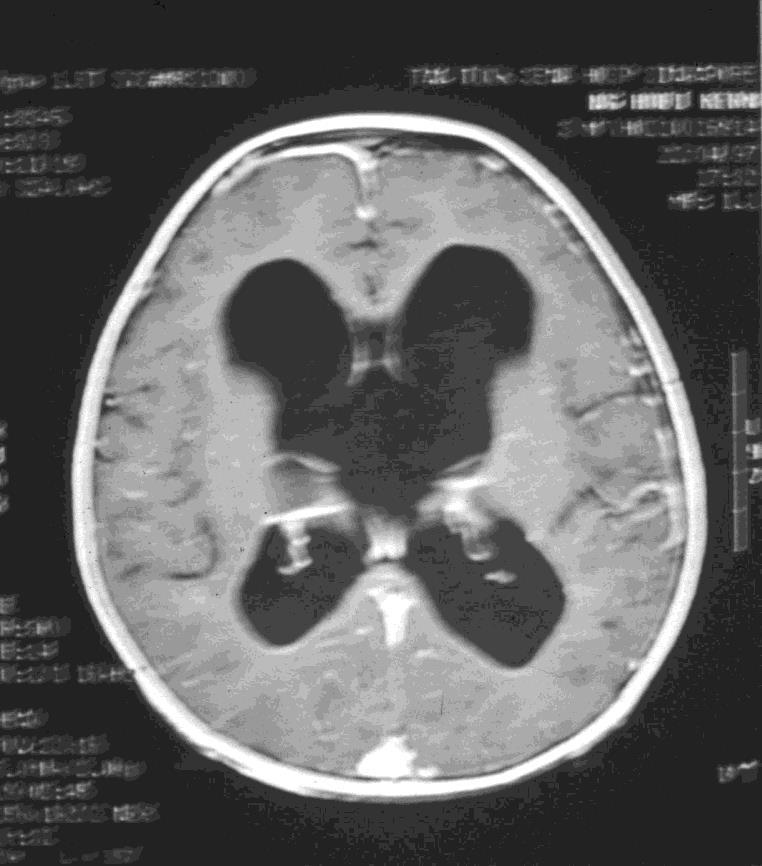
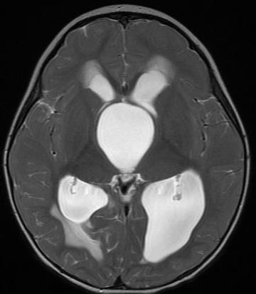
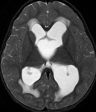
4 Suprasellar Arachnoid Cysts Most common presentation is usually with hydrocephalus May present with endocrinopathies and visual field/acuity deficit Most common endocrinologic symptoms is isosexual precocious puberty (10-40% in pts with suprasellar cysts) Growth hormone deficiency Bitemporal visual field deficit Decreased visual acuity Optic atrophy/papilloedema
5 Bobble-head doll syndrome - rhythmic flexion and extension movement of the head, neck and trunk decreased during periods of concentration, disappears during sleep and increased on standing and walking for cysts in III ventricle Hypothalamic syndromes : failure to thrive, eating disturbance, emotional liability, psychomotor retardation, excessive obesity

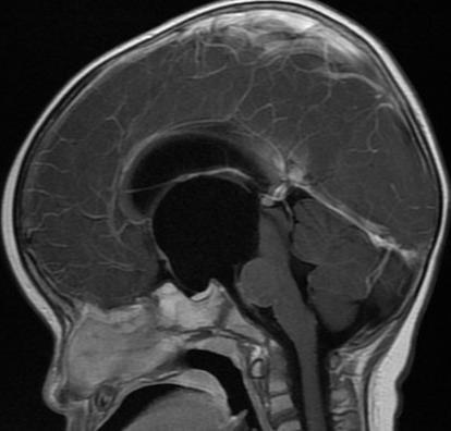
6 Suprasellar arachnoid cyst
7 Surgical anatomy Anatomy of the interpeduncular cistern to- gether with its relationships to other adjacent cisterns and of the Liliequist membrane are of paramount importance for understanding suprasellar cysts The cyst is a lobulated arachnoid complex, and is composed of 2 distinct arachnoid sheets the diencephalic membrane and the mesencephalic membrane Liliequist membrane is located between the interpeduncular and chiasmatic cisterns
8 Miyajima divided the suprasellar cysts into 2 different subtypes: A. cystic dilatation of the interpeduncular cistern B. intra-arachnoid cysts of the diencephalic membrane of Liliequist Ozek World Neurosurgery, 2013
9 The differentiation among these 2 types is very important during the surgical approach because the position of the basilar artery changes in each of them.

10 Where cystic dilation of the interpeduncular cistern had occurred, the diencephalic membrane would constitute the dome and the mesencephalic membrane the bottom of the cyst. The basilar artery bifurcation would remain inside the cyst. Ozek World Neurosurgery, 2013

11 With the intra-arachnoid cystic lesion of the diencephalic membrane, the interpeduncular cistern would be compressed, leaving the basilar artery bifurcation behind the posterior wall of the cyst Ozek World Neurosurgery, 2013
12 Treatment of Suprasellar Arachnoid Cysts Treated in variety of methods Stereotactic drainage Stereotactic intracavitary injection of radioactive isotopes Cyst-ventricular shunting Open fenestration transcortically, transcallosally, pterional or subfrontal approached Endoscopic fenestration via foramen of Monro
13
14 Suprasellar Arachnoid Cysts- Open approaches For transcallosal approach, cyst is often immediately encountered after surgically passing through the corpus callosum; can be difficult if hydrocephalus is absent; hence subfrontal may be safer Major difficulty with open fenestration is in the creating more than one opening in the cyst Transcallosal approach usually succeeds in fenestrating the cyst to ventricle but subfrontal approach only fenestrates to the basal cisterns Transcortical approach risks brain injury and seizure Subfrontal approach risks injury to olfactory tracts with low success rate
15 Shunting Suprasellar Arachnoid Cysts Sole ventricular shunting can lead to cyst enlargement in 40% of time Shunting of suprasellar cyst is difficult without fluoroscopic, stereotactic or endoscopic guidance; but generally wise to leave a catheter inside the cyst as an insurance policy after open/endoscopic fenestration
16 Endoscopic approaches Ventriculo-cystostomy Communicating cyst to the lateral ventricle Ventriculo-cysto-cisternostomy Communicating cyst to the lateral ventricle and then performing a third ventriculostomy through the inferior cyst wall (communicating cyst cavity with the prepontine cistern)

17 Aqueduct is now opened
18 Ventriculo-cysto-cisternostomy Endoscopic fenestration of a suprasellar cyst into both the ventricular system and basal cistern is the more effective treatment It allows communication with a CSF-containing space even if one fenestration closes Decq found endoscopic ventriculo-cystostomy closed in 2 patients with ventriculo-cysto-cisternostomy but opening into the basal cisterns open This phenomenon is due to collapse of upper wall of the cyst
19 Ventriculo-cysto-cisternostomy Fenestrate cyst through Foramen of Monro Fenestrate wall of cyst on floor of 3 rd ventricle Fenestrate floor of 3 rd ventricle (3 rd ventriculostomy)
20
21
22 Summary Not very common lesion Presents with hydrocephalus Cyst is a lobulated arachnoid complex, and is composed of 2 distinct arachnoid sheets the diencephalic membrane and the mesencephalic membrane Best treated with ventriculo-cysto-cisternostomy
Residence of Discipline of Neurosurgery of Hospital da Santa Casa de Misericórdia of Sao Paulo Sao Paulo, Brazil
 Cronicon OPEN ACCESS NEUROLOGY Research Article Efficacy of the Lamina Terminalis Fenestration Associated With the Liliequist Membrane Fenestration in Reducing Shunt-Dependent Hydrocephalus Following Aneurysm
INTRACRANIAL ARACHNOID CYSTS: CLASSIFICATION AND MANAGEMENT. G. Tamburrini, Rome
 INTRACRANIAL ARACHNOID CYSTS: CLASSIFICATION AND MANAGEMENT G. Tamburrini, Rome Incidence 2% of occasional neuroradiological findings From clinical studies (1960 s): 0.4-1% of intracranial space occupying
Complex Hydrocephalus
 2012 Hydrocephalus Association Conference Washington, DC - June 27-July1, 2012 Complex Hydrocephalus Marion L. Walker, MD Professor of Neurosurgery & Pediatrics Primary Children s Medical Center University
Anatomical observations of the subarachnoid cisterns of the brain during surgery
 Anatomical observations of the subarachnoid cisterns of the brain during surgery M. GAZI YASARGIL~ M.D., KONSTANTIN KASDAGLIS, M.D., KEWAL K. JAIN, M.D., AND HANS'PETER WEBER University Neurosurgical Clinic,
For Emergency Doctors. Dr Suzanne Smallbane November 2011
 For Emergency Doctors Dr Suzanne Smallbane November 2011 A: Orbit B: Sphenoid Sinus C: Temporal Lobe D: EAC E: Mastoid air cells F: Cerebellar hemisphere A: Frontal lobe B: Frontal bone C: Dorsum sellae
Brain Meninges, Ventricles and CSF
 Brain Meninges, Ventricles and CSF Lecture Objectives Describe the arrangement of the meninges and their relationship to brain and spinal cord. Explain the occurrence of epidural, subdural and subarachnoid
Optic Pathway Gliomas, Germinomas, Spinal Cord Tumours. Colin Kennedy March 2015
 Optic Pathway Gliomas, Germinomas, Spinal Cord Tumours Colin Kennedy March 2015 Glioma of the optic chiasm. T1-weighted MRI with gadolinium enhancement, showing intense irregular uptake of contrast. The
Anatomy, Histology and general pathology of the Pineal gland. Uri Shiri 1 st year, Int. medicine B
 Anatomy, Histology and general pathology of the Pineal gland Uri Shiri 1 st year, Int. medicine B The Pineal gland A small, ~8mm sized Endocrine gland. The Pineal gland A small, ~8mm sized Endocrine gland.
Aria Fallah MD, MSc, FRCSC
 Aria Fallah MD, MSc, FRCSC Department of Neurosurgery David Geffen School of Medicine at UCLA Pineal Region Tumors Brain Tumor Symposium August 22, 2015 Disclosures None Pineal Gland Arises from an invagination
Microsurgical third ventriculocisternostomy as an alternative to ETV: report of two cases
 Childs Nerv Syst (2008) 24:757 761 DOI 10.1007/s00381-007-0572-6 CASE REPORT Microsurgical third ventriculocisternostomy as an alternative to ETV: report of two cases Erik J. van Lindert Received: 10 August
CSF. Cerebrospinal Fluid(CSF) System
 System") Cerebrospinal Fluid(CSF) System By the end of the lecture, students must be able to describe Physiological Anatomy of CSF Compartments Composition Formation Circulation Reabsorption CSF Pressure Functions
Cerebrospinal Fluid(CSF) System By the end of the lecture, students must be able to describe Physiological Anatomy of CSF Compartments Composition Formation Circulation Reabsorption CSF Pressure Functions
Cavum velum interpositum cyst causing symptomatic trapped ventricle: A case report
 Cavum velum interpositum cyst causing symptomatic trapped ventricle: A case report Poster No: R-0286 Congress: 2014 CSM Type: Scientific Exhibit Authors: T Singh, S Dupre; NAMBOUR/AU Keywords: CNS, Neuroradiology
PHYSIOLOGY OF CSF AND PATHOPHYSIOLOGY OF HYDROCEPHALUS
 PHYSIOLOGY OF CSF AND PATHOPHYSIOLOGY OF HYDROCEPHALUS Introduction Dynamic component of CNS Invaluable tool to diagnosis Physiological reservoir of human proteome Reflects the physiologic state of CNS
displacement of the brain-stem in benign aqueduct stenosis
 Journal of Neurology, Neurosurgery, and Psychiatry, 1972, 35, 114-123 Axial enlargement of the 3rd ventricle, and displacement of the brain-stem in benign aqueduct stenosis JAN JAKUBOWSKI AND ANTONY JEFFERSON
Meninges and Ventricles
 Meninges and Ventricles Irene Yu, class of 2019 LEARNING OBJECTIVES Describe the meningeal layers, the dural infolds, and the spaces they create. Name the contents of the subarachnoid space. Describe the
The anatomy of the subarachnoid cisterns
 Acta Radiologica ISSN: 0001-6926 (Print) (Online) Journal homepage: http://www.tandfonline.com/loi/iaro20 The anatomy of the subarachnoid cisterns B. Liliequist To cite this article: B. Liliequist (1956)
Slide 1. Slide 2. Slide 3. Tomography vs Topography. Computed Tomography (CT): A simplified Topographical review of the Brain. Learning Objective
: A simplified Topographical review of the Brain. Learning Objective") Slide 1 Computed Tomography (CT): A simplified Topographical review of the Brain Jon Wheiler, ACNP-BC Slide 2 Tomography vs Topography Tomography: A technique for displaying a representation of a cross
Slide 1 Computed Tomography (CT): A simplified Topographical review of the Brain Jon Wheiler, ACNP-BC Slide 2 Tomography vs Topography Tomography: A technique for displaying a representation of a cross
Intracranial arachnoid cysts: radiological study of the incidental, the symptomatic and the complicated.
 Intracranial arachnoid cysts: radiological study of the incidental, the symptomatic and the complicated. Poster No.: C-1092 Congress: ECR 2015 Type: Educational Exhibit Authors: C. Ospina Moreno, I. Montejo
Neurosonography: State of the art
 Neurosonography: State of the art Lisa H Lowe, MD, FAAP Professor and Academic Chair, University MO-Kansas City Pediatric Radiologist, Children s Mercy Hospitals and Clinics Learning objectives After this
Microsurgical anatomy of Liliequist s membrane demonstrating three-dimensional configuration
 Acta Neurochir (2011) 153:191 200 DOI 10.1007/s00701-010-0823-2 CLINICAL ARTICLE Microsurgical anatomy of Liliequist s membrane demonstrating three-dimensional configuration Shou-Sen Wang & He-Ping Zheng
The "filling defect" sign helps localise the site of intracranial aneurysm rupture on an unenhanced CT
 The "filling defect" sign helps localise the site of intracranial aneurysm rupture on an unenhanced CT Poster No.: C-3380 Congress: ECR 2010 Type: Topic: Authors: Keywords: DOI: Educational Exhibit Neuro
5. COMMON APPROACHES. Each of the described approaches is also demonstrated on supplementary videos, please see Appendix 2.
 5. COMMON APPROACHES Each of the described approaches is also demonstrated on supplementary videos, please see Appendix 2. 5.1. LATERAL SUPRAORBITAL APPROACH The most common craniotomy approach used in
MR Evaluation of Hydrocephalus
 591 MR Evaluation of Hydrocephalus Taher EI Gammal 1 Marshall B. Allen, Jr. 2 Betty Sue Brooks 1 Edward K. Mark2 An analysis of sagittal T1-weighted MR studies was performed in 23 patients with hydrocephalus,
Sectional Anatomy Head Practice Problems
 1. Which of the following is illustrated by #3? (Fig. 5-42) A) maxillary sinus B) vomer C) septal cartilage D) perpendicular plate of ethmoid bone 2. What number illustrates the cornea? (Fig. 5-42) A)
HEAD AND NECK IMAGING. James Chen (MS IV)
") HEAD AND NECK IMAGING James Chen (MS IV) Anatomy Course Johns Hopkins School of Medicine Sept. 27, 2011 OBJECTIVES Introduce cross sectional imaging of head and neck Computed tomography (CT) Review head
HEAD AND NECK IMAGING James Chen (MS IV) Anatomy Course Johns Hopkins School of Medicine Sept. 27, 2011 OBJECTIVES Introduce cross sectional imaging of head and neck Computed tomography (CT) Review head
Transfontanelar Ultrasound Technique, Normal Anatomy, Anatomic Variants and Classification Review
 Transfontanelar Ultrasound Technique, Normal Anatomy, Anatomic Variants and Classification Review Poster No.: C-2615 Congress: ECR 2013 Type: Educational Exhibit Authors: S. E. Vazquez, R. E. Ochoa Albíztegui
In March of 2000, my family and I moved to Mbale,
 CHPTER 13 Endoscopic Third Ventriculostomy and Choroid Plexus Cauterization for Pediatric Benjamin C. Warf, M.D. In March of 2, my family and I moved to Mbale, Uganda, to help Children s United Rehabilitation
Transcortical Endoscopic Surgery for Intraventricular Lesions
 Review Article J Korean Neurosurg Soc 60 (3) : 327-334, 2017 https://doi.org/10.3340/jkns.2017.0101.008 pissn 2005-3711 eissn 1598-7876 Transcortical Endoscopic Surgery for Intraventricular Lesions Myung-Hyun
World's largest Science, Technology & Medicine Open Access book publisher
 PUBLISHED BY World's largest Science, Technology & Medicine Open Access book publisher 2750+ OPEN ACCESS BOOKS 96,000+ INTERNATIONAL AUTHORS AND EDITORS 88+ MILLION DOWNLOADS BOOKS DELIVERED TO 151 COUNTRIES
Intraventricular neoplasms are uncommon, Intraventricular Tumor: An. Analysis of 18 Cases Dhaka, Bangladesh
 Original Article Nepal Journal of Neuroscience 13:23-29, 2016 Shamsul Alam, MS Intraventricular Tumor: An Analysis of 18 Cases Abu N W Uddin, MS Mashiur R Majumder, MS Comilla Medical College Comilla,
Endoscopic Third Ventriculostomy. Dr Kanwaljeet Garg
 Endoscopic Third Ventriculostomy Dr Kanwaljeet Garg Introduction Endoscopic third ventriculostomyis a technique to treat non communicating hydrocephalus. Involves making a hole in the floor of the third
Blood Supply of the CNS
 Blood Supply of the CNS Lecture Objectives Describe the four arteries supplying the CNS. Follow up each artery to its destination. Describe the circle of Willis and its branches. Discuss the principle
Neurosurgery Department, Cork University Hospital, Cork, Republic of Ireland
 J Neurosurg 103:848 852, 2005 Use of a simple intraoperative hydrostatic pressure test to assess the relationship between mobility of the ventricular stoma and success of third ventriculostomy MAHMOUD
Surgical management of the fourth ventricle arachnoid cysts
 Annals of Medical Research DOI: 10.5455/annalsmedres.2018.09.199 2019;26(1):42-6 Original Article Surgical management of the fourth ventricle arachnoid cysts Sukru Oral 1, Hanifi Bayarogullari 2, Atilla
CNS Embryology 5th Menstrual Week (Dorsal View)
") Imaging of the Fetal Brain; Normal & Abnormal Alfred Abuhamad, M.D. Eastern Virginia Medical School CNS Embryology 5th Menstrual Week (Dorsal View) Day 20 from fertilization Neural plate formed in ectoderm
Imaging of the Fetal Brain; Normal & Abnormal Alfred Abuhamad, M.D. Eastern Virginia Medical School CNS Embryology 5th Menstrual Week (Dorsal View) Day 20 from fertilization Neural plate formed in ectoderm
SOP: Cerebral Ultrasound
 SOP: Cerebral Ultrasound Version Author(s) Date Changes Approved by 1.0 Cornelia Hagmann Manon Benders 29.5.2012 Initial Version Gorm Greisen 1.1 Cornelia Hagmann 18.6.2012 Minor changes Gorm Greisen 1.2
Intracranial arachnoid cyst: an institutional experience
 136 Sharma et al Intracranial arachnoid cyst Intracranial arachnoid cyst: an institutional experience Mukesh Sharma, R.S. Mittal, Rajeev Bansal, Achal Sharma Department of Neurosurgery,S.M.S. Medical College
NANOS Patient Brochure
 NANOS Patient Brochure Pseudotumor Cerebri Copyright 2016. North American Neuro-Ophthalmology Society. All rights reserved. These brochures are produced and made available as is without warranty and for
The Egyptian Journal of Hospital Medicine (July 2018) Vol. 72 (9), Page
 Vol. 72 (9), Page") The Egyptian Journal of Hospital Medicine (July 2018) Vol. 72 (9), Page 5259-5269 Role of Neuroendoscopy in Management of Intraventricular Lesions Shehab Mohamed El Khadrawy, Mohamed Ahmed Ellabbad, Ahmed
The Egyptian Journal of Hospital Medicine (July 2018) Vol. 72 (9), Page 5259-5269 Role of Neuroendoscopy in Management of Intraventricular Lesions Shehab Mohamed El Khadrawy, Mohamed Ahmed Ellabbad, Ahmed
Central nervous system (CNS): brain and spinal cord Collections of cell body and dendrites (grey matter) are called nuclei/nucleus Nucleus can also
: brain and spinal cord Collections of cell body and dendrites (grey matter) are called nuclei/nucleus Nucleus can also") Chapter 3 Part 1 Orientation Directions in the nervous system are described relatively to the neuraxis An imaginary line drawn through the center of the length of the central nervous system, from the bottom
Chapter 3 Part 1 Orientation Directions in the nervous system are described relatively to the neuraxis An imaginary line drawn through the center of the length of the central nervous system, from the bottom
2/20/2019 BRAIN DISSECTION CODING AND DOCUMENTATION OBJECTIVES INTRODUCTION
 BRAIN DISSECTION CODING AND DOCUMENTATION Diana R. Phelps, CPC, CPC-I, CEMC OBJECTIVES Identify general structure of the human brain Describe how the different parts work Recognized the two hemispheres
Original Article Clinical Outcome after Transcortical Approach for Lateral Ventricular Neoplasms
 Egyptian Journal of Neurosurgery Volume 29 / No. 2 / April - June 2014 19-24 Original Article Clinical Outcome after Transcortical Approach for Lateral Ventricular Neoplasms ARTICLE INFO Received: 1 March
Central Nervous System (CNS) -> brain and spinal cord. Major Divisions of the nervous system:
 -> brain and spinal cord. Major Divisions of the nervous system:") Central Nervous System (CNS) -> brain and spinal cord Major Divisions of the nervous system: Afferent (sensory input) -> cell bodies outside of the central nervous system (CNS), carry info into the CNS
Central Nervous System (CNS) -> brain and spinal cord Major Divisions of the nervous system: Afferent (sensory input) -> cell bodies outside of the central nervous system (CNS), carry info into the CNS
Sample page. Radiology. Cross Coder. Essential links from CPT codes to ICD-10-CM and HCPCS
 Cross Coder 2018 Radiology Essential links from CPT codes to ICD-10-CM and HCPCS POWER UP YOUR CODING with Optum360, your trusted coding partner for 32 years. Visit optum360coding.com. Contents Introduction...
NEURO IMAGING 2. Dr. Said Huwaijah Chairman of radiology Dep, Damascus Univercity
 NEURO IMAGING 2 Dr. Said Huwaijah Chairman of radiology Dep, Damascus Univercity I. EPIDURAL HEMATOMA (EDH) LOCATION Seventy to seventy-five percent occur in temporoparietal region. CAUSE Most likely caused
Attenuation value in HU From -500 To HU From -10 To HU From 60 To 90 HU. From 200 HU and above
 Brain Imaging Common CT attenuation values Structure Air Fat Water Brain tissue Recent hematoma Calcifications Bone Brain edema and infarction Normal liver parenchyma Attenuation value in HU From -500
Supra- and infratentorial brain tumors from childhood to maternity
 Supra- and infratentorial brain tumors from childhood to maternity What to expect? I am going to show you the characteristic imaging findings of following tumors: Thierry A.G.M. Huisman, MD, FICIS, EQNR
Table of Contents: SKULL AND BRAIN. Scalp, Skull. Anatomically Based Differentials. Skull Normal Variants. Scalp Mass, Child.
 Table of Contents: SKULL AND BRAIN Scalp, Skull Skull Normal Variants Scalp Mass, Child Scalp Mass, Adult Congenital Anomalies of Skull Base Sellar/Parasellar Mass With Skull Base Invasion "Hair on End"
Ventricles, CSF & Meninges. Steven McLoon Department of Neuroscience University of Minnesota
 Ventricles, CSF & Meninges Steven McLoon Department of Neuroscience University of Minnesota 1 Coffee Hour Thursday (Sept 14) 8:30-9:30am Surdyk s Café in Northrop Auditorium Stop by for a minute or an
Table of Contents: Section I. Introduction. 1. Assessing Surgical Innovation. Section II. Trauma to the Scalp, Skull, and Brain
 Table of Contents: Section I. Introduction 1. Assessing Surgical Innovation Section II. Trauma to the Scalp, Skull, and Brain 2. Surgical Repair of Major Defects of the Scalp and Skull 3. Perioperative
Spontaneous Ventriculostomy: Report of Three Cases Revealed by Flow-Sensitive Phase-Contrast Cine MR Imaging
 AJNR Am J Neuroradiol 20:1647 1652, October 1999 Case Report Spontaneous Ventriculostomy: Report of Three Cases Revealed by Flow-Sensitive Phase-Contrast Cine MR Imaging Alex Rovira, Jaume Capellades,
Hydrocephalus in children. Eva Brichtova, M.D., Ph.D., Department of Pediatric Sugery, Orthopaedics and Traumatology, University Hospital Brno
 Hydrocephalus in children Eva Brichtova, M.D., Ph.D., Department of Pediatric Sugery, Orthopaedics and Traumatology, University Hospital Brno Ventricle system Ventricle system, cerebral cisterns Hydrocephalus
Tumours of the third ventricle in children
 Journal of Neurology, Neurosurgery, and Psychiatry, 1972, 35, 776-788 Tumours of the third ventricle in children BENNETT M. STEIN, RICHARD A. R. FRASER, AND MICHAEL S. TENNER From the Department of Neurological
Neuroendoscopic management of interhemispheric cysts in children
 J Neurosurg (3 Suppl Pediatrics) 105:194 202, 2006 Neuroendoscopic management of interhemispheric cysts in children GIUSEPPE CINALLI, M.D., PAOLA PERETTA, M.D., PIETRO SPENNATO, M.D., LUCIANO SAVARESE,
Chapter 5: Fetal Central Nervous System 71
 71 Chapter 5 Fetal Central Nervous System Embryology NEURULATION begins with the formation of the neural plate, the neural folds and their ultimate fusion and closure as the NEURAL TUBE. NEURAL PLATE -
intracranial anomalies
 Chapter 5: Fetal Central Nervous System 84 intracranial anomalies Hydrocephaly Dilatation of ventricular system secondary to an increase in the amount of CSF. Effects of hydrocephalus include flattening
EXPERT DIFFERENTIAL DIAGNOSIS:
 EXPERT DIFFERENTIAL DIAGNOSIS: Sellar Region Anne G. Osborn, M.D. DISCLOSURE: Published RSNA 2008 SELLA, PITUITARY: Normal Gross, 3T Anatomy SELLA, PITUITARY: Anatomically-Based Differential Diagnoses
Clinical anatomy of the fourth ventricle foramina
 Clinical Anatomy Page 1 of 5 Clinical anatomy of the fourth ventricle foramina IN Mavridis 1* Abstract Introduction The three foramina of the fourth ventricle of the human brain were first described during
Acute stroke. Ischaemic stroke. Characteristics. Temporal classification. Clinical features. Interpretation of Emergency Head CT
 Ischaemic stroke Characteristics Stroke is the third most common cause of death in the UK, and the leading cause of disability. 80% of strokes are ischaemic Large vessel occlusive atheromatous disease
Craniopharyngioma. Michael Gottschalk, MD,PhD University of California San Diego Rady Children s Hospital
 Craniopharyngioma Michael Gottschalk, MD,PhD University of California San Diego Rady Children s Hospital Objectives Incidence Clinical Presentation Treatment Options Perioperative concerns Long-term endocrine
Brainstem. By Dr. Bhushan R. Kavimandan
 Brainstem By Dr. Bhushan R. Kavimandan Development Ventricles in brainstem Mesencephalon cerebral aqueduct Metencephalon 4 th ventricle Mylencephalon 4 th ventricle Corpus callosum Posterior commissure
Case Studies in Sella/Parasellar Region. Child thirsty, increased urination. Imaging. Suprasellar Germ Cell Tumor (Germinoma) No Disclosures
 No Disclosures") Case Studies in Sella/Parasellar Region No Disclosures 2018 Head and Neck Imaging Conference Child thirsty, increased urination Suprasellar Germ Cell Tumor (Germinoma) Midline Pineal >> Suprasellar > Other
Case Studies in Sella/Parasellar Region No Disclosures 2018 Head and Neck Imaging Conference Child thirsty, increased urination Suprasellar Germ Cell Tumor (Germinoma) Midline Pineal >> Suprasellar > Other
CNS TUMORS. D r. Ali Eltayb ( U. of Omdurman. I ). M. Path (U. of Alexandria)
. M. Path (U. of Alexandria)") CNS TUMORS D r. Ali Eltayb ( U. of Omdurman. I ). M. Path (U. of Alexandria) CNS TUMORS The annual incidence of intracranial tumors of the CNS ISmore than intraspinal tumors May be Primary or Secondary
CNS TUMORS D r. Ali Eltayb ( U. of Omdurman. I ). M. Path (U. of Alexandria) CNS TUMORS The annual incidence of intracranial tumors of the CNS ISmore than intraspinal tumors May be Primary or Secondary
Prenatal Prediction of The Neurologically Impaired Neonate By Ultrasound
 Prenatal Prediction of The Neurologically Impaired Neonate By Ultrasound Robert H. Debbs, D.O.,F.A.C.O.O.G. Professor of OB-GYN Perelman School of Medicine, University of Pennsylvania Director, Pennsylvania
PRENATAL DIAGNOSIS OF ARACHNOID CYSTS
 REVIEW ARTICLE PRENATAL DIAGNOSIS OF ARACHNOID CYSTS Chih-Ping Chen* Department of Obstetrics and Gynecology, Mackay Memorial Hospital, Taipei, and Department of Biotechnology, Asia University, Taichung,
EANS Training Course Moscow, 5 th 8 th May 2019 Tumours
 EANS Training Course Moscow, 5 th 8 th May 2019 Tumours SUNDAY, MAY 5 th, 2019 09:00 09:10 Welcome 09:15 10:30 Tumours Chair: Meling 09:15 09:30 Molecular biology of brain tumours Reinert 09:30 09:45 Imaging
Head CT Scan Interpretation: A Five-Step Approach to Seeing Inside the Head Lawrence B. Stack, MD
 Head CT Scan Interpretation: A Five-Step Approach to Seeing Inside the Head Lawrence B. Stack, MD Five Step Approach 1. Adequate study 2. Bone windows 3. Ventricles 4. Quadrigeminal cistern 5. Parenchyma
The second most common reason for being sued for negligence in neurosurgery is a problem
 Correspondence to: Mr Ian K Pople, Department of Neurosurgery, Frenchay Hospital, Frenchay Park Road, Bristol BS16 1LE, UK: ikpople@hotmail.com HYDROCEPHALUS AND SHUNTS: WHAT THE NEUROLOGIST SHOULD KNOW
Cerebral hemisphere. Parietal Frontal Occipital Temporal
 Cerebral hemisphere Sulcus / Fissure Central Precental gyrus Postcentral gyrus Lateral (cerebral) Parieto-occipital Cerebral cortex Frontal lobe Parietal lobe Temporal lobe Insula Amygdala Hippocampus
Subarachnoid space. Subarachnoid space size
 80 Subarachnoid space Martin M. Mortazavi 1, Nimer Adeeb 2, Fareed Rizq 2 and R. Shane Tubbs 3 1 University of Washington School of Medicine, Seattle, Washington, United States 2 Children s of Alabama,
What the pediatric lateral ventricles tell us.
 What the pediatric lateral ventricles tell us. Poster No.: C-2069 Congress: ECR 2017 Type: Educational Exhibit Authors: R. Dominguez Piedra, M. I. Martinez-Leon, V. Romero Laguna, 1 1 2 1 1 2 M. Sánchez-Porro
Laser-assisted neuroendoscopy using a neodymium yttrium aluminum garnet or diode contact laser with pretreated fiber tips
 J Neurosurg 88:82 92, 1998 Laser-assisted neuroendoscopy using a neodymium yttrium aluminum garnet or diode contact laser with pretreated fiber tips WILLIAM P. VANDERTOP, M.D., PH.D., RUDOLF M. VERDAASDONK,
Student Lab #: Date. Lab: Gross Anatomy of Brain Sheep Brain Dissection Organ System: Nervous Subdivision: CNS (Central Nervous System)
") Lab: Gross Anatomy of Brain Sheep Brain Dissection Organ System: Nervous Subdivision: CNS (Central Nervous System) Student Lab #: Date 1 Objectives: 1. Learn the main components making up a motor neuron.
Lab: Gross Anatomy of Brain Sheep Brain Dissection Organ System: Nervous Subdivision: CNS (Central Nervous System) Student Lab #: Date 1 Objectives: 1. Learn the main components making up a motor neuron.
Craniopharyngiomas (from Greek: κρανίον, skull
 J Neurosurg 119:1194 1207, 2013 AANS, 2013 Endoscopic endonasal surgery for craniopharyngiomas: surgical outcome in 64 patients Clinical article Maria Koutourousiou, M.D., 1 Paul A. Gardner, M.D., 1 Juan
The optimal treatment for arachnoid cysts is controversial. Cyst-ventricle stent as primary or salvage treatment for posterior fossa arachnoid cysts
 J Neurosurg Pediatrics 7:000 000, 7:549 556, 2011 Cyst-ventricle stent as primary or salvage treatment for posterior fossa arachnoid cysts Clinical article Daniel H. Fulkerson, M.D., 1 Todd D. Vogel, M.D.,
Cross sectional imaging of Intracranial cystic lesions Abdel Razek A
 Cross sectional imaging of Intracranial cystic lesions Abdel Razek A Department of Radiology. Mansoura Faculty of Medicine, Mansoura. Egypt. arazek@mans.edu.eg Introduction Intracranial cystic lesions
Quick practical guide to Cranial Ultrasound in the newborn
 Quick practical guide to Cranial Ultrasound in the newborn Introduction A standard set of views is taken to assist with consistent visualisation of structures and in the interpretation of possible abnormalities.
The authors discuss their surgical approaches
 GENERAL SCIENTIFIC SESSION 3 GENERAL SCIENTIFIC SESSION 3 Open vs Endoscopic: When To Use Which Laligam Sekhar, MD* Alessandra Mantovani, MD* Martin Mortazavi, MD* Theodore H. Schwartz, MD WIlliam T. Couldwell,
Cerebro-vascular stroke
 Cerebro-vascular stroke CT Terminology Hypodense lesion = lesion of lower density than the normal brain tissue Hyperdense lesion = lesion of higher density than normal brain tissue Isodense lesion = lesion
INTRACRANIAL PRESSURE -!!
 INTRACRANIAL PRESSURE - Significance raised ICP main cause of death in severe head injury main cause of morbidity in moderate and mild head injury main target and prognostic indicator in the ITU setting
Journal of Neurological Sciences [Turkish] 30:(2)# 36; , Case Report
![Journal of Neurological Sciences [Turkish] 30:(2)# 36; , Case Report](/thumbs/91/107308952.jpg "Journal of Neurological Sciences [Turkish] 30:(2)# 36; , Case Report") Journal of Neurological Sciences [Turkish] 30:(2)# 36; 440-444, 2013 http://www.jns.dergisi.org/text.php3?id=677 Case Report Endoscopic Treatment of a Suprasellar Arachnoid Cyst Causing Precocious Puberty
Journal of Neurological Sciences [Turkish] 30:(2)# 36; 440-444, 2013 http://www.jns.dergisi.org/text.php3?id=677 Case Report Endoscopic Treatment of a Suprasellar Arachnoid Cyst Causing Precocious Puberty
Paul Gigante HMS IV Gillian Lieberman, MD. Sept Mr. T s T s Headache. Paul Gigante,, Harvard Medical School Year IV Gillian Lieberman, MD
 Sept 2005 Mr. T s T s Headache Paul Gigante,, Harvard Medical School Year IV Mr. T s T s Presentation 45 year-old welder complains of sudden severe headache and witnessed seizure with loss of consciousness
Surgical Treatment of Olfactory Groove Meningioma
 Med. J. Cairo Univ., VoL 81, No. 1, March: 133-137, 2013 www.medicaljournalofcairouniversity.com Surgical Treatment of Olfactory Groove Meningioma AHMED ELSAWAF, M.D., Ph.D. The Department of Neurosurgery,
Department of Cognitive Science UCSD
 Department of Cognitive Science UCSD Verse 1: Neocortex, frontal lobe, Brain stem, brain stem, Hippocampus, neural node, Right hemisphere, Pons and cortex visual, Brain stem, brain stem, Sylvian fissure,
Empty sella syndrome: does it exist in children?
 Neurosurg Focus 7 (2):Clinical Pearl 2, 1999 Empty sella syndrome: does it exist in children? Ahmed Ammar, M.B., Ch.B., D.M.Sc., Ali Al-Sultan, M.D., F.R.C.P.(C), Fatma Al Mulhim, M.B., Ch.B., and Abdulla
Ventriculo-Peritoneal/ Lumbo-Peritoneal Shunts
 Ventriculo-Peritoneal/ Lumbo-Peritoneal Shunts Exceptional healthcare, personally delivered Ventriculo-Peritoneal/ Lumbo-Peritoneal Shunts What is hydrocephalus? Hydrocephalus is the build up of an excess
MECHANICAL 3-D MODEL FOR NEURO-ENDOSCOPIC SURGERY SIMULATION
 MECHANICAL 3-D MODEL FOR NEURO-ENDOSCOPIC SURGERY SIMULATION Anyi Wang Leader Jeff Groskopf Communicator Michael Rossmiller BSAC Nicholas Schapals BWIG Client: Bermans J. Iskandar, MD. Advisor: Professor
Intrasphenoidal Rathke's Cleft Cyst: Case presentation and review of the literature
 Romanian Neurosurgery Volume XXX Number 4 2016 October - December Article Intrasphenoidal Rathke's Cleft Cyst: Case presentation and review of the literature Umit Kocaman, Muhammet Bahadir Yilmaz, Hakan
Pediatric Brain Tumors Pre, Intra & Post Op Evaluation and Management. Timothy M. George, MD, FACS, FAAP
 Pediatric Brain Tumors Pre, Intra & Post Op Evaluation and Management Timothy M. George, MD, FACS, FAAP PEDIATRIC BRAIN TUMORS BACKGROUND: Incidence: Third most common pediatric tumor type (leukemia, neuroblastoma,
Corpus Callosal Signal Changes in Patients with Obstructive Hydrocephalus after Ventriculoperitoneal Shunting
 AJNR Am J Neuroradiol 22:158 162, January 2001 Corpus Callosal Signal Changes in Patients with Hydrocephalus after Ventriculoperitoneal Shunting John I. Lane, Patrick H. Luetmer, and John L. Atkinson BACKGROUND
CODING SHEET HYDROCEPHALUS REIMBURSEMENT. All Medicare information is current as of the time of printing.
 CODING SHEET HYDROCEPHALUS REIMBURSEMENT All Medicare information is current as of the January 2014 Hydrocephalus ing Coding Options Commonly Billed Codes for Physicians, Hospitals, and Ambulatory Surgery
Lab Photo Review Sheet
 9 8 0. Posterior Median Sulcus. Central Canal. Dorsal (Posterior) Horn. Ventral (Anterior) Horn. Grey Matter. White Matter. Anterior Median Fissure 8. Ventral (Anterior) Root (ramus) 9. Dorsal (Posterior)
Surgery of posterior fossa arachnoid cyst causing hydrocephalus unmasking the cause of epilepsy
 CASE REPORTS ALBANIAN MEDICAL JOURNAL Surgery of posterior fossa arachnoid cyst causing hydrocephalus unmasking the cause of epilepsy Florian Dashi 1, Arben Rroji 1, Eugen Enesi 1, Suzana Gjeci 1, Ridvan
Naples, Italy IFNE-JSNE-GLEN-ISNE. Hands-on Workshop on CEREBRAL AND VENTRICULAR NEUROENDOSCOPY. January 22-24, 2018 PRELIMINARY PROGRAM
 Under the patronage of International Federation of NeuroEndoscopy (IFNE) Japanese Society for NeuroEndoscopy (JSNE) Grupo Latinoamericano de Estudios en Neuroendoscopia (GLEN) Indian Society for Neuroendoscopy
Skullbase Lesions. Skullbase Surgery Open vs endoscopic. Choice Of Surgical Approaches 12/28/2015. Skullbase Surgery: Evolution
 Skullbase Lesions Skullbase Surgery Open vs endoscopic Prof Asim Mahmood,FRCS,FACS,FICS,FAANS, Professor of Neurosurgery Henry Ford Hospital Detroit, MI, USA Anterior Cranial Fossa Subfrontal meningioma
We are IntechOpen, the world s leading publisher of Open Access books Built by scientists, for scientists. International authors and editors
 We are IntechOpen, the world s leading publisher of Open Access books Built by scientists, for scientists 3,900 116,000 120M Open access books available International authors and editors Downloads Our
Arachnoid cysts account for 1% of all intracranial. Endoscopic treatment of intraparenchymal arachnoid cysts in children.
 J Neurosurg Pediatrics 14:501 507, 2014 AANS, 2014 Endoscopic treatment of intraparenchymal arachnoid cysts in children Clinical article Nasser M. F. El-Ghandour, M.D. Department of Neurosurgery, Faculty
Anatomy Lab (1) Theoretical Part. Page (2 A) Page (2B)
 Theoretical Part. Page (2 A) Page (2B)") Anatomy Lab (1) This sheet only includes the extra notes for the lab handout regarding the theoretical part, as for the practical part it includes everything the doctor mentioned. Theoretical Part Page
Anatomy Lab (1) This sheet only includes the extra notes for the lab handout regarding the theoretical part, as for the practical part it includes everything the doctor mentioned. Theoretical Part Page
The Human Brain. I Think Therefore I am
 The Human Brain I Think Therefore I am The Beginning The simplest creatures have very simple nervous systems made up of nothing but a bunch of nerve cells They have neural nets, individual neurons linked
Long- term outcome for patients with craniopharyngiomas
 1 Long- term outcome for patients with craniopharyngiomas Eric Tande Håland Tutor: Jon Berg- Johnsen Department of Neurosurgery Oslo University Hospital Faculty of Medicine UNIVERSITY OF OSLO 2017 2 Contents