EXPERT DIFFERENTIAL DIAGNOSIS:
|
|
|
- Ashlyn Gallagher
- 5 years ago
- Views:
Transcription
1 EXPERT DIFFERENTIAL DIAGNOSIS: Sellar Region Anne G. Osborn, M.D.

2 DISCLOSURE: Published RSNA 2008
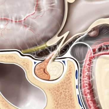
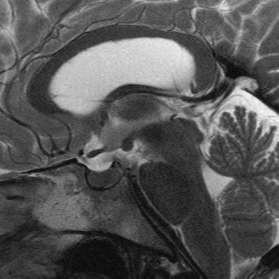
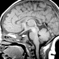
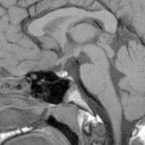
3 SELLA, PITUITARY: Normal Gross, 3T Anatomy
4 SELLA, PITUITARY: Anatomically-Based Differential Diagnoses Intrasellar Sella/pituitary, normal variants Enlarged pituitary gland Intrasellar lesion Cystic intrasellar mass Suprasellar Suprasellar mass, general Suprasellar mass, pediatric Suprasellar cystic mass Calcified suprasellar mass Thick infundibular stalk
5 SELLA/PITUITARY: Normal Variants Common Pituitary hyperplasia (physiologic) Pituitary incidentaloma Empty sella Less common Bright pituitary gland Absent posterior pituitary bright spot Small sella turcica J -shaped sella Rare but important Paramedian ( kissing ) internal carotid arteries


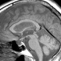
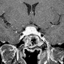
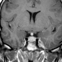
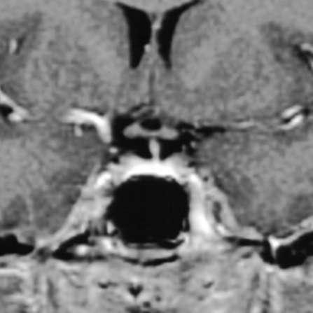
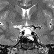
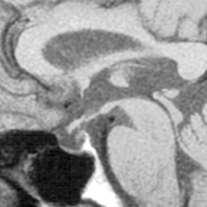
6 PITUITARY HYPERPLASIA (PHYSIOLOGIC) Must know age, gender!! Physiologic mm Convex upwards Strong, uniform enhancement Can be indistinguishable from Macroadenoma Lymphocytic hypophitis Metastasis, lymphoma Beware: Macroadenoma in prepubescent males! Postpartum lactating 14 mm 11 mm 21y menstruating


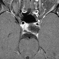
7 PITUITARY INCIDENTALOMA Inhomogeneous or nonenhancing filling defect 15-20% normal MRs Can also be transient Etiology Nonneoplastic cyst (e.g., pars intermedia, Rathke cleft) Nonfunctioning microadenoma (common at autopsy)


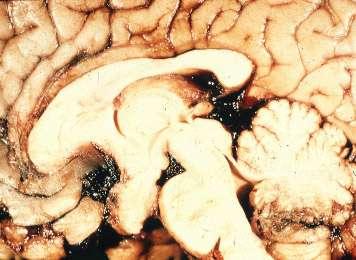
8 EMPTY SELLA Primary (i.e., not post-surgical) Courtesy M. Sage
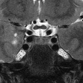
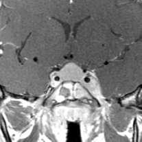
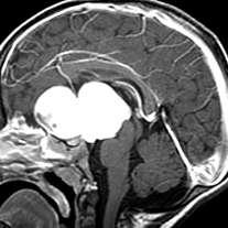
9 MISCELLANEOUS NORMAL VARIANTS Bright pituitary Shallow sella Small sella Kissing carotids
10 ENLARGED PITUITARY Common Pituitary hyperplasia Microadenoma Macroadenoma Less common Neurosarcoid LCH Lymphocytic hypophysitis Macroadenoma mimics Rare but important Intracranial hypotension Meningioma Metastasis davf Pituicytoma Pseudotumor Lymphoma Leukemia

11 ENLARGED PITUITARY Neurosarcoid LCH Lymphoma Pseudotumor


12 ENLARGED PITUITARY Macro mimic ( shallow sella ) Intracranial hypotension Lymphocytic hypophysitis Metastasis
13 INTRASELLAR LESION Common Pituitary hyperplasia Microadenoma Empty sella Less common Macroadenoma Rathke cleft cyst Craniopharyngioma Neurosarcoid Rare but important Lymphocytic hypophysitis Intracranial hypotension Kissing carotids Aneurysm Meningioma Metastasis Lymphoma davf CNS siderosis ( black pituitary) Hepatic encephalopathy ( white pituitary )

14 Variable histology Prolactinoma 30% GH 20% Null cell 20% ACTH 10% FSH/LH 10% PRL-GH 5% Mixed, TSH 1-5% MICROADENOMA Incidental pituitary lesions are common 17% in autopsy series
 Enhances slower than normal")
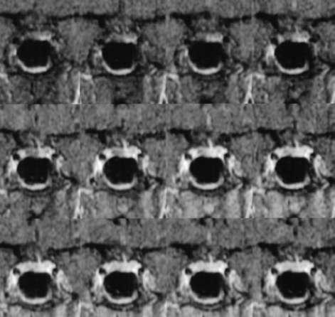
15 MICROADENOMA Pituitary Microadenoma 10mm or less 10-20% of autopsies Micro >>> Macro Dynamic Imaging Increases sensitivity (10-30% seen only on dynamic MR) Enhances slower than normal gland
16 MICROADENOMA


17 RATHKE CLEFT CYST Intrasellar 40% Suprasellar extent 60% 3mm 3cm Most incidental Symptomatic Pituitary dysfunction Visual change, HA Look for Intracystic nodule Claw sign


18 MISCELLANEOUS INTRASELLAR LESIONS Craniopharyngioma Completely intrasellar is rare Variable signal Craniopharyngioma CNS siderosis Black pituitary on T2* Iron overload states >> SAH Thalassemia Hemochromatosis Siderosis
19 CYSTIC-APPEARING INTRASELLAR MASS Common Empty sella Intracranial hypertension, idiopathic Less common Obstructive hydrocephalus Rathke cleft cyst Craniopharyngioma Arachnoid cyst Epidermoid cyst Neurocysticercosis Rare but important Pituitary apoplexy Saccular aneurysm (thrombosed)
20 CYSTIC INTRASELLAR MASS Key question Cystic mass originating WITHIN sella? Intrasellar extension from suprasellar cystic lesion Intrasellar extension of suprasellar lesion >> cystic intrasellar mass


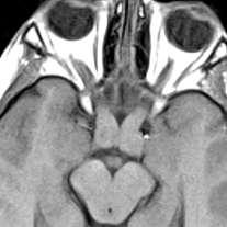
21 INTRACRANIAL HYPERTENSION (IDIOPATHIC) A.k.a. pseudotumor cerebri ICP without underlying pathology Young obese female Imaging Partial empty sella Dilation/tortuosity of optic n. sheath Posterior globe flattened Subarachnoid spaces, sulci Small ( slit-like ) ventricles seen in only 10%

22 INTRASELLAR CYSTIC LESIONS Hydrocephalus Craniopharyngioma Epidermoid cyst Rathke cleft cyst


23 INTRASELLAR CYSTIC LESIONS Arachnoid cyst Neurocysticercosis Pituitary apoplexy (Sheehan) Thrombosed aneurysm
24 SUPRASELLAR MASS, GENERAL Common Macroadenoma Meningioma Saccular aneurysm Craniopharyngioma Pilocytic astrocytoma Less common Dilated 3 rd ventricle Arachnoid cyst Neurocysticercosis Rathke cleft cyst Neurosarcoid LCH Germinoma Dermoid cyst Lipoma Rare but important Lymphocytic hypophysitis Pituitary apoplexy Tuber cinereum hamartoma Epidermoid cyst Pituicytoma Diffuse astrocytoma Pilomyxoid astrocytoma Ectopic neurohypophysis Metastasis Lymphoma Leukemia Cavernous malformation Tuberculoma Pituitary abscess

25 SUPRASELLAR MASS, GENERAL Big 5 = 75% The big kahuna Macroadenoma (35%-50%) Approximately 10% Meningioma Aneurysm Craniopharyngioma Astrocytoma (hypothalamicchiasmatic)
26 SUPRASELLAR MASS, GENERAL: Key Questions to Consider Is the patient adult or child? Is the mass intra- or extra-axial? If extra-axial, does it arise from pituitary? Can you identify pituitary gland separate from mass? Or is the gland the mass? Does it mostly involve the infundibular stalk? Intra-axial masses arise from Optic chiasm, hypothalamus 3 rd ventricle Is the mass cystic or solid? If cystic, is it exactly like CSF?
27 SUPRASELLAR MASS, PEDIATRIC Common Pilocytic astrocytoma Craniopharyngioma Pituitary hyperplasia Hydrocephalus ( 3 rd v.) Less common Germinoma Tuber cinereum hamartoma Arachnoid cyst LCH Stalk anomalies Teratoma Rare but important Lipoma Macroadenoma Dermoid cyst Pilomyxoid aneurysm Saccular aneurysm Trilateral retinoblastoma Lymphocytic hypophysitis Lymphoma/leukemia Rathke cleft cyst
28 PILOCYTIC ASTROCYTOMA


29 CRANIOPHARYNGIOMA 2nd most common suprasellar mass in children Peak incidence 5-15 yrs Second peak yrs M = F Visual changes Endocrine dysfunction Mass effect H/A, N, V, papilledema Imaging 90% Ca++, 90% cystic 90% enhance Cysts variable intensity

30 Suprasellar region is second most common site M = F suprasellar 90% present < 20 yrs Endocrine dysfunction Diabetes insipidus Panhypopituitarism Radiosensitive Up to 90% 10 survival GERMINOMA


31 GERMINOMA Imaging Combined lesion typical but may affect only infundibular stalk May be hyperdense (CT) Isointense T1WI Hyper- to isointense T2WI Enhances homogeneously CSF dissemination common


32 TUBER CINEREUM HAMARTOMA Clinical Precocious puberty Usually < 2yrs Gelastic seizures M > F Pallister-Hall Facial anomalies Polydactyly Imperforate anus Pathology Congenital nonneoplastic heterotopia Between infundibular stalk, mamillary bodies

33 TUBER CINEREUM HAMARTOMA
34 SUPRASELLAR CYSTIC MASS Common Hydrocephalus ( 3 rd v) Arachnoid cyst Craniopharyngioma Neurocysticercosis Less common Rathke cleft cyst Dermoid cyst Epidermoid cyst Enlarged PVSs Rare but important Macroadenoma Pituitary apoplexy Astrocytoma (pilocytic, pilomyxoid) Ependymal cyst Saccular aneurysm (partially/completely thrombosed)


35 Arachnoid cyst HYDROCEPHALUS vs. ARACHNOID CYST Hydrocephalus 3 rd v easily identified, dilated May project into sella Signal CSF-like Hydrocephalus Arachnoid cyst 10% suprasellar 3 rd v elevated/compressed, often difficult to identify CSF may be slightly different signal from cyst


36 RATHKE CLEFT CYST 60% suprasellar Variable size Can be tiny (intrapituitary) Can be huge!! May widen, erode sella Variable signal Look for Intracystic nodule Claw sign

37 MISCELLANEOUS SUPRASELLAR CYSTS, CYSTIC-APPEARING MASSES Craniopharyngioma Neurocysticercosis Astrocytoma Pituitary apoplexy
38 CALCIFIED SUPRASELLAR MASS Common Atherosclerosis Craniopharyngioma Meningioma Aneurysm (saccular, fusiform) Less common Neurocysticercosis Pilocytic astrocytoma Dermoid cyst Rare but important Macroadenoma Tuberculosis Chondroid tumor
39 CALCIFIED SUPRASELLAR MASS Key questions Is Ca++ curvilinear, punctate, globular? Does lesion enhance?

")
40 CALCIFIED SUPRASELLAR MASS: Atherosclerosis Atherosclerosis Older patient Curvilinear Ca++ Bilateral, multifocal Aneurysm Saccular (ring, arc) Fusiform (curvilinear) Ca++


41 CALCIFIED SUPRASELLAR MASS: Miscellaneous Craniopharyngioma Meningioma Dermoid cyst Macroadenoma


42 THICK INFUNDIBULAR STALK: Key Issues Know what normal stalk looks like! Tapers from top to bottom 2 mm or less diameter Thick stalk > 2mm Nontapering Patient age extremely important Child = LCH, germinoma Adult = sarcoid, hypophysitis, pituicytoma, metastasis, lymphoma


43 THICK STALK Child Adult Germinoma Sarcoid Histiocytosis Lymphoma
44 SUMMARY Know patient age, clinical/lab Determine lesion sublocation Intrasellar Suprasellar Infundibular stalk Remember the Big Five Find pituitary separate from mass? No (if gland is mass, consider macroadenoma, metastasis, hypophysitis, lymphoma) Yes (consider meningioma, aneurysm, cyst, etc.)
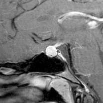
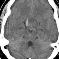
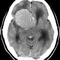
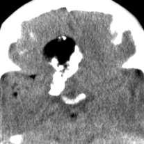
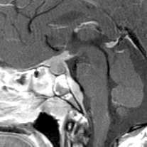
Metastasis. 57 year old with progressive Headache and Right Sided Visual Loss
 Metastasis 1% of sellar/parasellar masses Usually occurs with known primary Can involve third ventricle, hypothalamus, infundibular stalk May be both supra-, intrasellar 57 year old with progressive Headache
PITUITARY PARASELLAR LESIONS. Kim Learned, MD
 PITUITARY PARASELLAR LESIONS Kim Learned, MD DIFFERENTIALS Pituitary Sella Clivus, Sphenoid Sinus Suprasellar Optic chiasm, Hypothalamus, Circle of Willis Parasellar Cavernous Sinus Case 1 17 YEAR-OLD
Case Studies in Sella/Parasellar Region. Child thirsty, increased urination. Imaging. Suprasellar Germ Cell Tumor (Germinoma) No Disclosures
 No Disclosures") Case Studies in Sella/Parasellar Region No Disclosures 2018 Head and Neck Imaging Conference Child thirsty, increased urination Suprasellar Germ Cell Tumor (Germinoma) Midline Pineal >> Suprasellar > Other
Case Studies in Sella/Parasellar Region No Disclosures 2018 Head and Neck Imaging Conference Child thirsty, increased urination Suprasellar Germ Cell Tumor (Germinoma) Midline Pineal >> Suprasellar > Other
Laurie A. Loevner, MD
 Laurie A. Loevner, MD Chief, Division of Neuroradiology UPHS Professor of Radiology, Otorhinolaryngology: Head & Neck Surgery, Neurosurgery, and Ophthalmology University of Pennsylvania Health System Disclosures
Imaging The Turkish Saddle. Russell Goodman, HMS III Dr. Gillian Lieberman
 Imaging The Turkish Saddle Russell Goodman, HMS III Dr. Gillian Lieberman Learning Objectives Review the anatomy of the sellar region Discuss the differential diagnosis of sellar masses Discuss typical
Where Has My Vision Gone? Evaluation of Sellar Lesions. Caleb Stowell,, HMS III Gillian Lieberman, MD November 2008
 Where Has My Vision Gone? Evaluation of Sellar Lesions Caleb Stowell,, HMS III Gillian Lieberman, MD November 2008 Objectives Present a case highlighting the clinical presentation and evaluation of a sellar
What we will cover. Evaluation of the Child with Suspected Pituitary Disease. ituitary
 Evaluation of the Child with Suspected Pituitary Disease Craig Alter, MD University of Pennsylvania Children s Hospital of Philadelphia What we will cover * What laboratory tests to order * MRI: common
DISCLOSURES LEARNING OBJECTIVES WE WILL NOT DISCUSS. CSB: Birdseye View MESSAGE NAVIGATING THE SELLA AND CENTRAL SKULL BASE
 NAVIGATING THE SELLA AND CENTRAL SKULL BASE Christopher P. Hess, M.D., Ph.D. DISCLOSURES Research Support, General Electric SLIDES: http://www.radiology.ucsf.edu/research/meetings/rsna LEARNING OBJECTIVES
Pediatric CNS Tumors. Disclosures. Acknowledgements. Introduction. Introduction. Posterior Fossa Tumors. Whitney Finke, MD
 Pediatric CNS Tumors Disclosures Whitney Finke, MD Neuroradiology Fellow PGY-6 University of Utah Health Sciences Center Salt Lake City, Utah None Acknowledgements Introduction Nicholas A. Koontz, MD Luke
Neuro - imaging. Sella. ssregypt.com
 Neuro - imaging Sella ssregypt.com Bony Sella AP diameter Depth Contents 16mm 14mm Pituitary gland, part of infundibular stalk, CSF CT Technique 5 mm slices Axial and coronal Contrast injection Bone and
Part II - Revising the sellar and parasellar region: differential diagnosis of a sellar region mass
 Part II - Revising the sellar and parasellar region: differential diagnosis of a sellar region mass Poster No.: C-1390 Congress: ECR 2015 Type: Educational Exhibit Authors: I. Candelaria, C. Figueira,
MRI of the Pituitary Gland
 MRI of the Pituitary Gland Jean- François Bonneville Fabrice Bonneville Françoise Cattin Sonia Nagi MRI of the Pituitary Gland With a Foreword by A. Beckers Jean-François Bonneville, MD Department of
Radiology of hypothalamic lesions: A pictorial essay depicting characteristic hypothalamic pathologies
 Radiology of hypothalamic lesions: A pictorial essay depicting characteristic hypothalamic pathologies Poster No.: C-2713 Congress: ECR 2010 Type: Scientific Exhibit Topic: Neuro Authors: A. J. B. Baxi,
Imaging pituitary gland tumors
 November 2005 Imaging pituitary gland tumors Neel Varshney,, Harvard Medical School Year IV Two categories of presenting signs of a pituitary mass Functional tumors present with symptoms due to excess
Supra- and infratentorial brain tumors from childhood to maternity
 Supra- and infratentorial brain tumors from childhood to maternity What to expect? I am going to show you the characteristic imaging findings of following tumors: Thierry A.G.M. Huisman, MD, FICIS, EQNR
RINGS N THINGS: Imaging Patterns in Differential Diagnosis. Anne G. Osborn, M.D.
 RINGS N THINGS: Imaging Patterns in Differential Diagnosis Anne G. Osborn, M.D. ExpDDxs: Intra-axial (Parenchymal) Lesions Ring-enhancing lesions, solitary 1 Ring-enhancing lesion crossing corpus callosum
Brain Tumors. Medulloblastoma. Pilocytic astrocytoma: Ahmed Koriesh, MD. Pathological finding
 NeuroPathology Page 8 Brain Tumors Pathological finding Pseudorosette Rosenthal fibers Rosettes Wet Keratin Psammoma bodies Fried egg Tumor Ependymoma, SEGA Pilocytic astrocytoma Medulloblastoma Craniopharyngioma
TABLES. Imaging Modalities Evidence Tables Table 1 Computed Tomography (CT) Imaging. Conclusions. Author (Year) Classification Process/Evid ence Class
 Imaging. Conclusions. Author (Year) Classification Process/Evid ence Class") TABLES Imaging Modalities Evidence Tables Table 1 Computed Tomography (CT) Imaging Author Clark (1986) 9 Reformatted sagittal images in the differential diagnosis meningiomas and adenomas with suprasellar
TABLES Imaging Modalities Evidence Tables Table 1 Computed Tomography (CT) Imaging Author Clark (1986) 9 Reformatted sagittal images in the differential diagnosis meningiomas and adenomas with suprasellar
Sellar and Parasellar Lesions: over and above adenomas.
 Sellar and Parasellar Lesions: over and above adenomas. Poster No.: C-2052 Congress: ECR 2013 Type: Educational Exhibit Authors: S. Paz Maya, P. Lemercier, I. lópez blasco, D. Soriano Mena, J. P. Ruiz
panhypopituitarism Pattawan Wongwijitsook Maharat Nakhon Ratchasima hospital 17 Nov 2013
 panhypopituitarism Pattawan Wongwijitsook Maharat Nakhon Ratchasima hospital 17 Nov 2013 PITUITARY GLAND (HYPOPHYSIS CEREBRI) The master of endocrine glands master of endocrine glands It is a small oval
Visual pathways in the chiasm
 Visual pathways in the chiasm Intracranial relationships of the optic nerve Fixation of the chiasm Chiasmatic pathologies The function of the optic chiasm may be altered by the presence of : 4) Artero
Imaging of Hearing Loss
 Contemporary Imaging of Sensorineural Hearing Loss Imaging of Hearing Loss Discussion Outline (SNHL) Imaging Approaches Anatomic Relationships Lesions: SNHL KL Salzman, MD University of Utah School of
Cross sectional imaging of Intracranial cystic lesions Abdel Razek A
 Cross sectional imaging of Intracranial cystic lesions Abdel Razek A Department of Radiology. Mansoura Faculty of Medicine, Mansoura. Egypt. arazek@mans.edu.eg Introduction Intracranial cystic lesions
Diseases of pituitary gland
 Diseases of pituitary gland A brief introduction Anterior lobe = adenohypophysis Posterior lobe = neurohypophysis The production of most pituitary hormones is controlled in large part by positively and
CT & MRI Evaluation of Brain Tumour & Tumour like Conditions
 CT & MRI Evaluation of Brain Tumour & Tumour like Conditions Dr. Anjana Trivedi 1, Dr. Jay Thakkar 2, Dr. Maulik Jethva 3, Dr. Ishita Virda 4 1 M.D. Radiology, Professor and Head, P.D.U. Medical College
ANATOMY AND IMAGING APPEARANCES OF COMMON PATHOLOGIES OF THE PITUITARY REGION: A PICTORIAL REVIEW
 ANATOMY AND IMAGING APPEARANCES OF COMMON PATHOLOGIES OF THE PITUITARY REGION: A PICTORIAL REVIEW Sitheeque F 1, Udupihille JJKH 2, Amarasinghe VGPS 1 1 Department of Radiology and Medical Imaging, Teaching
Making Sense of Sellar Region Pathology: Image-Based Diagnostic Algorithm
 Volume 38 Number 22 October 31, 2015 Making Sense of Sellar Region Pathology: Image-Based Diagnostic Algorithm Ammar A. Chaudhry, MD, Rajesh Gupta, MD, Luboslav Woroch, DO, Alexander Filatov, MD, Robert
DIFFERENTIAL DIAGNOSIS OF SELLAR MASSES
 ~~ ~~ ~ ADVANCES IN PITUITARY TUMOR THERAPY 0889-8529/99 $8.00 +.OO DIFFERENTIAL DIAGNOSIS OF SELLAR MASSES Pamela U. Freda, MD, and Kalmon D. Post, MD Pituitary adenomas are the most common cause of a
RADIOANATOMY OF SELLA TURCICA
 RADIOANATOMY OF SELLA TURCICA O.BAKKACHA, H.MALAJATI, M.RHISSASSI, H. BENCHAABOUNE, N.CHAKIR, My R. EL HASSANI,M.JIDDANE Department of Neuroradiology specialties Hospital. Rabat Objective: New imaging
Sellar and Parasellar pathologies: a comprehensive review on MRI
 Sellar and Parasellar pathologies: a comprehensive review on MRI Poster No.: C-1854 Congress: ECR 2016 Type: Educational Exhibit Authors: S. Sahni, K. Saggar, K. GUPTA, C. Kakkar, A. Banerjee ; 1 1 2 2
Pituitary gland diseases
 Pituitary gland diseases Pituitary Gland Weight 600 mg Is located within the sella turcica Anatomically and functionally distinct anterior and posterior lobes Pituitary Development The pituitary originate
Adult Brain Tumours: an approach based on imaging findings
 Adult Brain Tumours: an approach based on imaging findings Robert J Sevick, MD, FRCPC, FACR Professor, Radiology and Clinical Neurosciences Cumming School of Medicine University of Calgary Learning objectives:
Transplanum Approach for Suprasellar pathology
 Transplanum Approach for Suprasellar pathology Omar A. El-Banhawy Prof. of otorhinolaryngology El Menoufyia University, Egypt Why Endoscopic Approach For Suprasellar Pathology Constant improvements in
Update on Sellar & Orbital Imaging
 Update on Sellar & Orbital Imaging Gabriella Szatmáry, MD, PhD Director of Neuro-Ophthalmology Neuroimager Hattiesburg Clinic, PA American Society of Neuroimaging 36 th Annual Meeting 01/19/2013 2 Intra
The central nervous system
 Sectc.qxd 29/06/99 09:42 Page 81 Section C The central nervous system CNS haemorrhage Subarachnoid haemorrhage Cerebral infarction Brain atrophy Ring enhancing lesions MRI of the pituitary Multiple sclerosis
MRI findings in childhood neurohypophyseal germinomas
 MRI findings in childhood neurohypophyseal germinomas Poster No.: C-1587 Congress: ECR 2015 Type: Scientific Exhibit Authors: C. Laganâ, S. I. Sirvent, M. A. Lopez-Pino, G. Albi, I. Solis Muniz, E. García
Craniopharyngioma. Michael Gottschalk, MD,PhD University of California San Diego Rady Children s Hospital
 Craniopharyngioma Michael Gottschalk, MD,PhD University of California San Diego Rady Children s Hospital Objectives Incidence Clinical Presentation Treatment Options Perioperative concerns Long-term endocrine
Non-Functioning Tumours and Pituitary Hormone Testing. Miguel Debono Consultant in Endocrinology
 Non-Functioning Tumours and Pituitary Hormone Testing Miguel Debono Consultant in Endocrinology Agenda Pituitary masses Non functioning pituitary adenomas Testing pituitary function Pituitary Hormone Replacement
Neuroimaging Core Curriculum
 Neuroimaging Core Curriculum Program Content The purpose of the training program is to prepare the physician for the independent practice of neuroimaging. Neuroimaging is the subspecialty of Neurology
Benign brain lesions
 Benign brain lesions Diagnostic and Interventional Radiology Hung-Wen Kao Department of Radiology, Tri-Service General Hospital, National Defense Medical Center Computed tomography Hounsfield unit (HU)
We are IntechOpen, the world s leading publisher of Open Access books Built by scientists, for scientists. International authors and editors
 We are IntechOpen, the world s leading publisher of Open Access books Built by scientists, for scientists 4,000 116,000 120M Open access books available International authors and editors Downloads Our
Pituitary Adenomas: Evaluation and Management. Fawn M. Wolf, MD 10/27/17
 Pituitary Adenomas: Evaluation and Management Fawn M. Wolf, MD 10/27/17 Over 18,000 pituitaries examined at autopsy: -10.6% contained adenomas (1.5-27%) -Frequency similar for men and women and across
MRI of the Pituitary Gland. Jean-François Bonneville Fabrice Bonneville Françoise Cattin Sonia Nagi
 MRI of the Pituitary Gland Jean-François Bonneville Fabrice Bonneville Françoise Cattin Sonia Nagi 123 MRI of the Pituitary Gland Jean- François Bonneville Fabrice Bonneville Françoise Cattin Sonia Nagi
Differential diagnosis of intracranial cystic lesions.
 Differential diagnosis of intracranial cystic lesions. Poster No.: C-0215 Congress: ECR 2015 Type: Educational Exhibit Authors: S. P. G. Alandete, M. A. Meseguer, E. De la Via, D. Uceda, C. Poyatos; Valencia/ES
Anatomic and Pathologic Spectrum of Pituitary Infundibulum Lesions
 Hamilton et al. natomy and Pathology of Pituitary Lesions Neuroradiology Pictorial Essay w223.fm 2/7/07 ronwyn E. Hamilton 1,2 Karen L. Salzman 1 nne G. Osborn 1 Hamilton E, Salzman KL, Osborn G Keywords:
CONTENTS. Section 1 Bilateral Predominantly Symmetric Abnormalities. Cases. Other Relevant Cases
 Edited by,, and List of contributors xi List of abbreviations xii Preface xv Section 1 Bilateral Predominantly Symmetric Abnormalities 1 Hepatic Encephalopathy 2 2 Neurofibromatosis Type 1 UBOs 4 3 Carbon
Describe the epidemiology and clinical presentations of pituitary tumours:
 Pituitary Tumours: Describe the epidemiology and clinical presentations of pituitary tumours: 10-15% of all primary brain tumours More common in females Unselected autopsy studies 20-25% of population
MR Imaging of the ellar and Juxtasellar
 .... S MR Imaging of the ellar and Juxtasellar. 1,: Regions DavidE.Jobnsen, MD. William W. Woodruff MD Ira S. Allen, MD PeterJ. Cera, MD George R. Funkbouser, MD Linda L. Coleman, MD. Multiplanar capability
S Alandete, M Meseguer, CR Poyatos, D Uceda, E de la Via, J Sales, J Vilar. H.U. Dr Peset, Valencia (Spain)
") S Alandete, M Meseguer, CR Poyatos, D Uceda, E de la Via, J Sales, J Vilar. H.U. Dr Peset, Valencia (Spain) Introduction Cystic lesions are usually a common finding in clinical practice and you can find
S Alandete, M Meseguer, CR Poyatos, D Uceda, E de la Via, J Sales, J Vilar. H.U. Dr Peset, Valencia (Spain) Introduction Cystic lesions are usually a common finding in clinical practice and you can find
Intrasphenoidal Rathke's Cleft Cyst: Case presentation and review of the literature
 Romanian Neurosurgery Volume XXX Number 4 2016 October - December Article Intrasphenoidal Rathke's Cleft Cyst: Case presentation and review of the literature Umit Kocaman, Muhammet Bahadir Yilmaz, Hakan
Table of Contents Section I Pituitary and Hypothalamus 1. Development of the Pituitary Gland 2. Divisions of the Pituitary Gland and Relationship to
 Table of Contents Section I Pituitary and Hypothalamus 1. Development of the Pituitary Gland 2. Divisions of the Pituitary Gland and Relationship to the Hypothalamus 3. Blood Supply of the Pituitary Gland
Pediatric Ocular Sonography
 Pediatric Ocular Sonography Cicero J Torres A Silva, MD Associate Professor of Radiology 2016 SPR Pediatric Ultrasound Course Yale University School of Medicine None Disclosures Objectives of Presentation
Characterization of sellar and parasellar
 NEURORADIOLOGY REVIEW SERIES Sellar and Parasellar Imaging Carlos Zamora, MD, PhD Mauricio Castillo, MD Department of Radiology, Division of Neuroradiology, University of North Carolina School of Medicine,
Pituitary Tumors and Incidentalomas. Bijan Ahrari, MD, FACE, ECNU Palm Medical Group
 Pituitary Tumors and Incidentalomas Bijan Ahrari, MD, FACE, ECNU Palm Medical Group Background Pituitary incidentaloma: a previously unsuspected pituitary lesion that is discovered on an imaging study
intracranial anomalies
 Chapter 5: Fetal Central Nervous System 84 intracranial anomalies Hydrocephaly Dilatation of ventricular system secondary to an increase in the amount of CSF. Effects of hydrocephalus include flattening
Pituitary Apoplexy: A Pictorial Review.
 Pituitary Apoplexy: A Pictorial Review. Poster No.: C-1655 Congress: ECR 2015 Type: Educational Exhibit Authors: D. Palominos Pose 1, J. J. Sanchez Fernandez 2, P. Puyalto 2, Keywords: DOI: D. Rodriguez
Pathology of pituitary gland. By: Shifaa Qa qa
 Pathology of pituitary gland By: Shifaa Qa qa Sella turcica Adenohypophysis (80%): - epithelial cells - acidophil, basophil, chromophobe - Somatotrophs, Mammosomatotrophs, Corticotrophs, Thyrotrophs, Gonadotrophs
Typical idiopathic intracranial hypertension Optic nerve appearance and brain MRI findings. Jonathan A. Micieli, MD Valérie Biousse, MD
 Typical idiopathic intracranial hypertension Optic nerve appearance and brain MRI findings Jonathan A. Micieli, MD Valérie Biousse, MD A 24 year old African American woman is referred for bilateral optic
MR Imaging of Central Diabetes Insipidus: A Pictorial Essay
 MR Imaging of Central Diabetes Insipidus: Pictorial Essay Ji Hoon Shin, MD 1 Ho Kyu Lee, MD 1 Choong Gon Choi, MD 1 Dae Chul Suh, MD 1 Chang Jin Kim, MD 2 Sung Kwan Hong, MD 3 Dong Gyu Na, MD 4 Central
Traditional Approach. Pathways for Skull Base Pathology. Special Pathways Approach. 1. Traditional Approach. Central Skull Base. Anterior Skull Base
 Traditional Approach Pathways for Skull Base Pathology Anatomy Local Pathology Wade Wong DO FACR Professor of Radiology University of California, San Diego Special Pathways Approach Perineural Perivascular
The pituitary stalk is a funnel-like structure connecting the
 ORIGINAL RESEARCH N. Satogami Y. Miki T. Koyama M. Kataoka K. Togashi Normal Pituitary Stalk: High-Resolution MR Imaging at 3T BACKGROUND AND PURPOSE: Knowing the normal imaging appearance of the pituitary
Pituitary Apoplexy: Early Detection with Diffusion-Weighted MR Imaging
 AJNR Am J Neuroradiol 23:1240 1245, August 2002 Case Report Pituitary Apoplexy: Early Detection with Diffusion-Weighted MR Imaging Jeffrey M. Rogg, Glenn A. Tung, Gordon Anderson, and Selina Cortez Summary:
A survey of pituitary incidentaloma in Japan
 European Journal of Endocrinology (2003) 149 123 127 ISSN 0804-4643 CLINICAL STUDY A survey of pituitary incidentaloma in Japan Naoko Sanno, Ken ichi Oyama, Shigeyuki Tahara, Akira Teramoto and Yuzuru
Imaging the Spinal Cord & Intradural Disease
 Department of Radiology University of California San Diego Imaging the Spinal Cord & Intradural Disease John R. Hesselink, M.D. Spinal Cord Diseases Tumors Syringohydromyelia Trauma Ischemia / Infarction
Marc Norman, Ph.D. - Do Not Use without Permission 1. Cerebrovascular Accidents. Marc Norman, Ph.D. Department of Psychiatry
 Cerebrovascular Accidents Marc Norman, Ph.D. Department of Psychiatry Neuropsychiatry and Behavioral Medicine Neuropsychology Clinical Training Seminar 1 5 http://www.nlm.nih.gov/medlineplus/ency/images/ency/fullsize/18009.jpg
INDEX INTRODUCTION & PATHOLOGY NORMAL ANATOMY PITUITARY MICROADENOMA PITUITARY MACROADENOMA
 INDEX www.yassermetwally.com INTRODUCTION & PATHOLOGY NORMAL ANATOMY PITUITARY MICROADENOMA PITUITARY MACROADENOMA CONTRAST ISSUES IN PITUITARY ADENOMAS PITUITARY APOPLEXY EMPTY SELLA SYNDROME INTRODUCTION
Case Report. Michael H. Goldman, MD; Alison T. Gruber; Marc A. Herman, MD ABSTRACT
 Case Report CONCURRENT PANHYPOPITUITARISM AND HYPERPROLACTINEMIA DUE TO A GIANT INTERNAL CAROTID ANEURYSM REVEALED BY THYROID HORMONE WITHDRAWAL DURING FOLLOW-UP MANAGEMENT OF THYROID CANCER Michael H.
Introduction to Endocrinology. Hypothalamic and Pituitary diseases Prolactinoma + Acromegaly
 Introduction to Endocrinology. Hypothalamic and Pituitary diseases Prolactinoma + Acromegaly Dr. Peter Igaz MD PhD DSc 2nd Department of Medicine Semmelweis University Fields of Endocrinology Diseases
Pediatric Brain Tumors Pre, Intra & Post Op Evaluation and Management. Timothy M. George, MD, FACS, FAAP
 Pediatric Brain Tumors Pre, Intra & Post Op Evaluation and Management Timothy M. George, MD, FACS, FAAP PEDIATRIC BRAIN TUMORS BACKGROUND: Incidence: Third most common pediatric tumor type (leukemia, neuroblastoma,
C.L. Davis 2017 Northeastern Veterinary Pathology Conference. Sponsored by the Davis-Thompson Foundation, MedImmune, and Charles River Laboratories
 C.L. Davis 2017 Northeastern Veterinary Pathology Conference Sponsored by the Davis-Thompson Foundation, MedImmune, and Charles River Laboratories CONTRIBUTOR(S)/INSTITUTION: Johns Hopkins University,
Sharon maslovitz Lis Maternity Hospital
 Sharon maslovitz Lis Maternity Hospital Case report Chief complaint 27 yo, with PMC @ 31+3w, BCBA twins Complaints of severe rt parietal and retrobulbar headaches Medical background Healthy until 24yo
GLMS CME- Cell Group 5 10 April Greenlane Medical Specialists Pui-Ling Chan Endocrinologist
 GLMS CME- Cell Group 5 10 April 2018 Greenlane Medical Specialists Pui-Ling Chan Endocrinologist Pituitary case one Mrs Z; 64F Seen ORL for tinnitus wax impaction MRI Head Pituitary microadenoma (3mm)
Optic Pathway Gliomas, Germinomas, Spinal Cord Tumours. Colin Kennedy March 2015
 Optic Pathway Gliomas, Germinomas, Spinal Cord Tumours Colin Kennedy March 2015 Glioma of the optic chiasm. T1-weighted MRI with gadolinium enhancement, showing intense irregular uptake of contrast. The
PHARMACY POLICY STATEMENT Indiana Medicaid
 DRUG NAME BILLING CODE BENEFIT TYPE SITE OF SERVICE ALLOWED COVERAGE REQUIREMENTS LIST OF DIAGNOSES CONSIDERED NOT MEDICALLY NECESSARY PHARMACY POLICY STATEMENT Indiana Medicaid Norditropin (somatropin)
PHARMACY POLICY STATEMENT Indiana Medicaid
 DRUG NAME BILLING CODE BENEFIT TYPE SITE OF SERVICE ALLOWED COVERAGE REQUIREMENTS LIST OF DIAGNOSES CONSIDERED NOT MEDICALLY NECESSARY PHARMACY POLICY STATEMENT Indiana Medicaid Zomacton (somatropin) Must
JACK L. SNITZER, DO INTERNAL MEDICINE BOARD REVIEW COURSE 2018 PITUITARY
 JACK L. SNITZER, DO INTERNAL MEDICINE BOARD REVIEW COURSE 2018 PITUITARY JACK L. SNITZER, D.O. Peninsula Regional Endocrinology 1415 S. Division Street Salisbury, MD 21804 Phone:410-572-8848 Fax:410-572-6890
Imaging of the Pituitary: Microsurgical Anatomy, Mass Lesions and Differential Diagnosis
 Imaging of the Pituitary: Microsurgical Anatomy, Mass Lesions and Differential Diagnosis Poster No.: R-0115 Congress: 2015 ASM Type: Educational Exhibit Authors: C. (. Vo, S. Bhuta; Gold Coast/AU Keywords:
RING ENCHANCING LESION BY M.S. HEMHNATH
 RING ENCHANCING LESION BY M.S. HEMHNATH A 21 YRS FEMALE CAME WITH H/O HEADACHE AND SEIZURE FOR THE PAST ONE MONTH. NO OTHER FOCAL NEUROLOGICAL DEFICIT. DIFFERENTIAL DIAGNOSIS For this case are Neurocysticerosis
Pituitary Region Tumours: Not Always An Adenoma
 Pituitary Region Tumours: Not Always An Adenoma Poster No: C-0944 Congress: ECR 2014 Type: Educational Exhibit Authors: C W Oh, F Gaillard, Y D Weerakkody ; Melbourne/AU, 1 2 1 2 1 Perth/AU Keywords: CNS,
Table of Contents: SKULL AND BRAIN. Scalp, Skull. Anatomically Based Differentials. Skull Normal Variants. Scalp Mass, Child.
 Table of Contents: SKULL AND BRAIN Scalp, Skull Skull Normal Variants Scalp Mass, Child Scalp Mass, Adult Congenital Anomalies of Skull Base Sellar/Parasellar Mass With Skull Base Invasion "Hair on End"
Index. radiologic.theclinics.com. Note: Page numbers of article titles are in boldface type.
 Index Note: Page numbers of article titles are in boldface type. A ACC. See Adrenal cortical carcinoma. Acromegaly and the pituitary gland, 551 Acute suppurative thyroiditis, 405, 406 Addison, Thomas and
Anatomy of Pituitary Gland
 Anatomy of Pituitary Gland Please view our Editing File before studying this lecture to check for any changes. Color Code Important Doctors Notes Notes/Extra explanation Objectives At the end of the lecture,
CNS TUMORS. D r. Ali Eltayb ( U. of Omdurman. I ). M. Path (U. of Alexandria)
. M. Path (U. of Alexandria)") CNS TUMORS D r. Ali Eltayb ( U. of Omdurman. I ). M. Path (U. of Alexandria) CNS TUMORS The annual incidence of intracranial tumors of the CNS ISmore than intraspinal tumors May be Primary or Secondary
CNS TUMORS D r. Ali Eltayb ( U. of Omdurman. I ). M. Path (U. of Alexandria) CNS TUMORS The annual incidence of intracranial tumors of the CNS ISmore than intraspinal tumors May be Primary or Secondary
Lecture 03. Hyophyseal Cerebri or Pituitary Gland. By: Dr Farooq Khan PMC Date: 16 th March. 2018
 Lecture 03 Hyophyseal Cerebri or Pituitary Gland By: Dr Farooq Khan PMC Date: 16 th March. 2018 The pituitary gland Also called as Hypophyseal Cerebri. Hypo.Under. Physis..Growth Cerebri Cerebrum. Small
Topical Diagnosis of Chiasmal and Retrochiasmal Disorders
 Topical Diagnosis of Chiasmal and Retrochiasmal Disorders Leonard A. Levin CHAPTER 12 TOPICAL DIAGNOSIS OF OPTIC CHIASMAL LESIONS Visual Field Defects Etiologies of the Optic Chiasmal Syndrome Masqueraders
What Are We Going to Do? Fourth Year Meds Clinical Neuroanatomy. Hydrocephalus and Effects of Interruption of CSF Flow. Tube Blockage Doctrine
 Fourth Year Meds Clinical Neuroanatomy Ventricles, CSF, Brain Swelling etc. David A. Ramsay, Neuropathologist, LHSC What Are We Going to Do? Hydrocephalus and some effects of the interruption of CSF flow
Disclosures. Visual Pathways. Visual Pathways. Visual Loss Understanding the Patterns. I have no financial disclosures. Tabby A.
 Visual oss Understanding the Patterns Tabby A. Kennedy, MD University of Wisconsin Department of adiology I have no financial disclosures Acknowledgements: indell Gentry Greg Avey JP Yu Judy Chen Disclosures
Role of Diffusion weighted Imaging in the Evaluation of Intracranial Tumors
 IOSR Journal of Dental and Medical Sciences (IOSR-JDMS) e-issn: 2279-0853, p-issn: 2279-0861.Volume 15, Issue 12 Ver. IX (December. 2016), PP 99-104 www.iosrjournals.org Role of Diffusion weighted Imaging
NANOS Patient Brochure
 NANOS Patient Brochure Pituitary Tumor Copyright 2015. North American Neuro-Ophthalmology Society. All rights reserved. These brochures are produced and made available as is without warranty and for informational
Suprasellar Arachnoid Cysts. Wan Tew SEOW FRACS Singapore
 Suprasellar Arachnoid Cysts Wan Tew SEOW FRACS Singapore Distribution Intracranial Arachnoid Cysts Sylvian fissure 49% CPA 11% Quadrigeminal 10% Vermian 9% Sellar and suprasellar 9% Interhemispheric 5%
Mechanism of hyperprolactinemia
 Hyperprolactinemia Mechanism of hyperprolactinemia Causes of hyperprolactinemia Hormone-producing pituitary tumors Prolactinoma Acromegaly Hypothalamic/pituitary stalk lesion Tumors, cysts (craniopharyngeoma,
Essentials of Clinical MR, 2 nd edition. 51. Primary Neoplasms
 51. Primary Neoplasms As with spinal central canal neoplasms in other regions, those of the lumbar spine may be classified as extradural, intradural extramedullary, and medullary. If an extradural lesion
PITUITARY: JUST THE BASICS PART 2 THE PATIENT
 PITUITARY: JUST THE BASICS PART 2 THE PATIENT DISCLOSURE Relevant relationships with commercial entities none Potential for conflicts of interest within this presentation none Steps taken to review and
NEURORADIOLOGY DIL part 3
 NEURORADIOLOGY DIL part 3 Bleeds and hemorrhages K. Agyem MD, G. Hall MD, D. Palathinkal MD, Alexandre Menard March/April 2015 OVERVIEW Introduction to Neuroimaging - DIL part 1 Basic Brain Anatomy - DIL
Orbit and Sellar Region
 Orbit and Sellar Region 2 Francisco de Asís Bravo-Rodríguez and Rocío Díaz-Aguilera Contents Case 1 Choroidal Melanoma....................................... 24 Case 2 Grave s Ophthalmopathy....................................
(3) Pituitary tumours
 Pituitary tumours") Hypopituitarism Diabetes Insipidus Pituitary tumours (2) Dr T Kemp - Endocrinology and Metabolism Unit - Steve Biko Academic Hospital (3) Pituitary tumours Pituitary microadenoma - intrasellar adenoma
Hypopituitarism Diabetes Insipidus Pituitary tumours (2) Dr T Kemp - Endocrinology and Metabolism Unit - Steve Biko Academic Hospital (3) Pituitary tumours Pituitary microadenoma - intrasellar adenoma
Pituitary Tumors: adenoma, craniopharyngioma, rathke cyst
 Pituitary Tumors: adenoma, craniopharyngioma, rathke cyst Overview Tumors that grow from the pituitary gland can affect the whole body by interfering with normal hormone levels. They can also cause headaches
Complex Hydrocephalus
 2012 Hydrocephalus Association Conference Washington, DC - June 27-July1, 2012 Complex Hydrocephalus Marion L. Walker, MD Professor of Neurosurgery & Pediatrics Primary Children s Medical Center University
Pituitary apoplexy 台北榮總內分泌新陳代謝科主治醫師林怡君
 Pituitary apoplexy 台北榮總內分泌新陳代謝科主治醫師林怡君 Williams text book of endocrinology 11 th e Anterior pituitary hormone 10-20% of pituitary cells, increase to 40% during AP PRL releasing factors: TRH, oxytocin,
Unknown Cases. Financial Disclosures & Disclaimers. Unknown Cases Case #2. Unknown Cases Case #1. Differentials in Pediatric Brain Imaging
 Financial Disclosures & Disclaimers Differentials in Pediatric Brain Imaging William T. O Brien, Sr., D.O. Program Director, Diagnostic Radiology Residency, DGMC Associate Clinical Professor of Radiology