M. PIEMONTE SOC O.R.L. Az. Ospedaliero-Universitaria S.M.M., Udine
|
|
|
- Amanda Newman
- 5 years ago
- Views:
Transcription


1 M. PIEMONTE SOC O.R.L. Az. Ospedaliero-Universitaria S.M.M., Udine LIMITS OF ENDOSCOPIC RESECTIONS IN ANTERIOR SKULL BASE TUMORS Limiti delle resezioni endoscopiche nei tumori della rinobase anteriore

2 The limits of endonasal skull base surgery have not yet been realized The limits of yesterday. have already been overtaken and we are going further


3 Improvement in technical instrumentations allowed Expanded Endonasal Approach Modern endonasal drills Ultrasonic aspirator Microdebrider Intraoperative navigator Nerve monitoring High resolution cameras & monitors Hemostatic materials Endonasal bipolar forceps Materials for dural reconstruction


4 Advantages of Endoscopic Approaches to Anterior Skull Base Avoidance of facial incisions No craniotomies No facial osteotomies Avoidance of brain retraction Decreased pain Shorter hospital stay Faster recovery Etc. DIRECT APPROACH THROUGH AERIAL SPACES
5 Anterior skull base surgery: the surgical choice The principle of skull base surgery is to choose the best pathway to the pathology The endonasal corridor can provide the best and most direct approach to many tumors Other tumor shall be better approached by external surgery Other tumors shall be better approached by a combined approach (endoscopic+external)
6 Extended Endoscopic Approach (EEA) to anterior skull base shall foresee: 1. A target 2. A skull base approach 3. A nasal corridor Three pre-operative questions: a) Where are we going? b) How will we get there? c) Where do we start? Schwartz TH, Fraser JF et al: Neurosurgery 62, , 2008

7 Schwartz TH, Fraser JF et al: Neurosurgery 62, , 2008
 Transsphenoid 5) Transclival 6) Transodontoid Snyderman CH, Pant H et al.")
8 Expanded endonasal approach (EEA) can be performed in the sagittal plane 1) Transfrontal 2) Transcribriform 3) Transplanum 4) Transsphenoid 5) Transclival 6) Transodontoid Snyderman CH, Pant H et al.: Keio J Med 58, , 2009
 Anterior Supraorbital Transorbital")

9 Expanded endonasal approach (EEA) can be performed in the coronal plane 1) Anterior Supraorbital Transorbital 2) Middle Midclival/paraclival Transpterygoid 3) Posterior Snyderman CH, Pant H et al.: Keio J Med 58, , 2009

10 Present limits in the endoscopic approach to anterior skull base tumors Anatomy Pathology Oncological principles Patient Surgeon Technical Equipment Surgery

 Lateral")
11 ANATOMY Bony wall of paranasal sinuses and bony palate Soft tissues of the face Orbit Major vessels ICA (parapharyngeal, intrapetrous, parasellar) Vertebral artery Optic Nerve and chiasma (Eustachian Tube) Lateral Infratemporal region Dura

12 Bony wall of paranasal sinuses and bony palate Only maxillary sinus medial wall, sphenoidal sinus anterior wall and lateral ethmoid wall can be properly resected by endoscopy Tumor invasion of all other bony wall of paranasal sinuses and of bony palate are still considered specific contraindications to endoscopy Invasion of soft tissues of the face cannot be treated by endoscopy Orbit Limited periorbital tissue invasion can be endoscopically treated Invasion of soft tissues of the orbit, with need for Exenteratio Orbitae, requires open surgery

 highly dangerous for vascular break Vertebral artery Cavernous Sinus Optic Nerve and chiasma")
13 Major vessels Internal Carotid Artery ICA (parapharyngeal, intrapetrous, parasellar) It is a major limit to endoscopic surgery of the anterior skull base, with main importance in specific approaches (transplanum, transphenoid, etc.) highly dangerous for vascular break Vertebral artery Cavernous Sinus Optic Nerve and chiasma CAROTID (INTRACAVERN. TRACT) CAROTID A. UNCOVERED IN SPHEN. SINUS CAVERNOUS SINUS
14 (Eustachian Tube) Can be resected to gain full access to the infrapetrous area Lateral Infratemporal region Cannot be reached by endoscopic approach alone Dura A wide tumoral invasion of the dura (which cannot entirely and safely exposed by endoscopic approach) is a contraindication to endoscopic removal of the tumor
15 PATHOLOGY Location of the tumor Most part of the tumors of the nasoparanasal cavities and the anterior skull base show a medial to lateral growth, facilitating an endoscopic treatment Diagnosis Imaging Histotype Vascularity NON SECRETING MACROADENOMA

16 Age PATIENT Pediatric patients Narrow nasal spaces Low tolerance to blood losses Geriatric Patients (*) Most part of elderly people presents at least 1 chronic comorbidity About 33% of geriatric patients present 4 or more comorbidities Medical Comorbidities (*)
17 SURGEON & TECHNICAL EQUIPMENT Surgeon Training Expertise Surgical approach Tumor removal Bleeding and Haemorrhage control Dural reconstruction Technical equipment Full technical equipment Manoeuvre angle of the instruments Direct manouvre capability Pre- Post-

18 Increase of the risk in endoscopic operations Significant suprasellar extension Lateral extension Retrosellar extension Dural and/or brain invasion with brain oedema Firm tumor consistency Involvement or vasospasm of the arteries of the Circle of Willis Encasement of the optic apparatus or invasion of the optic foramina Zada G, Laws ER; J. Neurosurg 114, , 2011


19 ONCOLOGICAL PRINCIPLES Open Block resection vs. Endoscopic Piecemeal resection No difference in results Nicolai P, 2008; Hanna E., 2009; McCaffrey TV, 1994; Goffart Y, 2000; Zimmer LA, 2009 SURGERY PLANNING Duration of Surgery (two stage surgery?)
20 Do not forget! Endoscopic surgery of anterior skull base is not only and always. a radical therapeutic practice a one man/one way surgery It can also be: a part of a combined approach to surgical resection a palliation therapy in radically unoperable tumors
21 AN EXAMPLE: L.P., male, 39 years old NON SECRETING HYPOPHYISEAL MACROADENOMA

22 NASAL TIME OPTICS: 0 TRANSNASAL ENDOSCOPIC APPROACH NASAL SEPTUM UPPER TURBINATE SPHENOID OSTIUM


23 INTER-SPHENOIDAL SEPTUM ANTERIOR WALL OF SELLA TURCICA SPHENOIDAL TIME: REMOVAL OF THE SPHENOIDAL SEPTUM AND ACCESS TO ANTERIOR WALL OF SELLA TURCICA

24 CRESTA INTERSPHEN. ANT. WALL OF SELLA TURCICA

25 THIN BONY LAYER DURA

26 DURA OF THE SELLA
27
28 DECOMPRESSION AND COLLAPSE OF SELLAR CEILING
29 DECOMPRESSION OF INTRACRANIAL NERVOUS STRUCTURES


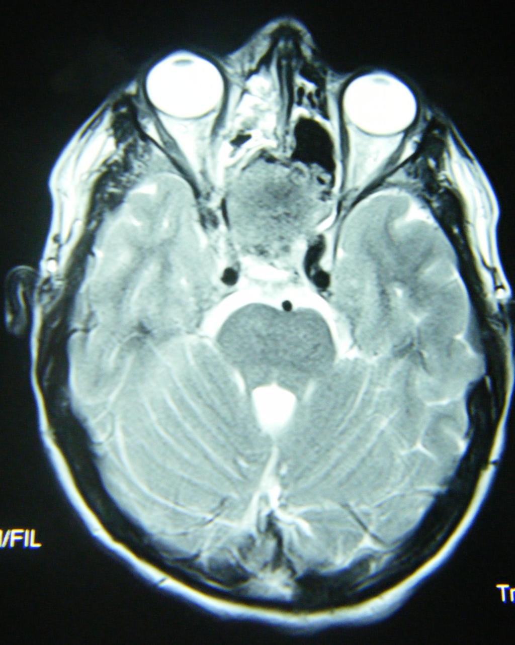
30 BRAIN MRI 5 MONTHS AFTER SURGERY


 Male, 17 y.")
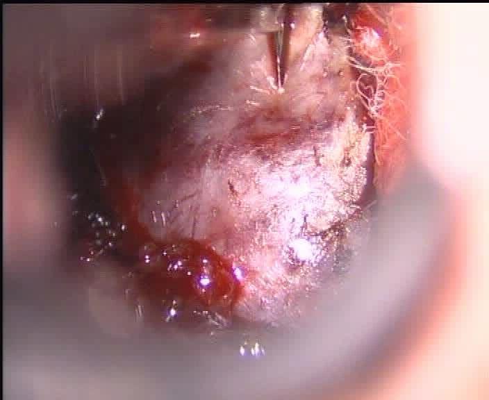
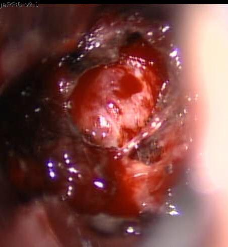
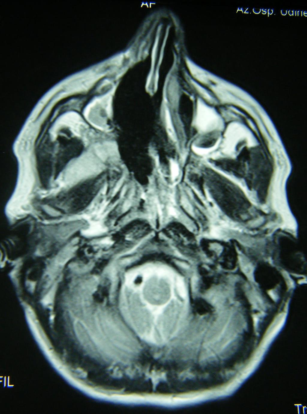
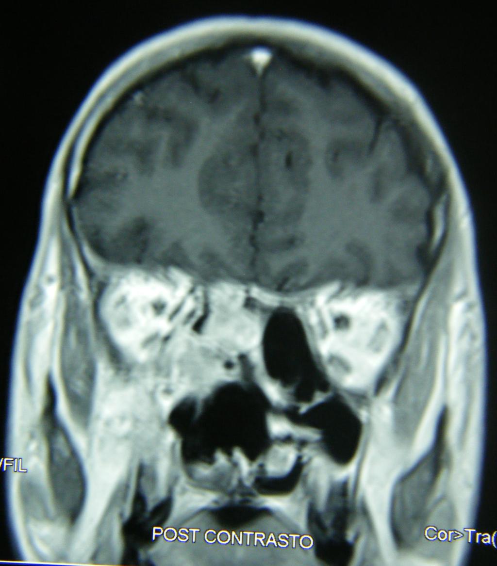
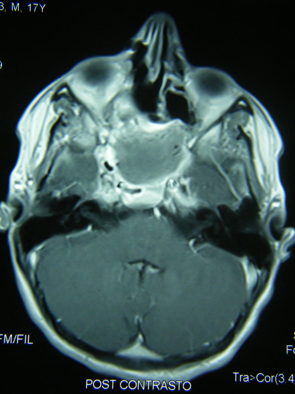
31 GIANT NASOPHARYNGEAL ANGIOFIBROMA WITH SKULL BASE EXTENSION (Extradural Type III acc. to Andrews, 1989) Male, 17 y. old TC imaging

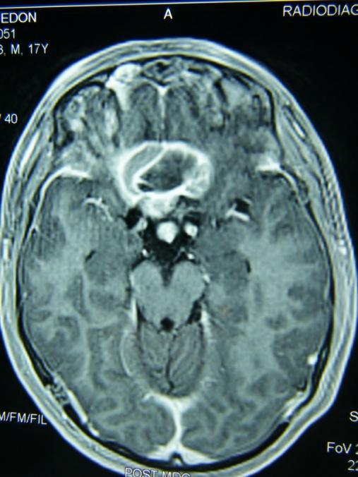
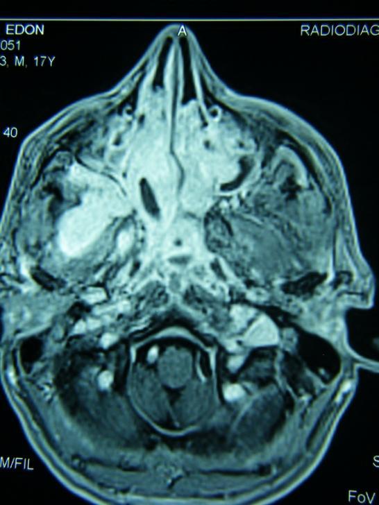
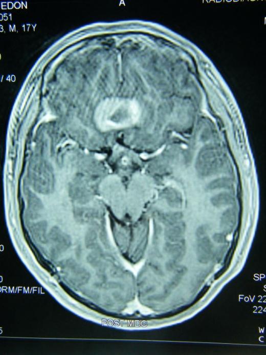
32 MR imaging

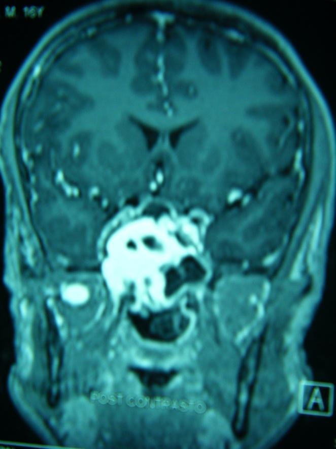
33 MR imaging

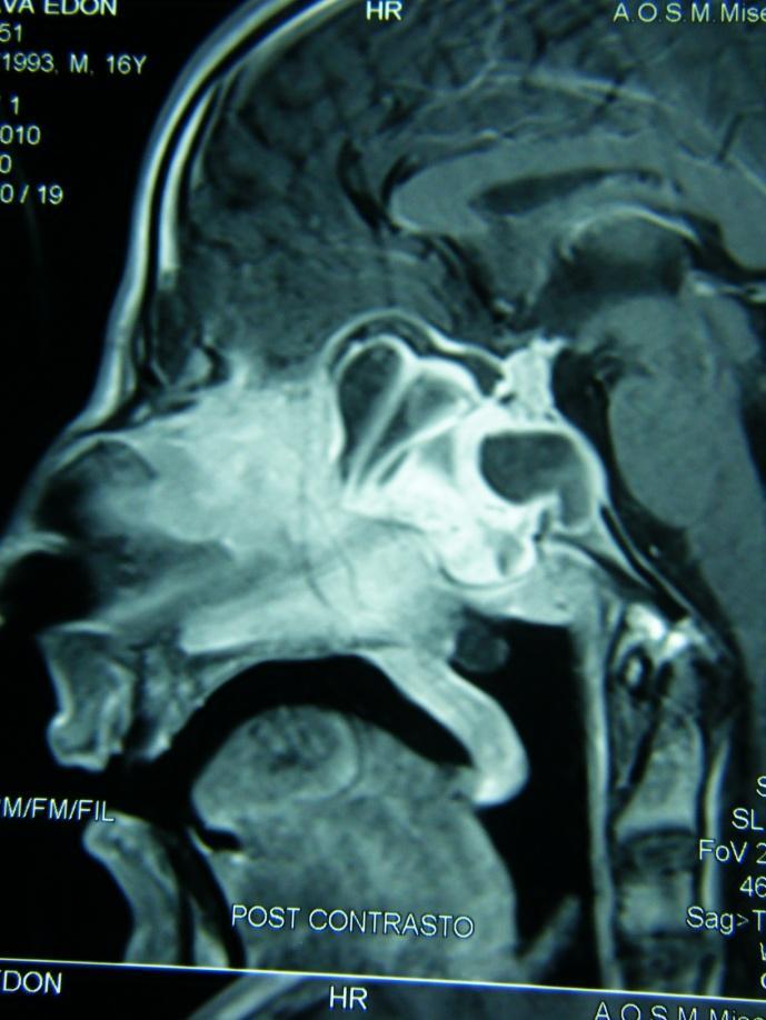
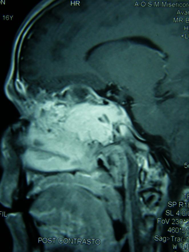
34 MR imaging


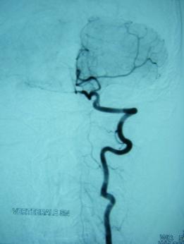

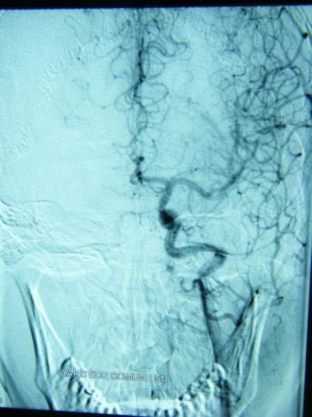
35 Selective right carotid artery angiography Left carotid artery Right and left vertebral arteries

36 Pre-operative angiographic embolization of the right internal maxillary artery Post Pre

37 Surgical navigator
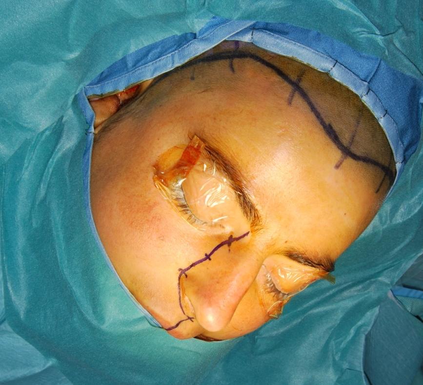
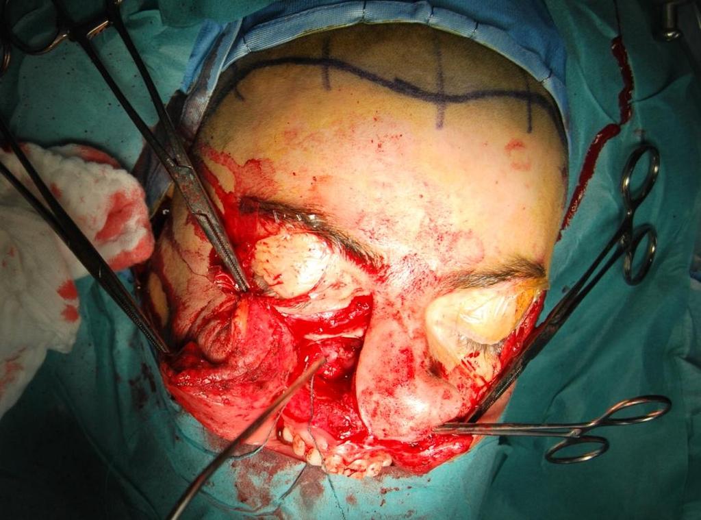
38 Paralatero-nasal approach through right maxillary bone volet


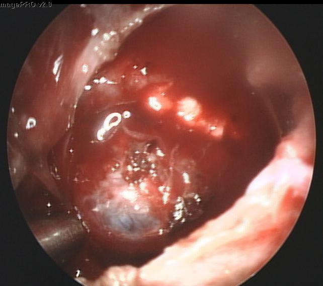
39 Uncovered dural lining of the clivus behind the posterior wall of the sphenoidal sinus

40 Left internal carotid artery sheath uncovered in lateral sphenoidal sinus wall Anterior sphenoidal sinus wall eroded by tumor growth


41 Post-operative closure of the surgical breach Intraoperative blood loss: 510 ml
42 Post-operative MRI check (7 PO day) PrePrePost- Post-
43 The future of endoscopic treatment of Anterior Skull Base Tumors: Where do we will draw the line? The limits of endonasal skull base surgery have not yet been fully realized 3D stereo-endoscopy is already adding new chances to endoscopic surgery, but surely other technical innovations will early enhance and enrich this surgical field in the next future.
44 CONCLUSIONS So we will move the line forward in EEA according to the constant improvements of surgical skill and technical progress Probably, many anterior skull base tumor shall be resected by open techniques also in the future, but the field of EEA is destined to always wider and safer applications


45 THANK YOU FOR YOUR KIND ATTENTION
10/23/2010. Excludes Single Surgeon Pituitary (N=~140) Skull Base Volume 12 Month UC SF. Patients. Anterior/Midline. Pituitary CSF Leak.
 Skull Base Volume 12 Month UC SF. Patients. Anterior/Midline. Pituitary CSF Leak.") Advances in Pituitary Surgery Ivan El-Sayed MD, FACS Director- Otolaryngology Minimally Invasive Skull Base Surgery Program Otolaryngology-Head and Neck Surgery University of California-San Francisco Minimally
Advances in Pituitary Surgery Ivan El-Sayed MD, FACS Director- Otolaryngology Minimally Invasive Skull Base Surgery Program Otolaryngology-Head and Neck Surgery University of California-San Francisco Minimally
What Are the Limits of Endoscopic Sinus Surgery?: The Expanded Endonasal Approach to the Skull Base
 REVIEW What Are the Limits of Endoscopic Sinus Surgery?: The Expanded Endonasal Approach to the Skull Base Carl H. Snyderman, 1 Harshita Pant, 1 Ricardo L. Carrau, 1 Daniel Prevedello, 2 Paul Gardner 2
Skull Base Volume 12 Month. Patients. Anterior/Midline. Pituitary CSF Leak. Lateral. Craniocervical Junction
 UC SF 2 11/7/2009 Skull Base Surgery in 2009 Ivan El-Sayed MD, FACS Director- Otolaryngology Minimally Invasive Skull Base Surgery Program Department Otolaryngology-Head and Neck Surgery University of
DRAFT PROGRAMME SKULL BASE 360 : ENDO/MICRO SKULL BASE COURSE Pre-congress workshop of AOSBS 2018 September 17-20, 2018
 DRAFT PROGRAMME SKULL BASE 360 : ENDO/MICRO SKULL BASE COURSE Pre-congress workshop of AOSBS 2018 September 17-20, 2018 Chairman MH. Huang Show Chwan Memorial Hospital Changhua, Taïwan President Director
Skullbase Lesions. Skullbase Surgery Open vs endoscopic. Choice Of Surgical Approaches 12/28/2015. Skullbase Surgery: Evolution
 Skullbase Lesions Skullbase Surgery Open vs endoscopic Prof Asim Mahmood,FRCS,FACS,FICS,FAANS, Professor of Neurosurgery Henry Ford Hospital Detroit, MI, USA Anterior Cranial Fossa Subfrontal meningioma
Tips and Tricks in Ventral Skull Base Dissection Narayanan Janakiram, Dharambir S. Sethi, Onkar K. Deshmukh, and Arvindh K.
 05 Tips and Tricks in Ventral Skull Base Dissection Narayanan Janakiram, Dharambir S. Sethi, Onkar K. Deshmukh, and Arvindh K. Gananathan Introduction...75 General Principles...76 Tips and Tricks in Ventral
DOWNLOAD OR READ : THE ENDOSCOPIC APPROACH TO VESTIBULAR SCHWANNOMAS AND POSTEROLATERAL SKULL BASE PATHOLOGY PDF EBOOK EPUB MOBI
 DOWNLOAD OR READ : THE ENDOSCOPIC APPROACH TO VESTIBULAR SCHWANNOMAS AND POSTEROLATERAL SKULL BASE PATHOLOGY PDF EBOOK EPUB MOBI Page 1 Page 2 the endoscopic approach to vestibular schwannomas and posterolateral
Research Article Expanded Endoscopic Endonasal Treatment of Primary Intracranial Tumors within the Paranasal Sinuses
 ISRN Minimally Invasive Surgery Volume 2013, Article ID 129780, 5 pages http://dx.doi.org/10.1155/2013/129780 Research Article Expanded Endoscopic Endonasal Treatment of Primary Intracranial Tumors within
Pituitary Macroadenoma with Superior Orbital Fissure Syndrome
 1 CASE REPORT OPEN ACCESS Pituitary Macroadenoma with Superior Orbital Fissure Syndrome Tapan Nagpal, Ankit Singhania ABSTRACT Introduction: Pituitary adenomas are benign tumours which arise within the
1 st International Advanced Sinus Dissection Course, 30 th - 31 st March 2017, Berlin. in Berlin
 1 st International Advanced Sinus Dissection Course, 30 th - 31 st March 2017, Berlin in Berlin Dear colleagues, We are very happy to invite you to the 1 st International Advanced Sinus Dissection Course
ENT NAVIGATION SIMPLE AND INTUITIVE
 ENT NAVIGATION SIMPLE AND INTUITIVE REVOLUTIONARY NAVIGATION TECHNOLOGY FOR ROUTINE AND COMPLEX ENT PROCEDURES We at Fiagon believe patient care should be centered around the patient. For us, patient-centered-care
The View through the Nose: ENT considerations for Pituitary/Skull Base Surgery
 The View through the Nose: ENT considerations for Pituitary/Skull Base Surgery Edsel Kim, M.D. Otolaryngology-Head and Neck Surgery The Oregon Clinic Providence Brain and Spine Institute Pituitary, Thyroid
Head & Neck Clinical Sub Group. Network Agreed Imaging Guidelines for UAT and Thyroid Cancer. Measure Nos: 11-1C-105i & 11-1C-106i
 Greater Manchester, Lancashire & South Cumbria Strategic Clinical Network & Senate Head & Neck Clinical Sub Group Network Agreed Imaging Guidelines for UAT and Thyroid Cancer Measure Nos: 11-1C-105i &
The pediatric skull base undergoes an intricate developmental
 J Neurosurg Pediatrics 13:155 169, 2014 AANS, 2014 Impact of skull base development on endonasal endoscopic surgical corridors Clinical article Matei A. Banu, M.D., M.S., 1 Amancio Guerrero-Maldonado,
Skull-2. Norma Basalis Interna Norma Basalis Externa. Dr. Heba Kalbouneh Associate Professor of Anatomy and Histology
 Skull-2 Norma Basalis Interna Norma Basalis Externa Dr. Heba Kalbouneh Associate Professor of Anatomy and Histology Norma basalis interna Base of the skull- superior view The interior of the base of the
Juvenile Angiofibroma
 Juvenile Angiofibroma Disclaimer The pictures used in this presentation have been obtained from a number of sources. Their use is purely for academic and teaching purposes. The contents of this presentation
NIH Public Access Author Manuscript J Neurosurg. Author manuscript; available in PMC 2014 August 13.
 NIH Public Access Author Manuscript Published in final edited form as: J Neurosurg. 2011 May ; 114(5): 1319 1330. doi:10.3171/2010.11.jns10768. The neurosurgical anatomy of the sphenoid sinus and sellar
How to Choose? Endoscopic Skull Base Reconstructive Options and Limitations
 ORIGINAL ARTICLE How to Choose? Endoscopic Skull Base Reconstructive Options and Limitations Mihir R. Patel, M.D., 1 Michael E. Stadler, M.D., 1 Carl H. Snyderman, M.D., 2,3 Ricardo L. Carrau, M.D., 4
Surgical approaches to PITUITARY ADENOMAS
 Surgical approaches to PITUITARY ADENOMAS HISTORY OF PITUITARY SURGERY In 1893,Caton and Paul attempted to explore sella turcica via lateral sub temporal route along with orbital exenteration. The transfrontal
Exposure techniques in endoscopic skull base surgery: Posterior septectomy, medial maxillectomy, transmaxillary and transpterygoid approach
 European Annals of Otorhinolaryngology, Head and Neck diseases (2012) 129, 284 288 Available online at www.sciencedirect.com TECHNICAL NOTE Exposure techniques in endoscopic skull base surgery: Posterior
Chapter 7: Head & Neck
 Chapter 7: Head & Neck Osteology I. Overview A. Skull The cranium is composed of irregularly shaped bones that are fused together at unique joints called sutures The skull provides durable protection from
Endoscopic Endonasal Approach to the Maxillary Strut: Anatomical Review and Case Series
 The Laryngoscope VC 2014 The American Laryngological, Rhinological and Otological Society, Inc. Endoscopic Endonasal Approach to the Maxillary Strut: Anatomical Review and Case Series Sanjeet S. Grewal,
Faculty of Medicine Khon Kaen University Thailand
 THE 13 th KHON KAEN FESS COURSE: HANDS-ON DISSECTION IN FRESH FROZEN CADAVERS January 16 th, 2018 & The 6 th KHON KAEN INTERNATIONAL COURSE IN ADVANCED ENDOSOCPIC SINUS AND SKULL BASE SURGERY: HANDS-ON
Unit 18: Cranial Cavity and Contents
 Unit 18: Cranial Cavity and Contents Dissection Instructions: The calvaria is to be removed without damage to the dura mater which is attached to the inner surface of the calvaria. Cut through the outer
182 Ligia Tataranu et al Endoscopic endonasal transsphenoidal approach
 182 Ligia Tataranu et al Endoscopic endonasal transsphenoidal approach Endoscopic endonasal transsphenoidal approach in the management of sellar and parasellar lesions: alternative surgical techniques,
Chapter 7 Part A The Skeleton
 Chapter 7 Part A The Skeleton Why This Matters Understanding the anatomy of the skeleton enables you to anticipate problems such as pelvic dimensions that may affect labor and delivery The Skeleton The
OPEN ACCESS ATLAS OF OTOLARYNGOLOGY, HEAD & NECK OPERATIVE SURGERY
 OPEN ACCESS ATLAS OF OTOLARYNGOLOGY, HEAD & NECK OPERATIVE SURGERY INFERIOR MAXILLECTOMY Tumours of the hard palate and superior alveolus may be resected by inferior maxillectomy (Figure 1). A Le Fort
PRINCIPLES OF ENDOSCOPIC MANAGEMENT OF NASAL AND. Frontier Steven D. Schaefer, MD, FACS
 PRINCIPLES OF ENDOSCOPIC MANAGEMENT OF NASAL AND SKULL : A New Frontier Steven D. Schaefer, MD, FACS Professor and Chair Department of Otolaryngology New York keye and dear Infirmary New York Medical College
Dr.Ban I.S. head & neck anatomy 2 nd y جامعة تكريت كلية طب االسنان مادة التشريح املرحلة الثانية أ.م.د. بان امساعيل صديق 6102/6102
 جامعة تكريت كلية طب االسنان مادة التشريح املرحلة الثانية أ.م.د. بان امساعيل صديق 6102/6102 Pterygopalatine fossa: The pterygopalatine fossa is a cone-shaped depression, It is located between the maxilla,
Unresectable (T4b) When Medical Professionals Opt Not to Treat. What s Resectable? The Current State of Sino-nasal Tumors
 When Medical Professionals Opt Not to Treat. What s Resectable? The Current State of Sino-nasal Tumors") UC SF 2 What s Resectable? The Current State of Sino-nasal Tumors When Medical Professionals Opt Not to Treat Truly Unresectable? Incurable? Ivan El-Sayed MD, FACS Otolaryngology Minimally Invasive Skull
UC SF 2 What s Resectable? The Current State of Sino-nasal Tumors When Medical Professionals Opt Not to Treat Truly Unresectable? Incurable? Ivan El-Sayed MD, FACS Otolaryngology Minimally Invasive Skull
January th, 2012
 Basic Endoscopic Sinus Surgery for Residents & Novices and 1st International Advanced Course in Endoscopic Sinus Surgery & Skull Base Surgery January 17-20 th, 2012 January 17 th, 2012: Basic Endoscopic
We are IntechOpen, the world s leading publisher of Open Access books Built by scientists, for scientists. International authors and editors
 We are IntechOpen, the world s leading publisher of Open Access books Built by scientists, for scientists 3,500 108,500 1.7 M Open access books available International authors and editors Downloads Our
5. COMMON APPROACHES. Each of the described approaches is also demonstrated on supplementary videos, please see Appendix 2.
 5. COMMON APPROACHES Each of the described approaches is also demonstrated on supplementary videos, please see Appendix 2. 5.1. LATERAL SUPRAORBITAL APPROACH The most common craniotomy approach used in
Analysis of the Petrous Portion of the Internal Carotid Artery: Landmarks for an Endoscopic Endonasal Approach
 The Laryngoscope VC 2014 The American Laryngological, Rhinological and Otological Society, Inc. Analysis of the Petrous Portion of the Internal Carotid Artery: Landmarks for an Endoscopic Endonasal Approach
Bones of the skull & face
 Bones of the skull & face Cranium= brain case or helmet Copyright The McGraw-Hill Companies, Inc. Permission required for reproduction or display. The cranium is composed of eight bones : frontal Occipital
Skull-2. Norma Basalis Interna. Dr. Heba Kalbouneh Assistant Professor of Anatomy and Histology
 Skull-2 Norma Basalis Interna Dr. Heba Kalbouneh Assistant Professor of Anatomy and Histology Norma basalis interna Base of the skull- superior view The interior of the base of the skull is divided into
RADIOANATOMY OF SELLA TURCICA
 RADIOANATOMY OF SELLA TURCICA O.BAKKACHA, H.MALAJATI, M.RHISSASSI, H. BENCHAABOUNE, N.CHAKIR, My R. EL HASSANI,M.JIDDANE Department of Neuroradiology specialties Hospital. Rabat Objective: New imaging
PITUITARY PARASELLAR LESIONS. Kim Learned, MD
 PITUITARY PARASELLAR LESIONS Kim Learned, MD DIFFERENTIALS Pituitary Sella Clivus, Sphenoid Sinus Suprasellar Optic chiasm, Hypothalamus, Circle of Willis Parasellar Cavernous Sinus Case 1 17 YEAR-OLD
PTERYGOPALATINE FOSSA
 PTERYGOPALATINE FOSSA Outline Anatomical Structure and Boundaries Foramina and Communications with other spaces and cavities Contents Pterygopalatine Ganglion Especial emphasis on certain arteries and
Three-Dimensional Volumetric Display of the Nasal Ostiomeatal Channels and Paranasal Sinuses
 Downloaded from www.ajronline.org by 37.44.202.192 on 12/22/17 from IP address 37.44.202.192. Copyright RRS. For personal use only; all rights reserved Three-Dimensional Volumetric Display of the Nasal
Radiological anatomy of frontal sinus By drtbalu
 2009 Radiological anatomy of frontal sinus By drtbalu Anatomy of frontal sinus is highly variable. Precise understanding of these variables will help a surgeon to avoid unnecessary complications during
We describe some of the conceptual nuances and
 Suprasellar Meningiomas Ivan Ciric, M.D., Sami Rosenblatt, M.D. Division of Neurosurgery, Evanston Northwestern Healthcare, Evanston Hospital, Northwestern University Medical School, Evanston, Illinois
The surgical approach to the sphenoid sinus continues to
 A comparison of two sphenoidotomy approaches using a novel computerized tomography grading system Heitham Gheriani, F.R.C.S.C., F.R.C.S.I., David Flamer, B.Sc., Trent Orton, M.D., Brad Mechor, F.R.C.S.C.,
Boundaries Septum Turbinates & Meati Lamellae Drainage Pathways Variants
 The Fastest 20 Minutes in Michelle A. Michel, MD Professor of Radiology and Otolaryngology Medical College of Wisconsin, Milwaukee Overview Nasal cavity Anterior skull base Ostiomeatal complex Frontal
1. BRIEF DESCRIPTION OF TRAINING
 RHINOLOGY 1. BRIEF DESCRIPTION OF TRAINING Exposure to clinical rhinology is provided in each of the four ORL years over the course of several rotations in a graduated approach. MEE General Otolaryngology
QUANTITATIVE ANALYSIS AND COMPUTER AIDED SIMULATION OF MINIMALLY INVASIVE APPROACHES FOR INTRACRANIAL VASCULAR LESIONS
 QUANTITATIVE ANALYSIS AND COMPUTER AIDED SIMULATION OF MINIMALLY INVASIVE APPROACHES FOR INTRACRANIAL VASCULAR LESIONS Alberto Prats Galino Facultat de Medicina UB 1. Project Summary The main goal of this
Eyebrow craniotomy for anterior skull base lesions: how I do it
 Acta Neurochir (2013) 155:99 106 DOI 10.1007/s00701-012-1552-5 HOW I DO IT - NEUROSURGICAL TECHNIQUES Eyebrow craniotomy for anterior skull base lesions: how I do it Zsolt Zador & Kanna Gnanalingham Received:
 www.oralradiologists.com CONE BEAM CT REPORT CASE XXXX Patient information Patient Name: - Referring Doctor: - Patient DOB: - Scan Date: [Start date] Reason for Exam: Maxillary facial pain Doctor Notes:
Endoscopic anatomical study on anterior communicating artery aneurysm surgery by endonasal transphenoidal approach
 Ma et al. Chinese Neurosurgical Journal (2016) 2:27 DOI 10.1186/s41016-016-0042-7 CHINESE NEUROSURGICAL SOCIETY RESEARCH CHINESE MEDICAL ASSOCIATION Endoscopic anatomical study on anterior communicating
*in general the blood supply of the nose comes from branches of the internal and external carotid arteries.
 In the previous lecture we talked about the anatomy of the nasal cavity, today we will talk about its blood supply, venous drainage, innervations, and finally about the paranasal sinuses. When we describe
Bisection of Head & Nasal Cavity 頭部對切以及鼻腔. 解剖學科馮琮涵副教授 分機
 Bisection of Head & Nasal Cavity 頭部對切以及鼻腔 解剖學科馮琮涵副教授 分機 3250 E-mail: thfong@tmu.edu.tw Outline: The structure of nose The concha and meatus in nasal cavity The openings of paranasal sinuses Canals, foramens
Malformations of the nose, Clefts & Tumors of the nose. Summer School David Holzmann
 Malformations of the nose, Clefts & Tumors of the nose Summer School 2018 David Holzmann Department of Otorhinolaryngology Head & Neck Surgery University Hospital Zurich University Zurich Switzerland Content
The authors discuss their surgical approaches
 GENERAL SCIENTIFIC SESSION 3 GENERAL SCIENTIFIC SESSION 3 Open vs Endoscopic: When To Use Which Laligam Sekhar, MD* Alessandra Mantovani, MD* Martin Mortazavi, MD* Theodore H. Schwartz, MD WIlliam T. Couldwell,
The KASSAM Range of Bipolar Forceps
 EndoWorld NEURO 9-1-E/04-2008 The KASSAM Range of Bipolar Forceps for Bleeding Management especially designed for Brain and Skull Base Surgery acc. to Dr. Kassam, Dr. Snydermann The KASSAM Range of Bipolar
Biology 218 Human Anatomy. Adapted from Martini Human Anatomy 7th ed. Chapter 6 The Skeletal System: Axial Division
 Adapted from Martini Human Anatomy 7th ed. Chapter 6 The Skeletal System: Axial Division Introduction The axial skeleton: Composed of bones along the central axis of the body Divided into three regions:
Imaging of the Paranasal Sinuses
 14. Sommerschule Imaging of the Paranasal Sinuses Bettlach 24.08.2018 Christoph Schlegel Conventional Radiology NNH-Status: okzipito-frontal: frontal sinus, anterior ethmoid okzipito-nasal : maxillary
Transplanum Approach for Suprasellar pathology
 Transplanum Approach for Suprasellar pathology Omar A. El-Banhawy Prof. of otorhinolaryngology El Menoufyia University, Egypt Why Endoscopic Approach For Suprasellar Pathology Constant improvements in
Transnasal Corridors to Skull Base and Orbit
 Transnasal Corridors to Skull Base and Orbit 3 nd International Hands-on Course Wien 11-13 April 2012 Anatomy Training Center Department of Systematic Anatomy University of Wien Course directors: P. Castelnuovo
Omran Saeed. Luma Taweel. Mohammad Almohtaseb. 1 P a g e
 2 Omran Saeed Luma Taweel Mohammad Almohtaseb 1 P a g e I didn t include all the photos in this sheet in order to keep it as small as possible so if you need more clarification please refer to slides In
Variations in sphenoid sinus anatomy with special emphasis on pneumatization and endoscopic anatomic distances
 Variations in sphenoid sinus anatomy with special emphasis on pneumatization and endoscopic anatomic distances Gulgun Kayalioglu, MD, Mete Erturk, MD, Tuncay Varol, MD. ABSTRACT Objectives: The purpose
CSF Rhinorrhoea after Transsphenoidal Surgery
 ISPUB.COM The Internet Journal of Neurosurgery Volume 5 Number 1 CSF Rhinorrhoea after Transsphenoidal Surgery E Elgamal Citation E Elgamal. CSF Rhinorrhoea after Transsphenoidal Surgery. The Internet
Benign Neoplasms of the Nose
 Department of Otolaryngology Head and Neck Surgery Pursuing Wellness Through Teaching, Learning and Healing Benign Neoplasms of the Nose Ivan El Sayed, MD Disclosure Principal Investigator: Grant Support
Superior View of the Skull (Norma Verticalis) Anteriorly the frontal bone articulates with the two parietal bones AT THE CORONAL SUTURE
 Anteriorly the frontal bone articulates with the two parietal bones AT THE CORONAL SUTURE") Superior View of the Skull (Norma Verticalis) Anteriorly the frontal bone articulates with the two parietal bones AT THE CORONAL SUTURE 1 The two parietal bones articulate in the midline AT THE SAGITTAL
Superior View of the Skull (Norma Verticalis) Anteriorly the frontal bone articulates with the two parietal bones AT THE CORONAL SUTURE 1 The two parietal bones articulate in the midline AT THE SAGITTAL
HEAD AND NECK IMAGING. James Chen (MS IV)
") HEAD AND NECK IMAGING James Chen (MS IV) Anatomy Course Johns Hopkins School of Medicine Sept. 27, 2011 OBJECTIVES Introduce cross sectional imaging of head and neck Computed tomography (CT) Review head
HEAD AND NECK IMAGING James Chen (MS IV) Anatomy Course Johns Hopkins School of Medicine Sept. 27, 2011 OBJECTIVES Introduce cross sectional imaging of head and neck Computed tomography (CT) Review head
PROBLEM RECOMMENDATION
 PREVENTION (MINIMIZING) IN ENDOSCOPIC Steven D. Schaefer, MD Professor and Chair Department of Otolaryngology PREVENTION AND Intraoperative Hemorrhage Loss of Orientation Inability to Identify/Preserve
Imaging The Turkish Saddle. Russell Goodman, HMS III Dr. Gillian Lieberman
 Imaging The Turkish Saddle Russell Goodman, HMS III Dr. Gillian Lieberman Learning Objectives Review the anatomy of the sellar region Discuss the differential diagnosis of sellar masses Discuss typical
Role of the superior turbinate when performing endoscopic endonasal transsphenoidal approach
 O R I G I N A L A R T I C L E Folia Morphol. Vol. 73, No. 1, pp. 73 78 DOI: 10.5603/FM.2014.0010 Copyright 2014 Via Medica ISSN 0015 5659 www.fm.viamedica.pl Role of the superior turbinate when performing
Binostril Endoscopic Trans-Sphenoidal Approach for Pituitary Adenomas
 Med. J. Cairo Univ., Vol. 85, No. 4, June: 1593-1600, 2017 www.medicaljournalofcairouniversity.net Binostril Endoscopic Trans-Sphenoidal Approach for Pituitary Adenomas HESHAM ABO RAHMA, M.D. and AHMED
Incisionless Brain Surgery
 Transcript Details This is a transcript of an educational program accessible on the ReachMD network. Details about the program and additional media formats for the program are accessible by visiting: https://reachmd.com/programs/medical-breakthroughs-from-penn-medicine/incisionless-brainsurgery/3873/
Skull and Axial Skeleton
 Published on Second Faculty of Medicine, Charles University (http://www.lf2.cuni.cz ) Skull and Axial Skeleton Description of the test The examination of the skull skeleton is in oral format. It consists
The aim of this project is to use 3-dimensional
 Surgical Anatomy and Technique A 3-Dimensional Transnasal Endoscopic Journey Through the Paranasal Sinuses and Adjacent Skull Base: A Practical and Surgery-Oriented Perspective Andrea Bolzoni Villaret,
10/4/2013. Sinonasal and Skull Base Cancer Progress, Challenges, and Future Directions
 Ehab Hanna, M.D. Head and Neck Surgery MD Anderson Cancer Center Sinonasal and Skull Base Cancer Progress, Challenges, and Future Directions Context Advances in Diagnosis Office endoscopy High Resolution
SINUS ANATOMY AND FUNCTION
 EMBRYOLOGY AND DEVELOPMENT SINUS ANATOMY AND FUNCTION -4 th week gestation: -frontonasal process identified, arises over developing forebrain -ectodermal -contributes to nasal capsule -9 th and 10 th week
The dura is sensitive to stretching, which produces the sensation of headache.
 Dural Nerve Supply Branches of the trigeminal, vagus, and first three cervical nerves and branches from the sympathetic system pass to the dura. Numerous sensory endings are in the dura. The dura is sensitive
Case Report Recurrent Massive Epistaxis from an Anomalous Posterior Ethmoid Artery
 Case Reports in Otolaryngology Volume 2016, Article ID 8504348, 4 pages http://dx.doi.org/10.1155/2016/8504348 Case Report Recurrent Massive Epistaxis from an Anomalous Posterior Ethmoid Artery Marco Giuseppe
4 days. From Pituitary to Skull Base course. November 27 th, 30 th, 2017 Istituto delle Scienze Neurologiche Ospedale Bellaria, Bologna
 Save the Date From Pituitary to Skull Base course November 27 th, 30 th, 2017 Istituto delle Scienze Neurologiche Ospedale Bellaria, Bologna 4 days 2 days, anatomy dissection 2 days, live surgery Chairman
Nasal region. cartilages: septal cartilage (l); lateral nasal cartilage (2); greater alar cartilages (2); lesser alar cartilages (?
; lateral nasal cartilage (2); greater alar cartilages (2); lesser alar cartilages (?") Nasal region skull bones: nasal and frontal processes of maxilla cartilages: septal cartilage (l); lateral nasal cartilage (2); greater alar cartilages (2); lesser alar cartilages (?) 1 Nasal cavity Roof
Nasal region skull bones: nasal and frontal processes of maxilla cartilages: septal cartilage (l); lateral nasal cartilage (2); greater alar cartilages (2); lesser alar cartilages (?) 1 Nasal cavity Roof
Neuroradiology MR Protocols
 Neuroradiology MR Protocols Brain protocols N 1: Brain MRI without contrast N 2: Pre- and post-contrast brain MRI N 3 is deleted N 4: Brain MRI without or pre-/post-contrast (seizure protocol) N 5: Pre-
Sphenoid rhinosinusitis associated with abducens nerve palsy Case report
 Romanian Journal of Rhinology, Volume 8, No. 30, April-June 2018 CASE REPORT Sphenoid rhinosinusitis associated with abducens nerve palsy Case report Lucian Lapusneanu 1, Marlena Radulescu 1, Florin Ghita
The orbit-1. Dr. Heba Kalbouneh Assistant Professor of Anatomy and Histology
 The orbit-1 Dr. Heba Kalbouneh Assistant Professor of Anatomy and Histology Orbital plate of frontal bone Orbital plate of ethmoid bone Lesser wing of sphenoid Greater wing of sphenoid Lacrimal bone Orbital
Surgical Anatomy for the Endoscopic Endonasal Approach to the Ventrolateral Skull Base
 REVIEW ARTICLE Neurol Med Chir (Tokyo) 57, 534 541, 2017 doi: 10.2176/nmc.ra.2017-0039 Online August 25, 2017 Surgical Anatomy for the Endoscopic Endonasal Approach to the Ventrolateral Skull Base Kenichi
Pedicled Nasoseptal Flap as Final Layer of Reconstruction for Skull Base Defects
 Med. J. Cairo Univ., Vol. 79, No. 2, June: 123-128, 2011 www.medicaljournalofcairouniversity.com Pedicled Nasoseptal Flap as Final Layer of Reconstruction for Skull Base Defects SAMEH M. AMIN, M.D.*; AHMED
Coding Update: Rhinology
 Michael J. Sillers, M.D., F.A.C.S Alabama Nasal and Sinus Center Clinical Professor The University of Alabama-Birmingham Birmingham, Alabama Teaching Objectives Understand basic coding principles Recognize
Cranium Facial bones. Sternum Rib
 Figure 7.1 The human skeleton. Skull Thoracic cage (ribs and sternum) Cranium Facial bones Sternum Rib Bones of pectoral girdle Vertebral column Sacrum Vertebra Bones of pelvic girdle (a) Anterior view
Endoscopic endonasal pituitary adenomas surgery: the surgical experience of 178 consecutive patients and learning curve of two neurosurgeons
 Shou et al. BMC Neurology (2016) 16:247 DOI 10.1186/s12883-016-0767-0 RESEARCH ARTICLE Open Access Endoscopic endonasal pituitary adenomas surgery: the surgical experience of 178 consecutive patients and
ANATOMY & PHYSIOLOGY I Laboratory Version B Name Section. REVIEW SHEET Exercise 10 Axial Skeleton
 ANATOMY & PHYSIOLOGY I Laboratory Version B Name Section REVIEW SHEET Exercise 10 Axial Skeleton 1 POINT EACH. THE SKULL MULTIPLE CHOICE 1. The major components of the axial skeleton include the 7. The
Chapter 7. Skeletal System
 Chapter 7 Skeletal System 1 Skull A. The skull is made up of 22 bones: 8 cranial bones, 13 facial bones, and the mandible. B. The Cranium encloses and protects the brain, provides attachments for muscles,
Endoscopic Management Of A Giant Ethmoid Mucocele
 ISPUB.COM The Internet Journal of Otorhinolaryngology Volume 6 Number 1 S Ceylan, F Bora Citation S Ceylan, F Bora.. The Internet Journal of Otorhinolaryngology. 2006 Volume 6 Number 1. Abstract We present
Multimodality approach for advanced-stage juvenile nasopharyngeal angiofibromas
 ORIGINAL ARTICLE Multimodality approach for advanced-stage juvenile nasopharyngeal angiofibromas Fernando Lopez Alvarez, MD, PhD,* Vanessa Suarez, MD, PhD, Carlos Suarez, MD, PhD, Jose L. Llorente, MD,
Endoscopic Sellar, Suprasellar and Parasellar Surgery with Image Guidance
 Med. J. Cairo Univ., Vol. 83, No. 1, December: 881-886, 2015 www.medicaljournalofcairouniversity.net Endoscopic Sellar, Suprasellar and Parasellar Surgery with Image Guidance HESHAM M. NEGM, M.D.*; HUSSAM
Pure endoscopic endonasal odontoidectomy: anatomical study
 Neurosurg Rev (2007) 30:189 194 DOI 10.1007/s10143-007-0084-6 ORIGINAL ARTICLE Pure endoscopic endonasal odontoidectomy: anatomical study Andrea Messina & Maria Carmela Bruno & Philippe Decq & Andre Coste
YOU MUST BRING YOUR OWN GLOVES FOR THIS ACTIVITY.
 ACTIVITY 3: AXIAL SKELETON AND LONG BONE DISSECTION Objectives: 1) How to get ready: Read Chapter 7, McKinley et al., Human Anatomy, 5e. All text references are for this textbook. Learning the meanings
Journal of Clinical Neuroscience
 Journal of Clinical Neuroscience 17 (2010) 746 750 Contents lists available at ScienceDirect Journal of Clinical Neuroscience journal homepage: www.elsevier.com/locate/jocn Neuroanatomical Study Surgical
Transnasal Endoscopic Sinonasal Surgery
 Reda kamel, Cadaveric dissection 1 Transnasal Endoscopic Sinonasal Surgery Cadaver Dissection Guide For Endoscopic Sinus Surgery Cairo University Egypt Reda Kamel Professor of Rhinology Cairo University
TABLES. Imaging Modalities Evidence Tables Table 1 Computed Tomography (CT) Imaging. Conclusions. Author (Year) Classification Process/Evid ence Class
 Imaging. Conclusions. Author (Year) Classification Process/Evid ence Class") TABLES Imaging Modalities Evidence Tables Table 1 Computed Tomography (CT) Imaging Author Clark (1986) 9 Reformatted sagittal images in the differential diagnosis meningiomas and adenomas with suprasellar
TABLES Imaging Modalities Evidence Tables Table 1 Computed Tomography (CT) Imaging Author Clark (1986) 9 Reformatted sagittal images in the differential diagnosis meningiomas and adenomas with suprasellar
A Cross Sectional Study of Normal Variations of Sphenoid Sinus Through CT-Scan in North Karnataka, India
 Original Article A Cross Sectional Study of Normal Variations of Sphenoid Sinus Through CT-Scan in North Karnataka, India DOI: 10.7860/IJARS/2016/20940.2180 Anatomy Section Manisha S. Chougule, Preetam
ACTIVITY 3: AXIAL SKELETON AND LONG BONE DISSECTION COW BONE DISSECTION
 ACTIVITY 3: AXIAL SKELETON AND LONG BONE DISSECTION Objectives: 1) How to get ready: Read Chapter 7, McKinley et al., Human Anatomy, 4e. All text references are for this textbook. Learning the meanings
Anatomical and Imaging Studies of Endoscopic Optic Nerve Decompression
 The Journal of Bioscience and Medicine 2, 1 (2012) Anatomical and Imaging Studies of Endoscopic Optic Nerve Decompression Tao Wang 1#,Zhuang Kang 2,Peng Li 1, Qintai Yang 1*,Xian Liu 1,Gehua Zhang 1,Yuan