Transplanum Approach for Suprasellar pathology
|
|
|
- Nora Willis
- 5 years ago
- Views:
Transcription


1 Transplanum Approach for Suprasellar pathology Omar A. El-Banhawy Prof. of otorhinolaryngology El Menoufyia University, Egypt Why Endoscopic Approach For Suprasellar Pathology Constant improvements in diagnostic imaging techniques Experience gained from CSF closure & endoscope in transsphenoidal pituitary surgery Use of image guidance systems lad to constant surgical orientation in an anatomically complex area Collaboration between ES surgeons and neurosurgeons 1

2 Why Endoscopic Approach For Suprasellar Pathology different areas of the midline skull base exposed through the endoscopic endonasal approach. to the olfactory groove; to the sella turcica and planum sphenoidale; to the clivus to the craniovertebral junction and foramen magnum. Why Endoscopic Approach For Suprasellar Pathology different areas of the midline skull base exposed through the endoscopic endonasal approach. to the olfactory groove; to the sella turcica and planum sphenoidale; to the clivus to the craniovertebral junction and foramen magnum. 2
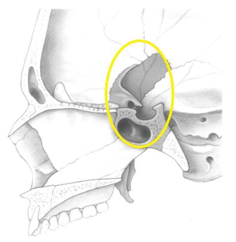

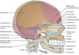
3 Planum Sphenoidale Suprasellar Area Sella Turcica 3


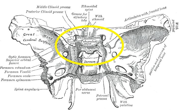
4 Sella Turcica Sella Turcica: tuberculum sellae 4


5 Planum Sphenoidale (PS) It is the roof of the anterior part of the SS Its bone thickness is a mean of 0.6 mm Laterally is marked by optic canals and the medial orbital wall Anteriorly it is in continuation to the cells of the posterior Ethmoidal sinus (PES) The junction of the PS and pituitary fossa is called the tuberculum sellae (bone thickness is 1 mm) Planum Sphenoidale (PS) 5


6 Planum Sphenoidale (PS) Bone covering the anterior vertical segment of the carotid artery is 0.5 mm thicknes in 90% of patients Bone covering the optic nerves is 0.5 mm thicknes in 80% of patients Planum Sphenoidale (PS) 6

7 Planum Sphenoidale (PS) a the widest lateral length of resection area mm b, the widest anteroposterior length of resection area mm c, resection area d, at the level of optic canal, the length between the medial sides of the optic nerves mm e, the length between the medial aspects of both internal carotid arteries mm f, the angle between optic nerves in the planum sphenoidale region g, the length between medial opticocarotid recess and lateral opticocarotid recess (inter- recess length) gr, right inter-recess length 3.75 mm gl, left inter-recess length 4.13 mm Planum Sphenoidale (PS) 7


8 Planum Sphenoidale (PS) Diaphragma Sellae A sheet of dura separating the pituitary gland in its fossa from the brain above. 8
9 Posterior Ethmoidectomy live posterior ethmoidectomy Sphenoidotomy Sphenoidotomy live 9
10 Planum Sphenoidale (PS) After bony resorption wall of the sphenoid sinus walls Suprasellar pathology Pituitary macroadenomas that extend high above the planum Midline suprasellar craniopharyngiomas Rathke cleft cysts Planum sphenoidale and tuberculum sellae meningiomas, Other pathologies: Chiasmatic-hypothalamic glioma Germ cell tumor. Langerhans cell histiocytosis Hypothalamic hamartoma Leukemia 10

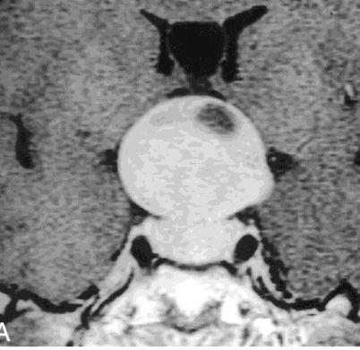
11 Pituitary Adenomas Etiology is unknown 10-15% of all primary brain tumors 20-25% of pituitary glands at autopsy found to have adenomas Pituitary adenomas have long natural history Vary in size and direction of spread 70% of adenomas are endocrinogically secreting Microadenomas < 10 mm may cause focal bulging Macroadenomas > 10 mm cause problems due to mass effect Endocrine-Active Pituitary Adenomas Prolactin Amenorrhea, galactorrhea, impotence Growth hormone Gigantism and acromegaly Corticotropin Cushing s disease, Nelson s syndrome post adrenalectomy TSH - Hyperthyroidism 11


12 Pituitary adenomas Cushing syndrome ACTH Chromophobe PROLACTIN Growth hormone Acromegaly Gigantism Amenorrhoea Infertility Galactorrhoea Hypoglandism Impotence Infertility Gynaecomastia Galactorrhoea Omar A. El-Banhawy, December 2013 Clinical Presentation Most common are endocrine abnormalities hyper- /hyposecretion of ant. pituitary hormones Vision changes bitemporal hemianopsia and superior temporal defects 12

13 Visual field defects in pituitary adenomas LE RE HM CF Anteriorly Decussating fibres are most vunerable Non-functioning Adenomas % of patients do not have classical hypersecretory syndromes May grow to a large size before they are detected Present due to mass effect Visual deficits Hormone deficiency 13


14 Supra sellar Pituitary Adenomas Omar A. El-Banhawy, December

15 Suprasellar pathology Midline suprasellar craniopharyngiomas Rathke cleft cysts planum sphenoidale and tuberculum sellae meningiomas, other pathologies: chiasmatic-hypothalamic glioma germ cell tumor. Langerhans cell histiocytosis hypothalamic hamartoma Leukemia Craniopharyngioma It is a low-grade extra-axial, epithelialsquamous calcified, and cystic tumor It arising from remnants of the craniopharyngeal duct and/or Rathke cleft The most common sellar/suprasellar tumor in children Accounting for approximately 6-9 % of CNS tumors. Two main hypotheses embryogenetic and metaplastic explain the origin of craniopharyngioma. 15
 The")

.")

16 Craniopharyngioma There are two pathologic variants determine the age of presentation (bimodal) The adamantinomatous In children peak between10 and 14 years It presents as a cystic and solid sellar/suprasellar mass Peripheral calcifications (>90% of cases). The papillary variant Middle-aged and older adults typically a solid enhancing sellar/suprasellar mass Cystic changes and calcifications are less common. Craniopharyngioma 16
17 Craniopharyngioma Symptoms frequently develops after the tumor attains a diameter of about 3cm. The time interval between the onset of symptoms and diagnosis usually ranges from 1-2 years. The most common presenting symptoms are Dull headache slowly progressive, (55-86%) endocrine dysfunction (66-90%) visual disturbances (37-68%) Supra sellar Craniopharyngioma In the anatomic location of the craniopharyngioma Prechiasmal localization visual problems Retrochiasmal location - Commonly is associated with hydrocephalus Intrasellar craniopharyngioma - as headache and/or endocrinopathy 17

18 Craniopharyngioma LE RE Presents In children with endocrine dysfunction In adults with visual field defects CF HM The posteriorly crossing fibres are most vunerable Craniopharyngioma Suprasellar pathology Rathke cleft cysts planum sphenoidale and tuberculum sellae meningiomas, other pathologies: chiasmatic-hypothalamic glioma germ cell tumor. Langerhans cell histiocytosis hypothalamic hamartoma Leukemia 18

19 Rathke cleft cyst Rathke s cleft describes the region that forms between the adenohypophysis and neurohypophysis during the third or fourth week of gestation. RCC arise when the Rathke s cleft does not regress fully and remains patent The cyst wall is formed by an epithelial membrane The inside is filled with a mucinous, gelatinous, or caseous cystic fluid Most RCC are asymptomatic Common symptoms are headache visual endocrine disturbances RCC, surgical decompression through a transsphenoidal approach is commonly performed Complex cases of RCC may necessitate an open ranscranial approach Rathke cleft cyst 19


20 Suprasellar pathology other pathologies: tuberculum sellae meningiomas chiasmatic-hypothalamic glioma germ cell tumor. Langerhans cell histiocytosis hypothalamic hamartoma Leukemia Suprasellar pathology Tuberculum sellae meningiomas 20
21 Transplanum Approach for Suprasellar pathology Transplanum Approach for Suprasellar pathology 21

22 Transplanum Approach Endoscopic view obtained in a cadaver showing the removal of the superior portion of the nasal septum and the anterior sphenoidotomy. SphS Sphenoidal sinus, ST superior turbinate, IT inferior turbinate, V vomer, EB ethmoid bulla, NS nasal septum, UP uncinate process, CO choana, CP carotid protuberance, OCR optocarotid recess, SF sellar floor, SphA sphenopalatine artery Transplanum Approach Removal of sella floor and planum 22


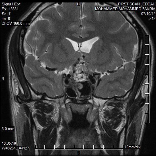
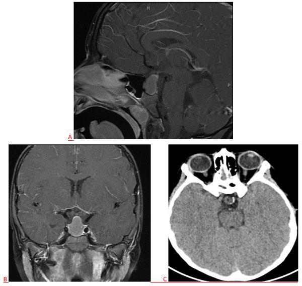
23 Transplanum Approach Transplanum Approach A 28 years old male c/o: Sever headache in the last 10 months bilateral watery rhinorrhea that was confirmed as CSF by β2-transferrin test Nasal endoscopy NAD Radiology 23
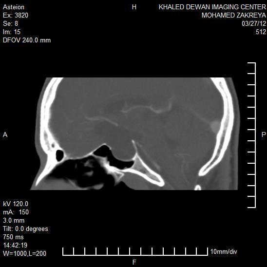
24 CT MRI 24
25 An Approach for Suprasellar pathology Follow up 25

26 Follow up Omar A. El-Banhawy, December 2013 Thank You 26
27 27
.")
28 Chiasmatic-Hypothalamic Glioma. Gliomas are common CNS tumors in children, the majority of which are low grade. Common locations in pediatric patients include the posterior fossa and optic pathways. Gliomas may occur sporadically or in association with neurofibromatosis type 1 (NF-1). Chiasmatichypothalamic gliomas have no sex predilection and are typically seen in children; they rarely occur in adults.1 Presenting symptoms include painless proptosis, visual impairment, diabetes insipidus, diencephalic syndrome, and precocious puberty. Radiographs demonstrate a classic J-shaped sella turcica, as well as enlarged optic canals. On MRI, gliomas are typically T1 isointense to mildly hypointense, T2 hyperintense, and show variable degrees of enhancement. Meningioma Typically affect middle-aged women LE RE Junctional scotoma Tuberculum Sella meningioma Sphenoid ridge meningioma Olfactory groove meningioma 28

29 Classsification of intracranial germ cell tumor Benign: Mature teratoma Malignant: Germinoma (60%) Embryonal carcinoma/endodermal sinus tumor Choriocarcinoma Immature teratoma Anatomy of chiasm and pituitary gland Upper nasal fibres Macular fibres Lower nasal fibres III rd ventricle Craniopharyngioma Optic chiasm Diaphragma sellae Anterior clinoid Pituitary gland Posterior clinoid Dorsum sellae 29


30 Pathology- histogenesis Primordial germ cells become disseminated widely throughout the embryo. Failure of the normal involution of these migrated totipotent cells leaves rests of cells that are susceptible to neoplastic transformation. optic chiasm 30
31 31
; c, resection area (mm2); d, the distance between medial sides of optic nerves at the level of proximal aperture of optic canal (mm); e,")
32 the widest lateral length of resection area b, the widest anteroposterior length of resection area (mm); c, resection area (mm2); d, the distance between medial sides of optic nerves at the level of proximal aperture of optic canal (mm); e, the distance between medial aspects of both internal carotid arteries next to the medial opticocarotid recess (mm); f, the angle between optic nerves in the planum sphenoidale region (degree); gr, right inter-recess length (mm); gl, left inter-recess length (mm); SD, standard deviation. 32

33 33

34 Endoscopic view obtained in a cadaver showing the removal of the superior portion of the nasal septum and the anterior sphenoidotomy. SphS Sphenoidal sinus, ST superior turbinate, IT inferior turbinate, V vomer, EB ethmoid bulla, NS nasal septum, UP uncinate process, CO choana, CP carotid protuberance, OCR optocarotid recess, SF sellar floor, SphA sphenopalatine artery The wide anterior sphenoidotomy provide a full view of the sella, cavernous sinus, and optic and carotid protuberances. Using the Kerrison punch, the sellar floor is resected to the limits of the cavernous sinus laterally and to the intercavernous sinus superiorly. The bony removal proceeds along the planum sphenoidale and anterior cranial fossa floor removal of bony sellar floor and planum sphenoidale 34

35 removing only the bone necessary to expose the tumor to avoid risk of a postoperative CSF leak. the width of bone removal along the planum sphenoidale is approximately 1.5 cm After the intracapsular removal is complete, the dura of the anterior cranial fossa is opened widely- to expose the chiasmal and suprasellar compartments if needed. After the intracapsular removal is complete, the dura of the anterior cranial fossa is opened widely 35
36 36

37 sellar_and_parasellar_region:endonasal-intracranial_correlation 37


38 Introduction Facts About This Surgery Tumors can now be resected through a patient's nostril using an endoscope without any skin incisions. less invasive because they bypass potentially extensive face-disfiguring surgical approaches. These methods are innovative because they evolutionalize the known frontier of skull base surgery by incorporating new technologies. These techniques over good visibility and accessibility for completion of lesion resection These procedures reduce duration of surgery and hospital stay and allow quicker to return to work by eliminating extensive bone resection or skin incisions. 38


, the sella and the lateral parasellar regions")
39 Sagittal section of a fresh anatomical preparation showing the lateral wall of the right nasal fossa and the rhinobase. Using the transsphenoid route, the different inclination of the endoscope and surgical instruments, together with removal of the nasosinusal structures located in the foreground, permits reaching the skull base at the ethmoidsphenoid planum (yellow), the sella and the lateral parasellar regions (green) and the clivus and petrousoccipital clival areas (blue 39

40 40
Laurie A. Loevner, MD
 Laurie A. Loevner, MD Chief, Division of Neuroradiology UPHS Professor of Radiology, Otorhinolaryngology: Head & Neck Surgery, Neurosurgery, and Ophthalmology University of Pennsylvania Health System Disclosures
Metastasis. 57 year old with progressive Headache and Right Sided Visual Loss
 Metastasis 1% of sellar/parasellar masses Usually occurs with known primary Can involve third ventricle, hypothalamus, infundibular stalk May be both supra-, intrasellar 57 year old with progressive Headache
Imaging The Turkish Saddle. Russell Goodman, HMS III Dr. Gillian Lieberman
 Imaging The Turkish Saddle Russell Goodman, HMS III Dr. Gillian Lieberman Learning Objectives Review the anatomy of the sellar region Discuss the differential diagnosis of sellar masses Discuss typical
Visual pathways in the chiasm
 Visual pathways in the chiasm Intracranial relationships of the optic nerve Fixation of the chiasm Chiasmatic pathologies The function of the optic chiasm may be altered by the presence of : 4) Artero
Craniopharyngioma. Michael Gottschalk, MD,PhD University of California San Diego Rady Children s Hospital
 Craniopharyngioma Michael Gottschalk, MD,PhD University of California San Diego Rady Children s Hospital Objectives Incidence Clinical Presentation Treatment Options Perioperative concerns Long-term endocrine
Where Has My Vision Gone? Evaluation of Sellar Lesions. Caleb Stowell,, HMS III Gillian Lieberman, MD November 2008
 Where Has My Vision Gone? Evaluation of Sellar Lesions Caleb Stowell,, HMS III Gillian Lieberman, MD November 2008 Objectives Present a case highlighting the clinical presentation and evaluation of a sellar
Part II - Revising the sellar and parasellar region: differential diagnosis of a sellar region mass
 Part II - Revising the sellar and parasellar region: differential diagnosis of a sellar region mass Poster No.: C-1390 Congress: ECR 2015 Type: Educational Exhibit Authors: I. Candelaria, C. Figueira,
PITUITARY PARASELLAR LESIONS. Kim Learned, MD
 PITUITARY PARASELLAR LESIONS Kim Learned, MD DIFFERENTIALS Pituitary Sella Clivus, Sphenoid Sinus Suprasellar Optic chiasm, Hypothalamus, Circle of Willis Parasellar Cavernous Sinus Case 1 17 YEAR-OLD
10/23/2010. Excludes Single Surgeon Pituitary (N=~140) Skull Base Volume 12 Month UC SF. Patients. Anterior/Midline. Pituitary CSF Leak.
 Skull Base Volume 12 Month UC SF. Patients. Anterior/Midline. Pituitary CSF Leak.") Advances in Pituitary Surgery Ivan El-Sayed MD, FACS Director- Otolaryngology Minimally Invasive Skull Base Surgery Program Otolaryngology-Head and Neck Surgery University of California-San Francisco Minimally
Advances in Pituitary Surgery Ivan El-Sayed MD, FACS Director- Otolaryngology Minimally Invasive Skull Base Surgery Program Otolaryngology-Head and Neck Surgery University of California-San Francisco Minimally
Imaging pituitary gland tumors
 November 2005 Imaging pituitary gland tumors Neel Varshney,, Harvard Medical School Year IV Two categories of presenting signs of a pituitary mass Functional tumors present with symptoms due to excess
The View through the Nose: ENT considerations for Pituitary/Skull Base Surgery
 The View through the Nose: ENT considerations for Pituitary/Skull Base Surgery Edsel Kim, M.D. Otolaryngology-Head and Neck Surgery The Oregon Clinic Providence Brain and Spine Institute Pituitary, Thyroid
Case Studies in Sella/Parasellar Region. Child thirsty, increased urination. Imaging. Suprasellar Germ Cell Tumor (Germinoma) No Disclosures
 No Disclosures") Case Studies in Sella/Parasellar Region No Disclosures 2018 Head and Neck Imaging Conference Child thirsty, increased urination Suprasellar Germ Cell Tumor (Germinoma) Midline Pineal >> Suprasellar > Other
Case Studies in Sella/Parasellar Region No Disclosures 2018 Head and Neck Imaging Conference Child thirsty, increased urination Suprasellar Germ Cell Tumor (Germinoma) Midline Pineal >> Suprasellar > Other
DISCLOSURES LEARNING OBJECTIVES WE WILL NOT DISCUSS. CSB: Birdseye View MESSAGE NAVIGATING THE SELLA AND CENTRAL SKULL BASE
 NAVIGATING THE SELLA AND CENTRAL SKULL BASE Christopher P. Hess, M.D., Ph.D. DISCLOSURES Research Support, General Electric SLIDES: http://www.radiology.ucsf.edu/research/meetings/rsna LEARNING OBJECTIVES
182 Ligia Tataranu et al Endoscopic endonasal transsphenoidal approach
 182 Ligia Tataranu et al Endoscopic endonasal transsphenoidal approach Endoscopic endonasal transsphenoidal approach in the management of sellar and parasellar lesions: alternative surgical techniques,
RADIOANATOMY OF SELLA TURCICA
 RADIOANATOMY OF SELLA TURCICA O.BAKKACHA, H.MALAJATI, M.RHISSASSI, H. BENCHAABOUNE, N.CHAKIR, My R. EL HASSANI,M.JIDDANE Department of Neuroradiology specialties Hospital. Rabat Objective: New imaging
Intrasphenoidal Rathke's Cleft Cyst: Case presentation and review of the literature
 Romanian Neurosurgery Volume XXX Number 4 2016 October - December Article Intrasphenoidal Rathke's Cleft Cyst: Case presentation and review of the literature Umit Kocaman, Muhammet Bahadir Yilmaz, Hakan
EXPERT DIFFERENTIAL DIAGNOSIS:
 EXPERT DIFFERENTIAL DIAGNOSIS: Sellar Region Anne G. Osborn, M.D. DISCLOSURE: Published RSNA 2008 SELLA, PITUITARY: Normal Gross, 3T Anatomy SELLA, PITUITARY: Anatomically-Based Differential Diagnoses
Boundaries Septum Turbinates & Meati Lamellae Drainage Pathways Variants
 The Fastest 20 Minutes in Michelle A. Michel, MD Professor of Radiology and Otolaryngology Medical College of Wisconsin, Milwaukee Overview Nasal cavity Anterior skull base Ostiomeatal complex Frontal
Neuro - imaging. Sella. ssregypt.com
 Neuro - imaging Sella ssregypt.com Bony Sella AP diameter Depth Contents 16mm 14mm Pituitary gland, part of infundibular stalk, CSF CT Technique 5 mm slices Axial and coronal Contrast injection Bone and
Pituitary Tumors: adenoma, craniopharyngioma, rathke cyst
 Pituitary Tumors: adenoma, craniopharyngioma, rathke cyst Overview Tumors that grow from the pituitary gland can affect the whole body by interfering with normal hormone levels. They can also cause headaches
Pediatric CNS Tumors. Disclosures. Acknowledgements. Introduction. Introduction. Posterior Fossa Tumors. Whitney Finke, MD
 Pediatric CNS Tumors Disclosures Whitney Finke, MD Neuroradiology Fellow PGY-6 University of Utah Health Sciences Center Salt Lake City, Utah None Acknowledgements Introduction Nicholas A. Koontz, MD Luke
Skull Base Volume 12 Month. Patients. Anterior/Midline. Pituitary CSF Leak. Lateral. Craniocervical Junction
 UC SF 2 11/7/2009 Skull Base Surgery in 2009 Ivan El-Sayed MD, FACS Director- Otolaryngology Minimally Invasive Skull Base Surgery Program Department Otolaryngology-Head and Neck Surgery University of
Skull-2. Norma Basalis Interna. Dr. Heba Kalbouneh Assistant Professor of Anatomy and Histology
 Skull-2 Norma Basalis Interna Dr. Heba Kalbouneh Assistant Professor of Anatomy and Histology Norma basalis interna Base of the skull- superior view The interior of the base of the skull is divided into
Skull-2. Norma Basalis Interna Norma Basalis Externa. Dr. Heba Kalbouneh Associate Professor of Anatomy and Histology
 Skull-2 Norma Basalis Interna Norma Basalis Externa Dr. Heba Kalbouneh Associate Professor of Anatomy and Histology Norma basalis interna Base of the skull- superior view The interior of the base of the
WADE H. RENN, M.D., AND ALBERT L. RHOTON, JR., M.D.
 Microsurgical anatomy of the sellar region WADE H. RENN, M.D., AND ALBERT L. RHOTON, JR., M.D. Division of Neurological Surgery, University of Florida Health Center, Gainesville, Florida v' Fifty adult
Pituitary Macroadenoma with Superior Orbital Fissure Syndrome
 1 CASE REPORT OPEN ACCESS Pituitary Macroadenoma with Superior Orbital Fissure Syndrome Tapan Nagpal, Ankit Singhania ABSTRACT Introduction: Pituitary adenomas are benign tumours which arise within the
Optic Pathway Gliomas, Germinomas, Spinal Cord Tumours. Colin Kennedy March 2015
 Optic Pathway Gliomas, Germinomas, Spinal Cord Tumours Colin Kennedy March 2015 Glioma of the optic chiasm. T1-weighted MRI with gadolinium enhancement, showing intense irregular uptake of contrast. The
Diseases of pituitary gland
 Diseases of pituitary gland A brief introduction Anterior lobe = adenohypophysis Posterior lobe = neurohypophysis The production of most pituitary hormones is controlled in large part by positively and
Impact of Gamma Knife Radiosurgery on the neurosurgical management of skull-base lesions: The Combined Approach
 Radiosurgery as part of the neurosurgical armamentarium: Educational Symposium November 24 th 2011 Impact of Gamma Knife Radiosurgery on the neurosurgical management of skull-base lesions: The Combined
Pathology of pituitary gland. By: Shifaa Qa qa
 Pathology of pituitary gland By: Shifaa Qa qa Sella turcica Adenohypophysis (80%): - epithelial cells - acidophil, basophil, chromophobe - Somatotrophs, Mammosomatotrophs, Corticotrophs, Thyrotrophs, Gonadotrophs
Skullbase Lesions. Skullbase Surgery Open vs endoscopic. Choice Of Surgical Approaches 12/28/2015. Skullbase Surgery: Evolution
 Skullbase Lesions Skullbase Surgery Open vs endoscopic Prof Asim Mahmood,FRCS,FACS,FICS,FAANS, Professor of Neurosurgery Henry Ford Hospital Detroit, MI, USA Anterior Cranial Fossa Subfrontal meningioma
(3) Pituitary tumours
 Pituitary tumours") Hypopituitarism Diabetes Insipidus Pituitary tumours (2) Dr T Kemp - Endocrinology and Metabolism Unit - Steve Biko Academic Hospital (3) Pituitary tumours Pituitary microadenoma - intrasellar adenoma
Hypopituitarism Diabetes Insipidus Pituitary tumours (2) Dr T Kemp - Endocrinology and Metabolism Unit - Steve Biko Academic Hospital (3) Pituitary tumours Pituitary microadenoma - intrasellar adenoma
NIH Public Access Author Manuscript J Neurosurg. Author manuscript; available in PMC 2014 August 13.
 NIH Public Access Author Manuscript Published in final edited form as: J Neurosurg. 2011 May ; 114(5): 1319 1330. doi:10.3171/2010.11.jns10768. The neurosurgical anatomy of the sphenoid sinus and sellar
CSF Rhinorrhoea after Transsphenoidal Surgery
 ISPUB.COM The Internet Journal of Neurosurgery Volume 5 Number 1 CSF Rhinorrhoea after Transsphenoidal Surgery E Elgamal Citation E Elgamal. CSF Rhinorrhoea after Transsphenoidal Surgery. The Internet
Biology 218 Human Anatomy. Adapted from Martini Human Anatomy 7th ed. Chapter 6 The Skeletal System: Axial Division
 Adapted from Martini Human Anatomy 7th ed. Chapter 6 The Skeletal System: Axial Division Introduction The axial skeleton: Composed of bones along the central axis of the body Divided into three regions:
M. PIEMONTE SOC O.R.L. Az. Ospedaliero-Universitaria S.M.M., Udine
 M. PIEMONTE SOC O.R.L. Az. Ospedaliero-Universitaria S.M.M., Udine LIMITS OF ENDOSCOPIC RESECTIONS IN ANTERIOR SKULL BASE TUMORS Limiti delle resezioni endoscopiche nei tumori della rinobase anteriore
Surgical approaches to PITUITARY ADENOMAS
 Surgical approaches to PITUITARY ADENOMAS HISTORY OF PITUITARY SURGERY In 1893,Caton and Paul attempted to explore sella turcica via lateral sub temporal route along with orbital exenteration. The transfrontal
TABLES. Imaging Modalities Evidence Tables Table 1 Computed Tomography (CT) Imaging. Conclusions. Author (Year) Classification Process/Evid ence Class
 Imaging. Conclusions. Author (Year) Classification Process/Evid ence Class") TABLES Imaging Modalities Evidence Tables Table 1 Computed Tomography (CT) Imaging Author Clark (1986) 9 Reformatted sagittal images in the differential diagnosis meningiomas and adenomas with suprasellar
TABLES Imaging Modalities Evidence Tables Table 1 Computed Tomography (CT) Imaging Author Clark (1986) 9 Reformatted sagittal images in the differential diagnosis meningiomas and adenomas with suprasellar
Radiology of hypothalamic lesions: A pictorial essay depicting characteristic hypothalamic pathologies
 Radiology of hypothalamic lesions: A pictorial essay depicting characteristic hypothalamic pathologies Poster No.: C-2713 Congress: ECR 2010 Type: Scientific Exhibit Topic: Neuro Authors: A. J. B. Baxi,
Pituitary adenomas in childhood and adolescence ISABELLE L. RICHMOND, M.D., PH.D., AND CHARLES B. WILSON, M.D.
 J Neurosurg 49:163-168, 1978 Pituitary adenomas in childhood and adolescence ISABELLE L. RICHMOND, M.D., PH.D., AND CHARLES B. WILSON, M.D. Department of Neurological Surgery, University of California
Meningioma tumor. Meningiomas are named according to their location (Fig. 1) and cause various symptoms: > 1
 and cause various symptoms: > 1") Meningioma tumor Overview A meningioma is a type of tumor that grows from the protective membranes, called meninges, which surround the brain and spinal cord. Most meningiomas are benign (not cancer) and
Meningioma tumor Overview A meningioma is a type of tumor that grows from the protective membranes, called meninges, which surround the brain and spinal cord. Most meningiomas are benign (not cancer) and
Topical Diagnosis of Chiasmal and Retrochiasmal Disorders
 Topical Diagnosis of Chiasmal and Retrochiasmal Disorders Leonard A. Levin CHAPTER 12 TOPICAL DIAGNOSIS OF OPTIC CHIASMAL LESIONS Visual Field Defects Etiologies of the Optic Chiasmal Syndrome Masqueraders
Mechanism of hyperprolactinemia
 Hyperprolactinemia Mechanism of hyperprolactinemia Causes of hyperprolactinemia Hormone-producing pituitary tumors Prolactinoma Acromegaly Hypothalamic/pituitary stalk lesion Tumors, cysts (craniopharyngeoma,
Professor Dr.Muhammad Ajmal Dr.Tehmina Nazir. HOLY FAMILY HOSPITAL Rawalpindi
 Professor Dr.Muhammad Ajmal Dr.Tehmina Nazir HOLY FAMILY HOSPITAL Rawalpindi SCHEME OF PRESENTATION PLAIN X-RAYS CT SCAN MRI CONCLUSION IMAGING MODALITIES PLAIN X-RAYS CT SCAN MRI OCCIPITOMENTAL/WATER
25/06/2010. Scaricato da 1
 Approcci chirurgici al Clivus DIPARTIMENTO DI NEUROCHIRURGIA SECONDA UNIVERSITÀ DI NAPOLI Prof. Aldo Moraci Surgical Anatomy of the Clivus Scaricato da www.sunhope.it 1 Midsagittal Section of the Skull
Transnasal Endoscopic Sinonasal Surgery
 Reda kamel, Cadaveric dissection 1 Transnasal Endoscopic Sinonasal Surgery Cadaver Dissection Guide For Endoscopic Sinus Surgery Cairo University Egypt Reda Kamel Professor of Rhinology Cairo University
Exposure techniques in endoscopic skull base surgery: Posterior septectomy, medial maxillectomy, transmaxillary and transpterygoid approach
 European Annals of Otorhinolaryngology, Head and Neck diseases (2012) 129, 284 288 Available online at www.sciencedirect.com TECHNICAL NOTE Exposure techniques in endoscopic skull base surgery: Posterior
NANOS Patient Brochure
 NANOS Patient Brochure Pituitary Tumor Copyright 2015. North American Neuro-Ophthalmology Society. All rights reserved. These brochures are produced and made available as is without warranty and for informational
Radiological anatomy of frontal sinus By drtbalu
 2009 Radiological anatomy of frontal sinus By drtbalu Anatomy of frontal sinus is highly variable. Precise understanding of these variables will help a surgeon to avoid unnecessary complications during
Pituitary gland Pituitary fossa Mass: 5 gms DIMENSIONS 7mm (Ht) 9mm (AP) 11m(transverse) originates from Rathke s pouch and infundibulum
 9mm (AP) 11m(transverse) originates from Rathke s pouch and infundibulum") Pituitary gland Pituitary fossa Mass: 5 gms DIMENSIONS 7mm (Ht) 9mm (AP) 11m(transverse) originates from Rathke s pouch and infundibulum Cell type hormone Clinical syndrome Tumor type Somatotroph Growth
Pituitary gland Pituitary fossa Mass: 5 gms DIMENSIONS 7mm (Ht) 9mm (AP) 11m(transverse) originates from Rathke s pouch and infundibulum Cell type hormone Clinical syndrome Tumor type Somatotroph Growth
Anatomy of Pituitary Gland
 Anatomy of Pituitary Gland Please view our Editing File before studying this lecture to check for any changes. Color Code Important Doctors Notes Notes/Extra explanation Objectives At the end of the lecture,
Sellar and Parasellar Lesions: over and above adenomas.
 Sellar and Parasellar Lesions: over and above adenomas. Poster No.: C-2052 Congress: ECR 2013 Type: Educational Exhibit Authors: S. Paz Maya, P. Lemercier, I. lópez blasco, D. Soriano Mena, J. P. Ruiz
Non-Functioning Tumours and Pituitary Hormone Testing. Miguel Debono Consultant in Endocrinology
 Non-Functioning Tumours and Pituitary Hormone Testing Miguel Debono Consultant in Endocrinology Agenda Pituitary masses Non functioning pituitary adenomas Testing pituitary function Pituitary Hormone Replacement
DIFFERENTIAL DIAGNOSIS OF SELLAR MASSES
 ~~ ~~ ~ ADVANCES IN PITUITARY TUMOR THERAPY 0889-8529/99 $8.00 +.OO DIFFERENTIAL DIAGNOSIS OF SELLAR MASSES Pamela U. Freda, MD, and Kalmon D. Post, MD Pituitary adenomas are the most common cause of a
PRINCIPLES OF ENDOSCOPIC MANAGEMENT OF NASAL AND. Frontier Steven D. Schaefer, MD, FACS
 PRINCIPLES OF ENDOSCOPIC MANAGEMENT OF NASAL AND SKULL : A New Frontier Steven D. Schaefer, MD, FACS Professor and Chair Department of Otolaryngology New York keye and dear Infirmary New York Medical College
Coincidental aneurysms with tumours of pituitary origin
 Journal ofneurology, Neurosurgery, and Psychiatry, 1978, 41, 972-979 Coincidental aneurysms with tumours of pituitary origin JAN JAKUBOWSKI AND BRIAN KENDALL From the Gough Cooper Department of Neurological
Anatomic Relations Summary. Done by: Sohayyla Yasin Dababseh
 Anatomic Relations Summary Done by: Sohayyla Yasin Dababseh Anatomic Relations Lecture 1 Part-1 - The medial wall of the nose is the septum. - The vestibule lies directly inside the nostrils (Nares). -
SKULL AS A WHOLE + ANTERIOR CRANIAL FOSSA
 SKULL AS A WHOLE + ANTERIOR CRANIAL FOSSA LEARNING OBJECTIVES At the end of this lecture, the student should be able to know: Parts of skeleton (axial and appendicular) Parts of skull Sutures of skull
PROBLEM RECOMMENDATION
 PREVENTION (MINIMIZING) IN ENDOSCOPIC Steven D. Schaefer, MD Professor and Chair Department of Otolaryngology PREVENTION AND Intraoperative Hemorrhage Loss of Orientation Inability to Identify/Preserve
panhypopituitarism Pattawan Wongwijitsook Maharat Nakhon Ratchasima hospital 17 Nov 2013
 panhypopituitarism Pattawan Wongwijitsook Maharat Nakhon Ratchasima hospital 17 Nov 2013 PITUITARY GLAND (HYPOPHYSIS CEREBRI) The master of endocrine glands master of endocrine glands It is a small oval
The authors discuss their surgical approaches
 GENERAL SCIENTIFIC SESSION 3 GENERAL SCIENTIFIC SESSION 3 Open vs Endoscopic: When To Use Which Laligam Sekhar, MD* Alessandra Mantovani, MD* Martin Mortazavi, MD* Theodore H. Schwartz, MD WIlliam T. Couldwell,
ORIGINAL ARTICLE. Age and sex related morphology and morphometry of sellar region of sphenoid in prenatal and postnatal human cadavers ANATOMY
 ORIGINAL ARTICLE www.ijrdh.com ISSN: 2321-1431 i Age and sex related morphology and morphometry of sellar region of sphenoid in prenatal and postnatal human cadavers ANATOMY Subhadra Devi V*, Baburao S
Dr. Sami Zaqout, IUG Medical School
 The skull The skull is composed of several separate bones united at immobile joints called sutures. Exceptions? Frontal bone Occipital bone Vault Cranium Sphenoid bone Zygomatic bones Base Ethmoid bone
What we will cover. Evaluation of the Child with Suspected Pituitary Disease. ituitary
 Evaluation of the Child with Suspected Pituitary Disease Craig Alter, MD University of Pennsylvania Children s Hospital of Philadelphia What we will cover * What laboratory tests to order * MRI: common
Journal of Clinical Neuroscience
 Journal of Clinical Neuroscience 17 (2010) 746 750 Contents lists available at ScienceDirect Journal of Clinical Neuroscience journal homepage: www.elsevier.com/locate/jocn Neuroanatomical Study Surgical
Hypothalamus & Pituitary Gland
 Hypothalamus & Pituitary Gland Hypothalamus and Pituitary Gland The hypothalamus and pituitary gland form a unit that exerts control over the function of several endocrine glands (thyroid, adrenals, and
THE OPTIC CHIASM MAY BE DAMAGED BY A VARIETY
 Clinical Features Associated With Lesions Other Than Pituitary Adenoma in Patients With an Optic Chiasmal Syndrome LUIS J. MEJICO, MD, NEIL R. MILLER, MD, AND LI MING DONG, PHD PURPOSE: Pituitary adenomas
SINUS ANATOMY AND FUNCTION
 EMBRYOLOGY AND DEVELOPMENT SINUS ANATOMY AND FUNCTION -4 th week gestation: -frontonasal process identified, arises over developing forebrain -ectodermal -contributes to nasal capsule -9 th and 10 th week
Introduction to Neurosurgical Subspecialties:
 Introduction to Neurosurgical Subspecialties: Tumor and Skull Base Neurosurgery Brian L. Hoh, MD 1 and Gregory J. Zipfel, MD 2 1 University of Florida, 2 Washington University Tumor / Skull Base Neurosurgery
Bones of the skull & face
 Bones of the skull & face Cranium= brain case or helmet Copyright The McGraw-Hill Companies, Inc. Permission required for reproduction or display. The cranium is composed of eight bones : frontal Occipital
Surgical anatomy of the juxtadural ring area
 Surgical anatomy of the juxtadural ring area Susumu Oikawa, M.D., Kazuhiko Kyoshima, M.D., and Shigeaki Kobayashi, M.D. Department of Neurosurgery, Shinshu University School of Medicine, Matsumoto, Japan
View of a Skull, 1489 by Leonardo Da Vinci. Kaan Yücel M.D., Ph.D Tuesday
 View of a Skull, 1489 by Leonardo Da Vinci Kaan Yücel M.D., Ph.D. 26.11.2013 Tuesday 1.SKULL skeleton of the head cranium 22 bones excluding ossicles of the ear 1.SKULL Mandible Lower jaw bone Neurocranium
Endoscopic Assisted resection for congenital Midline Nasal Mass
 Endoscopic Assisted resection for congenital Midline Nasal Mass Ahmed Aly Ibrahim A.prof ORL Department Alexandria University Emad. A Magdy prof ORL Department Alexandria University Haytham Morsi,MD Mohammad
Gross Morphology of the Endocrine Glands
 Gross Morphology of the Endocrine Glands A Pituitary Gland (Hypophysis Cerebri) Hypo means below, and physis means growth, so it is the gland that grows from below because it is located below the brain.
Surgical anatomy of the juxta dural ring area
 J Neurosurg 89:250 254, 1998 Surgical anatomy of the juxta dural ring area SUSUMU OIKAWA, M.D., KAZUHIKO KYOSHIMA, M.D., AND SHIGEAKI KOBAYASHI, M.D. Department of Neurosurgery, Shinshu University School
See the latest estimates for new cases of pituitary tumors in the US and what research is currently being done.
 About Pituitary Tumors Overview and Types If you have been diagnosed with a pituitary tumor or worried about it, you likely have a lot of questions. Learning some basics is a good place to start. What
Research Article Expanded Endoscopic Endonasal Treatment of Primary Intracranial Tumors within the Paranasal Sinuses
 ISRN Minimally Invasive Surgery Volume 2013, Article ID 129780, 5 pages http://dx.doi.org/10.1155/2013/129780 Research Article Expanded Endoscopic Endonasal Treatment of Primary Intracranial Tumors within
Unit 18: Cranial Cavity and Contents
 Unit 18: Cranial Cavity and Contents Dissection Instructions: The calvaria is to be removed without damage to the dura mater which is attached to the inner surface of the calvaria. Cut through the outer
Tuberculum sellae meningiomas
 Neurosurg Focus 14 (6):Article 6, 2003, Click here to return to Table of Contents Tuberculum sellae meningiomas JOHN H. CHI, M.D. M.P.H., AND MICHAEL W. MCDERMOTT, M.D. F.R.C.S.(C) Department of Neurological
Complex Hydrocephalus
 2012 Hydrocephalus Association Conference Washington, DC - June 27-July1, 2012 Complex Hydrocephalus Marion L. Walker, MD Professor of Neurosurgery & Pediatrics Primary Children s Medical Center University
Hypothalamic glioma masquerading as craniopharyngioma
 1 di 7 24/01/2014 18.58 J Neurosci Rural Pract. 2013 Jul-Sep; 4(3): 323 325. doi: 10.4103/0976-3147.118790 PMCID: PMC3821425 Hypothalamic glioma masquerading as craniopharyngioma Sameer Vyas, Nidhi Prabhakar,
Binostril Endoscopic Trans-Sphenoidal Approach for Pituitary Adenomas
 Med. J. Cairo Univ., Vol. 85, No. 4, June: 1593-1600, 2017 www.medicaljournalofcairouniversity.net Binostril Endoscopic Trans-Sphenoidal Approach for Pituitary Adenomas HESHAM ABO RAHMA, M.D. and AHMED
Major Anatomic Components of the Orbit
 Major Anatomic Components of the Orbit 1. Osseous Framework 2. Globe 3. Optic nerve and sheath 4. Extraocular muscles Bony Orbit Seven Bones Frontal bone Zygomatic bone Maxillary bone Ethmoid bone Sphenoid
EANS Training Course Moscow, 5 th 8 th May 2019 Tumours
 EANS Training Course Moscow, 5 th 8 th May 2019 Tumours SUNDAY, MAY 5 th, 2019 09:00 09:10 Welcome 09:15 10:30 Tumours Chair: Meling 09:15 09:30 Molecular biology of brain tumours Reinert 09:30 09:45 Imaging
Nasal region. cartilages: septal cartilage (l); lateral nasal cartilage (2); greater alar cartilages (2); lesser alar cartilages (?
; lateral nasal cartilage (2); greater alar cartilages (2); lesser alar cartilages (?") Nasal region skull bones: nasal and frontal processes of maxilla cartilages: septal cartilage (l); lateral nasal cartilage (2); greater alar cartilages (2); lesser alar cartilages (?) 1 Nasal cavity Roof
Nasal region skull bones: nasal and frontal processes of maxilla cartilages: septal cartilage (l); lateral nasal cartilage (2); greater alar cartilages (2); lesser alar cartilages (?) 1 Nasal cavity Roof
Microsurgical Treatment of Tuberculum Sellae Meningiomas with Visual Impairments: A Chinese Experience of 56 Cases
 DOI: 10.5137/1019-5149.JTN.11476-14.1 Received: 15.09.2014 / Accepted: 08.01.2015 Original Investigation Microsurgical Treatment of Tuberculum Sellae Meningiomas with Visual Impairments: A Chinese Experience
Variations in sphenoid sinus anatomy with special emphasis on pneumatization and endoscopic anatomic distances
 Variations in sphenoid sinus anatomy with special emphasis on pneumatization and endoscopic anatomic distances Gulgun Kayalioglu, MD, Mete Erturk, MD, Tuncay Varol, MD. ABSTRACT Objectives: The purpose
Chapter 7: Head & Neck
 Chapter 7: Head & Neck Osteology I. Overview A. Skull The cranium is composed of irregularly shaped bones that are fused together at unique joints called sutures The skull provides durable protection from
SIPAP: A new MR classification for pituitary adenomas
 Acta Radiologica ISSN: 0284-1851 (Print) 1600-0455 (Online) Journal homepage: https://www.tandfonline.com/loi/iard20 SIPAP: A new MR classification for pituitary adenomas A. L. Edal, K. Skjödt & H. J.
PITUITARY: JUST THE BASICS PART 2 THE PATIENT
 PITUITARY: JUST THE BASICS PART 2 THE PATIENT DISCLOSURE Relevant relationships with commercial entities none Potential for conflicts of interest within this presentation none Steps taken to review and
Outcome of Visual Functions in Surgery for Meningiomas of the Jugum Sphenoidale and Tuberculum Sellae
 Med. J. Cairo Univ., Vol. 85, No. 4, June: 1635-1640, 2017 www.medicaljournalofcairouniversity.net Outcome of Visual Functions in Surgery for Meningiomas of the Jugum Sphenoidale and Tuberculum Sellae
Superior View of the Skull (Norma Verticalis) Anteriorly the frontal bone articulates with the two parietal bones AT THE CORONAL SUTURE
 Anteriorly the frontal bone articulates with the two parietal bones AT THE CORONAL SUTURE") Superior View of the Skull (Norma Verticalis) Anteriorly the frontal bone articulates with the two parietal bones AT THE CORONAL SUTURE 1 The two parietal bones articulate in the midline AT THE SAGITTAL
Superior View of the Skull (Norma Verticalis) Anteriorly the frontal bone articulates with the two parietal bones AT THE CORONAL SUTURE 1 The two parietal bones articulate in the midline AT THE SAGITTAL
MR Imaging of the ellar and Juxtasellar
 .... S MR Imaging of the ellar and Juxtasellar. 1,: Regions DavidE.Jobnsen, MD. William W. Woodruff MD Ira S. Allen, MD PeterJ. Cera, MD George R. Funkbouser, MD Linda L. Coleman, MD. Multiplanar capability
Perspectives on endoscopic transsphenoidal surgery
 Neurosurg Focus 19 (6):E2, 2005 Perspectives on endoscopic transsphenoidal surgery JOHN A. JANE JR., M.D., JOSEPH HAN, M.D., DANIEL M. PREVEDELLO, M.D., JAY JAGANNATHAN, M.D., AARON S. DUMONT, M.D., AND
Table of Contents: Section I. Introduction. 1. Assessing Surgical Innovation. Section II. Trauma to the Scalp, Skull, and Brain
 Table of Contents: Section I. Introduction 1. Assessing Surgical Innovation Section II. Trauma to the Scalp, Skull, and Brain 2. Surgical Repair of Major Defects of the Scalp and Skull 3. Perioperative
Omran Saeed. Luma Taweel. Mohammad Almohtaseb. 1 P a g e
 2 Omran Saeed Luma Taweel Mohammad Almohtaseb 1 P a g e I didn t include all the photos in this sheet in order to keep it as small as possible so if you need more clarification please refer to slides In
Cross sectional imaging of Intracranial cystic lesions Abdel Razek A
 Cross sectional imaging of Intracranial cystic lesions Abdel Razek A Department of Radiology. Mansoura Faculty of Medicine, Mansoura. Egypt. arazek@mans.edu.eg Introduction Intracranial cystic lesions
Reasons for Failure and Surgical Revisions. Stil Kountakis, MD, PhD Professor and Chief, Division of Rhinology
 Reasons for Failure and Surgical Revisions Stil Kountakis, MD, PhD Professor and Chief, Division of Rhinology Medical College of Georgia of Georgia Regents University Department of Otolaryngology / Head
Making Sense of Sellar Region Pathology: Image-Based Diagnostic Algorithm
 Volume 38 Number 22 October 31, 2015 Making Sense of Sellar Region Pathology: Image-Based Diagnostic Algorithm Ammar A. Chaudhry, MD, Rajesh Gupta, MD, Luboslav Woroch, DO, Alexander Filatov, MD, Robert