COPYRIGHTED MATERIAL. Adrenal Imaging. 1.1 Introduction. Khaled M. Elsayes 1, Isaac R. Francis 1, Melvyn Korobkin 1 and Gerard M.
|
|
|
- Calvin Short
- 5 years ago
- Views:
Transcription
1 1 Adrenal Imaging Khaled M. Elsayes 1, Isaac R. Francis 1, Melvyn Korobkin 1 and Gerard M. Doherty 2 1 Department of Radiology, University of Michigan 2 Department of Radiology and Surgery, University of Michigan 1.1 Introduction Most adrenal adenomas are initially detected incidentally by computed tomography (CT), in patients who undergo the examination for other indications. But CT and magnetic resonance imaging (MRI) are also used in the investigation of adrenal hyperfunction. Adrenal adenoma is the most common adrenal mass that is seen on cross-sectional imaging, usually CT and MRI. The majority of these lesions contain abundant lipid and can be seen on unenhanced CT as low density masses measuring less than 10 Hounsfield units [HU], and exhibit loss of signal intensity on out-of-phase (opposed phase) gradient-echo MR images. Adenomas also exhibit rapid intravenous iodinated contrast enhancement washout and therefore can be distinguished from malignant lesions which do not exhibit this feature. CT and MRI can be used to stage adrenal cortical carcinomas and detect pheochromocytomas. FDG PET scans can help differentiate adrenal metastases from adenomas by their strong avidity for FDG, but some adenomas show mild tracer uptake. There are several masses such as uncomplicated adrenal cysts, adrenal myelolipomas and acute adrenal hemorrhage which can be readily characterized on CT and MRI. Utility of various imaging modalities in diagnosis of adrenal gland masses: COPYRIGHTED MATERIAL Ultrasound: Ultrasound is sensitive but not specific for diagnosis adrenal masses Computed Tomography (CT): Most commonly used modality for detection and characterization of adrenal masses Measuring the unenhanced attenuation value of adrenal mass is important for diagnosing lipid rich adenoma Urogenital Imaging: A Problem-oriented Approach 2009 John Wiley & Sons, Ltd Edited by Sameh K. Morcos and Henrik S. Thomsen
2 2 CH 1 ADRENAL IMAGING Use of contrast enhancement washout values are also useful in distinguishing between adenomas and malignant lesions The absolute per cent enhancement washout can be calculated by measuring the enhanced attenuation, the delayed enhanced, the unenhanced values and using the following formula: Absolute enhancement Washout Enhanced attenuation value Delayed attenuation value = Enhanced attenuation value Unenhanced attenuation value When non-contrast scans have not been obtained, and only contrast enhanced scans have been obtained, delayed images of the adrenal mass can be performed at 15 min following initial injection of intravenous contrast, and relative enhancement washout calculated as follows: Relative enhancement Washout Enhanced attenuation value Delayed attenuation value = Enhanced attenuation value Threshold values of greater 60% for absolute and 40% for relative enhancement washout have been found to be over 90% specific for adenoma diagnosis. Magnetic Resonance Imaging (MRI): Qualitative analysis: The most important sequence of the adrenal MR imaging protocol is chemical shift imaging sequence. Chemical shift imaging is performed with in-phase and out-of-phase sequences. Loss of signal intensity of the adrenal mass using the spleen as reference organ, on out-of-phase, compared with in-phase pulse sequence is diagnostic for the presence of intracellular lipid Quantitative Analysis: SI on in-phase SI on opposed-phase Percentage loss of signal = 100 SI on in-phase SI: Signal Intensity >16.5% loss of SI on out-of-phase images as compared with in-phase images has >90% specificity for adenoma diagnosis. 1.2 Cushing s syndrome The most common cause of adrenocortical steroid hormone excess is Cushing s disease due to pituitary hypersecretion of ACTH. However, primary adrenal causes are an important part of the differential diagnosis, and diagnostic plan. Clinical features Facial plethora, dorsocervical fat pad, supraclavicular fat pad, truncal obesity, easy bruisabilility, purple striae, hirsutism, impotence or amenorrhea, muscle weakness, and psychosis.
3 1.2 CUSHING S SYNDROME 3 Hypertension. Hyperglycemia. Includes Cushing s disease (excess adrenocorticotropic hormone [ACTH] produced by pituitary adenomas) and Cushing s syndrome (ectopic ACTH syndrome or primary adrenal disease resulting in glucocorticoid secretion independent of ACTH stimulation). Symptoms and Signs Truncal obesity, hirsutism, moon facies, acne, buffalo hump, purple striae Hypertension Hyperglycemia Weakness Depression Growth retardation or arrest in children. Laboratory Findings Overnight, low-dose dexamethasone suppression test and measurement of urinary free cortisol establishes diagnosis No suppression and elevated urinary cortisol suggest Cushing s syndrome. Detection of elevated midnight cortisol level suggests Cushing s syndrome (midnight plasma level or late-night salivary cortisol sampling). Once Cushing s syndrome established, measure plasma ACTH level A normal or elevated ACTH level suggests pituitary adenoma or ectopic ACTH secretion. Suppressed ACTH is diagnostic of hyperadrenocorticism due to primary adrenal disease. If ACTH-dependent Cushing s disease and no clear pituitary lesion on MRI, may proceed to petrosal sinus sampling with corticotropin-releasing hormone (CRH) stimulation: a central to peripheral ACTH gradient suggests Cushing s disease, while no gradient suggests ectopic ACTH secretion. Pathophysiology Rare forms of ACTH-independent Cushing s syndrome include macronodular hyperplasia. Pigmented micronodular hyperplasia is associated with the syndrome of Carney complex (also includes cardiac myxomas and lentigines). Rarely, ectopic adrenal tissue can be the source for excess cortisol secretion; most common location is along the abdominal aorta. Ectopic ACTH syndrome usually caused by small-cell lung cancers or carcinoids but can result from tumors of the pancreas, thyroid, thymus, prostate, esophagus, colon, ovaries, pheochromocytoma, and malignant melanoma.

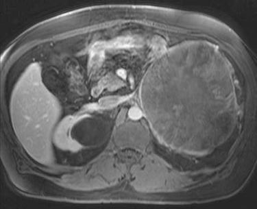
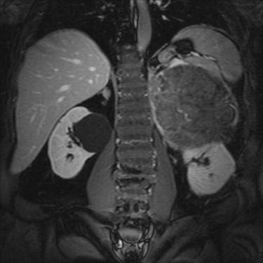
4 4 CH 1 ADRENAL IMAGING Treatment Resection is best treatment for cortisol-producing adrenal tumors or ACTH-producing tumors. Pituitary irradiation may be necessary if pituitary surgery fails. Medical treatment may be indicated to control hypercortisolism, or when patients not cured by resection or when complete resection is impossible. Imaging findings Adrenal hyperplasia Often seen in patients with Cushing s syndrome and less commonly in Conn s syndrome. May be diffuse or nodular and is typically bilateral (Figs 1.1 and 1.2). Adrenal adenoma Most are less than 3 cm in size. Can be of varying density on CT and MRI. Lipid-rich adenoma. Attenuation value of 10 HU or less at unenhanced CT (Fig. 1.3). Adenomas usually have absolute enhancement washout of >60% (Fig. 1.4) and relative enhancement washout of >40%. Greater than 16.5% loss of signal intensity on out-of-phase, compared with in-phase MRI pulse sequences (Fig. 1.5). Figure 1.1 Bilateral adrenal cortical hyperplasia. Axial contrast-enhanced CT image shows nodular thickening of adrenal glands bilaterally in patient with Cushing s syndrome.

. 1.")
5 1.3 PRIMARY HYPERALDOSTERONISM 5 Figure 1.2 Bilateral adrenal cortical hyperplasia. Axial out-of-phase MR image shows nodular thickening of adrenal glands bilaterally in patient with Cushing s syndrome. Figure 1.3 8HU. Lipid-rich adenoma. Axial unenhanced CT shows a right adrenal mass measuring Functioning and non-functioning adenomas, appear similar based on imaging as do Cushing s and Conn s adenomas. Adrenal cortical carcinomas can also cause Cushing s syndrome (see below for imaging appearances of adrenal cortical carcinoma). 1.3 Primary hyperaldosteronism Introduction Primary hyperaldostronism is a relatively common and underdiagnosed condition that contributes to hypertension in about 1% of hypertensive people. The condition is very effectively treated, and so screening programs have become routine in some places.

 (a).")
 (b) and 50 HU (arrow) on delayed")

 (a) Figure 1.5 Adrenal adenoma.")
6 6 CH 1 ADRENAL IMAGING (a) (b) Figure 1.4 Lipid-poor adenoma. Axial unenhanced CT shows a right adrenal mass measuring 27 HU (arrow) (a). Following intravenous contrast enhancement the mass measures 96 HU (arrow) (b) and 50 HU (arrow) on delayed images (c), respectively. This mass had an absolute enhancement washout of 67%. Absolute Washout = 96 50/ = 67%. (c) (a) Figure 1.5 Adrenal adenoma. Coronal in-phase (a) and out-of-phase (b) MR images show an adrenal mass (arrow) which exhibits a typical decrease in signal intensity on the out-of-phase image. (b)
7 1.3 PRIMARY HYPERALDOSTERONISM 7 Clinical features Hypertension with or without hypokalemia. Elevated aldosterone secretion and suppressed plasma renin activity. Metabolic alkalosis, relative hypernatremia. Weakness, polyuria, paresthesias, tetany, cramps due to hypokalemia. Common subtypes of primary hyperaldosteronism: aldosteronoma (75%) and bilateral adrenal hyperplasia (25%). Rare subtypes of primary hyperaldosteronism: unilateral primary adrenal hyperplasia, aldosterone-producing adrenocortical carcinoma, glucocorticoid-remediable hyperaldosteronism (familial hyperaldosteronism type 1). Symptoms and signs Hypertension Headaches Malaise Muscle weakness Polyuria Polydipsia Cramps Paresthesias Hypokalemic paralysis (rare). Laboratory findings Hypokalemia Hypernatremia Metabolic alkalosis Elevated plasma aldosterone to renin ratio ({ } 20) Elevated plasma aldosterone concentration ({ } 15 ng/dl) Elevated urine/serum aldosterone level with PO or IV sodium challenge. Treatment Surgical therapy for patients with aldosteronoma and unilateral primary adrenal hyperplasia. Medical therapy for bilateral adrenal hyperplasia, or poor surgical candidates. Surgery Nearly always laparoscopic approach. Unilaterality best defined by adrenal vein sampling for aldosterone and cortisol
8 8 CH 1 ADRENAL IMAGING Indications Unilateral aldosteronoma Unilateral primary adrenal hyperplasia. Contraindications Bilateral adrenal hyperplasia. Removal of aldosteronoma normalizes potassium, but hypertension is not always cured. 33% of patients have persistent, mild hypertension (easier to control than before operation). Medications Spironolactone: competitive aldosterone antagonist. Amiloride: potassium-sparing diuretic. Other antihypertensive agents such as ACE inhibitors and calcium channel blockers. Imaging findings Adrenal hyperplasia May be diffuse or nodular and is typically bilateral (Figs 1.1 and 1.2). Adrenal adenoma Most are small and less than 2 cm in size. Usually much smaller than Cushing s adenoma. Can have varying appearances of CT and MRI. Lipid-rich adenoma- Attenuation value of 10 HU or less at unenhanced CT (Fig. 1.3). Absolute enhancement washout >60% (Fig. 1.4) and relative enhancement washout >40%. Greater than 16.5% loss of signal intensity on out-of-phase, compared with in-phase MRI pulse sequences (Fig. 1.5). Functioning and non-functioning adenomas, appear similar based on imaging as do Cushing s and Conn s adenomas. Adrenal cortical carcinomas rarely cause Conn s syndrome. 1.4 Pheochromocytoma Introduction Pheochromocytomas are tumors that develop from the adrenal medulla. The hormonal function typically includes production of catecholamines, and the characteristic syndrome that follows. These tumors can be benign or malignant.
9 1.4 PHEOCHROMOCYTOMA 9 Clinical features Episodic headache, excessive sweating, palpitations, and visual blurring. Hypertension, frequently sustained, with or without paroxysms. Postural tachycardia and hypotension. Elevated urinary catecholamines or their metabolites, hypermetabolism, hyperglycemia. Early recognition during pregnancy is key because if left untreated, half of fetuses and nearly half of the mothers will die. Epidemiology. Found in <0.1% of patients with hypertension. 5% of tumors discovered incidentally on CT scan. Most occur sporadically. Associated with familial syndromes, such as: Multiple endocrine neoplasia type 2A (MEN 2A) MEN 2B Recklinghausen disease von Hippel-Lindau disease. Pheochromocytomas are present in 40% of patients with MEN 2 90% of patients with pheochromocytoma are hypertensive. Rule of 10s: 10% malignant 10% familial 10% bilateral 10% multiple tumors 10% extra-adrenal. Hypertension less common in children. In children, 50% of patients have multiple or extra-adrenal tumors. Symptoms and signs Episodic or sustained hypertension. Triad of palpitation, headache, and diaphoresis. Anxiety, tremors. Weight loss. Dizziness, nausea, and vomiting. Abdominal discomfort, constipation, diarrhea. Visual blurring. Tachycardia, postural hypotension. Hypertensive retinopathy.
10 10 CH 1 ADRENAL IMAGING Laboratory findings Hyperglycemia. Elevated plasma metanephrines. Elevated 24-h urine metanephrines and free catecholamines. Elevated urinary vanillylmandelic acid (VMA). Elevated plasma catecholamines. Avoid arteriography or fine-needle aspiration as they can precipitate a hypertensive crisis. Treatment α-adrenergic blocking agents should be started as soon as the biochemical diagnosis is established to restore blood volume, to prevent a severe crisis, and to allow recovery from the cardiomyopathy. β-blockade should generally be established after α-blockade, and prior to operation surgery Indications All resectable pheochromocytomas should be excised. Contraindications Unresectable systemic disease. Inadequate medical preparation (α-blockade). Medications α-adrenergic blocking agents, such as phenoxybenzamine. Other agents include metyrosine, prazosin, and calcium channel blockers. β-adrenergic blocking agents can be used only after full α-blockade has been achieved. Avoid opioids as they stimulate histamine release. Prognosis Operative mortality is 1 2%. Mild to moderate essential hypertension may persist after surgery. Treatment with 131 I-MIBG may help patients with metastatic or recurrent malignant pheochromocytomas. Imaging findings May be homogeneous or heterogeneous, solid or cystic complex masses. May show calcification. Smaller tumors tend to have a more uniform attenuation.
.")

11 1.4 PHEOCHROMOCYTOMA 11 Figure 1.6 Adrenal pheochromocytoma. Axial contrast enhanced CT shows a heterogeneously enhancing right adrenal mass (arrow). Typically enhance avidly with intravenous contrast administration but can be heterogeneous (Fig. 1.6). Most have an absolute enhancement washout of less than 60%, and a relative enhancement of less than 40%, but washout features are variable. Show less than 16.5% loss of signal intensity on out-of-phase, compared with in-phase MRI pulse sequences. The classic light bulb bright signal on T2-weighted images is infrequently seen. Pheochromocytomas occur in association with various syndromes such as; multiple endocrine neoplasias (MEN2), Von Recklinghausen neurofibromatosis (NF1), and Von Hippel-Lindau disease (VHL) (Fig. 1.7). Figure 1.7 Adrenal pheochromocytoma. Axial contrast enhanced CT shows a heterogeneously enhancing right adrenal mass (arrow). Also note visualization of a left renal cyst (arrow) in this patient with Von Hippel Lindau disease.

12 12 CH 1 ADRENAL IMAGING Figure 1.8 Adrenal pheochromocytoma. Radionuclide MIBG scan shows increased tracer uptake in the region of a right adrenal mass. Pheochromocytomas usually exhibit high uptake on MIBG scintigraphic examination (Fig. 1.8). 1.5 Adrenal cortical carcinoma Introduction Adrenocortical carcinoma is typically an aggressive malignancy with a poor prognosis, though less aggressive forms do occur. The tumors can present either due to hormone production, or due to mass effect from the primary or metastatic lesions. Clinical features Variety of clinical symptoms through excess production of adrenal hormones. Complete surgical removal of the primary lesion and symptomatic metastatic sites, if possible, has been the mainstay of treatment. Epidemiology These tumors are rare; 1 2 cases per million persons in the USA. Less than 0.05% of newly diagnosed cancers per year. Bimodal occurrence, with tumors developing in children less than 5 years of age and in adults in their fifth through seventh decade of life. Male:female ratio is 2:1, with functional tumors being more common in women.
13 1.5 ADRENAL CORTICAL CARCINOMA 13 Left adrenal involved slightly more often than the right (53% vs. 47%); bilateral tumors are rare (2%) % of patients have symptoms related to hypersecretion of hormones (most commonly Cushing s syndrome and virilization). Feminizing and purely aldosterone-secreting carcinomas are more rare. 50% of patients have metastases at the time of diagnosis. Symptoms and signs Symptoms of specific hormone excess (cortisol excess, virilization, feminization). Palpable abdominal mass. Abdominal pain. Fatigue, weight loss, fever, hematuria. Laboratory findings All laboratory abnormalities depend on hormonal status of tumor. Elevated urinary free cortisol or steroid precursors. Loss of normal circadian rhythm for serum cortisol. Low serum adrenocorticotropic hormone (ACTH). Abnormal dexamethasone suppression test. Elevated serum testosterone, estradiol, or aldosterone levels. Treatment Surgery is the only treatment that can cure or prolong survival. Laparoscopic surgery not recommended because of spread of tumor, fragility of tumor, and the possible need to resect adjacent involved organs. For local recurrent disease, reoperation is the only effective therapy and may prolong life. Surgery Indications Disease localized to the adrenal, or local spread. Contraindications Widely metastatic disease. Medications Mitotane (an adrenolytic agent) can be used as adjuvant therapy; controls endocrine symptoms in 50% of patients but does not generally affect survival. Prognosis Tumor stage at the initial operation predicts survival. Median survival is 25 months. 5-year actuarial survival is 25%. 5-year survival with grossly complete surgical resection is 50%.

 (b)")
")
14 14 CH 1 ADRENAL IMAGING (a) (b) (c) (d) (e)
15 Imaging findings 1.6 ADRENAL INCIDENTALOMA 15 Usually large tumors at diagnosis, most larger than 6 cm. Heterogeneous on CT and MRI, owing to the presence of internal hemorrhage, calcification and necrosis (Fig. 1.9). Large tumors tend to invade the adrenal vein and inferior vena cava. 1.6 Adrenal incidentaloma Introduction Incidentally identified adrenal tumors found on studies performed for other indications than symptomatic adrenal disease are common. Evaluation of patients who have these tumors can depend on the age of the patient and the size of the tumor. Although adrenal cortical adenomas are the most common lesions that present as incidentalomas, a wide variety of benign and malignant masses as well as non-hyperfunctioning and subclinical hormonally active masses can present in this manner. Clinical features Incidence of diagnosis has increased with use of ultrasonography, CT, and MRI for various, non-related conditions of the abdomen. Diagnosis includes such conditions as non-functioning adrenocortical adenoma, functioning adenoma, pheochromocytomas with subclinical secretion of hormones, and adrenocortical carcinomas. Major issues are to determine whether the tumor is hormonally active, or a carcinoma, or neither. Most simple adrenal cysts, myelolipomas, and adrenal hemorrhages can be identified by the imaging characteristics alone. Adrenal cysts can be very large. Since most tumors are non-functioning adenomas, the work-up should avoid unnecessary procedures and expense. Non-functioning adrenal tumors that are greater than 5 cm have a higher risk of cancer. An adrenal mass { } 3 cm in a patient with a previously treated malignancy is very likely a metastasis. Figure 1.9 Adrenal cortical carcinoma. (a) Axial contrast enhanced CT shows a large heterogeneously enhancing left adrenal mass containing calcification (black arrow) and low attenuation area of necrosis (white arrow). (b-e) MR images in a different case of adrenal carcinoma shows heterogeneous regions (arrows) prior to and following intravenous gadolinium administration due to degeneration and necrosis.
16 16 CH 1 ADRENAL IMAGING Primary tumors that metastasize to the adrenal gland include: lung, breast, colon, renal cell carcinoma, malignant melanoma, uterine, and prostate. Epidemiology Found in 1 4% of CT scans. Found in 6% of random autopsies. Incidence increases with age. Over 80% are non-functioning cortical adenomas. 5% each are preclinical Cushing s syndrome, pheochromocytoma, and adrenocortical carcinoma. 2% are metastatic carcinoma. 1% are aldosteronoma. 25% of pheochromocytomas are found incidentally. Workup and treatment Complete history and physical exam, with specific reference to previous malignancies, symptoms of Cushing s syndrome, hypertension, virilization, or feminization. All patients, even those without hypertension, should have plasma metanephrines and 24-h urinary fractionated catecholamines determined to evaluate for pheochromocytoma. All patients should have a serum cortisol, 24-h urine collection for cortisol, and an overnight dexamethasone suppression test. Patients who are hypertensive should have serum potassium, and plasma aldosterone and renin activity measured. Consider obtaining a dehydroepiandrosterone (DHEA) level (potential marker for adrenocortical carcinoma). If above studies show the tumor to be non-functional, the size of the tumor and the patient s overall medical condition determine management. If metastasis is suspected and pheochromocytoma is ruled out, then CT-guided biopsy is useful. Imaging findings Most commonly these are non-functioning adrenal cortical adenomas, but functioning tumors such as adrenal cortical carcinoma and pheochromocytoma may also present as incidental adrenal masses. Adrenal adenoma Most are less than 3 cm in size. Can have varying appearances on CT.

. Hypointense on T1-weighted images and hyperintense on T2-weighted images, with no soft-tissue component and no internal enhancement (Fig. 1.11).")
17 1.6 ADRENAL INCIDENTALOMA 17 Lipid-rich adenoma. Attenuation value of 10 HU or less on unenhanced CT. Absolute enhancement washout >60% and relative enhancement washout >40%. Greater than 16.5% loss of signal intensity on out-of-phase, compared with in-phase MRI pulse sequences. Other masses include the following: Adrenal cyst Can have features of simple cyst such as attenuation of less than 20 HU on unenhanced image and no enhancement following intravenous contrast administration (Fig. 1.10). Hypointense on T1-weighted images and hyperintense on T2-weighted images, with no soft-tissue component and no internal enhancement (Fig. 1.11). Pseudocysts typically arise after an episode of adrenal hemorrhage or trauma. Adrenal pseudocysts may have a complicated appearance on CT and MR images, with septations, blood products, soft-tissue component secondary to hemorrhage or hyalinized thrombus, and curvilinear calcification (Fig. 1.12). Pseudocysts can be indistinguishable from malignant tumors. Adrenal myelolipoma Benign tumor composed of marrow elements such as mature adipose tissue (fat), hematopoietic tissue and calcification/ossification. Fat can be diagnosed by the presence of areas of negative attenuation value by CT (Fig. 1.13) and on MR, by suppression of signal intensity on fat suppressed images, when compared with non-fat suppressed images. (a) Figure 1.10 Adrenal cyst. Axial unenhanced CT shows a left adrenal mass measuring 18 HU (arrow) (a). Following intravenous contrast enhancement the mass did not show significant enhancement (arrow) (b). (b)
, and axial fat suppressed T2 weighted (b) MR show a left adrenal mass exhibiting a hypointense")
 Figure 1.12 Adrenal pseudocyst. Axial contrast enhanced CT shows a right calcified adrenal mass measuring 19 HU.")

18 18 CH 1 ADRENAL IMAGING (a) Figure 1.11 Adrenal cyst. Axial contrast enhanced T1 weighted (a), and axial fat suppressed T2 weighted (b) MR show a left adrenal mass exhibiting a hypointense signal on T1-weighted images (arrow) and hyperintense signal on T2-weighted images (arrow) with no soft-tissue component and no internal enhancement. (b) Figure 1.12 Adrenal pseudocyst. Axial contrast enhanced CT shows a right calcified adrenal mass measuring 19 HU. Adrenal hemorrhage Usually bilateral. Can be seen in post-operative states, trauma, sepsis and in patients with blood dyscrasias or receiving anticoagulant therapy. If unilateral usually due to trauma or following liver transplantation (right sided) High density on unenhanced images (Fig. 1.14). Appearance overlaps that of other lesions following contrast enhancement.
.")
 with high attenuation. Metastases (Fig. 1.")
19 1.6 ADRENAL INCIDENTALOMA 19 Figure 1.13 Adrenal myelolipoma. Axial contrast enhanced CT shows a left adrenal mass with an intralesional area of fat indicated by a negative attenuation value of 28 HU (arrow). Note that qualitatively this area is of similar attenuation to the adjacent intra-abdominal fat. Figure 1.14 Bilateral adrenal hemorrhage. Unenhanced CT shows bilateral adrenal masses (arrows) with high attenuation. Metastases (Fig. 1.15) Common primary tumors include the lung, breast, GI tract and pancreas. Can be of varying size. Attenuation higher than 10 HU on unenhanced CT, as they do not typically contain intracellular lipid.
. Absolute enhancement washout of less than 60%, and a relative enhancement of less than 40%. Usually heterogeneous.")
 from adenoma. Comparison to liver uptake is often useful for this assessment (Fig.")
20 20 CH 1 ADRENAL IMAGING Figure 1.15 Adrenal metastases. Contrast-enhanced CT shows diffuse hepatic and bilateral adrenal metastases (arrows) in patient with advanced melanoma. Figure 1.16 Adrenal metastasis. Fused PET/CT scan shows marked tracer uptake in a small left adrenal mass (arrow). Absolute enhancement washout of less than 60%, and a relative enhancement of less than 40%. Usually heterogeneous. Less than 16.5% loss of signal intensity on out-of-phase, compared with in-phase MRI pulse sequences, except in metastatic clear cell renal carcinoma which may contain foci of lipid. High tracer activity on FDG PET scans usually differentiates malignancy (metastasis or adrenal cortical carcinoma) from adenoma. Comparison to liver uptake is often useful for this assessment (Fig. 1.16).
ADRENAL INCIDENTALOMA. Jamii St. Julien
 ADRENAL INCIDENTALOMA Jamii St. Julien Outline Definition Differential Evaluation Treatment Follow up Questions Case Definition The phenomenon of detecting an otherwise unsuspected adrenal mass on radiologic
Mineralocorticoids: aldosterone Angiotensin II/renin regulation by sympathetic tone; High potassium will stimulate and ACTH Increase in aldosterone
 Disease of the Adrenals 1 Zona Glomerulosa Mineralocorticoids: aldosterone Angiotensin II/renin regulation by sympathetic tone; High potassium will stimulate and ACTH Increase in aldosterone leads to salt
Case Based Urology Learning Program
 Case Based Urology Learning Program Resident s Corner: UROLOGY Case Number 4 CBULP 2010 004 Case Based Urology Learning Program Editor: Associate Editors: Manager: Case Contributors: Steven C. Campbell,
The Work-up and Treatment of Adrenal Nodules
 The Work-up and Treatment of Adrenal Nodules Lawrence Andrew Drew Shirley, MD, MS, FACS Assistant Professor of Surgical-Clinical Department of Surgery Division of Surgical Oncology The Ohio State University
Endocrine MR. Jan 30, 2015 Michael LaFata, MD
 Endocrine MR Jan 30, 2015 Michael LaFata, MD Brief case 55-year-old female in ED PMH: HTN, DM2, HLD, GERD CC: Epigastric/LUQ abdominal pain, N/V x2 days AF, HR 103, BP 155/85, room air CMP: Na 133, K 3.6,
Diseases of the Adrenal gland
 Diseases of the Adrenal gland Adrenal insufficiency Cushing disease vs syndrome Pheochromocytoma Hyperaldostronism What are the layers of the adrenal gland?? And what does each layer produce?? What are
Adrenal Mass. Cynthia Kwong SUNY Downstate Medical Center Grand Rounds October 13, 2016
 Adrenal Mass Cynthia Kwong SUNY Downstate Medical Center Grand Rounds October 13, 2016 Case Presentation 65F found to have a 4cm left adrenal mass in 2012 now presents with 6.7cm left adrenal mass PMHx:
Evaluation of Thyroid Nodules
 Evaluation of Thyroid Nodules Stephan Kowalyk, MD January 25 28, 2018 1 Primary goal Exclude malignancy Incidental thyroid nodules If found on CT, MRI, PET scan, carotid Doppler ULTRASOUND!! January 25
Daniela Faivovich K., MS VII Universidad de Chile Gillian Lieberman, MD Harvard Medical School
 Daniela Faivovich K., MS VII Universidad de Chile Gillian Lieberman, MD Harvard Medical School May 21st, 2010 56 year old male patient History of hypertension, hyperlipidemia and insulin-resistance 2009:
ADRENAL LESIONS 10/09/2012. Adrenal + lesion. Introduction. Common causes. Anatomy. Financial disclosure. Dr. Boraiah Sreeharsha. Nothing to declare
 ADRENAL LESIONS Financial disclosure Nothing to declare Dr. Boraiah Sreeharsha MBBS;FRCR;FRCPSC Introduction Adrenal + lesion Adrenal lesions are common 9% of the population Increase in the detection rate
Adrenal gland Incidentaloma
 Adrenal gland Incidentaloma Topic review 17 sep 2008 Anatomy 1 Anatomical consideration Blood supply Artery: small branches from Inf. phrenic, renal artery and aorta Vein: Rt : medial aspect to IVC Lt
Approach to Adrenal Incidentaloma. Alice Y.Y. Cheng, MD, FRCP
 Approach to Adrenal Incidentaloma Alice Y.Y. Cheng, MD, FRCP Copyright 2017 by Sea Courses Inc. All rights reserved. No part of this document may be reproduced, copied, stored, or transmitted in any form
The Adrenal Glands. I. Normal adrenal gland A. Gross & microscopic B. Hormone synthesis, regulation & measurement. II.
 The Adrenal Glands Thomas Jacobs, M.D. Diane Hamele-Bena, M.D. I. Normal adrenal gland A. Gross & microscopic B. Hormone synthesis, regulation & measurement II. Hypoadrenalism III. Hyperadrenalism; Adrenal
CUSHING SYNDROME Dr. Muhammad Sarfraz
 Indep Rev Jul-Dec 2018;20(7-12) CUSHING SYNDROME Dr. Muhammad Sarfraz IR-655 Abstract: It is defined as clinical condition in which there are increased free circulating glucocorticoides casused by excessive
How to Recognize Adrenal Disease
 How to Recognize Adrenal Disease CME Away India & Sri Lanka March 23 - April 7, 2018 Richard A. Bebb MD, ABIM, FRCPC Consultant Endocrinologist Medical Subspecialty Institute Cleveland Clinic Abu Dhabi
Endocrine. Endocrine as it relates to the kidney. Sarah Elfering, MD University of Minnesota
 Endocrine Sarah Elfering, MD University of Minnesota Endocrine as it relates to the kidney Parathyroid gland Vitamin D Endocrine causes of HTN Adrenal adenoma PTH Bone Kidney Intestine 1, 25 OH Vitamin
Characterization of adrenal lesions on CT and MRI: all that a radiologist must know
 Characterization of adrenal lesions on CT and MRI: all that a radiologist must know Poster No.: C-2476 Congress: ECR 2013 Type: Educational Exhibit Authors: N. Benzina, S. MAJDOUB, C. H. ZARRAD, H. Zaghouani,
Adrenal incidentaloma guideline for Northern Endocrine Network
 Adrenal incidentaloma guideline for Northern Endocrine Network Definition of adrenal incidentaloma Adrenal mass detected on an imaging study done for indications that are not related to an adrenal problem
THE HIGHS AND LOWS OF ADRENAL GLAND PATHOLOGY
 THE HIGHS AND LOWS OF ADRENAL GLAND PATHOLOGY Symptoms of Adrenal Gland Disorders 2 Depends on whether it is making too much or too little hormone And on what you Google! Symptoms include obesity, skin
Indications for Surgical Removal of Adrenal Glands
 The adrenal glands are orange-colored endocrine glands which are located on the top of both kidneys. The adrenal glands are triangular shaped and measure about one-half inch in height and 3 inches in length.
Odise Cenaj, Harvard Medical School Year III. Gillian Lieberman, MD
 February 2012 Radiologic evaluation of adrenal masses and an atypical radiologic presentation of adrenocortical carcinoma in a patient with primary aldosteronism Odise Cenaj, Harvard Medical School Year
The Management of adrenal incidentaloma
 The Management of adrenal incidentaloma Dimitrios Linos, MD Director of Surgery, Hygeia Hospital, Athens, Greece Consultant in Surgery, Massachusetts General Hospital, Boston, USA 8 th Postgraduate Course
Adrenal incidentaloma
 Adrenal incidentaloma Prevalence 5% post-mortem series 4% CT series 6-20% CT series in patients with Hx extra-adrenal malignancy Commoner with increasing age Associated with adrenal hyperfunction in 15%
Pheochromocytoma AMERICAN ASSOCIATION OF CLINICAL ENDOCRINOLOGY ILLINOIS CHAPTER OCTOBER 13, 2018
 Pheochromocytoma AMERICAN ASSOCIATION OF CLINICAL ENDOCRINOLOGY ILLINOIS CHAPTER OCTOBER 13, 2018 Steven A. De Jong, M.D., FACS, FACE Professor and Vice Chair of Surgery Chief, Division of General Surgery
ADRENAL MEDULLARY DISORDERS: PHAEOCHROMOCYTOMAS AND MORE
 ADRENAL MEDULLARY DISORDERS: PHAEOCHROMOCYTOMAS AND MORE DR ANJU SAHDEV READER AND CONSULTANT RADIOLOGIST QUEEN MARY UNIVERSITY AND ST BARTHOLOMEW S HOSPITAL BARTS HEALTH, LONDON, UK DISCLOSURE OF CONFLICT
Adrenal Incidentaloma Management
 Adrenal Incidentaloma Management Full Title of Guideline: Author Management of Incidentally-discovered Adrenal Lesions ( Incidentalomas ) Mr David Chadwick Consultant Endocrine Surgeon david.chadwick2@nuh.nhs.uk
Pituitary Gland Disorders
 Pituitary Gland Disorders 1 2 (GH-RH) (CRH) (TRH) (TRH) (GTRH) (GTRH) 3 Classification of pituitary disorders: 1. Hypersecretory diseases: a. Acromegaly and gigantism: Usually caused by (GH)-secreting
THE FACTS YOU NEED TO KNOW
 PHEOCHROMOCYTOMA THE FACTS YOU NEED TO KNOW Pheochromocytoma is a part of the pheochromocytoma and paraganglioma group of syndromes. A pheochromocytoma is a tumor arising in the adrenal gland medulla.
REVIEW. Distinguishing benign from malignant adrenal masses
 Cancer Imaging (2003) 3, 102 110 DOI: 10.1102/1470-7330.2003.0006 CI REVIEW Distinguishing benign from malignant adrenal masses Isaac R Francis Professor of Radiology, Department of Radiology, University
Incidental Adrenal Nodules Differential Diagnosis
 Adrenal Stuff Richard J. Auchus, MD, PhD, FACE Division of Metabolism, Endocrinology & Diabetes Departments of Internal Medicine & Pharmacology University of Michigan/VA Ann Arbor Incidental Adrenal Nodules
RECURRENT ADRENAL DISEASE. Megan Applewhite Endorama 2/19/2015 SR , SC
 RECURRENT ADRENAL DISEASE Megan Applewhite Endorama 2/19/2015 SR 2412318, SC 3421561 Category: Adrenal Attendings: Angelos & Grogan PATIENT #1 36yo woman with a hx of Cushing s Syndrome and right adrenalectomy
Personal data. Age : 63 Gender : male
 Personal data Age : 63 Gender : male Chief complain No specific symptom or discomfort A hepatic mass, found by abdominal sonography of routine health exam on 88-12-08 Past history 1984-3-3 Old CVA with
John Sutton, DO, FACOI, FACE, CCD. Carson Tahoe Endocrinology Carson City, NV KCOM Class of 1989
 John Sutton, DO, FACOI, FACE, CCD Carson Tahoe Endocrinology Carson City, NV KCOM Class of 1989 Gonadal Physiology and Disease 3 No Disclosures Gonadal Axis Hypothalamic-pituitary-gonadal Feedback mechanisms
Incidental adrenal masses A primary care approach
 CLINICAL Incidental adrenal masses A primary care approach Rasha Gendy, Prem Rashid Background The common use of cross-sectional imaging for the investigation of abdominal and thoracic illness has resulted
Read the following article and answer the questions that follow. Refer to the Keys section to check your answers.
 ENGLISH 183 READING PRACTICE - Pheochromocytoma Read the following article and answer the questions that follow. Refer to the Keys section to check your answers. Pheochromocytoma is a tumor on the medulla
ESUR 2018, Sept. 13 th.-16 th., 2018 Barcelona, Spain
 ESUR 2018, Sept. 13 th.-16 th., 2018 Barcelona, Spain OUR APPROACH Incidental adrenal nodule/mass Isaac R Francis, M.B;B.S University of Michigan, Ann Arbor, Michigan Disclosures None (in memory) M Korobkin,
Dimitrios Linos, M.D., Ph.D. Professor of Surgery National & Kapodistrian University of Athens
 Dimitrios Linos, M.D., Ph.D. Professor of Surgery National & Kapodistrian University of Athens What is an adrenal incidentaloma? An adrenal incidentaloma is defined as an adrenal tumor initially diagnosed
Endocrine Emergencies: Recognition and Management
 Endocrine Emergencies: Recognition and Management John Wass Department of Endocrinology, Oxford University, UK An Update on Acute Medical Emergencies for Psychiatrists Royal College of Psychiatrists' address
adrenal and parathyroid glands Done by jehad abdel aziz
 15-11-09 prof. muhammed khammash adrenal and parathyroid glands Done by jehad abdel aziz The adrenal glands:- Anatomy:- The adrenal glands are flattened, yellowish structures that weigh less than 10g in
ULTIMATE BEAUTY OF BIOCHEMISTRY. Dr. Veena Bhaskar S Gowda Dept of Biochemistry 30 th Nov 2017
 ULTIMATE BEAUTY OF BIOCHEMISTRY Dr. Veena Bhaskar S Gowda Dept of Biochemistry 30 th Nov 2017 SUSPECTED CASE OF CUSHING S SYNDROME Clinical features Moon face Obesity Hypertension Hunch back Abdominal
Adrenal and retropetionium
 Adrenal and retropetionium Disorders of the Adrenal Cortex Hyperaldosteronism: Hyperaldosteronism may be secondary to stimulation of the renin-angiotensin system from renal artery stenosis and to low-flow
C h a p t e r 3 8 Cushing s Syndrome : Current Concepts in Diagnosis and Management
 C h a p t e r 3 8 Cushing s Syndrome : Current Concepts in Diagnosis and Management Padma S Menon Professor of Endocrinology, Seth G S Medical College & KEM Hospital, Mumbai A clinical syndrome resulting
Morbidity & Mortality. Mark H. Tseng MD SUNY Downstate Medical Center Lutheran Medical Center December 16, 2005
 Morbidity & Mortality Mark H. Tseng MD SUNY Downstate Medical Center Lutheran Medical Center December 16, 2005 Case presentation Pt is a xx year old Asian woman who present to the ED with cc of epigastric
The Case of the Adrenal Mass
 The Case of the Adrenal Mass Functional Adrenal Tumors Patricia Leung 10.2.14 Kings County Hospital Case presentation 62 year old F PMH: HTN, DM, arthritis PSH: none Meds: Metoprolol, Nifedipine, Losartan,
A case of micturition syncope
 A case of micturition syncope Kimberly Bundick, PA-S S L I D E 1 Agenda Purpose Utilize case to illustrate classic finding of an interesting pathology Agenda Case study Epidemiology, etiology of disease
A Woman with Long-Standing Hypertension Diagnosed with Metastatic Adrenal Carcinoma
 A Woman with Long-Standing Hypertension Diagnosed with Metastatic Adrenal Carcinoma Mir A. Alikhan, MD* Frank J. Pikul, MD Peter P. Toth, MD, PhD *Department of Hematology/Oncology, Sterling Rock Falls
Pediatric Retroperitoneal Masses Radiologic-Pathologic Correlation
 Acta Radiológica Portuguesa, Vol.XVIII, nº 70, pág. 61-70, Abr.-Jun., 2006 Pediatric Retroperitoneal Masses Radiologic-Pathologic Correlation Marilyn J. Siegel Mallinckrodt Institute of Radiology, Washington
Management of adrenal incidentalomas
 31 Management of adrenal incidentalomas KEVIN MURTAGH, NANA MUHAMMAD AND MAREK MILLER The return of a scan result with reference to an incidental finding of an adrenal mass is a common scenario. 1 The
27 F with new onset hypertension and weight gain. Rajesh Jain Endorama 10/01/2015
 27 F with new onset hypertension and weight gain Rajesh Jain Endorama 10/01/2015 HPI 27 F with hypertension x 1 year BP 130-140/90 while on amlodipine 5 mg daily She also reports weight gain, 7 LB, mainly
CUSHING S SYNDROME. Chapter 8. Case: A 43-year-old man with delusions
 Chapter 8 CUSHING S SYNDROME Case: A 43-year-old man with delusions A previously healthy 43-year-old man is brought to the emergency department for evaluation of confusion. The patient has complained to
The Evaluation of the Incidental Adrenal Mass and Not-So-Incidental Adrenal Hormone Excess
 The Evaluation of the Incidental Adrenal Mass and Not-So-Incidental Adrenal Hormone Excess Richard J. Auchus, MD, PhD, FACE Depts. Internal Medicine/MEND & Pharmacology Endocrinology Fellowship Program
SECONDARY HYPERTENSION
 SECONDARY HYPERTENSION Grand round for Medical student 25 October 2013 By Rungnapa Laortanakul, MD. OUTLINE Overview of HT Secondary HT Resistance HT Primary aldosteronism Pheochromocytoma Cushing s syndrome
SA CME Information SA CME INFORMATION. Target Audience
 SA CME INFORMATION SA CME Information Description Adrenal Imaging: A Three-category Approach To Managing The Adrenal "Incidentaloma" Imaging plays a critical role in the work-up and clinical management
Endocrine Topic Review. Sethanant Sethakarun, MD
 Endocrine Topic Review Sethanant Sethakarun, MD Definition Cushing's syndrome comprises a large group of signs and symptoms that reflect prolonged and in appropriately high exposure of tissue to glucocorticoids
Adrenal masses in infancy and childhood: A clinical and radiological overview M. Mearadji
 Adrenal masses in infancy and childhood: A clinical and radiological overview M. Mearadji International Foundation for Pediatric Imaging Aid Introduction Neoplastic adrenal masses usually originate from
Adrenal Gland. Zona Glomerulosa (mineralcorticoids) Zona Fasciculata (glucocorticoids) Zona Reticularis (sexual hormones) Adrenaline and noradrenaline
 Zona Fasciculata (glucocorticoids) Zona Reticularis (sexual hormones) Adrenaline and noradrenaline") n Cortex Zona Glomerulosa (mineralcorticoids) Zona Fasciculata (glucocorticoids) Zona Reticularis (sexual hormones) n Medulla Adrenaline and noradrenaline n Adrenocortical Hyperfunction Hyperaldosteronism
n Cortex Zona Glomerulosa (mineralcorticoids) Zona Fasciculata (glucocorticoids) Zona Reticularis (sexual hormones) n Medulla Adrenaline and noradrenaline n Adrenocortical Hyperfunction Hyperaldosteronism
CUSHING S SYNDROME THE FACTS YOU NEED TO KNOW
 CUSHING S SYNDROME THE FACTS YOU NEED TO KNOW Written by: Paul Margulies, MD, FACE, FACP, Medical Director, NADF. Clinical Associate Professor of Medicine, Zucker School of Medicine at Hofstra/Northwell.
Adrenocortical Scan. Quality Control. Adult Dose Range
 chapter 1 Adrenocortical Scan RADIOPHARMACY Radionuclide 131 I t 1/2 : 8.1 days Energies: 364 kev Type: β, γ, fission product Radiopharmaceutical 131 I-6-β-Iodomethyl-19-norcholesterol (NP-59). Available
Endogenous Cushing s syndrome: The Philippine general hospital experience
 ORIGINAL ARTICLE Endogenous Cushing s syndrome: The Philippine general hospital experience Tom Edward N. Lo, Joyce M. Cabradilla, Sue Ann Lim, Cecilia A. Jimeno Section of Endocrinology and Metabolism,
Role of imaging in RCC. Ultrasonography. Solid lesion. Cystic RCC. Solid RCC 31/08/60. From Diagnosis to Treatment: the Radiologist Perspective
 Role of imaging in RCC From Diagnosis to Treatment: the Radiologist Perspective Diagnosis Staging Follow up Imaging modalities Limitations and pitfalls Duangkamon Prapruttam, MD Department of Therapeutic
Adrenal Incidentalomas. G Stephen DeCherney, MD, MPH Clinical Professor of Medicine Division of Endocrinology UNC School of Medicine
 Adrenal Incidentalomas G Stephen DeCherney, MD, MPH Clinical Professor of Medicine Division of Endocrinology UNC School of Medicine Disclosures No financial, investment, or consulting relationship with
The endocrine system is made up of a complex group of glands that secrete hormones.
 1 10. Endocrinology I MEDCHEM 535 Diagnostic Medicinal Chemistry Endocrinology The endocrine system is made up of a complex group of glands that secrete hormones. These hormones control reproduction, metabolism,
Kingdom of Bahrain Arabian Gulf University College of Medicine and Medical Sciences. Endocrinology. (Review) Year 5 Internal Medicine
 Year 5 Internal Medicine") Kingdom of Bahrain Arabian Gulf University College of Medicine and Medical Sciences Endocrinology (Review) Year 5 Internal Medicine Presented by: Dr. Mona Arekat Prepared by: Ali Jassim Alhashli Case (1):
Kingdom of Bahrain Arabian Gulf University College of Medicine and Medical Sciences Endocrinology (Review) Year 5 Internal Medicine Presented by: Dr. Mona Arekat Prepared by: Ali Jassim Alhashli Case (1):
Endocrine Surgery When to Refer and What We Do
 Endocrine Surgery When to Refer and What We Do None Disclosures W. Heath Giles, M.D., F.A.C.S. Surgery Residency Program Director Assistant Professor of Surgery What is Endocrine Surgery? Who performs
PITUITARY: JUST THE BASICS PART 2 THE PATIENT
 PITUITARY: JUST THE BASICS PART 2 THE PATIENT DISCLOSURE Relevant relationships with commercial entities none Potential for conflicts of interest within this presentation none Steps taken to review and
PHEOCHROMOCYTOMA. Anita Chiu, MD Kings County Hospital Center January 13, 2011
 PHEOCHROMOCYTOMA Anita Chiu, MD Kings County Hospital Center January 13, 2011 Case Presentation 62 year old female from Grenada with longstanding HTN, DM, CRI Complaints of palpitations for years Abdominal
The Pathological l Basis of Disease
 Endocrine Diseases The Pathological l Basis of Disease - Graduate Course CMM5001 Qiao Li, MD, PhD Faculty of Medicine University of Ottawa qiaoli@uottawa.ca Outline Endocrine System Adrenal Gland Anatomy
ظظظ/ Omar Sami. Hussam Twaissi. Mousa Abbadi
 ظظظ/ 5 Omar Sami Hussam Twaissi Mousa Abbadi The doctor started this lecture by revising what we have taken in lecture number four, I won t re-write these stuff as it becomes boring so often. This sheet
Pathophysiology of Adrenal Disorders
 Pathophysiology of Adrenal Disorders PHCL 415 Hadeel Alkofide April 2010 Some slides adapted from Rania Aljizani MSc 1 Learning Objectives Describe the roles of the various zones of the adrenal cortex
ENDOCRINOLOGY 3. R. A. Benacka, MD, PhD, prof. Department of Pathophysiology Medical faculty, Safarik University, Košice
 Academic lectures for general medicine 3rd year 2005/2006, 2013/2014 ENDOCRINOLOGY 3 R. A. Benacka, MD, PhD, prof. Department of Pathophysiology Medical faculty, Safarik University, Košice Figures and
ADRENAL MR: PEARLS AND PITFALLS
 ADRENAL MR: PEARLS AND PITFALLS Frank Miller, M.D. Lee F. Rogers MD Professor of Medical Education Chief, Body Imaging Section and Fellowship Medical Director, MR Imaging Professor of Radiology Northwestern
Renal Parenchymal Neoplasms
 Renal Parenchymal Neoplasms د. BENIGN TUMORS : Benign renal tumors include adenoma, oncocytoma, angiomyolipoma, leiomyoma, lipoma, hemangioma, and juxtaglomerular tumors. Renal Adenomas : The adenoma is
Recommendations for cross-sectional imaging in cancer management, Second edition
 www.rcr.ac.uk Recommendations for cross-sectional imaging in cancer management, Second edition Renal and adrenal tumours Faculty of Clinical Radiology www.rcr.ac.uk Contents Renal cell carcinoma 3 Clinical
in Primary Care (Part 2) Jonathan R. Anolik, MD, FACP, FACE Lewis Katz School of Medicine at Temple University
 Jonathan R. Anolik, MD, FACP, FACE Lewis Katz School of Medicine at Temple University") Common Endocrine Problems Seen in Primary Care (Part 2) Lecture #34 Jonathan R. Anolik, MD, FACP, FACE Lewis Katz School of Medicine at Temple University None Conflict of Interest Topics to be Covered
Common Endocrine Problems Seen in Primary Care (Part 2) Lecture #34 Jonathan R. Anolik, MD, FACP, FACE Lewis Katz School of Medicine at Temple University None Conflict of Interest Topics to be Covered
Assistant Professor of Endocrinology
 Pathophysiology Of Adrenal Disorder Dr.Rezvan Salehidoost Assistant Professor of Endocrinology Pathophysiology Of Adrenal Disorder The adrenal glands lie at the superior pole of each kidney and are composed
Index. radiologic.theclinics.com. Note: Page numbers of article titles are in boldface type.
 Index Note: Page numbers of article titles are in boldface type. A ACC. See Adrenal cortical carcinoma. Acromegaly and the pituitary gland, 551 Acute suppurative thyroiditis, 405, 406 Addison, Thomas and
Endocrinology and VHL: The adrenal and the pancreas
 Overview Endocrinology and VHL: The adrenal and the pancreas LAUREN FISHBEIN MD, PHD UNIVERSITY OF COLORADO SCHOOL OF MEDICINE DIVISION OF ENDOCRINOLOGY, METABOLISM AND DIABETES DIVISION OF BIOMEDICAL
Sex: 女 Age: 51 Occupation: 無 Admission date:92/07/22
 Sex: 女 Age: 51 Occupation: 無 Admission date:92/07/22 Chief complaint Unknown fever for one month Hand tremor and left huge renal tumor was noted Present illness Suffered from fever for one month, hand
CPY 605 ADVANCED ENDOCRINOLOGY
 CPY 605 ADVANCED ENDOCRINOLOGY THE ADRENAL CORTEX PRESENTED BY WAINDIM NYIAMBAM YVONNE HS09A187 INTRODUCTION Two adrenal glands lie on top of each kidney. Each gland between 6 and 8g in weight is composed
THE ENDOCRINE AND REPRODUCTIVE SYSTEMS
 THE ENDOCRINE AND REPRODUCTIVE SYSTEMS The focus of this week s lab will be pathology of the endocrine and reproductive systems. There are a bunch of tissues and topics that can be covered in these systems,
Traumatic and Non Traumatic Adrenal Emergencies
 Traumatic and Non Traumatic Adrenal Emergencies Michael N. Patlas, MD, FRCPC (1), Christine O. Menias, MD (2), Douglas S. Katz, MD, FACR (3), Ania Z. Kielar, MD, FRCPC (4), Alla M. Rozenblit, MD (5), Jorge
Adrenal venous sampling as used in a patient with primary pigmented nodular adrenocortical disease
 Original Article on Translational Imaging in Cancer Patient Care Adrenal venous sampling as used in a patient with primary pigmented nodular adrenocortical disease Xiaoxin Peng 1, Yintao Yu 1, Yi Ding
Cushing s Syndrome. Diagnosis. GuidelineCentral.com. Key Points. Diagnosis
 Cushing s Syndrome Consultant: Endocrine Society of Cushing s Syndrome Clinical Practice Guideline Writing Committee Key Points GuidelineCentral.com Key Points The most common cause of Cushing s syndrome
Hyperadrenocorticism or Cushing's Syndrome in Dogs
 Customer Name, Street Address, City, State, Zip code Phone number, Alt. phone number, Fax number, e-mail address, web site Hyperadrenocorticism or Cushing's Syndrome in Dogs (Excessive Levels of Steroids
Disorders of the Adrenal Cortex
 Disorders of the Adrenal Cortex Cushing s Syndrome and Primary Aldosteronism 凌雁 Yan Ling Department of Endocrinology and Metabolism Zhongshan Hospital Fudan University Cushing s Syndrome Definition of
57-year-old man with anxiety, diaphoresis, fatigue and bilateral adrenal nodules. Celeste Thomas November 1, 2012
 57-year-old man with anxiety, diaphoresis, fatigue and bilateral adrenal nodules Celeste Thomas November 1, 2012 History of Present Illness 8 months prior to presentation developed intermittent right flank
Update in Pheochromocytoma/Paraganglioma: Focus on Diagnosis and Management
 Update in Pheochromocytoma/Paraganglioma: Focus on Diagnosis and Management Ohk-Hyun Ryu, MD. Associate Professor, Department of Internal Medicine Division of Endocrinology and Metabolism College of Medicine,
ADRENAL INCIDENTALOMAS _ A MANAGEMENT APPROACH Dr Tan Khai Tong
 T H E M E : A S T H M A ARENAL INCIENTALOMAS _ A MANAGEMENT APPROACH r Tan Khai Tong SUMMARY The adrenal incidentaloma is an increasingly common clinical problem. Although most of these masses are innocuous,
Karim Said. 41 year old farmer. Referred from the Uro-surgery Department because of uncontrolled hypertension prior to Lt. partial nephrectomy
 Case Presentation Karim Said Cardiology Departement Cairo University 41 year old farmer Referred from the Uro-surgery Department because of uncontrolled hypertension prior to Lt. partial nephrectomy ١
Nephtali R. Gomez, M.D. To The Incidental Adrenal Mass
 Nephtali R. Gomez, M.D. To The Incidental Adrenal Mass The Complete Idiot s Guide to The Incidental Adrenal Mass Defini:on Any adrenal mass 1cm or more in diameter discovered on a radiologic exam performed
Contrast Materials Patient Safety: What are contrast materials and how do they work?
 Contrast Materials Patient Safety: What are contrast materials and how do they work? Which imaging exams use contrast materials? How safe are contrast materials? How should I prepare for my imaging procedure
ID data. Sex: female Age: 46y/o Birthday: 1955/10/13
 ID data Sex: female Age: 46y/o Birthday: 1955/10/13 Chief Complain Right upper quadrate abdominal tenderness for one month. Present illness (1) This 46 years old female patient was in a healthy condition
Endocrine Hypertension
 Endocrine Hypertension 1 No Disclosures Endocrine Hypertension Objectives: 1. Understand Endocrine disorders causing hypertension 2. Understand clinical presentation of Pheochromocytoma and Hyperaldosteronism
performed to help sway the clinician in what the appropriate diagnosis is, which can substantially alter the treatment of management.
 Hello, I am Maura Polansky at the University of Texas MD Anderson Cancer Center. I am a Physician Assistant in the Department of Gastrointestinal Medical Oncology and the Program Director for Physician
Cushing's disease, Cushing's syndrome
 Greenville Veterinary Clinic LLC 409 E. Jamestown Rd. Greenville, PA 16125 (724) 588-5260 Canine hyperadrenocorticism Cushing's disease, Cushing's syndrome AffectedAnimals: Although dogs of almost every
AVS and IPSS: The Basics and the Pearls William F. Young, Jr., MD, MSc Professor of Medicine Mayo Clinic College of Medicine Rochester, MN, USA
 AVS and IPSS: The Basics and the Pearls William F. Young, Jr., MD, MSc Professor of Medicine Mayo Clinic College of Medicine Rochester, MN, USA 2016 Mayo Foundation for Medical Education and Research.
Year 2004 Paper two: Questions supplied by Megan 1
 Year 2004 Paper two: Questions supplied by Megan 1 QUESTION 96 A 32yo woman if found to have high blood pressure (180/105mmHg) at an insurance medical examination. She is asymptomatic. Clinical examination
Endocrine Testing. Alice Y.Y. Cheng, MD, FRCP October 14, 2015
 Endocrine Testing Alice Y.Y. Cheng, MD, FRCP October 14, 2015 Disclosure No disclosures relevant to the content of this workshop Learning Objectives By the end of this workshop, you will be able to: 1.
Hypertension: Who and How (and Why) to Investigate. Jessica Triay Andy Levy
 to Investigate. Jessica Triay Andy Levy") Hypertension: Who and How (and Why) to Investigate Jessica Triay Andy Levy What I'm not going to talk about Most Common: Renal Disease Renal USS Likely to be normal if bloods and urine normal Renal artery
Hypertension: Who and How (and Why) to Investigate Jessica Triay Andy Levy What I'm not going to talk about Most Common: Renal Disease Renal USS Likely to be normal if bloods and urine normal Renal artery
AVS and IPSS: The Basics and the Pearls
 AVS and IPSS: The Basics and the Pearls William F. Young, Jr., MD, MSc Professor of Medicine Mayo Clinic College of Medicine Rochester, MN, USA 2018 Mayo Foundation for Medical Education and Research.