Neuroblastoma Joseph Junewick, MD FACR
|
|
|
- Abner Morris
- 5 years ago
- Views:
Transcription
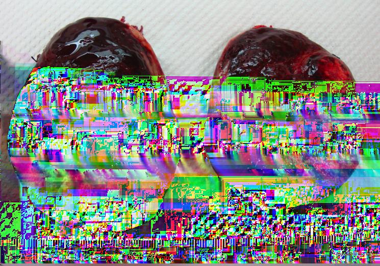
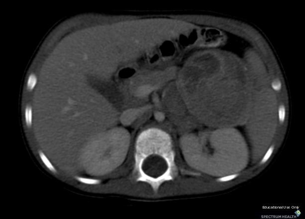
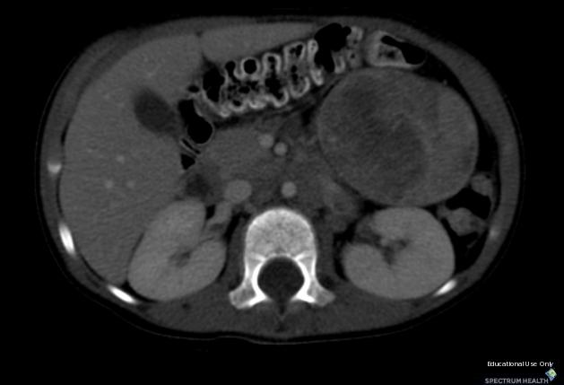
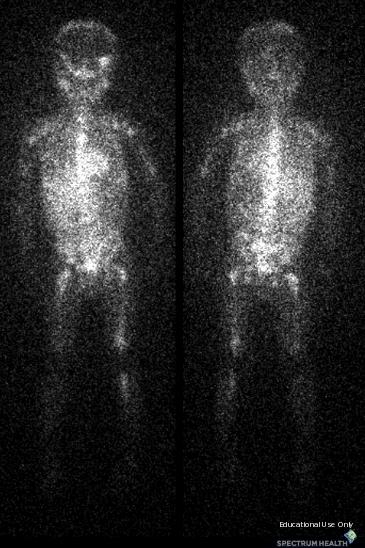
1 Neuroblastoma Joseph Junewick, MD FACR 03/18/2011 History 15 month old with anemia. Diagnosis Neuroblastoma Discussion Neuroblastic tumors derive from primordial neural crest cells destined for sympathetic differentiation. The most common sites of origin, in order, are the adrenal medulla (35% of cases), extraadrenal retroperitoneum (30% 35%), and posterior mediastinum (20%). Less common sites are the neck (1% 5% of cases) and pelvis (2% 3%) (23). Approximately 1% of patients present with metastatic disease but no discoverable primary tumor. Unusual locations such as the thymus, lung, kidney, anterior mediastinum, stomach, and cauda equina have also been described. MIBG is taken up by catecholamine-producing tumors (neuroblastoma, ganglioneuroblastoma, pheochromocytoma, carcinoid tumor and medullary thyroid carcinoma). Although 90% 95% of neuroblastomas secrete catecholamines, only about 70% are MIBG positive. MIBG scintigraphy is highly sensitive (88%) and specific (99%) for sympathetic tissue but does not discriminate between the catecholamine-producing tumors. Positive MIBG scans provide an excellent map of primary and metastatic disease. CT is the most commonly used imaging modality for assessment of neuroblastic tumors, because it reveals extent of tumor, organ of origin, regional invasion, vascular encasement, adenopathy, and calcification. CT of the chest, abdomen, and pelvis is standard for all newly diagnosed cases. Abdominal and pelvic tumors are usually large and heterogeneous. Approximately 80% 90% of neuroblastomas demonstrate calcification on CT scans. Low-attenuation areas of necrosis or hemorrhage are frequently noted. Vascular encasement and compression are common, although vascular invasion is rare. Regional invasion of psoas and paraspinal musculature may occur, and invasion of the neural foramen into the epidural space is also frequent. Adenopathy of the renal hilum, porta hepatis, and retroperitoneum may be seen. Metastatic disease of the liver and lung are readily evaluated with CT. Liver metastases may be diffuse infiltration or focal hypoenhancing masses. Lung metastases may appear as discreet nodules or confluent areas of parenchymal consolidation. Findings CT-Prominent left adrenal gland with large circumscribed mixed attenuation left upper quadrant mass. NM-Anterior and posterior whole body I-123 MIBG images after resection of abdominal disease show extensive marrow involvement. Reference Lonergan GJ, Schwab CM, Suarez ES, et al. From the Archives of the AFIP Neuroblastoma, Ganglioneuroblastoma, and Ganglioneuroma: Radiologic-Pathologic Correlation. Radiographics (2002); 22:
2
3
4

5
6
7 Sponsored By Disclaimer This teaching site is partially funded by an educational grant from GE Healthcare and Advanced Radiology Services, PC. The material on this site is independently controlled by Advanced Radiology Services, PC, and GE Healthcare and Spectrum Health have no influence over the content of this site Content Download Agreement The cases and images on this website are owned by Spectrum Health. Permission is granted (for nonprofit educational purposes) to download and print materials to distribute for the purpose of facilitating the education of health professionals. The authors retain all rights to the material and users are requested to acknowledge the source of the material. Site Disclaimer This site is developed to reach healthcare professionals and medical students. Nothing this site should be considered medical advice. Only your own doctor can help you make decisions about your medical care. If you have a specific medical question or are seeking medical care, please contact your physician. The information in this website is provided for general medical education purposes only and is not meant to substitute for the independent medical judgment of a physician relative to diagnostic and treatment options of a specific medical condition. The viewpoints expressed in these cases are those of the authors. They do not represent an endorsement. In no event will Advanced Radiology Associates, PC, Spectrum Health Hospitals (Helen Devos Children's Hospital) or GE Healthcare be liable for any decision made or action taken in reliance upon the information provided through this website.
Presacral Neuroblastoma Joseph Junewick, MD FACR
 Presacral Neuroblastoma Joseph Junewick, MD FACR 01/12/2010 History 16 month old male with irritability. Diagnosis Presacral Neuroblastoma Additional Clinical Initial US to evaluate for intussusception
Paraspinal Venous Malformation Joseph Junewick, MD FACR
 Paraspinal Venous Malformation Joseph Junewick, MD FACR 06/04/2010 History 2 year old with history of fall. Rule out spinal injury. Diagnosis Paraspinal Venous Malformation Additional Clinical CT of the
Spinal LCH Joseph Junewick, MD FACR
 Spinal LCH Joseph Junewick, MD FACR 05/16/2009 History 16 year old female with multiply recurrent Langerhans Cell Histiocytosis now with severe left sided neck pain. Diagnosis Langerhans Cell Histiocytosis
Diskitis Joseph Junewick, MD FACR
 Diskitis Joseph Junewick, MD FACR 09/20/2010 History 2 year old with fever, back pain and elevated sedimentation rate. Diagnosis Diskitis Discussion Diskitis is an inflammatory process of the intervertebral
Radiation Pneumonitis Joseph Junewick, MD FACR
 Radiation Pneumonitis Joseph Junewick, MD FACR 03/19/2010 History 16 year old with history of relapsed stage IV-A Hodgkin disease. Prior pulmonary involvement was irradiated. Diagnosis Radiation Pneumonitis
Retroperitoneal Teratoma Heather Borders, MD
 Retroperitoneal Teratoma Heather Borders, MD 03/04/2012 History Newborn with congenitally diagnosed mass. No other clinical symptoms. Diagnosis Retroperitoneal Teratoma; Immature teratoma, grade 1, with
Scrofula Joseph Junewick, MD FACR
 Scrofula Joseph Junewick, MD FACR 06/20/2012 History 4 year old male with refractory cervical adenopathy Diagnosis Scrofula Additional Clinical Positive PPD skin test. Discussion Scrofula refers to tuberculous
Pleural Empyema Joseph Junewick, MD FACR
 Pleural Empyema Joseph Junewick, MD FACR 03/19/2010 History Teenager with persistent fever and cough. Pneumonia diagnosed 1 week ago. Diagnosis Pleural Empyema Additional Clinical Surgery-Clear fluid with
Term Hypoxic Ischemic Injury Joseph Junewick, MD FACR
 Term Hypoxic Ischemic Injury Joseph Junewick, MD FACR 08/11/2010 History Term infant with perinatal distress and attempted forceps delivery. Diagnosis Term Hypoxic Ischemic Injury Discussion Encephalopathy
Vein of Galen Malformation Joseph Junewick, MD FACR
 Vein of Galen Malformation Joseph Junewick, MD FACR 04/14/2018 History Midline cystic intracranial mass on prenatal ultrasound. Diagnosis Vein of Galen Malformation Discussion In normal fetal development,
Atlanto-occipital Dislocation Joseph Junewick, MD FACR
 Atlanto-occipital Dislocation Joseph Junewick, MD FACR 09/23/2009 History 12 year old male restrained back seat passenger in a car hit by a snowplow. Diagnosis Atlanto-occipital Dislocation Discussion
Chance Fracture Joseph Junewick, MD FACR
 Chance Fracture Joseph Junewick, MD FACR 08/02/2010 History Restrained teenager involved in motor vehicle accident. Diagnosis Chance Fracture (Hyperflexion-Distraction Injury) Discussion Chance-type spinal
Ulcerative Colitis Joseph Junewick, MD FACR
 Ulcerative Colitis Joseph Junewick, MD FACR 06/04/2010 History 16 year old male with hematochezia and anemia. Diagnosis Ulcerative Colitis Additional Clinical History of ulcerative colitis. Discussion
Transverse Dural Sinus Thrombosis Joseph Junewick, MD FACR
 Transverse Dural Sinus Thrombosis Joseph Junewick, MD FACR 03/19/2010 History Child with headache and otomastoiditis. Diagnosis Dural venous thrombosis secondary to mastoiditis Discussion The cerebral
Bilateral Retinoblastoma Joseph Junewick, MD FACR
 Bilateral Retinoblastoma Joseph Junewick, MD FACR 06/11/2010 History 17 month old adopted female with proptosis. Diagnosis Bilateral Retinoblastoma Discussion Retinoblastoma is the most common pediatric
Fallopian tube torsion and paratubal cyst Heather Borders, MD
 Fallopian tube torsion and paratubal cyst Heather Borders, MD 01/24/2012 History 13 year old female with one week of pelvic pain Diagnosis Fallopian tube torsion with paratubal cyst Additional Clinical
Pituitary Macroadenoma Joseph Junewick, MD FACR
 Pituitary Macroadenoma Joseph Junewick, MD FACR 08/13/2010 History 12 year old female with headache and visual disturbance. Diagnosis Pituitary Macroadenoma Additional Clinical Markedly elevated growth
Gastrointestinal Hemangiomatosis Joseph Junewick, MD FACR
 Gastrointestinal Hemangiomatosis Joseph Junewick, MD FACR 03/06/2010 History 3 month old with protuberant abdomen and anemia. Diagnosis Gastrointestinal Hemangiomatosis Discussion Gastrointestinal hemangiomatosis
Chiari III Joseph Junewick, MD FACR
 Chiari III Joseph Junewick, MD FACR 07/02/2010 History Newborn with suboccipital mass. Diagnosis Chiari III Additional Clinical Surgery-Skin covered suboccipital cystic mass confined by the dura. Pathology-Leptomeningeal
Tuberculous Meningitis Joseph Junewick, MD FACR
 Tuberculous Meningitis Joseph Junewick, MD FACR 08/11/2010 History 14 month old with fever and increasing lethargy. Diagnosis Tuberculous Meningitis Additional Clinical Grandmother with active tuberculosis.
Thymic Involvement in Chronic Granulomatous Disease of Childhood
 Thymic Involvement in Chronic Granulomatous Disease of Childhood Joseph Junewick, MD FACR 07/16/2010 History 3 year old male with multifocal osteomyelitis. Diagnosis Thymic Involvement in Chronic Granulomatous
Testicular Microlithiasis related to McCune-Albright Syndrome Joseph Junewick, MD FACR
 Testicular Microlithiasis related to McCune-Albright Syndrome Joseph Junewick, MD FACR 04/25/2010 History 12 year old with McCune-Albright syndrome. Diagnosis Testicular Microlithiasis related to Mcune-Albright
Posterior Slipped Capital Femoral Epiphysis Joseph Junewick, MD FACR
 Posterior Slipped Capital Femoral Epiphysis Joseph Junewick, MD FACR 08/11/2010 History 6 year old male with intermittent hip pain for several months, acutely worsened after climbing the sand dunes. Diagnosis
Adrenal masses in infancy and childhood: A clinical and radiological overview M. Mearadji
 Adrenal masses in infancy and childhood: A clinical and radiological overview M. Mearadji International Foundation for Pediatric Imaging Aid Introduction Neoplastic adrenal masses usually originate from
ADRENAL MEDULLARY DISORDERS: PHAEOCHROMOCYTOMAS AND MORE
 ADRENAL MEDULLARY DISORDERS: PHAEOCHROMOCYTOMAS AND MORE DR ANJU SAHDEV READER AND CONSULTANT RADIOLOGIST QUEEN MARY UNIVERSITY AND ST BARTHOLOMEW S HOSPITAL BARTS HEALTH, LONDON, UK DISCLOSURE OF CONFLICT
From the Archives of the AFIP
 AFIP ARCHIVES 911 CME FEATURE See accompanying test at http:// www.rsna.org /education /rg_cme.html LEARNING OBJECTIVES FOR TEST 6 After reading this article and taking the test, the reader will be able
Daniela Faivovich K., MS VII Universidad de Chile Gillian Lieberman, MD Harvard Medical School
 Daniela Faivovich K., MS VII Universidad de Chile Gillian Lieberman, MD Harvard Medical School May 21st, 2010 56 year old male patient History of hypertension, hyperlipidemia and insulin-resistance 2009:
SELF-ASSESSMENT MODULE REFERENCE SPR 2018 Oncologic Imaging Course Adrenal Tumors November 10, :00 12:10 p.m.
 SELF-ASSESSMENT MODULE REFERENCE SPR 2018 Oncologic Imaging Course Adrenal Tumors November 10, 2018 10:00 12:10 p.m. Staging Susan E. Sharp, MD 1. In the International Neuroblastoma Risk Group Staging
ADRENAL LESIONS 10/09/2012. Adrenal + lesion. Introduction. Common causes. Anatomy. Financial disclosure. Dr. Boraiah Sreeharsha. Nothing to declare
 ADRENAL LESIONS Financial disclosure Nothing to declare Dr. Boraiah Sreeharsha MBBS;FRCR;FRCPSC Introduction Adrenal + lesion Adrenal lesions are common 9% of the population Increase in the detection rate
Wilms Tumor and Neuroblastoma
 Wilms Tumor and Neuroblastoma Wilm s Tumor AKA: Nephroblastoma the most common intra-abdominal cancer in children. peak incidence is 2 to 3 years of age Biology somatic mutations restricted to tumor tissue
PDF created with pdffactory Pro trial version
 Neuroblastoma Tumor derived from neural crest cell that form the sympathetic ganglia&adrenal medulla. Causes *unknown. *familial neuroblastoma has been reported but is rare. * The incidence is 1:100,000
Retroperitoneal Ganglioneuroma Encasing the Celiac and Superior Mesenteric Arteries
 Case Study TheScientificWorldJOURNAL (2004) 4, 974 977 ISSN 1537-744X; DOI 10.1100/tsw.2004.198 Retroperitoneal Ganglioneuroma Encasing the Celiac and Superior Mesenteric Arteries Justin K. Nelms, Eric
Pediatric Abdominal Masses. Andrew Phelps MD Assistant Professor of Pediatric Radiology UCSF Benioff Children's Hospital
 Pediatric Abdominal Masses Andrew Phelps MD Assistant Professor of Pediatric Radiology UCSF Benioff Children's Hospital No Disclosures Take Home Message All you need to remember are the 5 common masses
1/25/13 Right partial nephrectomy followed by completion right radical nephrectomy.
 History and Physical Case Scenario 1 45 year old white male presents with complaints of nausea, weight loss, and back pain. A CT of the chest, abdomen and pelvis was done on 12/8/12 that revealed a 12
Read the following article and answer the questions that follow. Refer to the Keys section to check your answers.
 ENGLISH 183 READING PRACTICE - Pheochromocytoma Read the following article and answer the questions that follow. Refer to the Keys section to check your answers. Pheochromocytoma is a tumor on the medulla
Pediatric Retroperitoneal Masses Radiologic-Pathologic Correlation
 Acta Radiológica Portuguesa, Vol.XVIII, nº 70, pág. 61-70, Abr.-Jun., 2006 Pediatric Retroperitoneal Masses Radiologic-Pathologic Correlation Marilyn J. Siegel Mallinckrodt Institute of Radiology, Washington
A case of micturition syncope
 A case of micturition syncope Kimberly Bundick, PA-S S L I D E 1 Agenda Purpose Utilize case to illustrate classic finding of an interesting pathology Agenda Case study Epidemiology, etiology of disease
MDCT signs differentiating retroperitoneal and intraperitoneal lesions- diagnostic pearls
 MDCT signs differentiating retroperitoneal and intraperitoneal lesions- diagnostic pearls Poster No.: C-0987 Congress: ECR 2015 Type: Educational Exhibit Authors: D. V. Bhargavi, R. Avantsa, P. Kala; Bangalore/IN
THE FACTS YOU NEED TO KNOW
 PHEOCHROMOCYTOMA THE FACTS YOU NEED TO KNOW Pheochromocytoma is a part of the pheochromocytoma and paraganglioma group of syndromes. A pheochromocytoma is a tumor arising in the adrenal gland medulla.
Prof. Dr. NAGUI M. ABDELWAHAB,M.D.; MARYSE Y. AWADALLAH, M.D. AYA M. BASSAM, Ms.C.
 Role of Whole-body Diffusion MR in Detection of Metastatic lesions Prof. Dr. NAGUI M. ABDELWAHAB,M.D.; MARYSE Y. AWADALLAH, M.D. AYA M. BASSAM, Ms.C. Cancer is a potentially life-threatening disease,
Neuroblastoma. Elizabeth Roberts. Data Coordinator CIBMTR Data Managers Mentor. Tandem Meeting February 18
 Neuroblastoma Elizabeth Roberts Data Coordinator CIBMTR Data Managers Mentor Tandem Meeting February 18 Objectives Know what neuroblastoma is, how it is diagnosed, and how it is treated Complete form 2026:
Nuclear medicine in endocrinology
 Nuclear medicine in endocrinology Thyroid gland: anatomy, function, inflammation, Nuclear medicine in endocrinology tumor dignitiy Parathyroid gland: localisation Adrenal cortex: function Adrenal medulla:
Personal data. Age : 63 Gender : male
 Personal data Age : 63 Gender : male Chief complain No specific symptom or discomfort A hepatic mass, found by abdominal sonography of routine health exam on 88-12-08 Past history 1984-3-3 Old CVA with
CSU Research Output
 This is the Author s version of the paper published as: Author: M. Crook, J. Wheat and G. Currie Author Address: jwheat@csu.edu.au gcurrie@csu.edu.au Title: Pheochromocytoma: an unexpected finding Year:
Dr.Dafalla Ahmed Babiker Jazan University
 Dr.Dafalla Ahmed Babiker Jazan University Brain tumors are the second commonest malignancy in children Infratentorial tumors are more common As a general rule they do not metastasize out of the CNS, but
SPECT/CT in Endocrine Diseases and Dosimetry
 SPECT/CT in Endocrine Diseases and Dosimetry Heather A. Jacene, MD Division of Nuclear Medicine Russell H. Morgan Dept. of Radiology and Radiological Science Johns Hopkins University Baltimore, MD Disclosures
Jeffrey C. Weinreb, MD, FACR Yale School of Medicine Yale-New Haven Hospital
 Jeffrey C. Weinreb, MD, FACR Yale School of Medicine Yale-New Haven Hospital jeffrey.weinreb@yale.edu 1991 1997 Whole body MRI: multistation approach x z Isocenter: Table Move: Multiple Steps Whole body
PedsCases Podcast Scripts
 PedsCases Podcast Scripts This is a text version of a podcast from Pedscases.com on Approach to Abdominal Mass Part 2. These podcasts are designed to give medical students an overview of key topics in
ID data. Sex: female Age: 46y/o Birthday: 1955/10/13
 ID data Sex: female Age: 46y/o Birthday: 1955/10/13 Chief Complain Right upper quadrate abdominal tenderness for one month. Present illness (1) This 46 years old female patient was in a healthy condition
Kidney Case 1 SURGICAL PATHOLOGY REPORT
 Kidney Case 1 Surgical Pathology Report February 9, 2007 Clinical History: This 45 year old woman was found to have a left renal mass. CT urography with reconstruction revealed a 2 cm medial mass which
Update on RECIST and Staging of Common Pediatric Tumors Ethan A. Smith, MD
 Update on RECIST and Staging of Common Pediatric Tumors Ethan A. Smith, MD Section of Pediatric Radiology C.S. Mott Children s Hospital University of Michigan ethans@med.umich.edu Disclosures No relevant
An Introduction to Radiology for TB Nurses
 An Introduction to Radiology for TB Nurses Garold O. Minns, MD September 14, 2017 TB Nurse Case Management September 12 14, 2017 EXCELLENCE EXPERTISE INNOVATION Garold O. Minns, MD has the following disclosures
Traumatic and Non Traumatic Adrenal Emergencies
 Traumatic and Non Traumatic Adrenal Emergencies Michael N. Patlas, MD, FRCPC (1), Christine O. Menias, MD (2), Douglas S. Katz, MD, FACR (3), Ania Z. Kielar, MD, FRCPC (4), Alla M. Rozenblit, MD (5), Jorge
A 64 y.o. man presents to the hospital with persistent cough and hemoptysis. Fernando Mut Montevideo - Uruguay
 A 64 y.o. man presents to the hospital with persistent cough and hemoptysis Fernando Mut Montevideo - Uruguay Teaching case Bone # 1 A 64 y.o. man presents to the hospital with persistent cough and hemoptysis.
MR Tumor Staging for Treatment Decision in Case of Wilms Tumor
 MR Tumor Staging for Treatment Decision in Case of Wilms Tumor G. Schneider, M.D., Ph.D.; P. Fries, M.D. Dept. of Diagnostic and Interventional Radiology, Saarland University Hospital, Homburg/Saar, Germany
Lecture 56 Kidney and Urinary System
 Lecture 56 Kidney and Urinary System The adrenal glands are located on the superomedial aspect of the kidney The right diagram shows a picture of the kidney with the abdominal walls and organs removed
Characterization of adrenal lesions on CT and MRI: all that a radiologist must know
 Characterization of adrenal lesions on CT and MRI: all that a radiologist must know Poster No.: C-2476 Congress: ECR 2013 Type: Educational Exhibit Authors: N. Benzina, S. MAJDOUB, C. H. ZARRAD, H. Zaghouani,
Exercise 15: CSv2 Data Item Coding Instructions ANSWERS
 Exercise 15: CSv2 Data Item Coding Instructions ANSWERS CS Tumor Size Tumor size is the diameter of the tumor, not the depth or thickness of the tumor. Chest x-ray shows 3.5 cm mass; the pathology report
The Adrenal Glands. I. Normal adrenal gland A. Gross & microscopic B. Hormone synthesis, regulation & measurement. II.
 The Adrenal Glands Thomas Jacobs, M.D. Diane Hamele-Bena, M.D. I. Normal adrenal gland A. Gross & microscopic B. Hormone synthesis, regulation & measurement II. Hypoadrenalism III. Hyperadrenalism; Adrenal
Primitive Neuroectodermal Tumor of Mediastinum in an Adult: A Case Report 1
 Primitive Neuroectodermal Tumor of Mediastinum in an Adult: A Case Report 1 Young Jae Sung, M.D., Jeung Sook Kim, M.D. A peripheral primitive neuroectodermal tumor (PNET) is a rare and aggressive malignant
Sectional Anatomy Quiz II
 Sectional Anatomy II Rashid Hashmi Rural Clinical School, University of New South Wales, Wagga Wagga, New South Wales, Australia A R T I C L E I N F O Article type: Article history: Received: 3 Aug 2017
Imaging of bone metastases
 Imaging of bone metastases Antoine Feydy Service de Radiologie B Hôpital Cochin APHP Université Paris Descartes antoine.feydy@aphp.fr MEXICO 2016 INTRODUCTION Diagnostic Imaging Imaging Modalities Strengths,
Case Scenario 1: Thyroid
 Case Scenario 1: Thyroid History and Physical Patient is an otherwise healthy 80 year old female with the complaint of a neck mass first noticed two weeks ago. The mass has increased in size and is palpable.
GUIDELINES FOR CANCER IMAGING Lung Cancer
 GUIDELINES FOR CANCER IMAGING Lung Cancer Greater Manchester and Cheshire Cancer Network Cancer Imaging Cross-Cutting Group April 2010 1 INTRODUCTION This document is intended as a ready reference for
Clinical summary. Male 30 year-old with past history of non-seminomous germ cell tumour. Presents with retroperitoneal lymphadenopathy on CT.
 Clinical summary Male 30 year-old with past history of non-seminomous germ cell tumour. Presents with retroperitoneal lymphadenopathy on CT. For restaging PET/CT. PET/CT findings No significant FDG uptake
Radiation Dosimetry for CT Protocols
 Radiation Dosimetry for CT Protocols This document contains radiation dosimetry information from CT scans and can be used by investigators to estimate the dosimetry information required by the JRSC or
CLASSIFICATION AND SELECTION OF IMAGE DEFINED RISK FACTORS IN NEUROBLASTOMA
 MEDICAL UNIVERSITY PROF. DR. PARASKEV STOYANOV VARNA GEORGI NIKOLAEV VALCHEV, MD CLASSIFICATION AND SELECTION OF IMAGE DEFINED RISK FACTORS IN NEUROBLASTOMA DISSERTATION Scientific supervisor: Prof. Boyan
ADRENAL INCIDENTALOMA. Jamii St. Julien
 ADRENAL INCIDENTALOMA Jamii St. Julien Outline Definition Differential Evaluation Treatment Follow up Questions Case Definition The phenomenon of detecting an otherwise unsuspected adrenal mass on radiologic
IMAGING IN FAMILY MEDICINE
 IMAGING IN FAMILY MEDICINE Imaging Methods Ultrasonography (USG) X-ray Computed tomography (CT) Magnetic resonance imaging (MRI) Scintigraphy Angiography SNC MEDIASTINUM LUNGS ABDOMEN PELVIS USG SNC -
Prostate Case Scenario 1
 Prostate Case Scenario 1 H&P 5/12/16: A 57-year-old Hispanic male presents with frequency of micturition, urinary urgency, and hesitancy associated with a weak stream. Over the past several weeks, he has
objectives Pitfalls and Pearls in PET/CT imaging Kevin Robinson, DO Assistant Professor Department of Radiology Michigan State University
 objectives Pitfalls and Pearls in PET/CT imaging Kevin Robinson, DO Assistant Professor Department of Radiology Michigan State University To determine the regions of physiologic activity To understand
Guidelines, Policies and Statements D5 Statement on Abdominal Scanning
 Guidelines, Policies and Statements D5 Statement on Abdominal Scanning Disclaimer and Copyright The ASUM Standards of Practice Board have made every effort to ensure that this Guideline/Policy/Statement
TB Radiology for Nurses Garold O. Minns, MD
 TB Nurse Case Management Salina, Kansas March 31-April 1, 2010 TB Radiology for Nurses Garold O. Minns, MD April 1, 2010 TB Radiology for Nurses Highway Patrol Training Center Salina, KS April 1, 2010
Alison Douglass Gillian Lieberman, MD. November. Colon Cancer. Alison Douglass, Harvard Medical School Year III Gillian Lieberman, MD
 November Colon Cancer Alison Douglass, Harvard Medical School Year III Our Patient Mr. K. is a 67 year old man with no prior medical problems other than hemorrhoids which have caused occasional rectal
Institution INSTRUCTIONS (I6) 1. This form is to be completed by a DESIGNATED STUDY NUCLEAR MEDICINE SPECIALIST
 1. This form is to be completed by a DESIGNATED STUDY NUCLEAR MEDICINE SPECIALIST") I6 ACRIN 6660 Whole Body MRI in the Evaluation of Pediatric Malignancies Conventional Scintigraphy Imaging Form If this is a revised or corrected form, indicate by checking box and fax to 215-717 - 0936.
I6 ACRIN 6660 Whole Body MRI in the Evaluation of Pediatric Malignancies Conventional Scintigraphy Imaging Form If this is a revised or corrected form, indicate by checking box and fax to 215-717 - 0936.
Mediastinal Tumors: Imaging
 Mediastinal Tumors: Imaging References Imaging in Oncology, Husband and Reznek Computed Tomography and Magnetic Resonance of the thorax, Naidich, Zerhouni, Siegelman, Mediastinal compartments Anterior:
pulmonary metastasis 80EE4727C6037E7F69A9981B7E55A238 Pulmonary Metastasis 1 / 6
 Pulmonary Metastasis 1 / 6 2 / 6 3 / 6 Pulmonary Metastasis Metastatic tumors in the lungs are cancers that developed at other places in the body (or other parts of the lungs). They then spread through
Index. Note: Page numbers of article titles are in boldface type.
 Magn Reson Imaging Clin N Am 12 (2004) 587 591 Index Note: Page numbers of article titles are in boldface type. A Adenoma(s), adrenal, gadolinium-enhanced MR imaging in, 533 534 hyperfunctioning versus
11/1/2014. Radiologic incidentalomas Ordering pitfalls Newer technology and applications
 Bilal Tahir, MD Gitasree Borthakur, MD Indiana University School of Medicine Department of Radiology & Imaging Sciences October 31, 2014 ACP 2014 Dr. V. Aaron Nuclear (vaaron@iupui.edu) Dr. S. Westphal
PREAMBLE GENERAL DIAGNOSTIC RADIOLOGY
 PREAMBLE The General Diagnostic Radiology category is intended to cover the body of knowledge a practicing board certified Diagnostic Radiologist should know. Since the range of content relevant to the
Sex: 女 Age: 51 Occupation: 無 Admission date:92/07/22
 Sex: 女 Age: 51 Occupation: 無 Admission date:92/07/22 Chief complaint Unknown fever for one month Hand tremor and left huge renal tumor was noted Present illness Suffered from fever for one month, hand
Multi-Organ Distant Metastases in Follicular Thyroid Cancer- Rare Case Report
 Multi-Organ Distant Metastases in Follicular Thyroid Cancer- Rare Case Report Dr. Mohammed Raza 1, Dr. Sindhuri K 2, Dr. Dinesh Reddy Y 3 1 Professor, Department of Surgery, JSS University, Mysore, India
Radio-Pathologic Workup of a Retroperitoneal Abdominal Mass
 Radio-Pathologic Workup of a Retroperitoneal Abdominal Mass Joe Carlson Advanced Radiology Clerkship Harvard Medical School Year IV September 12, 2002 84 year old Male Presented to PCP With Abdominal Pain
Case: The patient is a 62 year old woman with a history of renal cell carcinoma that was removed years ago. A 2.4 cm liver mass was found on CT
 Case: The patient is a 62 year old woman with a history of renal cell carcinoma that was removed years ago. A 2.4 cm liver mass was found on CT during follow- up. ALT, AST, Alk Phos and bilirubin were
American College of Radiology ACR Appropriateness Criteria
 American College of Radiology ACR Criteria Radiologic Management of Thoracic Nodules and Masses Variant 1: Middle-aged patient (35 60 years old) with an incidental 1.5-cm lung nodule. The lesion was smooth.
Mediastinal Paraganglioma: a challenge to the echocardiographic
 Case - based learning from ESC Cardiologists of Tomorrow Look for the answer outside the heart Mediastinal Paraganglioma: a challenge to the echocardiographic 1 diagnosis and endovascular treatment 1 On
Endometrial Stromal Sarcoma
 May 26, 2011 By Sushila Ladumor, MD [1] Endometrial stromal sarcoma (ESS) is a rare malignant tumor of the endometrium, occurring in the age group of 40-50 years. History The 50-year-old, female patient
Calcitonin. 1
 Calcitonin Medullary thyroid carcinoma (MTC) is characterized by a high concentration of serum calcitonin. Routine measurement of serum calcitonin concentration has been advocated for detection of MTC
Hematologic Malignancies of the Liver : Spectrum of Disease. Zhou Jian
 Hematologic Malignancies of the Liver : Spectrum of Disease Zhou Jian 2015-7-8 Hematologic malignancies include a wide spectrum of lymphoproliferative and myeloproliferative disorders with nodal and extranodal
Ovarian Lesion Benign vs Malignant?
 Ovarian Lesion Benign vs Malignant? Michele Keenan 1,2 Bernice Dunne 2 Mary Moran 1 Therese Herlihy 1 1. Radiography and Diagnostic Imaging, School of Medicine, University College Dublin, Ireland 2. Midland
MRI findings related to Image Defined Risk Factors (IDRFs) in children with neuroblastic tumours: Analysis for structured reporting
 in children with neuroblastic tumours: Analysis for structured reporting") Paediatric Radiology Pictorial Essay MRI findings related to Image Defined Risk Factors (IDRFs) in children with neuroblastic tumours: Analysis for structured reporting Androniki Kozana 1, Vasiliki Fragkiadoulaki
Paediatric Radiology Pictorial Essay MRI findings related to Image Defined Risk Factors (IDRFs) in children with neuroblastic tumours: Analysis for structured reporting Androniki Kozana 1, Vasiliki Fragkiadoulaki
Radiology- Pathology Conference 4/29/2012. Lymph Nodes. John McGrath
 Radiology- Pathology Conference 4/29/2012 Lymph Nodes John McGrath 1 Presentation material is for education purposes only. All rights reserved. 2012 URMC Radiology Page 1 of 24 Case 1: 51 year-old male
Role of imaging in evaluation of genitourinary i trauma Spectrum of GU injuries Relevance of imaging findings in determining management Focus on MDCT
 Genitourinary Tract Injuries 6 th Nordic Course Scott D. Steenburg, MD Assistant Professor University of Maryland Department of Radiology Division of Trauma and Emergency Radiology R Adams Cowley Shock
Sporadic Pheochromocytoma. Bertil Hamberger Professor of Surgery Karolinska Institutet, Stockholm, Sweden
 Sporadic Pheochromocytoma Bertil Hamberger Professor of Surgery Karolinska Institutet, Stockholm, Sweden 1 Pheochromocytoma Anatomy, physiology and pathology Symptoms and diagnosis Plasma metanephrines
Evaluation and Management of Thyroid Nodules. Nick Vernetti, MD, FACE Palm Medical Group Las Vegas, Nevada
 Evaluation and Management of Thyroid Nodules Nick Vernetti, MD, FACE Palm Medical Group Las Vegas, Nevada Disclosure Consulting Amgen Speaking Amgen Objectives Understand the significance of incidental
Metachronic solitary breast metastasis from renal cell carcinoma: case report
 Metachronic solitary breast metastasis from renal cell carcinoma: case report Abstract We describe the case of a patient with solitary and metachronic breast metastasis, 3 years after nephrectomy for renal
Chest Radiology Interpretation: Findings of Tuberculosis
 Chest Radiology Interpretation: Findings of Tuberculosis Get out your laptops, smart phones or other devices pollev.com/chestradiology Case #1 1 Plombage Pneumonia Cancer 2 Reading the TB CXR Be systematic!
Images In Gastroenterology
 Images In Gastroenterology Thong-Ngam D, et al. THAI J GASTROENTEROL 2005 Vol. 6 No. 2 May - Aug. 2005 105 Imaging of Gastrointestinal Stromal Tumors Pornpim Fuangtharnthip, M.D. Narumol Hargroove, M.D.
Nuclear Medicine in Oncology
 Radiopharmaceuticals Nuclear Medicine in Oncology Practice Pharmaceutical Radionuc lide Function Tumor type Diphosphonates Tc-99m Osteoblast Bone tumor & metast. Ga-citrate Ga-67 Fe-analogue Bronchogenous