TENSOR Based Tumor Tissue Differentiation Using Magnetic Resonance Spectroscopic Imaging
|
|
|
- Gary Bennett
- 5 years ago
- Views:
Transcription


1 TENSOR Based Tumor Tissue Differentiation Using Magnetic Resonance Spectroscopic Imaging HN Bharath, DM Sima, N Sauwen, U Himmelreich, L De Lathauwer, Sabine Van Huffel IEEE EMBC 2015 Milano, Italy August 25-29, 2015
2 Contents Overview Brain Tumor Tissue type differentiation using MRSI NMF Hierarchical(h) NMF Non-negative Tensor Factorization Conclusions and new directions

3 Metabolite quantification for MR Spectroscopy (MRS) Single-voxel MRS MRS quantification Metabolite concentrations

4 Metabolite quantification for MRS Imaging (MRSI) Multi-voxel MRS MRS quantification using spatial information NAA Myo Cr PCho Glu Lac Lip1 Lip2 Ala Glc Tau Metabolite concentrations = biomarkers of disease Metabolite maps


5 Unsupervised Brain Tumor Diagnosis using NMF MRSI Y = matrix of spectra, Y W H min Y - W H such that W 0, H 0 non-negative matrix factorization W = tissue-specific spectral patterns: H = spatial distribution of tissue types: normal normal tissue active tumor necrosis tumor necrosis glioblastoma multiforme patient
6 MRI features MRI features Non-negative matrix factorization (NMF) Y W H voxels voxels (tissue) abundance (tissue) source Non-negativity constraint: Y i,j, W i,j, H i,j 0, i,j Unsupervised: applicable patient-by-patient, tissue classes not a priori known NMF: 1. integrate ALL features into one vector 2. use NMF
 1)")
 area ( B ) A B 2) Correlation coefficients")

7 Validation Based on manual segmentation by radiologist (only pathological tissue types) 1) Dice-scores (based on H) area( A B) Dice 2x area ( A ) area ( B ) A B 2) Correlation coefficients (based on W). ab a :tissue source vector Corr a. b b :average feature vector over corresponding tissue region


 NMF (# tissues source1) Mask needed!")
8 Hierarchical NMF (hnmf) Improved results on MRSI data only (Li et al., NMR in BioMed. 2013) Y = Coregistered MP-MRI data NMF (2 sources) W source1, H source1 (tumour, necrosis,...) W source2, H source2 (white matter, CSF,...) NMF (# tissues source1) Mask needed! NMF (# tissues source2) W tumor, H tumor W necrosis, H necrosis W WM, H WM W GM, H GM...

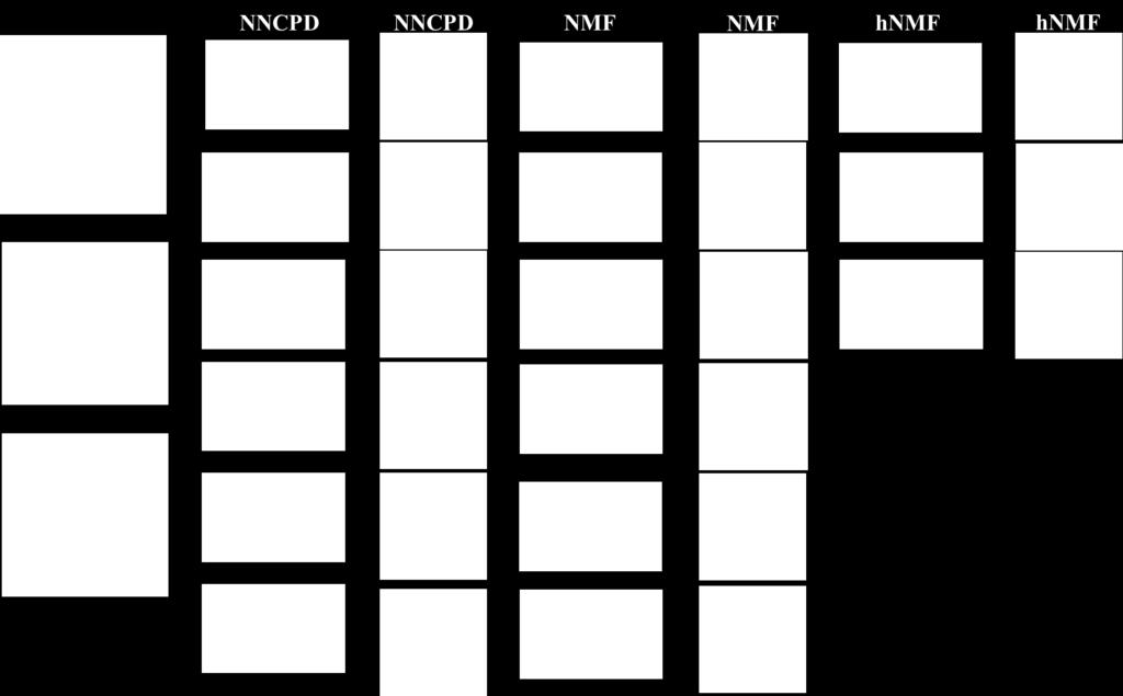
9 Case study: single stage NMF vs hnmf Single stage NMF hnmf Dice tumor = 71% Dice tumor+necrosis = 83% Dice complete tumor = 75% Corr tumor = 0.60 Corr necrosis = 0.97 Corr edema = 0.93 Dice tumor = 81% Dice tumor+necrosis = 92% Dice complete tumor = 83% Corr tumor = 0.78 Corr necrosis = 0.98 Corr edema = 0.97

10 Spatial Tensor Representation (MRSI only) Frontal slices (XY T ) representing the spatial distribution of a tissue type does not have low rank structure. Difficult to find the rank L R for a particular tissue type distribution. 10

11 XX T based Tensor Representation (MRSI only) L X(i) = j=1 S ij 2 Spectra reduced in length and denoised without losing vital information, Peaks get higher weights, 11 Peaks coupled because of XX T in the frontal slices HN Bharath et al, Proc. IEEE-EMB Symposium, Milan, Italy, Aug 2015, to appear


12 Non-negative CPD for Tumor Differentiation Constraints: 1. Non-negativity in all modes 2. Symmetry in frontal slices 3. Sparsity in H-factor apply Non-negative CPD with L 1 regularization using Tensorlab 1 [S, H ] = min S 0,H 0 K T r=1 S :, r о S :, r о H(:, r), Using H*, recover tissue-specific spectra W from Y via LS 2 2 +λ Vec H 1 Using W, recover tissue-type spatial distributions H from Y via NN-LS 12 1 Laurent Sorber, Marc Van Barel and Lieven De Lathauwer. Tensorlab v2.0, Available online, January URL:

13 Result: Patient-2 13
14 Source Correlation: Algorithm vs Expert labeling (MRSI only) W NCPD Single stage NMF hnmf Grade PATIENT-2 T 0.99 X X High PATIENT-2 N High Median/MAD T 0.98 / / / High Median/MAD N / / / Using 10 high-grade glioma (HGG) patients with MRSI dataset Abundance map Correlation: Algorithm vs Expert labeling H NCPD Single stage NMF hnmf Grade PATIENT-2 T 0.80 X X High PATIENT-2 N High Median/MAD T 0.79 / / / High Median/MAD N / / /
15 Contents Overview Brain Tumor Tissue type differentiation using MRSI Conclusions and new directions
16 o o Conclusions and new directions Many BSS problems in Smart Diagnostics are low rank solve via matrix or tensor factorization plus constraints Successful examples shown in brain tissue typing o Other BSS applications: bioinformatics (O. Alter, E. Acar), BCI (Cichocki, Mørup, Martinez-Montes), mobile EEG, multichannel ECG, EEG-fMRI, etc. New directions? o o Automate rank & structure estimation Extend to biomedical data fusion by adding other MR modalities such as anatomical MRI, DWI, PWI, DTI, DKI exploit full potential of Tensor toolbox


17 Acknowledgment University Hospitals Leuven Gasthuisberg ZNA Middelheim, Queen Paola Children s hospital EMC Rotterdam KU Leuven, Dept. Electrical Engineering-ESAT, division STADIUS & MICAS Ghent University, Dept. Telecommunication and Information Processing, TELIN-IPI Eindhoven University of Technology ERC advanced grant BIOTENSORS in collaboration with L. De Lathauwer and group Thank you!


18 TDA KU Leuven Workshop on Tensor Decompositions and Applications January 18-22, 2016, Leuven, Belgium Local Organisers: Sabine Van Huffel and Lieven De Lathauwer Confirmed Speakers Orly Alter Pierre Comon Eva Ceulemans Harm Derksen Nicolas Gilllis Daniel Kressner Lek-Heng Lim Ivan Markovsky Morten Mørup Nikos Sidiropoulos Bart Vandereycken Frank Verstraete
PARAFAC: a powerful tool in EEG monitoring
 Katholieke Universiteit Leuven K.U.Leuven PARAFAC: a powerful tool in EEG monitoring Sabine Van Huffel Dept. Electrical Engineering ESAT-SCD SCD Katholieke Universiteit Leuven, Belgium 1 Contents Overview
The power of Tensor algebra in medical diagnosis
 Katholieke Universiteit Leuven K.U.Leuven The power of Tensor algebra in medical diagnosis Sabine Van Huffel Dept. EE, ESAT-SCD iminds Future Health Dept KU Leuven, Belgium 1 Contents Overview Introduction
CLASSIFICATION OF BRAIN TUMORS BASED ON MAGNETIC RESONANCE SPECTROSCOPY
 CLASSIFICATION OF BRAIN TUMORS BASED ON MAGNETIC RESONANCE SPECTROSCOPY Jan Luts, Dirk Vandermeulen, Arend Heerschap, Uwe Himmelreich, Bernardo Celda, Johan A.K. Suykens, Sabine Van Huffel Department of
DATA-DRIVEN CLUSTERING OF P300 EEG DATA USING COUPLED TENSOR DECOMPOSITIONS
 DATA SCIENCE DATA-DRIVEN CLUSTERING OF P300 EEG DATA USING COUPLED TENSOR DECOMPOSITIONS Rob Zink 1,2, Borbála Hunyadi 1,2, Maarten De Vos 3, Sabine Van Huffel 1,2 1 KU Leuven, Department of Electrical
Comparison of manual and semi-manual delineations for classifying glioblastoma multiforme patients based on histogram and texture MRI features
 Comparison of manual and semi-manual delineations for classifying glioblastoma multiforme patients based on histogram and texture MRI features Adrian Ion-Ma rgineanu 1,2,, Sofie Van Cauter3, Diana M Sima1,2,
Removal of Nuisance Signal from Sparsely Sampled 1 H-MRSI Data Using Physics-based Spectral Bases
 Removal of Nuisance Signal from Sparsely Sampled 1 H-MRSI Data Using Physics-based Spectral Bases Qiang Ning, Chao Ma, Fan Lam, Bryan Clifford, Zhi-Pei Liang November 11, 2015 1 Synopsis A novel nuisance
Hierarchical non-negative matrix factorization (hnmf): a tissue pattern differentiation method for glioblastoma multiforme diagnosis using MRSI
: a tissue pattern differentiation method for glioblastoma multiforme diagnosis using MRSI") Research article Received: January, Revised: August, Accepted: August, Published online in Wiley Online Library: (wileyonlinelibrary.com) DOI:./nbm.5 Hierarchical non-negative matrix factorization (hmf):
Research article Received: January, Revised: August, Accepted: August, Published online in Wiley Online Library: (wileyonlinelibrary.com) DOI:./nbm.5 Hierarchical non-negative matrix factorization (hmf):
City, University of London Institutional Repository
 City Research Online City, University of London Institutional Repository Citation: Slabaugh, G.G., Asad, M. & Yang, G. (2016). Supervised Partial Volume Effect Unmixing for Brain Tumor Characterization
Heterogeneous Data Mining for Brain Disorder Identification. Bokai Cao 04/07/2015
 Heterogeneous Data Mining for Brain Disorder Identification Bokai Cao 04/07/2015 Outline Introduction Tensor Imaging Analysis Brain Network Analysis Davidson et al. Network discovery via constrained tensor
Borbála Hunyadi, Patrick Dupont, Wim Van Paesschen and Sabine Van Huffel (2015), The content is identical to the content of the published paper
, The content is identical to the content of the published paper") Citation/Reference Archived version Borbála Hunyadi, Patrick Dupont, Wim Van Paesschen and Sabine Van Huffel (15), Tensor decompositions and data fusion in epileptic electroencephalography and functional
Citation/Reference Archived version Borbála Hunyadi, Patrick Dupont, Wim Van Paesschen and Sabine Van Huffel (15), Tensor decompositions and data fusion in epileptic electroencephalography and functional
Differentiating Tumor and Edema in Brain Magnetic Resonance Images Using a Convolutional Neural Network
 Original Article Differentiating Tumor and Edema in Brain Magnetic Resonance Images Using a Convolutional Neural Network Aida Allahverdi 1, Siavash Akbarzadeh 1, Alireza Khorrami Moghaddam 2, Armin Allahverdy
Visualization strategies for major white matter tracts identified by diffusion tensor imaging for intraoperative use
 International Congress Series 1281 (2005) 793 797 www.ics-elsevier.com Visualization strategies for major white matter tracts identified by diffusion tensor imaging for intraoperative use Ch. Nimsky a,b,
International Journal of Engineering Trends and Applications (IJETA) Volume 4 Issue 2, Mar-Apr 2017
 Volume 4 Issue 2, Mar-Apr 2017") RESEARCH ARTICLE OPEN ACCESS Knowledge Based Brain Tumor Segmentation using Local Maxima and Local Minima T. Kalaiselvi [1], P. Sriramakrishnan [2] Department of Computer Science and Applications The Gandhigram
RESEARCH ARTICLE OPEN ACCESS Knowledge Based Brain Tumor Segmentation using Local Maxima and Local Minima T. Kalaiselvi [1], P. Sriramakrishnan [2] Department of Computer Science and Applications The Gandhigram
1) Diffusion weighted imaging DWI is a term used to describe moving molecules due to random thermal motion. This motion is restricted by boundaries
 Diffusion weighted imaging DWI is a term used to describe moving molecules due to random thermal motion. This motion is restricted by boundaries") 1) Diffusion weighted imaging DWI is a term used to describe moving molecules due to random thermal motion. This motion is restricted by boundaries such as ligaments, membranes and macro molecules. Diffusion
1) Diffusion weighted imaging DWI is a term used to describe moving molecules due to random thermal motion. This motion is restricted by boundaries such as ligaments, membranes and macro molecules. Diffusion
International Journal of Research (IJR) Vol-1, Issue-6, July 2014 ISSN
 Vol-1, Issue-6, July 2014 ISSN") Developing an Approach to Brain MRI Image Preprocessing for Tumor Detection Mr. B.Venkateswara Reddy 1, Dr. P. Bhaskara Reddy 2, Dr P. Satish Kumar 3, Dr. S. Siva Reddy 4 1. Associate Professor, ECE Dept,
Developing an Approach to Brain MRI Image Preprocessing for Tumor Detection Mr. B.Venkateswara Reddy 1, Dr. P. Bhaskara Reddy 2, Dr P. Satish Kumar 3, Dr. S. Siva Reddy 4 1. Associate Professor, ECE Dept,
Diffusion Tensor Imaging in brain tumours
 Diffusion Tensor Imaging in brain tumours @MarionSmits, MD PhD Associate Professor of Neuroradiology Dept. of Radiology, Erasmus MC, Rotterdam (NL) Honorary Consultant and Reader UCLH National Hospital
BRAIN STATE CHANGE DETECTION VIA FIBER-CENTERED FUNCTIONAL CONNECTIVITY ANALYSIS
 BRAIN STATE CHANGE DETECTION VIA FIBER-CENTERED FUNCTIONAL CONNECTIVITY ANALYSIS Chulwoo Lim 1, Xiang Li 1, Kaiming Li 1, 2, Lei Guo 2, Tianming Liu 1 1 Department of Computer Science and Bioimaging Research
Data-driven Structured Noise Removal (FIX)
") Hamburg, June 8, 2014 Educational Course The Art and Pitfalls of fmri Preprocessing Data-driven Structured Noise Removal (FIX) Ludovica Griffanti! FMRIB Centre, University of Oxford, Oxford, United Kingdom
Hamburg, June 8, 2014 Educational Course The Art and Pitfalls of fmri Preprocessing Data-driven Structured Noise Removal (FIX) Ludovica Griffanti! FMRIB Centre, University of Oxford, Oxford, United Kingdom
Discrete Wavelet Transform Based Whole-Spectral and Sub-Spectral Analysis for Improved Brain Tumour Clustering using Single Voxel MR Spectroscopy
 This is the author's version of an article that has been published in this journal. Changes were made to this version by the publisher prior to publication. Discrete Wavelet Transform Based Whole-Spectral
Detection of Mild Cognitive Impairment using Image Differences and Clinical Features
 Detection of Mild Cognitive Impairment using Image Differences and Clinical Features L I N L I S C H O O L O F C O M P U T I N G C L E M S O N U N I V E R S I T Y Copyright notice Many of the images in
A new Method on Brain MRI Image Preprocessing for Tumor Detection
 2015 IJSRSET Volume 1 Issue 1 Print ISSN : 2395-1990 Online ISSN : 2394-4099 Themed Section: Engineering and Technology A new Method on Brain MRI Preprocessing for Tumor Detection ABSTRACT D. Arun Kumar
Analysis of human biopsy specimens in a hospital by HR-MAS NMR Martial Piotto Bruker BioSpin France
 Analysis of human biopsy specimens in a hospital by HR-MAS NMR Martial Piotto Bruker BioSpin France Benutzertagung, Karlsruhe, November 8 rd -9 th, 2016 OUTLINE General principles of HR-MAS NMR Practical
Review of Longitudinal MRI Analysis for Brain Tumors. Elsa Angelini 17 Nov. 2006
 Review of Longitudinal MRI Analysis for Brain Tumors Elsa Angelini 17 Nov. 2006 MRI Difference maps «Longitudinal study of brain morphometrics using quantitative MRI and difference analysis», Liu,Lemieux,
POC Brain Tumor Segmentation. vlife Use Case
 Brain Tumor Segmentation vlife Use Case 1 Automatic Brain Tumor Segmentation using CNN Background Brain tumor segmentation seeks to separate healthy tissue from tumorous regions such as the advancing tumor,
Biomedical Imaging: Course syllabus
 Biomedical Imaging: Course syllabus Dr. Felipe Orihuela Espina Term: Spring 2015 Table of Contents Description... 1 Objectives... 1 Skills and Abilities... 2 Notes... 2 Prerequisites... 2 Evaluation and
Combining Outlier Detection with Random Walker for Automatic Brain Tumor Segmentation
 Combining Outlier Detection with Random Walker for Automatic Brain Tumor Segmentation Vasileios Kanas, Evangelia Zacharaki, Evangelos Dermatas, Anastasios Bezerianos, Kyriakos Sgarbas, Christos Davatzikos
Chapter 1. Introduction
 Chapter 1 Introduction 1.1 Motivation and Goals The increasing availability and decreasing cost of high-throughput (HT) technologies coupled with the availability of computational tools and data form a
Parameter Estimation in Block Term Decomposition for Noninvasive Atrial Fibrillation Analysis
 Parameter Estimation in Block Term Decomposition for Noninvasive Atrial Fibrillation Analysis Vicente Zarzoso Université Côte d Azur, CNRS, I3S Laboratory, CS 4121, 693 Sophia Antipolis Cedex, France Abstract
General Identification. Name: 江 X X Age: 29 y/o Gender: Male Height:172cm, Weight: 65kg Date of admission:95/09/27
 General Identification Name: 江 X X Age: 29 y/o Gender: Male Height:172cm, Weight: 65kg Date of admission:95/09/27 Chief Complaint Sudden onset of seizure for several minutes Present illness This 29-year
Vital Responder: Real-time Health Monitoring of First- Responders
 Vital Responder: Real-time Health Monitoring of First- Responders Ye Can 1,2 Advisors: Miguel Tavares Coimbra 2, Vijayakumar Bhagavatula 1 1 Department of Electrical & Computer Engineering, Carnegie Mellon
Comparative Study of K-means, Gaussian Mixture Model, Fuzzy C-means algorithms for Brain Tumor Segmentation
 Comparative Study of K-means, Gaussian Mixture Model, Fuzzy C-means algorithms for Brain Tumor Segmentation U. Baid 1, S. Talbar 2 and S. Talbar 1 1 Department of E&TC Engineering, Shri Guru Gobind Singhji
Brain Tumour Diagnostic Support Based on Medical Image Segmentation
 Brain Tumour Diagnostic Support Based on Medical Image Segmentation Z. Měřínský, E. Hošťálková, A. Procházka Institute of Chemical Technology, Prague Department of Computing and Control Engineering Abstract
Contributions to Brain MRI Processing and Analysis
 Contributions to Brain MRI Processing and Analysis Dissertation presented to the Department of Computer Science and Artificial Intelligence By María Teresa García Sebastián PhD Advisor: Prof. Manuel Graña
Image processing for cardiac and vascular applications
 Image processing for cardiac and vascular applications Isabelle Bloch Isabelle.Bloch@telecom-paristech.fr http://perso.telecom-paristech.fr/bloch LTCI, Télécom ParisTech Cardio-vascular imaging p.1/26
Introduction to Modern Imaging Physics and Techniques used in Clinical Neurology
 Introduction to Modern Imaging Physics and Techniques used in Clinical Neurology Benjamin M. Ellingson, Ph.D., M.S. Associate Professor of Radiology, Biomedical Physics, Bioengineering, and Psychiatry
Findings of DTI-p maps in comparison with T 2 /T 2 -FLAIR to assess postoperative hyper-signal abnormal regions in patients with glioblastoma
 Beigi et al. Cancer Imaging (2018) 18:33 https://doi.org/10.1186/s40644-018-0166-4 REGULAR ARTICLE Open Access Findings of DTI-p maps in comparison with T 2 /T 2 -FLAIR to assess postoperative hyper-signal
ONLINE SEIZURE DETECTION IN ADULTS WITH TEMPORAL LOBE EPILEPSY USING SINGLE-LEAD ECG
 ONLINE SEIZURE DETECTION IN ADULTS WITH TEMPORAL LOBE EPILEPSY USING SINGLE-LEAD ECG T. De Cooman [1,2], E. Carrette [3], P. Boon [3], A. Meurs [3], S. Van Huffel [1,2] [1] KU Leuven, Department of Electrical
Patterns of Brain Tumor Recurrence Predicted From DTI Tractography
 Patterns of Brain Tumor Recurrence Predicted From DTI Tractography Anitha Priya Krishnan 1, Isaac Asher 2, Dave Fuller 2, Delphine Davis 3, Paul Okunieff 2, Walter O Dell 1,2 Department of Biomedical Engineering
Metabolites 2017, 7, 20; doi: /metabo
 S1 of S7 Supplementary Materials: Metabolomics of Therapy Response in Preclinical Glioblastoma: A Multi-slice MRSI-Based Volumetric Analysis for Noninvasive Assessment of Temozolomide Treatment Nuria Arias-Ramos,
Real-time Functional Neuroimaging
 Real-time Functional Neuroimaging Stefan Posse, PhD Depts. of Psychiatry and Electrical & Computer Engineering, and Physics and Astronomy Univ. of New Mexico, Albuquerque, New Mexico, USA The Therapeutic
Studying structure-function relationships in the human brain. Lesley Fellows
 Studying structure-function relationships in the human brain Lesley Fellows lesley.fellows@mcgill.ca Studying structure-function relationships in the human brain Historical background Experimental design
Automated Volumetric Cardiac Ultrasound Analysis
 Whitepaper Automated Volumetric Cardiac Ultrasound Analysis ACUSON SC2000 Volume Imaging Ultrasound System Bogdan Georgescu, Ph.D. Siemens Corporate Research Princeton, New Jersey USA Answers for life.
Biomarkers Workshop In Clinical Trials Imaging for Schizophrenia Trials
 Biomarkers Workshop In Clinical Trials Imaging for Schizophrenia Trials Research focused on the following areas Brain pathology in schizophrenia and its modification Effect of drug treatment on brain structure
Chapter 7 Heart Rate Variability in Newborns with Hypoxic Brain Injury
 Chapter 7 Heart Rate Variability in Newborns with Hypoxic Brain Injury Vladimir Matić, Perumpillichira J. Cherian, Devy Widjaja, Katrien Jansen, Gunnar Naulaers, Sabine Van Huffel, and Maarten De Vos Abstract
Functional Elements and Networks in fmri
 Functional Elements and Networks in fmri Jarkko Ylipaavalniemi 1, Eerika Savia 1,2, Ricardo Vigário 1 and Samuel Kaski 1,2 1- Helsinki University of Technology - Adaptive Informatics Research Centre 2-
MRI-Based Classification Techniques of Autistic vs. Typically Developing Brain
 MRI-Based Classification Techniques of Autistic vs. Typically Developing Brain Presented by: Rachid Fahmi 1 2 Collaborators: Ayman Elbaz, Aly A. Farag 1, Hossam Hassan 1, and Manuel F. Casanova3 1Computer
Exploratory Identification of Cardiac Noise in fmri Images
 Exploratory Identification of Cardiac Noise in fmri Images Lilla Zöllei 1, Lawrence Panych 2, Eric Grimson 1, and William M. Wells III 1,2 1 Massachusetts Institute of Technology, Artificial Intelligence
arxiv: v1 [cs.cv] 17 Aug 2017
![arxiv: v1 [cs.cv] 17 Aug 2017](/thumbs/78/78278805.jpg "arxiv: v1 [cs.cv] 17 Aug 2017") Deep Learning for Medical Image Analysis Mina Rezaei, Haojin Yang, Christoph Meinel Hasso Plattner Institute, Prof.Dr.Helmert-Strae 2-3, 14482 Potsdam, Germany {mina.rezaei,haojin.yang,christoph.meinel}@hpi.de
Deep Learning for Medical Image Analysis Mina Rezaei, Haojin Yang, Christoph Meinel Hasso Plattner Institute, Prof.Dr.Helmert-Strae 2-3, 14482 Potsdam, Germany {mina.rezaei,haojin.yang,christoph.meinel}@hpi.de
Voxel-based Lesion-Symptom Mapping. Céline R. Gillebert
 Voxel-based Lesion-Symptom Mapping Céline R. Gillebert Paul Broca (1861) Mr. Tan no productive speech single repetitive syllable tan Broca s area: speech production Broca s aphasia: problems with fluency,
Exploring Peritumoral White Matter Fibers for Neurosurgical Planning
 Exploring Peritumoral White Matter Fibers for Sonia Pujol, Ph.D. Ron Kikinis, M.D. Surgical Planning Laboratory Harvard University Clinical Goal Diffusion Tensor Imaging (DTI) Tractography has the potential
Classification of Alzheimer s disease subjects from MRI using the principle of consensus segmentation
 Classification of Alzheimer s disease subjects from MRI using the principle of consensus segmentation Aymen Khlif and Max Mignotte 1 st September, Maynooth University, Ireland Plan Introduction Contributions
3D Deep Learning for Multi-modal Imaging-Guided Survival Time Prediction of Brain Tumor Patients
 3D Deep Learning for Multi-modal Imaging-Guided Survival Time Prediction of Brain Tumor Patients Dong Nie 1,2, Han Zhang 1, Ehsan Adeli 1, Luyan Liu 1, and Dinggang Shen 1(B) 1 Department of Radiology
MRI Image Processing Operations for Brain Tumor Detection
 MRI Image Processing Operations for Brain Tumor Detection Prof. M.M. Bulhe 1, Shubhashini Pathak 2, Karan Parekh 3, Abhishek Jha 4 1Assistant Professor, Dept. of Electronics and Telecommunications Engineering,
A deep learning model integrating FCNNs and CRFs for brain tumor segmentation
 A deep learning model integrating FCNNs and CRFs for brain tumor segmentation Xiaomei Zhao 1,2, Yihong Wu 1, Guidong Song 3, Zhenye Li 4, Yazhuo Zhang,3,4,5,6, and Yong Fan 7 1 National Laboratory of Pattern
SEPARATION OF ABDOMINAL FETAL ELECTROCARDIOGRAMS IN TWIN PREGNANCY 1. INTRODUCTION
 JOURNAL OF MEDICAL INFORMATICS & TECHNOLOGIES Vol. 12/28, ISSN 1642-637 Marian KOTAS, Janusz JEŻEWSKI ** Adam MATONIA **, Tomasz KUPKA ** SEPARATION OF ABDOMINAL FETAL ELECTROCARDIOGRAMS IN TWIN PREGNANCY
Automated Brain Tumor Segmentation Using Region Growing Algorithm by Extracting Feature
 Automated Brain Tumor Segmentation Using Region Growing Algorithm by Extracting Feature Shraddha P. Dhumal 1, Ashwini S Gaikwad 2 1 Shraddha P. Dhumal 2 Ashwini S. Gaikwad ABSTRACT In this paper, we propose
Combining fmri, ERP and SNP data with ICA: Introduction and examples Vince D. Calhoun, Ph.D.
 Combining fmri, ERP and SNP data with ICA: Introduction and examples Vince D. Calhoun, Ph.D. Director, Image Analysis & MR Research The Mind Research Network Associate Professor, Electrical and Computer
Methods of Sample Preparation for Analysis and Quality Assurance of Prostate MR Spectroscopy
 Methods of Sample Preparation for Analysis and Quality Assurance of Prostate MR Spectroscopy Kristina KRISTINAITYTĖ 1,2*, Jonas RAŽANSKAS 3, Vaida PAKETURYTĖ 3, Nomeda R. VALEVIČIENĖ 2,3, Vytautas BALEVIČIUS
K MEAN AND FUZZY CLUSTERING ALGORITHM PREDICATED BRAIN TUMOR SEGMENTATION AND AREA ESTIMATION
 K MEAN AND FUZZY CLUSTERING ALGORITHM PREDICATED BRAIN TUMOR SEGMENTATION AND AREA ESTIMATION Yashwanti Sahu 1, Suresh Gawande 2 1 M.Tech. Scholar, Electronics & Communication Engineering, BERI Bhopal,
The Central Nervous System
 The Central Nervous System Cellular Basis. Neural Communication. Major Structures. Principles & Methods. Principles of Neural Organization Big Question #1: Representation. How is the external world coded
Segmentation of Normal and Pathological Tissues in MRI Brain Images Using Dual Classifier
 011 International Conference on Advancements in Information Technology With workshop of ICBMG 011 IPCSIT vol.0 (011) (011) IACSIT Press, Singapore Segmentation of Normal and Pathological Tissues in MRI
PET in Radiation Therapy. Outline. Tumor Segmentation in PET and in Multimodality Images for Radiation Therapy. 1. Tumor segmentation in PET
 Tumor Segmentation in PET and in Multimodality Images for Radiation Therapy Wei Lu, Ph.D. Department of Radiation Oncology Mallinckrodt Institute of Radiology Washington University in St. Louis Outline
Primary Level Classification of Brain Tumor using PCA and PNN
 Primary Level Classification of Brain Tumor using PCA and PNN Dr. Mrs. K.V.Kulhalli Department of Information Technology, D.Y.Patil Coll. of Engg. And Tech. Kolhapur,Maharashtra,India kvkulhalli@gmail.com
DETECTION AND CORRECTION OF EYE BLINK ARTIFACT IN SINGLE CHANNEL ELECTROENCEPHALOGRAM (EEG) SIGNAL USING A SIMPLE k-means CLUSTERING ALGORITHM
 SIGNAL USING A SIMPLE k-means CLUSTERING ALGORITHM") Volume 120 No. 6 2018, 4519-4532 ISSN: 1314-3395 (on-line version) url: http://www.acadpubl.eu/hub/ http://www.acadpubl.eu/hub/ DETECTION AND CORRECTION OF EYE BLINK ARTIFACT IN SINGLE CHANNEL ELECTROENCEPHALOGRAM
Volume 120 No. 6 2018, 4519-4532 ISSN: 1314-3395 (on-line version) url: http://www.acadpubl.eu/hub/ http://www.acadpubl.eu/hub/ DETECTION AND CORRECTION OF EYE BLINK ARTIFACT IN SINGLE CHANNEL ELECTROENCEPHALOGRAM
MORNING COFFEE
 Final on-site schedule for 5. October MICCAI 2015 BrainLes: Brain-lesion workshop (BrainLes) Ischemic Stroke Lesion Segmentation Challenge (ISLES) Brain Tumor Image Segmentation Challenge (BRATS) MORNING
doi: /
 Yiting Xie ; Yu Maw Htwe ; Jennifer Padgett ; Claudia Henschke ; David Yankelevitz ; Anthony P. Reeves; Automated aortic calcification detection in low-dose chest CT images. Proc. SPIE 9035, Medical Imaging
DSS-oriented exploration of a multi-centre magnetic resonance spectroscopy brain tumour dataset through visualization
 DSS-oriented exploration of a multi-centre magnetic resonance spectroscopy brain tumour dataset through visualization Enrique Romero 1, Margarida Julià-Sapé 2,3 and Alfredo Vellido 1 1- Dept. de Llenguatges
Fusing Simultaneous EEG-fMRI by Linking Multivariate Classifiers
 Fusing Simultaneous EEG-fMRI by Linking Multivariate Classifiers Bryan R. Conroy 1, Jordan Muraskin 1, and Paul Sajda 1 Dept. of Biomedical Engineering, Columbia University, Columbia NY, USA Abstract.
Diagnosis of Liver Tumor Using 3D Segmentation Method for Selective Internal Radiation Therapy
 Diagnosis of Liver Tumor Using 3D Segmentation Method for Selective Internal Radiation Therapy K. Geetha & S. Poonguzhali Department of Electronics and Communication Engineering, Campus of CEG Anna University,
Measurement Denoising Using Kernel Adaptive Filters in the Smart Grid
 Measurement Denoising Using Kernel Adaptive Filters in the Smart Grid Presenter: Zhe Chen, Ph.D. Associate Professor, Associate Director Institute of Cyber-Physical Systems Engineering Northeastern University
doi: /
 Yiting Xie ; Matthew D. Cham ; Claudia Henschke ; David Yankelevitz ; Anthony P. Reeves; Automated coronary artery calcification detection on low-dose chest CT images. Proc. SPIE 9035, Medical Imaging
Large-scale classification of major depressive disorder via distributed Lasso
 Large-scale classification of major depressive disorder via distributed Lasso Dajiang Zhu a, Qingyang Li b, Brandalyn C. Riedel a, Neda Jahanshad a, Derrek P. Hibar a, Ilya M. Veer h, Henrik Walter h,
Myofascial Pain Syndrome Trigger Point Detection based on Ultrasound Image
 Myofascial Pain Syndrome Trigger Point Detection based on Ultrasound Image EKO SUPRIYANTO, JOANNE SOH ZI EN, SYED MOHD NOOH OMAR Advanced Diagnostics and Progressive Human Care Research Group Research
Brain Tumor Detection and Segmentation in MR images Using GLCM and. AdaBoost Classifier
 2015 IJSRSET Volume 1 Issue 3 Print ISSN : 2395-1990 Online ISSN : 2394-4099 Themed Section: Engineering and Technology Brain Tumor Detection and Segmentation in MR images Using GLCM and ABSTRACT AdaBoost
POSITION TITLE: Sr. Research Imaging Specialist, Vanderbilt University Institute of Imaging Science
 NAME: Allen Timothy Newton OMB No. 0925-0001/0002 (Rev. 08/12 Approved Through 8/31/2015) BIOGRAPHICAL SKETCH Provide the following information for the Senior/key personnel and other significant contributors.
MAGNETIC resonance imaging (MRI) has become a
 has become a") IEEE TRANSACTIONS ON MEDICAL IMAGING, VOL. 17, NO. 2, APRIL 1998 187 Automatic Tumor Segmentation Using Knowledge-Based Techniques Matthew C. Clark, Lawrence O. Hall,* Member, IEEE, Dmitry B. Goldgof,
IEEE TRANSACTIONS ON MEDICAL IMAGING, VOL. 17, NO. 2, APRIL 1998 187 Automatic Tumor Segmentation Using Knowledge-Based Techniques Matthew C. Clark, Lawrence O. Hall,* Member, IEEE, Dmitry B. Goldgof,
Case reports functional imaging in epilepsy
 Seizure 2001; 10: 157 161 doi:10.1053/seiz.2001.0552, available online at http://www.idealibrary.com on Case reports functional imaging in epilepsy MARK P. RICHARDSON Medical Research Council Fellow, Institute
Clusters, Symbols and Cortical Topography
 Clusters, Symbols and Cortical Topography Lee Newman Thad Polk Dept. of Psychology Dept. Electrical Engineering & Computer Science University of Michigan 26th Soar Workshop May 26, 2006 Ann Arbor, MI agenda
IEEE TRANSACTIONS ON MEDICAL IMAGING 1. GLISTR: Glioma Image Segmentation and Registration
 IEEE TRANSACTIONS ON MEDICAL IMAGING 1 GLISTR: Glioma Image Segmentation and Registration Ali Gooya*, Member, IEEE, Kilian M. Pohl, Member, IEEE, Michel Bilello, Luigi Cirillo, George Biros, Member, IEEE,
Development of an Epileptic Seizure Detection Application based on Parallel Computing
 Development of an Epileptic Seizure Detection Application based on Parallel Computing K. Sivasankari #1, Dr. K. Thanushkodi *2 #1 Department of ECE, Akshaya College of Engineering and Technology, Coimbatore
Redes Funcionales Cerebrales: Estructura y Deterioro
 Redes Funcionales Cerebrales: Estructura y Deterioro Javier M. Buldú http://www.complex.etsit.urjc.es/jmbuldu Complex Systems Group Universidad Rey Juan Carlos (Fuenlabrada, Madrid, Spain) & Center for
Brain Tumour Detection of MR Image Using Naïve Beyer classifier and Support Vector Machine
 International Journal of Scientific Research in Computer Science, Engineering and Information Technology 2018 IJSRCSEIT Volume 3 Issue 3 ISSN : 2456-3307 Brain Tumour Detection of MR Image Using Naïve
Interactive Semi-automated Method Using Non-negative Matrix Factorization and Level Set Segmentation for the BRATS Challenge
 Interactive Semi-automated Method Using Non-negative Matrix Factorization and Level Set Segmentation for the BRATS Challenge Dimah Dera 1, Fabio Raman 2, Nidhal Bouaynaya 1, and Hassan M. Fathallah-Shaykh
Toward a noninvasive automatic seizure control system with transcranial focal stimulations via tripolar concentric ring electrodes
 Toward a noninvasive automatic seizure control system with transcranial focal stimulations via tripolar concentric ring electrodes Oleksandr Makeyev Department of Electrical, Computer, and Biomedical Engineering
Publication for the Philips MRI Community Issue 39 December 2009
 FieldStrength Publication for the Philips MRI Community Issue 39 December 2009 32-channel coil boosts 3.0T neuro imaging at Kennedy Krieger Kennedy Krieger Institute sees significantly better fmri, DTI,
Table 1. Summary of PET and fmri Methods. What is imaged PET fmri BOLD (T2*) Regional brain activation. Blood flow ( 15 O) Arterial spin tagging (AST)
 Regional brain activation. Blood flow ( 15 O) Arterial spin tagging (AST)") Table 1 Summary of PET and fmri Methods What is imaged PET fmri Brain structure Regional brain activation Anatomical connectivity Receptor binding and regional chemical distribution Blood flow ( 15 O)
Table 1 Summary of PET and fmri Methods What is imaged PET fmri Brain structure Regional brain activation Anatomical connectivity Receptor binding and regional chemical distribution Blood flow ( 15 O)
NMF-Density: NMF-Based Breast Density Classifier
 NMF-Density: NMF-Based Breast Density Classifier Lahouari Ghouti and Abdullah H. Owaidh King Fahd University of Petroleum and Minerals - Department of Information and Computer Science. KFUPM Box 1128.
Activated Fibers: Fiber-centered Activation Detection in Task-based FMRI
 Activated Fibers: Fiber-centered Activation Detection in Task-based FMRI Jinglei Lv 1, Lei Guo 1, Kaiming Li 1,2, Xintao Hu 1, Dajiang Zhu 2, Junwei Han 1, Tianming Liu 2 1 School of Automation, Northwestern
PERFORMANCE ANALYSIS OF BRAIN TUMOR DIAGNOSIS BASED ON SOFT COMPUTING TECHNIQUES
 American Journal of Applied Sciences 11 (2): 329-336, 2014 ISSN: 1546-9239 2014 Science Publication doi:10.3844/ajassp.2014.329.336 Published Online 11 (2) 2014 (http://www.thescipub.com/ajas.toc) PERFORMANCE
Shape Modeling of the Corpus Callosum for Neuroimaging Studies of the Brain (Part I) Dongqing Chen, Ph.D.
 Dongqing Chen, Ph.D.") The University of Louisville CVIP Lab Shape Modeling of the Corpus Callosum for Neuroimaging Studies of the Brain (Part I) Dongqing Chen, Ph.D. Computer Vision & Image Processing (CVIP) Laboratory Department
The University of Louisville CVIP Lab Shape Modeling of the Corpus Callosum for Neuroimaging Studies of the Brain (Part I) Dongqing Chen, Ph.D. Computer Vision & Image Processing (CVIP) Laboratory Department
Joint Myocardial Registration and Segmentation of Cardiac BOLD MRI
 Joint Myocardial Registration and Segmentation of Cardiac BOLD MRI Ilkay Oksuz 1,2, Rohan Dharmakumar 3, and Sotirios A. Tsaftaris 2 1 IMT Institute for Advanced Studies Lucca, 2 The University of Edinburgh,
Tracking the language pathways in edema patients: Preliminary results.
 Tracking the language pathways in edema patients: Preliminary results. Sarah M. E. Gierhan 1,2, Peter Rhone 3,4, Alfred Anwander 1, Isabel Jost 3, Clara Frydrychowicz 3, Karl-Titus Hoffmann 4, Jürgen Meixensberger
Correlation of quantitative proton MR spectroscopy with local histology from stereotactic brain biopsy to evaluate heterogeneity of brain tumors
 Correlation of quantitative proton MR spectroscopy with local histology from stereotactic brain biopsy to evaluate heterogeneity of brain tumors Steve H. Fung, MD 1, Edward F. Jackson, PhD 2, Samuel J.
AUTOMATIC BRAIN TUMOR DETECTION AND CLASSIFICATION USING SVM CLASSIFIER
 AUTOMATIC BRAIN TUMOR DETECTION AND CLASSIFICATION USING SVM CLASSIFIER 1 SONU SUHAG, 2 LALIT MOHAN SAINI 1,2 School of Biomedical Engineering, National Institute of Technology, Kurukshetra, Haryana -
Characterizing Anatomical Variability And Alzheimer s Disease Related Cortical Thinning in the Medial Temporal Lobe
 Characterizing Anatomical Variability And Alzheimer s Disease Related Cortical Thinning in the Medial Temporal Lobe Long Xie, Laura Wisse, Sandhitsu Das, Ranjit Ittyerah, Jiancong Wang, David Wolk, Paul
THE INTERNATIONAL JOURNAL OF SCIENCE & TECHNOLEDGE
 THE INTERNATIONAL JOURNAL OF SCIENCE & TECHNOLEDGE Morphological Image Processing Approach for 2D to 3D Reconstruction of MRI Brain Tumor from MRI Images Naveenkumar R. PG Student, Department of CSE, AIT,
Grading of Brain Tumors by Mining MRS Spectrums Using LabVIEW
 Open Journal of Medical Imaging, 2017, 7, 17-27 http://www.scirp.org/journal/ojmi ISSN Online: 2164-2796 ISSN Print: 2164-2788 Grading of Brain Tumors by Mining MRS Spectrums Using LabVIEW Metabolite Peak
Effects of Contrast Material on Single-volume Proton MR Spectroscopy
 AJNR Am J Neuroradiol 21:1084 1089, June/July 2000 Effects of Contrast Material on Single-volume Proton MR Spectroscopy J. Keith Smith, Lester Kwock, and Mauricio Castillo BACKGROUND AND PURPOSE: Administration
A Review on Brain Tumor Detection in Computer Visions
 International Journal of Information & Computation Technology. ISSN 0974-2239 Volume 4, Number 14 (2014), pp. 1459-1466 International Research Publications House http://www. irphouse.com A Review on Brain
Functional MRI and Diffusion Tensor Imaging
 Functional MRI and Diffusion Tensor Imaging Andrew Steven March 23, 2018 Ochsner Neuroscience Symposium None Disclosure 1 Objectives Review basic principles of BOLD fmri and DTI. Discuss indications and