Shape Modeling of the Corpus Callosum for Neuroimaging Studies of the Brain (Part I) Dongqing Chen, Ph.D.
|
|
|
- Arleen Potter
- 6 years ago
- Views:
Transcription

 Do")


1 The University of Louisville CVIP Lab Shape Modeling of the Corpus Callosum for Neuroimaging Studies of the Brain (Part I) Dongqing Chen, Ph.D. Computer Vision & Image Processing (CVIP) Laboratory Department of Electrical & Computer Engineering University of Louisville Louisville, KY, April 8,
 for Autism Some Basic Concepts for Medical Imaging General Framework of Shape Modeling by CVIP Lab New Brain MRI")
2 Outlines Anatomy of Brain What is Autism? Medical Imaging (NeuroImaging Using MRI) for Autism Some Basic Concepts for Medical Imaging General Framework of Shape Modeling by CVIP Lab New Brain MRI Datasets (15 Autism & 30 Normal) Dataset Processing (Conversion, Segmentation and Registration) Some Results and Demo

3 Brain Structure Frontal Lobe: reasoning, planning, parts of speech, movement, emotions, and problem solving Parietal Lobe: movement, orientation, recognition, perception of stimuli Occipital Lobe: visual processing Temporal Lobe: perception and recognition of auditory stimuli, memory, and speech [
 fail to segment the corpus callosum only) consisting of 200-250 million")
4 Brain Structure The corpus callosum: a structure connecting the left and right cerebral hemispheres wide, flat bundle of axons beneath the cortex facilitating communication between the two hemispheres the largest white matter structure in the brain (most computer algorithms designed for segmentation (WM, GM, & CSF) fail to segment the corpus callosum only) consisting of million contralateral axonal projections [


5 Introduction to Autism Dustin Hoffman Tom Cruise [ [ [
6 Introduction to Autism Neuro-developmental disorder Impairments in social interaction, communication Unusual behaviors and interests According to Centers for Disease Control and Prevention (CDC), 1/150 American kids are autistic (4:1 ratio of boys to girls)
7 Challenges to Autism No definitive medical test for diagnosis No reliable cause is identified No cure. BUT: Therapies for specific symptoms Autism Movie


8 Introduction to Autism Brain Size Enlarged Brain weight & Head Circumference Normal brain size at birth and rapid expansion by age 2 By age 4 autistic children have brain the size of a typical 13-yrs old Corpus Callosum Corpus Callosum Deficits in the size of the Corpus Callosum (CC) and its sub-regions in patients with autism. Our work revealing distinctions in deformations of the CC regions

9 NeuroImaging NeuroImaging: an important diagnosis tool studying the brain s structure with MRI or CT: reveal tumors, evidence of small or large strokes, damage from severe head trauma or a buildup of fluid. NeuroImaging: functional and structural MRI A primary medical imaging technique Based on the principle of nuclear magnetic resonance (NMR) Visualizing the structure and function of the body
10 Introduction to Medical Imaging DICOM Image Format: Digital Imaging and Communications in Medicine (DICOM) Standard for handling, storing, printing, and transmitting information in medical imaging (CT, MRI, PET, SPECT, etc). Two Parts: DICOM Header and DICOM dataset 12 or 16 bits, higher than 8 bits in BMP, JPEG, TIFF, PGM Matlab and VTK can read, display, convert and save [

11 Supine and Prone Scanning Supine position: lying down with the face up Prone position: the body lying face down MRI Scan of a Patient on Supine Position


12 Sagittal, Coronal and Transverse Plane Sagittal plane: dividing the body into left and right parts. Coronal plane: dividing the body into anterior and posterior parts. Transverse plane: dividing the body into superior and inferior parts. [


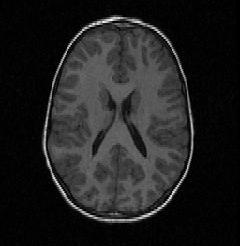
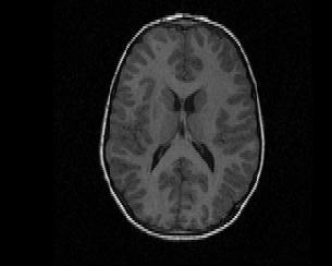
13 Examples Coronal view Sagittal view Transverse view Brain Transverse Movie Demo

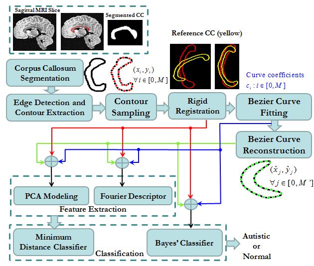
14 System Chart
15 MRI Brain Datasets Autism Normal Dataset Number Received from Dr. Casanova at the Department of Psychiatry GE 1.5T Signa HDxt MRI Scanner 124 Axial (Transverse), Coronal or Sagittal slices Spatial resolution: 1.5 mm in the axial or 2.0 mm in the coronal or Sagittal The spatial resolution for a pixel: mm 2.
16 Dataset Extraction or Conversion %This small program is coded to convert the Autistic MRI images to % the regular image format %based on the matlab codes given by Andrew %modified by Dongqing Chen, Ph.D. %This is the main file, which you just need to open each time close all; clc; clear all; %Autistic Brain MRI image folder path='d:\backup\new Autistic MRI data\autism Data\4.aut.cor\4\'; output_name='4_axi'; %Check either mri_read_signa or mri_read_genesis works x=mri_read_signa(path); %x=mri_read_genesis(path); %Create Mid-saggital slice x=x{1}; tf = maketform_slicer(0, 'axi'); img = mri_slice(x.data, x.ijk2ras, tf); savepgm([path 'mid_sag.pgm'],img); figure; imagesc(img); colormap(gray); title('middle saggital view'); %Create Coronal serial slices and save for i=1:slice_num i tf = maketform_slicer(i-1, 'axi'); img_tmp = mri_slice(x.data, x.ijk2ras, tf); figure; imagesc(img_tmp); colormap(gray); title(num2str(i)); truesize; axis off; pause(0.01); close all; %save to pgm file img_name = sprintf('%s%s.%03d.pgm',path,output_name,i); savepgm(img_name,img_tmp); end %Check the MRI data in x.data [row clm slice_num]=size(x.data); Show Code Demo
17 Dataset Extraction or Conversion %This small program is coded to convert the Autistic MRI images to % the regular image format %based on the matlab codes given by Andrew %modified by Dongqing Chen, Ph.D. %This is the main file, which you just need to open each time close all; clc; clear all; %Autistic Brain MRI image folder path='d:\backup\new Autistic MRI data\autism Data\4.aut.cor\4\'; output_name='4_axi'; %Check either mri_read_signa or mri_read_genesis works x=mri_read_signa(path); %x=mri_read_genesis(path); %Create Mid-saggital slice x=x{1}; tf = maketform_slicer(0, 'axi'); img = mri_slice(x.data, x.ijk2ras, tf); savepgm([path 'mid_sag.pgm'],img); figure; imagesc(img); colormap(gray); title('middle saggital view'); %Check the MRI data in x.data [row clm slice_num]=size(x.data); %Create Coronal serial slices and save for i=1:slice_num i tf = maketform_slicer(i-1, 'axi'); img_tmp = mri_slice(x.data, x.ijk2ras, tf); figure; imagesc(img_tmp); colormap(gray); title(num2str(i)); truesize; axis off; pause(0.01); close all; %save to pgm file img_name = sprintf('%s%s.%03d.pgm',path,output_name,i); savepgm(img_name,img_tmp); end


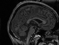
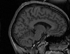
18 Examples of Saggital View

19 Segmentation of Corpus Callosum
20 Segmentation of Corpus Callosum Currently why Manual Segmentation? 1.Accurate 2.Small Datasets 3.Since corpus callosum is the largest white matter, most of the existing algorithm are designed to segment the whole white matter, and messed up the corpus collosum with other parts. (we tried level sets, statistical model (graph-cut), etc.) 4.Designing a 3D shape prior based segmentation Currently why sagittal view? 1.Clear corpus callosum region with closed boundary 2.Less slices numbers (9~12 compared with 50~70 slices along coronal view)
21 75 sampling points Contour Extraction & Sampling
22 75 sampling points Contour Extraction & Sampling
23 Topics for next talk and future work For next talk: Bezier curve fitting and reconstruction PCA based classification Classification results on 15 autism and 20 normal datasets For Future work: Idea on prior shape based 3D segmentation & Registration
MRI-Based Classification Techniques of Autistic vs. Typically Developing Brain
 MRI-Based Classification Techniques of Autistic vs. Typically Developing Brain Presented by: Rachid Fahmi 1 2 Collaborators: Ayman Elbaz, Aly A. Farag 1, Hossam Hassan 1, and Manuel F. Casanova3 1Computer
Brain anatomy tutorial. Dr. Michal Ben-Shachar 459 Neurolinguistics
 Brain anatomy tutorial Dr. Michal Ben-Shachar 459 Neurolinguistics The human brain Left hemisphere Right hemisphere http://www.brainmuseum.org/ Zoom out Zoom in Types of Brain Tissue Gray Matter: Cell
Ways we Study the Brain. Accidents Lesions CAT Scan PET Scan MRI Functional MRI
 The Brain Ways we Study the Brain Accidents Lesions CAT Scan PET Scan MRI Functional MRI Accidents Phineas Gage Story Personality changed after the accident. What this this tell us? That different part
SPAMALIZE s Cerebellum Segmentation routine.
 SPAMALIZE s Cerebellum Segmentation routine. Outline: - Introduction - Data Inputs - Algorithm Steps - Display Notes - Example with menu selections Introduction: This program attempts to segment the cerebellum
Regional and Lobe Parcellation Rhesus Monkey Brain Atlas. Manual Tracing for Parcellation Template
 Regional and Lobe Parcellation Rhesus Monkey Brain Atlas Manual Tracing for Parcellation Template Overview of Tracing Guidelines A) Traces are performed in a systematic order they, allowing the more easily
Visualization strategies for major white matter tracts identified by diffusion tensor imaging for intraoperative use
 International Congress Series 1281 (2005) 793 797 www.ics-elsevier.com Visualization strategies for major white matter tracts identified by diffusion tensor imaging for intraoperative use Ch. Nimsky a,b,
fmri (functional MRI)
") Lesion fmri (functional MRI) Electroencephalogram (EEG) Brainstem CT (computed tomography) Scan Medulla PET (positron emission tomography) Scan Reticular Formation MRI (magnetic resonance imaging) Thalamus
Lesion fmri (functional MRI) Electroencephalogram (EEG) Brainstem CT (computed tomography) Scan Medulla PET (positron emission tomography) Scan Reticular Formation MRI (magnetic resonance imaging) Thalamus
Myers Psychology for AP*
 Myers Psychology for AP* David G. Myers PowerPoint Presentation Slides by Kent Korek Germantown High School Worth Publishers, 2010 *AP is a trademark registered and/or owned by the College Board, which
Announcement. Danny to schedule a time if you are interested.
 Announcement If you need more experiments to participate in, contact Danny Sanchez (dsanchez@ucsd.edu) make sure to tell him that you are from LIGN171, so he will let me know about your credit (1 point).
CISC 3250 Systems Neuroscience
 CISC 3250 Systems Neuroscience Levels of organization Central Nervous System 1m 10 11 neurons Neural systems and neuroanatomy Systems 10cm Networks 1mm Neurons 100μm 10 8 neurons Professor Daniel Leeds
Basic Brain Structure
 The Human Brain Basic Brain Structure Composed of 100 billion cells Makes up 2% of bodies weight Contains 15% of bodies blood supply Uses 20% of bodies oxygen and glucose Brain Protection Surrounded by
Sample Copyright. Academic Group SELF 1 2. Syllabus Checklist. On completion of this chapter you should be able to understand:
 SELF 1 2 Syllabus Checklist On completion of this chapter you should be able to understand: 2.1 Biological influences/bases of behaviour functions of the major parts of the brain hindbrain midbrain forebrain
Biological Process 9/7/10. (a) Anatomy: Neurons have three basic parts. 1. The Nervous System: The communication system of your body and brain
 Anatomy: Neurons have three basic parts. 1. The Nervous System: The communication system of your body and brain") Biological Process Overview 1. The Nervous System: s (a) Anatomy, (b) Communication, (c) Networks 2. CNS/PNS 3. The Brain (a) Anatomy, (b) Localization of function 4. Methods to study the brain (Dr. Heidenreich)
Biological Process Overview 1. The Nervous System: s (a) Anatomy, (b) Communication, (c) Networks 2. CNS/PNS 3. The Brain (a) Anatomy, (b) Localization of function 4. Methods to study the brain (Dr. Heidenreich)
PsychoBrain. 31 st January Dr Christos Pliatsikas. Lecturer in Psycholinguistics in Bi-/Multilinguals University of Reading
 PsychoBrain 31 st January 2018 Dr Christos Pliatsikas Lecturer in Psycholinguistics in Bi-/Multilinguals University of Reading By the end of today s lecture you will understand Structure and function of
1. Processes nutrients and provides energy for the neuron to function; contains the cell's nucleus; also called the soma.
 1. Base of brainstem; controls heartbeat and breathing 2. tissue destruction; a brain lesion is a naturally or experimentally caused destruction of brain tissue 3. A thick band of axons that connects the
MRI and CT of the CNS
 MRI and CT of the CNS Dr.Maha ELBeltagy Assistant Professor of Anatomy Faculty of Medicine The University of Jordan 2018 Computed Tomography CT is used for the detection of intracranial lesions. CT relies
P. Hitchcock, Ph.D. Department of Cell and Developmental Biology Kellogg Eye Center. Wednesday, 16 March 2009, 1:00p.m. 2:00p.m.
 Normal CNS, Special Senses, Head and Neck TOPIC: CEREBRAL HEMISPHERES FACULTY: LECTURE: READING: P. Hitchcock, Ph.D. Department of Cell and Developmental Biology Kellogg Eye Center Wednesday, 16 March
Cerebrum-Cerebral Hemispheres. Cuneyt Mirzanli Istanbul Gelisim University
 Cerebrum-Cerebral Hemispheres Cuneyt Mirzanli Istanbul Gelisim University The largest part of the brain. Ovoid shape. Two incompletely separated cerebral hemispheres. The outer surface of the cerebral
Biocomputer Wired for Action MWABBYH CTBIR LOBES
 Biocomputer Wired for Action MWABBYH CTBIR LOBES 100 100 100 100 100 200 200 200 200 200 300 300 300 300 300 400 400 400 400 400 500 500 500 500 500 Biocomputer Wired for Action MWABBYH CTBIR LOBES 100
ASSUMPTION OF COGNITIVE UNIFORMITY
 The Human Brain cerebral hemispheres: two most important divisions of the brain, separated by the longitudinal fissure corpus callosum: a large bundle of axons that constitutes the major connection between
Specific Sulci/Fissures:
 Specific Sulci/Fissures: Central Sulcus Longitudinal Fissure Sylvian/Lateral Fissure Transverse Fissure http://www.bioon.com/book/biology/whole/image/1/1-8.tif.jpg http://www.dalbsoutss.eq.edu.au/sheepbrains_me/human_brain.gif
Student Lab #: Date. Lab: Gross Anatomy of Brain Sheep Brain Dissection Organ System: Nervous Subdivision: CNS (Central Nervous System)
") Lab: Gross Anatomy of Brain Sheep Brain Dissection Organ System: Nervous Subdivision: CNS (Central Nervous System) Student Lab #: Date 1 Objectives: 1. Learn the main components making up a motor neuron.
Lab: Gross Anatomy of Brain Sheep Brain Dissection Organ System: Nervous Subdivision: CNS (Central Nervous System) Student Lab #: Date 1 Objectives: 1. Learn the main components making up a motor neuron.
NIH Public Access Author Manuscript Proc SPIE. Author manuscript; available in PMC 2014 February 07.
 NIH Public Access Author Manuscript Published in final edited form as: Proc SPIE. 2007 March 5; 6512: 651236. doi:10.1117/12.708950. Semi-Automatic Parcellation of the Corpus Striatum Ramsey Al-Hakim a,
Gross Organization I The Brain. Reading: BCP Chapter 7
 Gross Organization I The Brain Reading: BCP Chapter 7 Layout of the Nervous System Central Nervous System (CNS) Located inside of bone Includes the brain (in the skull) and the spinal cord (in the backbone)
Online appendices are unedited and posted as supplied by the authors. SUPPLEMENTARY MATERIAL
 Appendix 1 to Sehmbi M, Rowley CD, Minuzzi L, et al. Age-related deficits in intracortical myelination in young adults with bipolar SUPPLEMENTARY MATERIAL Supplementary Methods Intracortical Myelin (ICM)
Announcements. Exam 1. VII. Imaging techniques of the brain. Anatomical/Structural Scans. Structural Scans: CT. Structural Scans: CT 2/17/2014
 Exam 1 None at the moment! Announcements Mean 78.0% Median 80% Mode 86% Min 26% Max 98% Std Dev 12.6% VII. Imaging techniques of the brain A. CT: anatomical B. MRI: anatomical C. fmri: functional D. SPECT
49a A&P: Nervous System -! Synaptic Transmission and Central Nervous System
 49a A&P: Nervous System -! Synaptic Transmission and Central Nervous System 49a A&P: Nervous System -! Synaptic Transmission and Central Nervous System! Class Outline" 5 minutes" "Attendance, Breath of
Exam 1. Mean 78.0% Median 80% Mode 86% Min 26% Max 98% Std Dev 12.6%
 Exam 1 Mean 78.0% Median 80% Mode 86% Min 26% Max 98% Std Dev 12.6% None at the moment! Announcements VII. Imaging techniques of the brain A. CT: anatomical B. MRI: anatomical C. fmri: functional D. SPECT
Use of Multimodal Neuroimaging Techniques to Examine Age, Sex, and Alcohol-Related Changes in Brain Structure Through Adolescence and Young Adulthood
 American Psychiatric Association San Diego, CA 24 May 2017 Use of Multimodal Neuroimaging Techniques to Examine Age, Sex, and Alcohol-Related Changes in Brain Structure Through Adolescence and Young Adulthood
CEREBRUM. Dr. Jamila EL Medany
 CEREBRUM Dr. Jamila EL Medany Objectives At the end of the lecture, the student should be able to: List the parts of the cerebral hemisphere (cortex, medulla, basal nuclei, lateral ventricle). Describe
Unit 3: The Biological Bases of Behaviour
 Unit 3: The Biological Bases of Behaviour Section 1: Communication in the Nervous System Section 2: Organization in the Nervous System Section 3: Researching the Brain Section 4: The Brain Section 5: Cerebral
Yin-Hui Siow MD, FRCPC Director of Nuclear Medicine Southlake Regional Health Centre
 Yin-Hui Siow MD, FRCPC Director of Nuclear Medicine Southlake Regional Health Centre Today Introduction to CT Introduction to MRI Introduction to nuclear medicine Imaging the dementias The Brain ~ 1.5
Detection of Mild Cognitive Impairment using Image Differences and Clinical Features
 Detection of Mild Cognitive Impairment using Image Differences and Clinical Features L I N L I S C H O O L O F C O M P U T I N G C L E M S O N U N I V E R S I T Y Copyright notice Many of the images in
Attenuation value in HU From -500 To HU From -10 To HU From 60 To 90 HU. From 200 HU and above
 Brain Imaging Common CT attenuation values Structure Air Fat Water Brain tissue Recent hematoma Calcifications Bone Brain edema and infarction Normal liver parenchyma Attenuation value in HU From -500
APPLICATION OF PHOTOGRAMMETRY TO BRAIN ANATOMY
 http://medifitbiologicals.com/central-nervous-system-cns/ 25/06/2017 PSBB17 ISPRS International Workshop APPLICATION OF PHOTOGRAMMETRY TO BRAIN ANATOMY E. Nocerino, F. Menna, F. Remondino, S. Sarubbo,
CNS Imaging. Dr Amir Monir, MD. Lecturer of radiodiagnosis.
 CNS Imaging Dr Amir Monir, MD Lecturer of radiodiagnosis www.dramir.net Types of radiological examinations you know Plain X ray X ray with contrast GIT : barium (swallow, meal, follow through, enema) ERCP
BIOL Dissection of the Sheep and Human Brain
 BIOL 2401 Dissection of the Sheep and Human Brain Laboratory Objectives After completing this lab, you should be able to: Identify the main structures in the sheep brain and to compare them with those
Homework Week 2. PreLab 2 HW #2 Synapses (Page 1 in the HW Section)
") Homework Week 2 Due in Lab PreLab 2 HW #2 Synapses (Page 1 in the HW Section) Reminders No class next Monday Quiz 1 is @ 5:30pm on Tuesday, 1/22/13 Study guide posted under Study Aids section of website
Homework Week 2 Due in Lab PreLab 2 HW #2 Synapses (Page 1 in the HW Section) Reminders No class next Monday Quiz 1 is @ 5:30pm on Tuesday, 1/22/13 Study guide posted under Study Aids section of website
To understand AD, it is important to
 To understand AD, it is important to know a bit about the brain. This part of Unraveling the Mystery gives an inside view of the normal brain, how it works, and what happens during aging. The brain is
Review of Longitudinal MRI Analysis for Brain Tumors. Elsa Angelini 17 Nov. 2006
 Review of Longitudinal MRI Analysis for Brain Tumors Elsa Angelini 17 Nov. 2006 MRI Difference maps «Longitudinal study of brain morphometrics using quantitative MRI and difference analysis», Liu,Lemieux,
Auditory and Vestibular Systems
 Auditory and Vestibular Systems Objective To learn the functional organization of the auditory and vestibular systems To understand how one can use changes in auditory function following injury to localize
Neuroimaging for Diagnosis of Psychiatric Disorders
 Psychiatric Disorder Neuroimaging for Diagnosis of Psychiatric Disorders JMAJ 45(12): 538 544, 2002 Yoshio HIRAYASU Associate Professor, Department of Neuropsychiatry Kyorin University School of Medicine
HEAD AND NECK IMAGING. James Chen (MS IV)
") HEAD AND NECK IMAGING James Chen (MS IV) Anatomy Course Johns Hopkins School of Medicine Sept. 27, 2011 OBJECTIVES Introduce cross sectional imaging of head and neck Computed tomography (CT) Review head
HEAD AND NECK IMAGING James Chen (MS IV) Anatomy Course Johns Hopkins School of Medicine Sept. 27, 2011 OBJECTIVES Introduce cross sectional imaging of head and neck Computed tomography (CT) Review head
Disorders affecting region: depression anxiety
 Amygdala Involved in learning, and the processing of emotional memories. Measures sensory input for potential threat level, then hypothalamus Regulates volatile emotions like fear and anger. Disorders
PSY 215 Lecture 17 (3/28/2010) (Lateralization in the Brain) Dr. Achtman PSY 215
 (Lateralization in the Brain) Dr. Achtman PSY 215") PSY 215 Lecture 17 Topic: Lateralization in the Brain Chapter 14.1, pages 403-414 Corrections: Lecture 16 (page 4) Broca s Area: trouble producing language, comprehension is okay. Announcements: Review
PSY 215 Lecture 17 Topic: Lateralization in the Brain Chapter 14.1, pages 403-414 Corrections: Lecture 16 (page 4) Broca s Area: trouble producing language, comprehension is okay. Announcements: Review
Wetware: The Biological Basis of Intellectual Giftedness
 Wetware: The Biological Basis of Intellectual Giftedness Why is "giftedness" such a puzzle for parents? Why is there so much confusion? The most common plea heard on TAGFAM is "my child is different; please
Chapter 6 Section 1. The Nervous System: The Basic Structure
 Chapter 6 Section 1 The Nervous System: The Basic Structure Essential Question: How does studying the biology of the brain give us an understanding of our behavior? Draw or type 2 things you already know
Prof. Greg Francis 5/23/08
 Brain parts The brain IIE 269: Cognitive Psychology Greg Francis Lecture 02 The source of cognition (consider transplant!) Weighs about 3 pounds Damage to some parts result in immediate death or disability
Human Nervous System
 Human Nervous System A network of interconnected parts that controls behavior & connects us to the world Central Nervous System consists of the brain and spinal cord Peripheral Nervous System consists
Chapter 3. Structure and Function of the Nervous System. Copyright (c) Allyn and Bacon 2004
 Allyn and Bacon 2004") Chapter 3 Structure and Function of the Nervous System 1 Basic Features of the Nervous System Neuraxis: An imaginary line drawn through the center of the length of the central nervous system, from the
Chapter 3 Structure and Function of the Nervous System 1 Basic Features of the Nervous System Neuraxis: An imaginary line drawn through the center of the length of the central nervous system, from the
Non-Invasive Techniques
 Non-Invasive Techniques Key: Does not hurt the organism Psychology 372 Physiological Psychology Steven E. Meier, Ph.D. Listen to the audio lecture while viewing these slides or view the video presentation
Non-Invasive Techniques
 Many Procedures Non-Invasive Techniques Key: Does not hurt the organism Psychology 372 Physiological Psychology Steven E. Meier, Ph.D. Listen to the audio lecture while viewing these slides or view the
mr brain volume analysis using brain assist
 mr brain volume analysis using brain assist This Paper describes the tool named BrainAssist, which can be used for the study and analysis of brain abnormalities like Focal Cortical Dysplasia (FCD), Heterotopia
Leah Militello, class of 2018
 Leah Militello, class of 2018 Objectives 1. Describe the general organization of cerebral hemispheres. 2. Describe the locations and features of the different functional areas of cortex. 3. Understand
Essentials of Human Anatomy & Physiology. Seventh Edition. The Nervous System. Copyright 2003 Pearson Education, Inc. publishing as Benjamin Cummings
 Essentials of Human Anatomy & Physiology Seventh Edition The Nervous System Copyright 2003 Pearson Education, Inc. publishing as Benjamin Cummings Functions of the Nervous System 1. Sensory input gathering
Organization of the nervous system. The withdrawal reflex. The central nervous system. Structure of a neuron. Overview
 Overview The nervous system- central and peripheral The brain: The source of mind and self Neurons Neuron Communication Chemical messengers Inside the brain Parts of the brain Split Brain Patients Organization
The Nervous System. Divisions of the Nervous System. Branches of the Autonomic Nervous System. Central versus Peripheral
 The Nervous System Divisions of the Nervous System Central versus Peripheral Central Brain and spinal cord Peripheral Everything else Somatic versus Autonomic Somatic Nerves serving conscious sensations
Big brains may hold clues to origins of autism
 VIEWPOINT Big brains may hold clues to origins of autism BY KONSTANTINOS ZARBALIS 23 FEBRUARY 2016 A persistent challenge to improving our understanding of autism is the fact that no single neurological
Okami Study Guide: Chapter 2 1
 Okami Study Guide: Chapter 2 1 Chapter Test 1. A cell that receives information and transmits it to other cells via an electrochemical process is called a(n) a. neuron b. hormone c. glia d. endorphin Answer:
Hemispheric Specialization (lateralization) Each lobe of the brain has specialized functions (Have to be careful with this one.)
 Each lobe of the brain has specialized functions (Have to be careful with this one.)") Cerebral Cortex Principles contralaterality the right half of your brain controls the left half of your body and vice versa. (contralateral control.) Localization of function Specific mental processes
Cerebral Cortex Principles contralaterality the right half of your brain controls the left half of your body and vice versa. (contralateral control.) Localization of function Specific mental processes
Chapter 3. Biological Processes
 Biological Processes Psychology, Fifth Edition, James S. Nairne What s It For? Biological Solutions Communicating internally Initiating and coordinating behavior Regulating growth and other internal functions
The Central Nervous System
 The Central Nervous System Cellular Basis. Neural Communication. Major Structures. Principles & Methods. Principles of Neural Organization Big Question #1: Representation. How is the external world coded
Explainer: This is your brain
 Explainer: This is your brain By The Conversation, adapted by Newsela staff on 03.24.17 Word Count 803 TOP: There are many different parts of the brain with their own specific function. There are times
Modules 4 & 6. The Biology of Mind
 Modules 4 & 6 The Biology of Mind 1 Neuron - 100 Billion - Communication System Glial cells Cell body (nucleus) Dendrites Axon Axon Terminals (terminal buttons) Synaptic cleft 3 4 Communication Within
Velocity Vector Imaging as a new approach for cardiac magnetic resonance: Comparison with echocardiography
 Velocity Vector Imaging as a new approach for cardiac magnetic resonance: Comparison with echocardiography Toshinari Onishi 1, Samir K. Saha 2, Daniel Ludwig 1, Erik B. Schelbert 1, David Schwartzman 1,
Organization of the nervous system. [See Fig. 48.1]
![Organization of the nervous system. [See Fig. 48.1]](/thumbs/90/103926552.jpg "Organization of the nervous system. [See Fig. 48.1]") Nervous System [Note: This is the text version of this lecture file. To make the lecture notes downloadable over a slow connection (e.g. modem) the figures have been replaced with figure numbers as found
Nervous System [Note: This is the text version of this lecture file. To make the lecture notes downloadable over a slow connection (e.g. modem) the figures have been replaced with figure numbers as found
Computer based delineation and follow-up multisite abdominal tumors in longitudinal CT studies
 Research plan submitted for approval as a PhD thesis Submitted by: Refael Vivanti Supervisor: Professor Leo Joskowicz School of Engineering and Computer Science, The Hebrew University of Jerusalem Computer
25/09/2012. Capgras Syndrome. Chapter 2. Capgras Syndrome - 2. The Neural Basis of Cognition
 Chapter 2 The Neural Basis of Cognition Capgras Syndrome Alzheimer s patients & others delusion that significant others are robots or impersonators - paranoia Two brain systems for facial recognition -
biological psychology, p. 40 The study of the nervous system, especially the brain. neuroscience, p. 40
 biological psychology, p. 40 The specialized branch of psychology that studies the relationship between behavior and bodily processes and system; also called biopsychology or psychobiology. neuroscience,
PROPHECY. Preoperative Navigation Guides ANKLE CT SCAN PROTOCOL
 PROPHECY Preoperative Navigation Guides ANKLE CT SCAN PROTOCOL 90 FIGURE 1 Examples FIGURE 1 Examples of neutral ankle positioning. PROPHECY Ankle CT Scan Protocol PROPHECY INBONE and PROPHECY INFINITY
Higher Cortical Function
 Emilie O Neill, class of 2016 Higher Cortical Function Objectives Describe the association cortical areas processing sensory, motor, executive, language, and emotion/memory information (know general location
Psych 56L/ Ling 51: Acquisition of Language
 Psych 56L/ Ling 51: Acquisition of Language Lecture 4 Biological Bases of Language II Announcements Be working on HW1 (due 1/26/12) Be working on bio bases review questions Check out the reference material
FRONTAL LOBE. Central Sulcus. Ascending ramus of the Cingulate Sulcus. Cingulate Sulcus. Lateral Sulcus
 FRONTAL LOBE Central Ascending ramus of the Cingulate Cingulate Lateral Lateral View Medial View Motor execution and higher cognitive functions (e.g., language production, impulse inhibition, reasoning
a) Central sulcus- shallow groove that runs across brain sagitally
 Central sulcus- shallow groove that runs across brain sagitally") KEY BRAIN Brain Gross Anatomy Terms 1) Explain each of the following in terms of structure of the brain a) Central sulcus- shallow groove that runs across brain sagitally b) Lateral fissure- deep groove
KEY BRAIN Brain Gross Anatomy Terms 1) Explain each of the following in terms of structure of the brain a) Central sulcus- shallow groove that runs across brain sagitally b) Lateral fissure- deep groove
Cerebral Cortex 1. Sarah Heilbronner
 Cerebral Cortex 1 Sarah Heilbronner heilb028@umn.edu Want to meet? Coffee hour 10-11am Tuesday 11/27 Surdyk s Overview and organization of the cerebral cortex What is the cerebral cortex? Where is each
Table 1. Summary of PET and fmri Methods. What is imaged PET fmri BOLD (T2*) Regional brain activation. Blood flow ( 15 O) Arterial spin tagging (AST)
 Regional brain activation. Blood flow ( 15 O) Arterial spin tagging (AST)") Table 1 Summary of PET and fmri Methods What is imaged PET fmri Brain structure Regional brain activation Anatomical connectivity Receptor binding and regional chemical distribution Blood flow ( 15 O)
Table 1 Summary of PET and fmri Methods What is imaged PET fmri Brain structure Regional brain activation Anatomical connectivity Receptor binding and regional chemical distribution Blood flow ( 15 O)
Test Bank. Multiple Choice
 Chapter 2: The Brain: An Overview of Structure and Function Test Bank Multiple Choice 1. Evolutionary structures within the are the most primitive. a. hindbrain b. thalamus c. forebrain d. midbrain Answer
Chapter 2 Test. 1. Evolutionary structures within the are the most primitive. *a. hindbrain b. thalamus c. forebrain d. midbrain e.
 Cognitive Psychology In and Out of the Laboratory 5th Edition Galotti TEST BANK Full clear download (no formatting errors) at: https://testbankreal.com/download/cognitive-psychology-laboratory-5thedition-galotti-test-bank/
Diffusion Tensor Imaging in Psychiatry
 2003 KHBM DTI in Psychiatry Diffusion Tensor Imaging in Psychiatry KHBM 2003. 11. 21. 서울대학교 의과대학 정신과학교실 권준수 Neuropsychiatric conditions DTI has been studied in Alzheimer s disease Schizophrenia Alcoholism
Biological Bases of Behavior. 3: Structure of the Nervous System
 Biological Bases of Behavior 3: Structure of the Nervous System Neuroanatomy Terms The neuraxis is an imaginary line drawn through the spinal cord up to the front of the brain Anatomical directions are
Biology 3201 Nervous System #2- Anatomy. Components of a Nervous System
 Biology 3201 Nervous System #2- Anatomy Components of a Nervous System In any nervous system, there are 4 main components: (1) sensors: gather information from the external environment (sense organs) (2)
DISSECTION OF THE SHEEP'S BRAIN
 Sheep Brain Dissection Guide Page 1 DISSECTION OF THE SHEEP'S BRAIN Introduction The purpose of the sheep brain dissection is to familiarize you with the threedimensional structure of the brain and teach
Stroke Awareness. Presented by: Duane Anderson, MD Snoqualmie Valley Hospital Emergency Department Medical Director
 Stroke Awareness Presented by: Duane Anderson, MD Snoqualmie Valley Hospital Emergency Department Medical Director What is a stroke? Stroke can happen to anyone. Stroke is the fourth leading cause of death
Definition of Anatomy. Anatomy is the science of the structure of the body and the relation of its parts.
 Definition of Anatomy Anatomy is the science of the structure of the body and the relation of its parts. Basic Anatomical Terms Anatomical terms for describing positions: Anatomical position: Supine position:
Whole brain CT perfusion maps with paradoxical low mean transit time to predict infarct core
 Whole brain CT perfusion maps with paradoxical low mean transit time to predict infarct core Poster No.: B-292 Congress: ECR 2011 Type: Scientific Paper Topic: Neuro Authors: S. Chakraborty, M. E. Ahmad,
The human brain. of cognition need to make sense gives the structure of the brain (duh). ! What is the basic physiology of this organ?
. ! What is the basic physiology of this organ?") The human brain The human brain! What is the basic physiology of this organ?! Understanding the parts of this organ provides a hypothesis space for its function perhaps different parts perform different
The human brain The human brain! What is the basic physiology of this organ?! Understanding the parts of this organ provides a hypothesis space for its function perhaps different parts perform different
Spatial localisation of EEG dipoles in MRI using the International System anatomical references
 Proc. of First Int'l Workshop on Image and Signal Processing and Analysis Spatial localisation of EEG dipoles in MRI using the 10-20 International System anatomical references J. Pascau a,b, M. Desco a,
Nervous System Task Exploration
 Nervous System Task Exploration Read It! Directions: Each member of the group will read the passage and answer the task questions. It is important to remember that the answers will come directly from the
A Comparison of IMRT and VMAT Technique for the Treatment of Rectal Cancer
 A Comparison of IMRT and VMAT Technique for the Treatment of Rectal Cancer Tony Kin Ming Lam Radiation Planner Dr Patricia Lindsay, Radiation Physicist Dr John Kim, Radiation Oncologist Dr Kim Ann Ung,
Brain and behaviour (Wk 6 + 7)
") Brain and behaviour (Wk 6 + 7) What is a neuron? What is the cell body? What is the axon? The basic building block of the nervous system, the individual nerve cell that receives, processes and transmits
Brain and behaviour (Wk 6 + 7) What is a neuron? What is the cell body? What is the axon? The basic building block of the nervous system, the individual nerve cell that receives, processes and transmits
Dissection of the Sheep Brain
 Dissection of the Sheep Brain Laboratory Objectives After completing this lab, you should be able to: 1. Identify the main structures in the sheep brain and to compare them with those of the human brain.
Cortical source analysis of infant spatial cueing International Conference on Infant Studies, 2012 John E. Richards University of South Carolina
 Cortical source analysis of infant spatial cueing International Conference on Infant Studies, 2012 John E. Richards University of South Carolina Cortical source analysis of infant spatial cueing This presentation
The Nervous System. Nerves, nerves everywhere!
 The Nervous System Nerves, nerves everywhere! Purpose of the Nervous System The information intake and response system of the body. Coordinates all body functions, voluntary and involuntary! Responds to
Chapter 3: 2 visual systems
 Chapter 3: 2 visual systems Overview Explain the significance of the turn to the brain in cognitive science Explain Mishkin and Ungerleider s hypothesis that there are two distinct visual systems Outline
Classification and Statistical Analysis of Auditory FMRI Data Using Linear Discriminative Analysis and Quadratic Discriminative Analysis
 International Journal of Innovative Research in Computer Science & Technology (IJIRCST) ISSN: 2347-5552, Volume-2, Issue-6, November-2014 Classification and Statistical Analysis of Auditory FMRI Data Using
Stuttering Research. Vincent Gracco, PhD Haskins Laboratories
 Stuttering Research Vincent Gracco, PhD Haskins Laboratories Stuttering Developmental disorder occurs in 5% of children Spontaneous remission in approximately 70% of cases Approximately 1% of adults with
Lab 12 Nervous System II
 Lab 12 Nervous System II Laboratory Objectives Identify the structural components of the central nervous system Label the functional areas of human cerebral cortex. Given a deficit affecting one or more
Prof. Greg Francis 1/2/19
 Brain scans PSY 200 Greg Francis Lecture 03 How to study the brain without killing someone. Scanning Technology provides insight into brain processes w EEG recordings w MRI w Non-invasive Maps of brain
Nervous System Task Exploration
 Nervous System Task Exploration Read It! Directions: Each member of the group will read the passage and answer the task questions. It is important to remember that the answers will come directly from the
Department of Cognitive Science UCSD
 Department of Cognitive Science UCSD Verse 1: Neocortex, frontal lobe, Brain stem, brain stem, Hippocampus, neural node, Right hemisphere, Pons and cortex visual, Brain stem, brain stem, Sylvian fissure,