New EAONO Cholesteatoma Classification with imaging illustration. Milan Profant, Katarina Sláviková
|
|
|
- Adrian Dalton
- 5 years ago
- Views:
Transcription
1 New EAONO Cholesteatoma Classification with imaging illustration Milan Profant, Katarina Sláviková
2 EAONO/JOS Joint Consensus Statements on the Definitions, Classification and Staging of Middle Ear Cholesteatoma Matthew Yung, Tetsuya Tono, Ewa Olszewska, Yutaka Yamamoto, Holger Sudhoff, Masafumi Sakagami, Jef Mulder, Hiromi Kojima, Armağan İncesulu, Franco Trabalzini, Nuri Özgirgin J Int Adv Otol 2017
3 Classification of cholesteatoma Acquired Congenital Unclassifiable (cholesteatoma whose origin cannot be accurately determined)
4 Congenital cholesteatoma Congenital cholesteatoma is typically an expanding cystic mass with keratinizing squamous epithelium located medial to the intact tympanic membrane It is assumed to be present at birth, but is usually diagnosed during infancy or in early childhood in patients with no prior history of otorrhea, perforation, or previous ear surgery. A history of previous bouts of otitis media or an effusion does not exclude congenital cholesteatoma. Congenital cholesteatoma is usually located at the anterosuperior quadrant of the middle ear. However, it may be located at the posterosuperior quadrant or other locations.
5 Acquired cholesteatoma 1. retraction pocket cholesteatoma a) pars flaccida (attic cholesteatoma) b) pars tensa cholesteatoma c) combination of pars flaccida and pars tensa cholesteatoma 2. non-retraction pocket cholesteatoma a) cholesteatoma secondary to tympanic perforation (the so-called secondary acquired cholesteatoma) b) cholesteatoma following trauma and/or otologic procedures
6 Cholesteatoma recidivism Residual cholesteatoma Residual cholesteatoma results from the incomplete surgical removal of the cholesteatoma matrix Recurrent cholesteatoma results from the reformation of the retraction pocket after a complete previous surgical cholesteatoma removal Post-surgical cholesteatoma may be residual or recurrent, although these are not mutually exclusive.
7
 S2, the sinus tympani Tympanic cavity (T) Attic (A) Mastoid")
8 Divisions of the middle ear space (STAM system) Difficult access sites (S) S1, the supratubal recess (also called the anterior epitympanum or protympanum) S2, the sinus tympani Tympanic cavity (T) Attic (A) Mastoid (M)
9 Cholesteatoma staging Stage I: Cholesteatoma localized in the primary site Stage II: Cholesteatoma involving two or more sites Stage III: Cholesteatoma with extracranial complications or pathologic conditions including Stage IV: Cholesteatoma with intracranial complications including
10 Stage I: Cholesteatoma localized in the primary site Pars flaccida cholesteatoma (attic cholest Pars tensa cholesteatoma Cholesteatoma secondary to a tensa perforation Congenital cholesteatoma
11 Stage II: Cholesteatoma involving two or more sites Pars flaccida cholesteatoma (attic cholest Pars tensa cholesteatoma Cholesteatoma secondary to a tensa perforation Congenital cholesteatoma
12 Stage III: Cholesteatoma with extracranial complications Facial palsy Labyrinthine fistula Labyrinthitis Postauricular abscess or fistula Zygomatic abscess Neck abscess Canal wall destruction: more than half the length of the bony ear canal Destruction of the tegmen
13 Stage IV: Cholesteatoma with intracranial complications Purulent meningitis Epidural abscess Subdural abscess Brain abscess Sinus thrombosis Brain herniation into the mastoid cavity


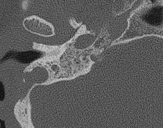
14 CT Gold standard to image middle ear pathology High space resolution (Temporal bone anatomy before surgery) High sensitivity imaging in normal middle ear cleft Low specificity in case of full middle ear cleft???
15 HRCT Axial and coronal plane with additional reconstruction Colimation (0,5 0,6mm), slice thicknes 1mm Bony algorithm Native imaging (+C in case of vascular middle ear lesion)
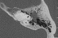


16 Stage I Cholesteatoma localised to the single primary site


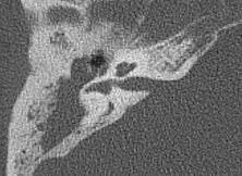


17 Cholesteatoma occupying 2 or more sites Stage II


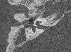
18 Cholesteatoma with extracranial complications Stage III


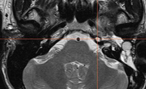
19 Stage IV Cholesteatoma with intracranial complications
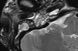
20 MR protocol 1,5 T Magnetom Avanto Sequence Thickness TR (ms) TE (ms) FOV (mm) Matrix b factor (mm) TRA tse T x216 TRA se T x168 TRA 3D tse T2 (spc T2) 0, x320 TRA HASTE DWI x144 0,1000 s/mm 2 COR tse T1 FS x180 TRA se T1 (Gd) x168 COR tse T1 FS (Gd) x180 3D T1 mp-rage (Gd) , x192
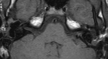
21 MR T1 Vo, T2 Vo CH after +C ring enhancement in periphery (perimatrix) / no enhancement CH equal to granulation tissue in T1 Vo and T2 Vo granulation tissue after +C significant enhancement DWI to differentiate postoperative changes from residual/recurrent cholesteatoma


22 Subtotal petrosectomy


23 CWU tympanomastoidectomy


24 Modified radical surgery


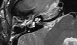
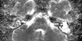
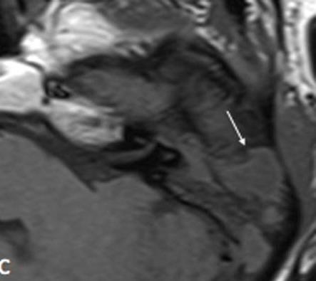
25 Cholesteatoma pearl


26 Cholesteatoma pearl in sinus tympani Sinus tympani

:878-83. doi: 10.1001/archoto.")
27 Localization (fusion of CT and MR) Plouin-Gaudon I. et al.,fusion of MRIs and CT scans for surgical treatment of cholesteatoma of the middle ear in children, Arch Otolaryngol Head Neck Surg., 2010 Sep;136(9): doi: /archoto
28 Differencial diagnosis in MR Pathology T1 Vo T2 Vo +C T1 Vo DWI ADC Cholesteatoma - (Ring) Cholesterol granuloma Glomus TU Slightly hyposi Slightly hypersi - - ( salt and spice ) Schwannoma Slightly hyposi Slightly hypersi - - Meningeoma Slightly hyposi Slightly hypersi - - Fresh granulation tissue Old scars Late Absces Ring Liquid -


29 Abscess
30 Author DWI P Size (mm) De Foer et al. (2008) Dhepnorrarat et al. (2009) nepi- DWI nepi- DWI Sensit. (%) Specific. (%) PPH (%) NPH (%) Khemani et al. (2011) Profant, Sláviková (2012) nepi- DWI nepi- DWI
31 Indications for imaging in cholesteatoma (our experience) Diagnosis CT MR (HASTE DWI) Chronic otitis with CH (primary dg, preoperative evaluation) Chronic otitis with CH (revision surgery to improve hearing) (complementary in complicated cases) Chronic otitis with CH (revision surgery second look ) Chronic otitis with CH (follow up) (negative MR, no revision)
32 Conclusions New classification will not change biological behaviour of cholesteatoma Nowadays there is no method of choice to manage cholesteatoma to be respected by all otosurgeons Rerurrence rate is varying from 0% to 30% Obliteration of mastoid cavity with bone dust and separation from the tympanic cleft is a hit of recent period Cholesteatom itself at the end is the master with the final word
EAONO/JOS Joint Consensus Statements on the Definitions, Classification and Staging of Middle Ear Cholesteatoma
 J Int Adv Otol 2016 DOI: 10.5152/iao.2017.3363 Review EAONO/JOS Joint Consensus Statements on the Definitions, Classification and Staging of Middle Ear Cholesteatoma Matthew Yung, Tetsuya Tono, Ewa Olszewska,
EAONO/JOS Joint Consensus Statements on the Definitions, Classification and Staging of Middle Ear Cholesteatoma
 J Int Adv Otol 2017; 13(1): 1-8 DOI: 10.5152/iao.2017.3363 Review EAONO/JOS Joint Consensus Statements on the Definitions, Classification and Staging of Middle Ear Cholesteatoma Matthew Yung, Tetsuya Tono,
Cholesteatoma-Pathogenesis and Surgical Management. Grand Rounds Presentation February 24, 1999 Kyle Kennedy, M.D. Jeffrey Vrabec,, M.D.
 Cholesteatoma-Pathogenesis and Surgical Management Grand Rounds Presentation February 24, 1999 Kyle Kennedy, M.D. Jeffrey Vrabec,, M.D. Introduction Cholesteatoma (keratoma)-essentially an accumulation
Clinical Utility of MRI for Cholesteatoma Recurrence
 Curr Surg Rep (2014) 2:63 DOI 10.1007/s40137-014-0063-0 EAR SURGERY (CJ LIMB, SECTION EDITOR) Clinical Utility of MRI for Cholesteatoma Recurrence Jonathan McJunkin Richard Chole Published online: 24 June
Cholesteatoma in children
 Cholesteatoma in children British Association of Paediatricians in Audiology London Conference, Jan.2012 Matthew Clark FRCS (ORL-HNS) Consultant Otologist Gloucestershire Royal Hospital Overview: Cholesteatoma
Introduction. Types of Cholesteatoma
 TITLE: Cholesteatoma SOURCE: Grand Rounds Presentation, UTMB, Dept. of Otolaryngology DATE: January 25, 2006 RESIDENT PHYSICIAN: Garrett Hauptman, MD FACULTY PHYSICIAN: Tomoko Makishima, MD, PhD SERIES
ORIGINAL ARTICLE. A New Staging System for Tympano-mastoid Cholesteatoma. Aziz Belal, Mahmoud Reda, Ahmed Mehana, Yousef Belal
 Int. Adv. Otol. 2012; 8:(1) 63-68 ORIGINAL ARTICLE A New Staging System for Tympano-mastoid Cholesteatoma Aziz Belal, Mahmoud Reda, Ahmed Mehana, Yousef Belal Alexandria Ear Hospital Alexandria Egypt (AB,
Complications of otitis media
 Chronic otitis media Definition:- Complications of otitis media Otitis media (OM) is broadly defined as inflammation from any cause of the middle ear.this may involve any of the contiguous pneumatized
UC SF. Safe Surgery Rule #1. Cholesteatoma. It s hard to have a surgical complication when you are not operating
 UC SF Cholesteatoma Chronic Ear Surgery: Staying Out of Trouble! Lawrence R. Lustig, MD Department of Oto-HNS University of California San Francisco Ligaments and folds Spaces NU Epitympanic Cholesteatoma
Chronic Ear Disease. Daekeun Joo Resident Lecture Series 11/18/09
 Chronic Ear Disease Daekeun Joo Resident Lecture Series 11/18/09 ETD URIs Viral-induced damage to ET lining resulting in decreased mucociliary clearance Viral invasion of ME mucosa results in inflamm Reflux
Middle ear CT imaging: Review of anatomy and common pathology
 Middle ear CT imaging: Review of anatomy and common pathology Poster No.: C-0665 Congress: ECR 2017 Type: Educational Exhibit Authors: M. R. Campos Arenas, M. C. Sánchez-Porro, J. Garrido Rull ; 1 1 2
ORIGINAL ARTICLE. Efficacy of the 2-Staged Procedure in the Management of Cholesteatoma. paradigms have been published
 Efficacy of the 2-Staged Procedure in the Management of Cholesteatoma Steven Y. Ho, MD; John F. Kveton, MD ORIGAL ARTICLE Objective: To demonstrate the efficacy of intact canal wall procedure coupled with
Congenital Middle Ear Cholesteatoma in Children; Retrospective Review of 35 Cases
 J Korean Med Sci 2009; 24: 126-31 ISSN 1011-8934 DOI: 10.3346/jkms.2009.24.1.126 Copyright The Korean Academy of Medical Sciences Congenital Middle Ear Cholesteatoma in Children; Retrospective Review of
Magnetic Resonance Imaging (MRI) and High Resolution Computed Tomography (HRCT): Can they improve the evaluation of Middle ear cholesteatoma?
 and High Resolution Computed Tomography (HRCT): Can they improve the evaluation of Middle ear cholesteatoma?") Magnetic Resonance Imaging (MRI) and High Resolution Computed Tomography (HRCT): Can they improve the evaluation of Middle ear cholesteatoma? Poster No.: C-1249 Congress: ECR 2013 Type: Educational Exhibit
Magnetic Resonance Imaging (MRI) and High Resolution Computed Tomography (HRCT): Can they improve the evaluation of Middle ear cholesteatoma? Poster No.: C-1249 Congress: ECR 2013 Type: Educational Exhibit
HASTE diffusion-weighted MRI for the reliable detection of cholesteatoma
 Diagn Interv Radiol 2012; 18:153 158 Turkish Society of Radiology 2012 HEAD AND NECK IMAGING ORIGINAL ARTICLE HASTE diffusion-weighted MRI for the reliable detection of A. Turan Ilıca, Yusuf Hıdır, Nail
ISSN: Volume 5 Issue CASE REPORT. Anju Chauhan, Vikram Wadhwa, Samuel Rajan, P.K. Rathore
 ISSN: 2250-0359 Volume 5 Issue 2 2015 CONGENITAL CHOLESTEATOMA ISOLATED TO MASTOID PROCESS : A CASE REPORT Anju Chauhan, Vikram Wadhwa, Samuel Rajan, P.K. Rathore Maulana Azad Medical College, New Delhi,
Acquired Cholesteatoma - a pictorial review.
 Acquired Cholesteatoma - a pictorial review. Award: NZ Radiology Education Trust Poster Prize Poster No.: R-0132 Congress: RANZCR ASM 2013 Type: Educational Exhibit Authors: V. Francis, N. Umaria; Auckland/NZ
Chronic otitis media with cholesteatoma: clinical presentation and surgical management
 International Journal of Otorhinolaryngology and Head and Neck Surgery http://www.ijorl.com pissn 2454-5929 eissn 2454-5937 Original Research Article DOI: http://dx.doi.org/10.18203/issn.2454-5929.ijohns20183454
Detection of Postoperative Residual Cholesteatoma With Non/EchoYPlanar Diffusion-Weighted Magnetic Resonance Imaging
 Otology & Neurotology 29:513Y517 Ó 2008, Otology & Neurotology, Inc. Detection of Postoperative Residual Cholesteatoma With Non/EchoYPlanar Diffusion-Weighted Magnetic Resonance Imaging *Bert De Foer,
Received 20 June 2006
 American Journal of Otolaryngology Head and Neck Medicine and Surgery 28 (2007) 230 234 www.elsevier.com/locate/amjoto Value of high-resolution computed tomography and magnetic resonance imaging in the
Surgical outcome in chronic otitis media with cholesteatoma
 International Journal of Otorhinolaryngology and Head and Neck Surgery http://www.ijorl.com pissn 2454-5929 eissn 2454-5937 Original Research Article DOI: http://dx.doi.org/10.18203/issn.2454-5929.ijohns20184260
Status of Ossicles in Cholesteatoma
 Bangladesh J Otorhinolaryngol 2015; 21(2): 97-101 Original Article Status of Ossicles in Cholesteatoma Lt Col Mohammad Delwar Hossain 1, Brig Gen Md Nasir Uddin Ahamed 2, Major Mohammad Misbah Al Kabir
Original Article Comparative study of tubotympanic and atticoantral variety of Chronic suppurative otitis media
 Bangladesh J Otorhinolaryngol 2010; 16(2): 113-119 Original Article Comparative study of tubotympanic and atticoantral variety of Chronic suppurative otitis media Md. Rafiqul Islam 1, Ahmmad Taous 2, Md.
Cholesteatoma and Non-cholesteatomatous Inflammatory Disease. Cholesteatoma. Disclosures. Overview EAC. Cholesteatoma. None
 Disclosures Cholesteatoma and Non-cholesteatomatous Inflammatory Disease None Amy F Juliano, MD Staff Radiologist, Massachusetts Eye and Ear Infirmary Assistant Professor of Radiology, Harvard Medical
Principles of Chronic Ear Surgery. Principles of Chronic Ear Surgery. Preoperative Considerations
 Principles of Chronic Ear Surgery Principles of Chronic Ear Surgery David S. Haynes, MD, FACS The Otology Group of Vanderbilt Professor, Dept. of Otolaryngology, Professor Dept. of Neurosurgery Professor,
Correlation of HRCT mastoid with clinical presentation and operative findings in ear diseases
 International Journal of Otorhinolaryngology and Head and Neck Surgery Chintale SG et al. Int J Otorhinolaryngol Head Neck Surg. 2017 Jul;3(3):656-660 http://www.ijorl.com pissn 2454-5929 eissn 2454-5937
CHOLESTEATOMA I DO IT?? TYMPANOPLASTY. Mohamed BADR-EL-DINE, M.D.
 CHOLESTEATOMA HOW AND I DO IT?? TYMPANOPLASTY Mohamed BADR-EL-DINE, M.D. Otologie et Neurotologie; Service d Otorhinolaryngologie Université d Alexandrie - EGYPTE. Panelists: Prof. Aziz Belal Prof. Douglas
High resolution computed tomography of temporal bone in the evaluation of otologic diseases
 International Journal of Otorhinolaryngology and Head and Neck Surgery Handi PS et al. Int J Otorhinolaryngol Head Neck Surg. 2018 Jan;4(1):87-92 http://www.ijorl.com pissn 2454-5929 eissn 2454-5937 Original
The Value of Computed Tomography Scanning in Assessment of Aditus ad Antrum Patency and Choice of Treatment Line in Revision Myringoplasty
 Med. J. Cairo Univ., Vol. 77, No. 2, September: 53-57, 2009 www.medicaljournalofcairouniversity.com The Value of Computed Tomography Scanning in Assessment of Aditus ad Antrum Patency and Choice of Treatment
Multidetector Computed Tomography Findings of Auto-Evacuated Secondary Acquired Cholesteatoma: A Morphologic and Quantitative Analysis
 J Int Adv Otol 2018; 14(3): 464-71 DOI: 10.5152/iao.2018.4859 Original Article Multidetector Computed Tomography Findings of Auto-Evacuated Secondary Acquired Cholesteatoma: A Morphologic and Quantitative
Key words: Chronic suppurative otitis media, High resolution Computed tomography, cholesteatoma,
 ORIGINAL ARTICLE Study of Radiological findings in High resolution computed tomography (HRCT) temporal bone in Chronic suppurative otitis Media (CSOM): A hospital Based cross sectional study. Vijay vaidya
Mastoid cavities CAUSES OF FAILURE?
 Management of troublesome mastoid cavities J. Magnan, Université de la Mediterranée Marseille, France ALEXANDRIA 2009 Mastoid cavities CAUSES OF FAILURE? Facial bridge Pneumatisation Skin Mucosa Meatoplasty
Pediatric Ear Diseases
 Pediatric Ear Diseases Yasushi Naito Pediatric Ear Diseases Diagnostic Imaging Atlas and Case Reports 242 figures, 7 in color and 5 tables, 2013 Basel Freiburg Paris London New York New Delhi Bangkok
Narrowest segment of the ear canal. Limited microscopic. Wide endoscopic. field of view. field of view
 Endoscopic Transcanal Management of Cholesteatoma M. Tarabichi American Hospital-Dubai The Endoscope in Otology Mostly for documentation. Mostly diagnostic. Exploration of old mastoid cavities Endoscopic
Sensorineural Hearing Loss in Complicated Cholesteatomatous Ear Disease
 Research in Otolaryngology 2014, 3(2): 29-35 DOI: 10.5923/j.otolaryn.20140302.04 Sensorineural Hearing Loss in Complicated Cholesteatomatous Ear Disease Borlingegowda Viswanatha 1,*, Khaja Naseeruddin
CT and MRI imaging of chronic otitis media complications.
 CT and MRI imaging of chronic otitis media complications. Poster No.: C-2131 Congress: ECR 2014 Type: Scientific Exhibit Authors: V. Bizimi, M. Tsitskari, K. Spyrou, V. Papalouka, A. Economou; Athens/GR
Value of Ear Endoscopy in Cholesteatoma Surgery
 Otology & Neurotology 23:631 635 2002, Otology & Neurotology, Inc. Value of Ear Endoscopy in Cholesteatoma Surgery M. Badr-el-Dine Ear, Nose, and Throat Department, Alexandria School of Medicine, University
ACUTE PAEDIATRIC EAR PRESENTATIONS PROF IAIN BRUCE PAEDIATRIC OTOLARYNGOLOGIST & ADULT OTOLOGIST
 www.manchesterchildrensent.com ACUTE PAEDIATRIC EAR PRESENTATIONS PROF IAIN BRUCE PAEDIATRIC OTOLARYNGOLOGIST & ADULT OTOLOGIST A CHILD WITH EARACHE UNCOMPLICATED AOM ACUTE OTITIS MEDIA Acute otitis media
ISOLATED CONGENITAL CHOLESTEATOMA OF THE MASTOID PROCESS: A CASE REPORT Shankar Tati 1, Shobhan Babu A 2, Nagaraj K 3, Srinivas K 4, Anjani Kumari K 5
 ISOLATED CONGENITAL CHOLESTEATOMA OF THE MASTOID PROCESS: A Shankar Tati 1, Shobhan Babu A 2, Nagaraj K 3, Srinivas K 4, Anjani Kumari K 5 HOW TO CITE THIS ARTICLE: Shankar Tati, Shobhan Babu A, Nagaraj
Cholesteatoma and the Destruction of Middle Ear and Mastoid Cavities in Ottorhoea
 ORIGINAL ARTICLLE Cholesteatoma and the Destruction of Middle Ear and Mastoid Cavities in Ottorhoea KHURSHID ANWAR, MOHAMMAD SAID*, BAKHT ZADA**, GULAB DIN***, IFTIKHAR AHMAD KHAN**** ABSTRACT Objective:
Surgical and Non-Surgical Causes of Progressive Hearing Loss in Children: What can be done about it?
 Surgical and Non-Surgical Causes of Progressive Hearing Loss in Children: What can be done about it? Daniela Carvalho, MD, MMM, FAAP Professor, Surgery Department UCSD Pediatric Otolaryngology Rady Children
OPEN CAVITY / CANAL WALL DOWN MASTOIDECTOMY. Bruce Black MD
 OPEN CAVITY / CANAL WALL DOWN MASTOIDECTOMY Plan of an open cavity (CWD: canal wall down surgery). The middle ear is essentially gutted/amputated to eliminate cholesteatoma. Plan of classic CWD (radical
Efficacy of pre-operative computed tomography scans on clinical management and temporal bone surgery in cases of chronic otitis media
 Boston University OpenBU Theses & Dissertations http://open.bu.edu Boston University Theses & Dissertations 2014 Efficacy of pre-operative computed tomography scans on clinical management and temporal
Clinical analysis of secondary acquired cholesteatoma.
 Research Article Clinical analysis of secondary acquired cholesteatoma. http://www.alliedacademies.org/archives-of-general-internal-medicine/ ISSN : 2591-7951 Takashi Yamatodani 1 *, Kunihiro Mizuta 2,
Characteristics of Patients Who Underwent Mastoidectomy: A Two Years Experience
 474 AMJ September 2017 AMJ. 2017;4(3):474 8 Characteristics of Patients Who Underwent Mastoidectomy: A Two Years Experience Ashwini Gunasekaran, 1 Sally Mahdiani, 2 Fifi Veronica 3 1 Faculty of Medicine
1. Axial view, left temporal bone. Plane through the upper antrum (A), superior semicircular canal (SSC) and IAC.
, superior semicircular canal (SSC) and IAC.") PA IAC SSC A 1. Axial view, left temporal bone. Plane through the upper antrum (A), superior semicircular canal (SSC) and IAC. IAC VII M I LSC Plane through the IAC, malleus head and incus and the lateral
PA IAC SSC A 1. Axial view, left temporal bone. Plane through the upper antrum (A), superior semicircular canal (SSC) and IAC. IAC VII M I LSC Plane through the IAC, malleus head and incus and the lateral
Kingdom of Bahrain Arabian Gulf University College of Medicine and Medical Sciences Year 6 ENT SMC Otitis Media (Dr.
 Kingdom of Bahrain Arabian Gulf University College of Medicine and Medical Sciences Year 6 ENT SMC Otitis Media (Dr. Jalal Almarzooq) - Anatomy of the ear: The ear is divided into 3 parts: External ear.
Cholesteatoma is a collection of keratinous debris lined by
 ORIGINAL RESEARCH M.H.G. Dremmen P.A.M. Hofman J.R. Hof R.J. Stokroos A.A. Postma The Diagnostic Accuracy of Non-Echo-Planar Diffusion-Weighted Imaging in the Detection of Residual and/or Recurrent Cholesteatoma
THE MANAGEMENT of COMPLICATED OTITIS MEDIA. IFOS, Lima, 2018
 THE MANAGEMENT of COMPLICATED OTITIS MEDIA IFOS, Lima, 2018 VINCENT C COUSINS ENT-Otoneurology Unit, The Alfred Hospital & Department of Surgery. Monash University MELBOURNE, AUSTRALIA Otologic Complications
Complications of At t i c o a n t ral Cholesteato ma:
 Complications of t t i c o a n t ral Cholesteato ma: MR Manifestations 1 Jeong Hyun Lee, M.D., Ho Kyu Lee, M.D., Soo Mi Lim, M.D., Ji Hoon Shin, M.D., Choong Gon Choi, M.D., Dae Chul Suh, M.D., Kwang Sun
High-resolution Computed Tomography Study of Temporal Bone Pathologies
 Original Article Print ISSN: 2321-6379 Online ISSN: 2321-595X DOI: 10.17354/ijss/2016/375 High-resolution Computed Tomography Study of Temporal Bone Pathologies Manjit Bagul Senior Resident, Department
Gerard J. Gianoli, MD, FACS The Ear and Balance Institute Baton Rouge, Louisiana
 Gerard J. Gianoli, MD, FACS The Ear and Balance Institute Baton Rouge, Louisiana SSCD is defined anatomically as the absence of bone between the SSC and the middle fossa dura PSCD is a defect of the PSC
RECONSTRUCTION OF POSTERIOR MEATAL AND/OR LATERAL ATTIC WALLS IN CHOLESTEATOMA SURGERY
 -857- RECONSTRUCTION OF POSTERIOR MEATAL AND/OR LATERAL ATTIC WALLS IN CHOLESTEATOMA SURGERY Mohammed Elsayed Elmaghawry, Mohammed Kamal Mobasher,Magdy Mohammed Abd-Elfattah, Adly Ahmed Tantawy,Atef Hamed
The Ear The ear consists of : 1-THE EXTERNAL EAR 2-THE MIDDLE EAR, OR TYMPANIC CAVITY 3-THE INTERNAL EAR, OR LABYRINTH 1-THE EXTERNAL EAR.
 The Ear The ear consists of : 1-THE EXTERNAL EAR 2-THE MIDDLE EAR, OR TYMPANIC CAVITY 3-THE INTERNAL EAR, OR LABYRINTH 1-THE EXTERNAL EAR Made of A-AURICLE B-EXTERNAL AUDITORY MEATUS A-AURICLE It consists
Gross Anatomy of the. TEMPORAL BONE, EXTERNAL EAR, and MIDDLE EAR. Assignment: Head to Toe Temporomandibular Joint (TMJ)
") Gross Anatomy the TEMPORAL BONE, EXTERNAL EAR, and MIDDLE EAR M1 Gross and Developmental Anatomy 9:00 AM, December 11, 2008 Dr. Milton M. Sholley Pressor Anatomy and Neurobiology Assignment: Head to Toe
Gross Anatomy the TEMPORAL BONE, EXTERNAL EAR, and MIDDLE EAR M1 Gross and Developmental Anatomy 9:00 AM, December 11, 2008 Dr. Milton M. Sholley Pressor Anatomy and Neurobiology Assignment: Head to Toe
ACUTE MASTOIDITIS IN CHILDREN: SUSCEPTIBILITY FACTORS AND MANAGEMENT
 & ACUTE MASTOIDITIS IN CHILDREN: SUSCEPTIBILITY FACTORS AND MANAGEMENT Slobodan Spremo*, Biljana Udovčić Clinic for Otorhinolaryngology, Clinic Center in Banja Luka, Zdrave Korde 1, 78000 Banja Luka, Bosnia
ORIGINAL ARTICLE. Temporal Lobe Injury in Temporal Bone Fractures. imaging (MRI) to evaluate lesions of the temporal
 to evaluate lesions of the temporal") ORIGINAL ARTICLE Temporal Lobe Injury in Temporal Bone Fractures Richard M. Jones, MD; Michael I. Rothman, MD; William C. Gray, MD; Gregg H. Zoarski, MD; Douglas E. Mattox, MD Objective: To determine the
ORIGINAL ARTICLE Temporal Lobe Injury in Temporal Bone Fractures Richard M. Jones, MD; Michael I. Rothman, MD; William C. Gray, MD; Gregg H. Zoarski, MD; Douglas E. Mattox, MD Objective: To determine the
Injury retrotympanic white, blue and red. Clinicalradiological
 Injury retrotympanic white, blue and red. Clinicalradiological correlation Poster No.: C-0211 Congress: ECR 2013 Type: Educational Exhibit Authors: R. Esteban Saiz, R. Castañón Martinez, M. Rebolledo Vicente,
Gross Anatomy of the. TEMPORAL BONE, EXTERNAL EAR, and MIDDLE EAR
 Gross Anatomy of the TEMPORAL BONE, EXTERNAL EAR, and MIDDLE EAR M1 Gross and Developmental Anatomy 9:00 AM, December 11, 2008 Dr. Milton M. Sholley Professor of Anatomy and Neurobiology Assignment: Head
CSF Leaks. Abnormal communication between the subarachnoid space and the tympanomastoid space or nasal cavity. Presenting symptoms:
 CSF Leaks Steven Wright, M.D. Faculty Advisor: Matthew Ryan, M.D. The University of Texas Medical Branch Department of Otolaryngology Grand Rounds Presentation January 5, 2005 CSF Leaks Abnormal communication
Bruce Black MD EAC TRAUMA
 EAC TRAUMA Bruising in the deep canal due to cotton bud/q-tip selfcleaning attempts. No action required. A granuloma of the deep Lt. EAC. Superficial trauma has become secondarily infected. Clean thoroughly,
The ear: some applied basic science
 Chapter 1 The ear: some applied basic science The pinna The external ear or pinna is composed of cartilage with closely adherent perichondrium and skin. It is developed from six tubercles of the first
Computed Tomography Staging of Middle Ear Cholesteatoma
 Signature: Pol J Radiol, 2015; 80: 328-333 DOI: 10.12659/PJR.894155 ORIGINAL ARTICLE Received: 2015.03.19 Accepted: 2015.04.09 Published: 2015.06.25 Authors Contribution: A Study Design B Data Collection
AUDIOLOGY/ OTOLOGY CLINICAL ASSESSMENT FORM (Includes history, examination, audiological testing and outcome)
") (A) DEMOGRAPHICS AUDIOLOGY/ OTOLOGY CLINICAL ASSESSMENT FORM (Includes history, examination, audiological testing and outcome) A1 ID Number A2 Name A3 Date of Birth dd/mm/yy / / A4 Hospital Number A5 Today
(A) DEMOGRAPHICS AUDIOLOGY/ OTOLOGY CLINICAL ASSESSMENT FORM (Includes history, examination, audiological testing and outcome) A1 ID Number A2 Name A3 Date of Birth dd/mm/yy / / A4 Hospital Number A5 Today
Bezold s Abcess Presenting as Parapharyngeal Abcessc
 ISSN: 2250-0359 Case Report Volume 6 Issue 2: 102 2016 Bezold s Abcess Presenting as Parapharyngeal Abcessc Ameya bihani*, Jyoti Dabholkar, Yogesh Dokhe and Priyanka Hardikar *Corresponding author: Ameya
Exposure of facial nerve and endolymphatic sac
 Exposure of facial nerve and endolymphatic sac 1 7 4 2 3 5 6 8 1 Vertical part of the facial nerve exposed 1 Second genu of facial nerve. 2 Vertical part of facial nerve. 3 Horizontal part of facial nerve.
Surgical intervention in middle-ear cholesterol granuloma
 Medicine Otorhinolaryngology fields Okayama University Year 2003 Surgical intervention in middle-ear cholesterol granuloma Manabu Maeta Ryusuke Saito Fumio Nakagawa Takakazu Miyahara Okayama University
Residual cholesteatoma: Prevalence and location. Follow-up strategy in adults
 European Annals of Otorhinolaryngology, Head and Neck diseases (2012) 129, 136 140 ORIGINAL ARTICLE Residual cholesteatoma: Prevalence and location. Follow-up strategy in adults L. Gaillardin a,c,, E.
Research Article Inferior Flap Tympanoplasty: A Novel Technique for Anterior Perforation Closure
 BioMed Research International Volume 2013, Article ID 758598, 4 pages http://dx.doi.org/10.1155/2013/758598 Research Article Inferior Flap Tympanoplasty: A Novel Technique for Anterior Perforation Closure
Cholesteatoma Definition and Classification: A Literature Review
 J Int Adv Otol 2017; 13(2): 266-71 DOI: 10.5152/iao.2017.3411 Review Cholesteatoma Definition and Classification: A Literature Review Justyna Rutkowska, Nuri Özgirgin, Ewa Olszewska Department of Otolaryngology,
Refresher Course EAR TUMOR. Sasikarn Chamchod, MD Chulabhorn Hospital
 Refresher Course EAR TUMOR Sasikarn Chamchod, MD Chulabhorn Hospital Reference: Perez and Brady s Principles and Practice of radiation oncology sixth edition Outlines Anatomy Epidemiology Clinical presentations
Outcome of intact canal wall mastoidectomy for limited attic cholesteatoma
 International Journal of Otorhinolaryngology and Head and Neck Surgery Reddy R. Int J Otorhinolaryngol Head Neck Surg. 2017 Jul;3(3):596-600 http://www.ijorl.com pissn 2454-5929 eissn 2454-5937 Original
JMSCR Vol 06 Issue 10 Page October 2018
 www.jmscr.igmpublication.org Impact Factor (SJIF): 6.379 Index Copernicus Value: 79.54 ISSN (e)-2347-176x ISSN (p) 2455-45 DOI: https://dx.doi.org/1.18535/jmscr/v6i1.145 Role of VR 3D Imaging in temporal
The Temporal Bone Anatomy & Pathology
 Department of Radiology University of California San Diego The Temporal Bone Anatomy & Pathology John R. Hesselink, M.D. Temporal Bone Axial View Temporal Bone Coronal View Longitudinal Fracture The Temporal
The Ear. Dr. Heba Kalbouneh Assistant Professor of Anatomy and Histology
 The Ear Dr. Heba Kalbouneh Assistant Professor of Anatomy and Histology The Ear The ear consists of the external ear; the middle ear (tympanic cavity); and the internal ear (labyrinth), which contains
8 External Ear Canal Surgery
 30 Chapter 8 8 External Ear Canal Surgery Henning Hildmann, Holger Sudhoff Surgery in the external auditory canal without surgery in the middle ear may be necessary: 1. After surgery 2. After trauma 3.
4 Cartilage Palisades in Underlay Tympanoplasty Techniques
 52 4 Cartilage Palisades in Underlay Tympanoplasty Techniques Definition Cartilage underlay palisade technique is the oldest and the most popular technique in cartilage tympanoplasty. As shown in Chapter
ORIGINAL ARTICLE. A 14-Year Prospective Follow-up Study of Children Treated Early in Life With Tympanostomy Tubes: Part 2: Hearing Outcomes
 ORIGINAL ARTICLE A 14-Year Prospective Follow-up Study of Children Treated Early in Life With Tympanostomy Tubes: Part 2: Hearing Outcomes Hannu Valtonen, MD, PhD; Henri Tuomilehto, MD, PhD; Yrjö Qvarnberg,
Evaluation of hearing loss in relation to site & size of tympanic membrane perforation
 Original article: Evaluation of hearing loss in relation to site & size of tympanic membrane perforation 1 Dr. Anup Agrawal, 2 Dr. Beni Prasad*, 3 Dr. Sunil Sharma 1Resident, 2 Head of Department, 3 Senior
Published May 8, 2014 as /ajnr.A3952. A. Alvo, C. Garrido, Á. Salas, G. Miranda, C.E. Stott, and P.H. Delano
 Published May 8, 2014 as 10.3174/ajnr.A3952 ORIGINAL RESEARCH HEAD & NECK Use of Non-Echo-Planar Diffusion-Weighted MR Imaging for the Detection of Cholesteatomas in High-Risk Tympanic Retraction Pockets
ORIGINAL ARTICLE. solid round white pearl in the anterior superior quadrant of the middle ear, just in
 Congenital Cholesteatoma ORIGINAL ARTICLE Classification, Management, and Outcome Marc Nelson, MD; Gilles Roger, MD; Peter J. Koltai, MD; Erea-Noel Garabedian, MD; Jean-Michel Triglia, MD; Stephane Roman,
Indications for canal wall down mastoidectomy in. Acetic Acid Instillation after Canal Wall Down Mastoidectomy. Main Article
 Acetic Acid Instillation after Canal Wall Down Mastoidectomy Hamsa Shetty, 1 Gangadhara K S 1 ABSTRACT Introduction Persistent otorrhoea and granulation tissue in the mastoid cavity are common post-operative
COM DEFINITION : TYPES
 COM DEFINITION : The diagnosis of chronic otitis media (COM) implies a permanent abnormality of the pars tensa or flaccida, most likely a result of earlier acute otitis media, negative middle ear pressure
Petrous Bone Normal anatomy
 Petrous Bone Normal anatomy By Mamdouh Mahfouz MD Prof. of Radiology Cairo University ssregypt.com Axial Coronal Petrous bone External ear Middle ear Inner ear External ear Cartilaginous part Bony part
EXPERIENCE WITH OVERLAY TYMPANOPLASTY IN 83 CHINESE PATIENTS
 JOURNAL OF OTOLOGY EXPERIENCE WITH OVERLAY TYMPANOPLASTY IN 83 CHINESE PATIENTS PENG Bengang, MIAO Xutao, WANG Xin, ZHU Sixiang, SUN Yiqing Abstract Background In many European and American hospitals,
DOI: / ORIGINAL ARTICLE. Tomographic evaluation of the contralateral ear in patients with severe chronic otitis media
 Braz J Otorhinolaryngol. 2013;79(4):475-9. DOI: 10.5935/1808-8694.20130085 ORIGINAL ARTICLE.org BJORL Tomographic evaluation of the contralateral ear in patients with severe chronic otitis media Maurício
Mastoid Cavity Obliteration with Combined Palva Flap and Bone Pâté
 Original Article Iranian Journal of Otorhinolaryngology, Vol. 27(1), Serial No.78, Jan 2015 Abstract Mastoid Cavity Obliteration with Combined Palva Flap and Bone Pâté * Samad Ghiasi Introduction: This
OTITIS MEDIA DR AKPALABA I.O.
 OTITIS MEDIA DR AKPALABA I.O. OTITIS MEDIA OUTLINE Introduction and Classification Brief Anatomy of the middle ear Acute Suppurative Otitis Media (ASOM) Chronic Suppurative Otitis Media (CSOM) Nonsuppurative
Combined TBH / GSH meeting. 11 September 2007 Eric F Post
 Combined TBH / GSH meeting 11 September 2007 Eric F Post Case 32 yo male Presenting: Vertigo x since a.m. Tinnitus 5/7 Hearing loss worsened 4/7 Otorhea (L) x 2/ 52 Case History Ear surgery 1990 @ Umtata
Comparison of visualization of the middle ear by microscope and endoscopes of 30 and 45 through posterior tympanotomy
 Case report Videosurgery Comparison of visualization of the middle ear by microscope and endoscopes of 30 and 45 through posterior tympanotomy Emilia B. Karchier, Kazimierz Niemczyk, Adam Orłowski Department
Management of Retraction Pockets of Pars Tensa and Pars Flaccida: A Systematic Review of Literature
 Int. Adv. Otol. 2012; 8:(2) 360-365 INVITED ARTICLE Management of Retraction Pockets of Pars Tensa and Pars Flaccida: A Systematic Review of Literature Codruta Neumann, Matthew Yung Department of Otolaryngology,
Dr. Vitthalrao Vikhe Patil Foundation s Medical College & Hospital, Ahmednagar, Maharashtra, India
 DOI: 10.21276/sjams.2017.5.3.17 Scholars Journal of Applied Medical Sciences (SJAMS) Sch. J. App. Med. Sci., 2017; 5(3B):770-779 Scholars Academic and Scientific Publisher (An International Publisher for
HRCT Temporal Bone Findings in CSOM: Our Experience in Rural Population of South India
 IOSR Journal of Dental and Medical Sciences (IOSR-JDMS) e-issn: 2279-0853, p-issn: 2279-0861.Volume 15, Issue 1 Ver. II (Jan. 2016), PP 49-53 www.iosrjournals.org HRCT Temporal Bone Findings in CSOM: Our
Original Research Article
 COMPARATIVE STUDY OF PALISADE CARTILAGE TYMPANOPLASTY WITH TEMPORALIS FASCIA TYMPANOPLASTY IN CSOM WITH SUBTOTAL PERFORATIONS Sathish Kumar K. N 1, M. K. Veenapani 2, Swathi V. M 3 1Assistant Professor,
ORIGINAL ARTICLE. and histological data. An additional confounding factor is infection, which oftendivertsthephysician sattention.
 ORIGINAL ARTICLE First Branchial Cleft Anomalies A Study of 39 Cases and a Review of the Literature Jean-Michel Triglia, MD; Richard Nicollas, MD; Vincent Ducroz, MD; Peter J. Koltai, MD; Erea-Noël Garabedian,
Selective Epitympanic Dysventilation Syndrome
 The Laryngoscope VC 2010 The American Laryngological, Rhinological and Otological Society, Inc. Selective Epitympanic Dysventilation Syndrome Daniele Marchioni, MD; Matteo Alicandri-Ciufelli, MD; Gabriele
Case Report Squamous Cell Carcinoma of the External Auditory Canal: ACaseReport
 Case Reports in Otolaryngology Volume 2011, Article ID 615210, 4 pages doi:10.1155/2011/615210 Case Report Squamous Cell Carcinoma of the External Auditory Canal: ACaseReport Harry Boamah, 1 Glenn Knight,
CT assessment of acute coalescent mastoiditis.
 CT assessment of acute coalescent mastoiditis. Poster No.: C-1794 Congress: ECR 2010 Type: Educational Exhibit Topic: Head and Neck Authors: A. Thomson, S. J. Thomas, A. Hutchings, E. Tilley; Portsmouth/UK
Tympanoplasty With Or Without Cortical Mastoidectomy
 ORIGINAL ARTICLE Tympanoplasty With Or Without Cortical Mastoidectomy Padam Singh Jamwal, Om Prakash, Monika Khajuria, Manish Sharma, Mohit Goel Abstract Tympanoplasty is the treatment of choice for non-cholesteatomatous
Temporal bone anatomy and imaging features of common conditions causing hearing loss: A pictorial review
 Temporal bone anatomy and imaging features of common conditions causing hearing loss: A pictorial review Poster No.: C-1892 Congress: ECR 2012 Type: Educational Exhibit Authors: A. Masukawa, H. Takeuchi,