Advanced multimodal imaging in malformations of cortical development
|
|
|
- Judith Snow
- 6 years ago
- Views:
Transcription

1 Advanced multimodal imaging in malformations of cortical development Seok Jun Hong NOEL Neuroimaging of Epilepsy Lab MICA Multimodal Imaging and Connectome Analysis Lab
2 w4 w5 w7 w8 w15 w18 w22 w25 w28 Budday S, et al. Fronti in Cell Neurosci 2015 Neuronal progenitor cell proliferation Neurogenesis Onset of neuronal migration Onset of cortical organization


3 Abnormal Blümcke I, et al. Epileptic disord, 2010 Barkovich J, et al. Brain, 2012 Guerrini R, et al. Lancet Neurol, 2014 Cortical organization Polymicrogyria Mild focal cortical dysplasia Cell migration Heterotopia (PVNH, SCH) Lissencephaly Cell proliferation Focal cortical dysplasia Micro-/Megalencephaly
4 Drug-resistant neocortical epilepsy Amenable to surgery if localized 40-60% good seizure control
 Activated Glia Pyramidal neuron Hypertrophic neuron IV V VI")
 Subcortical WM hypomyelination dyslamination adapted from Sisodiya, Lancet")
5 I Astrocytes Normal neocortex Focal cortical dysplasia Glia II III Balloon cell (Eosinophillic cytoplasm, and eccentric nuclei) Activated Glia Pyramidal neuron Hypertrophic neuron IV V VI Dysmorphic neuron (abnormal orientation and dendritic processes) Gliosis White matter (WM) Hexalaminar (6-layered) Subcortical WM hypomyelination dyslamination adapted from Sisodiya, Lancet Neurology (2009)


6 Blümcke I, et al. Epilepsia, 2011 Wang Z,I et al. Ann Neurol, 2015 FCD type II: Abnormal FCD Type I: Abnormal cortical dyslamination + Dysmorphic Neurons cortical dyslamination Normal Ia: radial Ib: tangential Subtle IIb Ia IIa: Without Balloon cells Histological spectrum Ib MRI-negative IIb: With Balloon cells Severe IIa MRI spectrum IIb MRI-positive

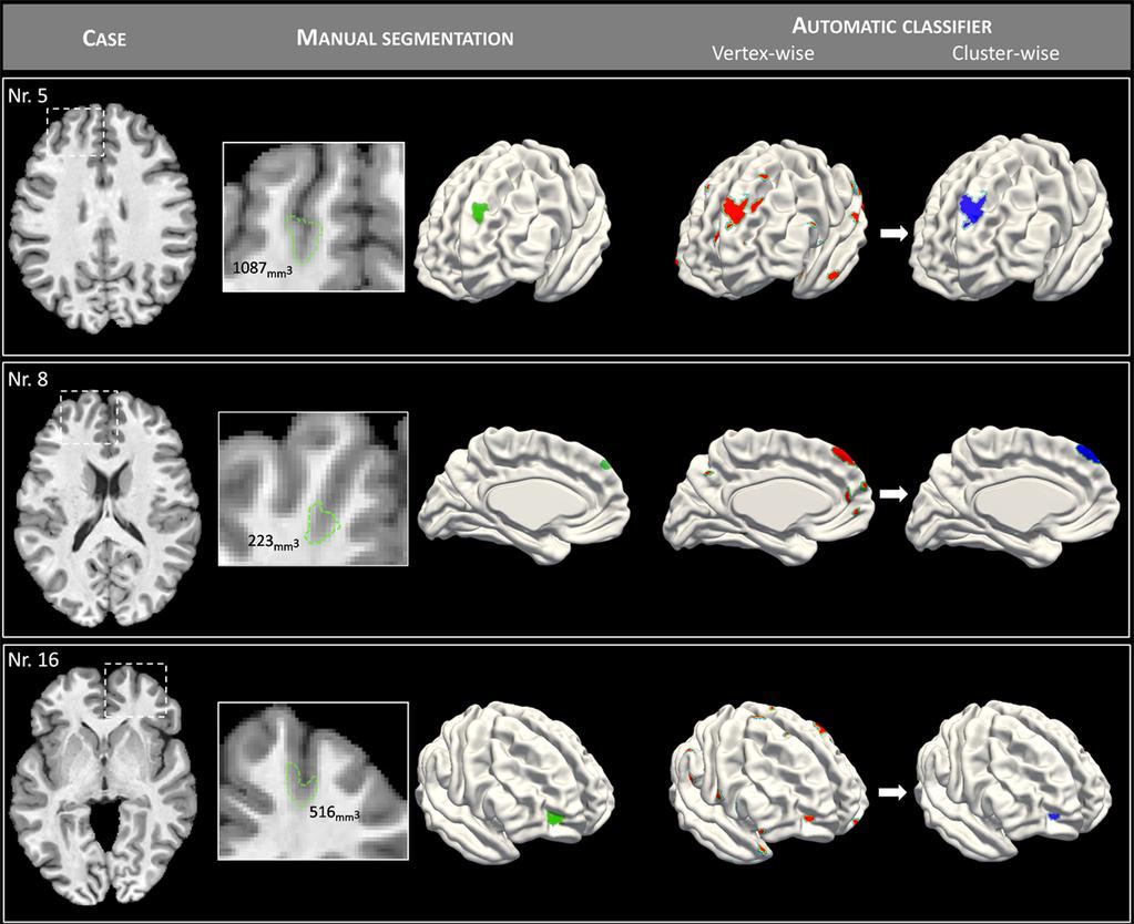
7 STUDY1 MULTIMODAL LESION CHARACTERIZATION
 2. Complement minimally invasive surgical procedures (e.g. thermal laser ablation) 3.")
8 Type-IIA Type-IIB Intracortical dyslamination + Dysmorphic neurons Intracortical dyslamination + Dysmorphic neurons + Balloon cell Identifying subtype-specific imaging signatures may have potential clinical utility 1. Optimize lesion detection (specific to certain histopathological types) 2. Complement minimally invasive surgical procedures (e.g. thermal laser ablation) 3. Guide and monitor pharmacological interventions (mtor signaling inhibitor)
9 In-vivo lesion profiling and subtype prediction in focal cortical dysplasia type-ii Consecutive patients with drug-resistant epilepsy and histologically verified 9 FCD Type-IIA and 24 IIB Multimodal MRIs (3T Siemens TimTrio, 32 channel head coil) - 3D T1w MPRAGE (1x1x1mm 3 ) - 3D FLAIR (0.9x0.9x0.9mm 3 ) - 2D EPI-DTI (2x2x2mm 3, 64 directions) - 2D rs-fmri (4x4x4mm 3, 150 volumes)
10

11 Multi-surface lesion profiling 1. Morphology 2. Intensity IIB: Abnormalities across all cortical and subcortical surfaces IIA: Abnormalities clustered around the GM-WM interface Distance-based lesion profiling Pathological infiltration beyond the visible lesion in both subtypes - Until 6-8 mm from the lesion - Nevertheless, anomalies more marked in Type-IIB - Except the reduced cortical FA specific to IIA Hong SJ, et al. MICCAI 2015, Munich, Germany Hong SJ, et al. Neurology 2016 (in press)
12 Histological subtype prediction Hong SJ, et al. MICCAI 2015, Munich, Germany Hong SJ, et al. Neurology 2016 (in press)
13 Multimodal, multiparametric MRI lesion profiling could dissociate FCD subtypes. Strikingly divergent subtype-specific patterns reflect different loads of underlying histopathology. In-vivo MRI prediction of histology could complement pre-surgical assessment (e.g., lesion detection) and possibly monitor emerging pharmacological interventions.


14 STUDY2 AUTOMATED DETECTION OF SUBTLE LESION
15 PURPOSE To detect automatically FCD type II in patients with extratemporal epilepsy initially diagnosed as MRI-negative on routine inspection, both at 1.5 and 3.0Tesla


16 Relative intensity Cortical thickness Gradient Sulcal depth & curvature
 Objective reduce false positives while maintaining high sensitivity Features")
 spatial priors Classifier Linear discriminant Hong")
17 1 st CLASSIFICATION (vertex-wise) Objective recognize lesional vertices with highest detection rate 2 nd CLASSIFICATION (cluster-wise) Objective reduce false positives while maintaining high sensitivity Features z-scores of GM thickness, GW-WM gradient, intensity, sulcal depth, sulco-gyral curvature Classifier Linear discriminant Features mean, SD skewness and kurtosis (texture) spatial priors Classifier Linear discriminant Hong SJ, et al Neurology, 2014
18 Hong SJ, et al Neurology, 2014 RESULTS 3.0 Tesla 1.5 Tesla 74% sensitivity 100% specificity 71% sensitivity 95% specificity
19
20 Substantially increased sensitivity and specificity Generalizability across different cohorts, scanners and field strengths Machine learning may assist presurgical decision-making

21 Extralesional findings Z-score Less abnormal features than primary FCD Most abnormal feature: sulcal depth No EEG correlates Z-score 50 % patients had 1-3 extra-lesional clusters Localized in frontal or central areas Same lobe as the primary lesion in 2 patients; contralateral hemisphere in 3; bilateral in 2 Almost all previous assessments have focused on primary lesion alone Integrity of whole-brain anomalies have not been systematically evaluated

22 STUDY3 WHOLE-BRAIN MORPHOMETRY
23 PURPOSE 1) To assess whole-brain morphology in patients with dysplasia-related frontal lobe epilepsy 2) To compare the cohor ts between FCD Type-I and II
24 Two frontal lobe epilepsy cohorts with histologically-verified FCD (13 Type-I; 28 Type-II) & closely-matched 41 controls 1.5 T, 3D T1-FFE (isotropic voxel size of 1mm3) Cortical thickness Gyral complexity (mean curvature) Cross-sectional group comparison analysis (patients vs. controls; FCD Type-I vs. Type-II)

25 S3. Group-level comparisons Hong SJ, et al Neurology, 2016 v

26 Hong SJ, et al Neurology, 2016 Subtype prediction Seizure focus lateralization Surgical Outcome prediction v
27 Extensive structural damage beyond the visible lesion Distinctive patterns between Type-I and Type-II. By successfully guiding multiple clinical tasks, our findings demonstrated high translational value for individualized diagnostics
28 P1. In vivo profiling and subtype prediction of FCD Type-II: - Reliable imaging markers to clearly dissociate histopathological subtypes P2. Automated detection of FCD Type II in MRI-negative epilepsy - Highly accurate detection performance across two different datasets P3. Whole-brain MRI phenotyping in dysplasia-related frontal lobe epilepsy - First demonstration that FCD is associated to whole-brain structural alterations. SIGNIFICANCE 1. The power of multimodal MRI and image postprocessing 2. A new avenue to better understand fundamental pathological mechanisms in MCD, and clinically improve lesion detection and treatment strategies.


29 THANKS TO ANDREA AND NEDA BERNASCONI BORIS BERNHARDT MIN LIU BENOIT C ALDAIROU RAVNOOR GILL THANKS AND ANY QUESTIONS?
New Classification of Focal Cortical Dysplasia: Application to Practical Diagnosis
 New Classification of Focal Cortical Dysplasia: Application to Practical Diagnosis Original Article Journal of Epilepsy Research pissn 2233-6249 / eissn 2233-6257 Yoon-Sung Bae, MD 1, Hoon-Chul Kang, MD,
Imaging of Pediatric Epilepsy MRI. Epilepsy: Nonacute Situation
 Imaging of Pediatric Epilepsy Epilepsy: Nonacute Situation MR is the study of choice Tailor MR study to suspected epileptogenic zone Temporal lobe Extratemporal A. James Barkovich, MD University of California
Invasive Evaluation for Epilepsy Surgery Lesional Cases NO DISCLOSURES. Mr. Johnson. Seizures at 29 Years of Age. Dileep Nair, MD Juan Bulacio, MD
 Invasive Evaluation for Epilepsy Surgery Lesional Cases NO DISCLOSURES Dileep Nair, MD Juan Bulacio, MD Mr. Johnson Seizures at 29 Years of Age Onset of seizures at 16 years of age bed wetting episodes
Automated detection of abnormal changes in cortical thickness: A tool to help diagnosis in neocortical focal epilepsy
 Automated detection of abnormal changes in cortical thickness: A tool to help diagnosis in neocortical focal epilepsy 1. Introduction Epilepsy is a common neurological disorder, which affects about 1 %
High Resolution MRI in the evaluation of cerebral focal cortical dysplasias
 High Resolution MRI in the evaluation of cerebral focal cortical dysplasias Poster No.: C-0966 Congress: ECR 2012 Type: Scientific Exhibit Authors: M. Stefanetti, E. Antichi, T. Tartaglione, F. Lanza,
Diffusion Tensor Imaging 12/06/2013
 12/06/2013 Beate Diehl, MD PhD FRCP University College London National Hospital for Neurology and Neurosurgery Queen Square London, UK American Epilepsy Society Annual Meeting Disclosure None Learning
MALFORMATIONS OF CORTICAL DEVELOPMENT: A PICTORIAL REVIEW
 MALFORMATIONS OF CORTICAL DEVELOPMENT: A PICTORIAL REVIEW Padmaja K. Naidu, M.D. Usha D. Nagaraj, M.D. William T. O Brien, Sr., D.O. AOCR Annual Conference 2018 Scottsdale, Arizona @CincyKidsRad facebook.com/cincykidsrad
Epilepsy surgery is an increasingly recognized therapeutic
 J Neurosurg Pediatrics 14:68 80, 2014 AANS, 2014 Magnetic resonance imaging abnormalities in the resection region correlate with histopathological type, gliosis extent, and postoperative outcome in pediatric
Challenges for multivariate and multimodality analyses in "real life" projects: Epilepsy
 Challenges for multivariate and multimodality analyses in "real life" projects: Epilepsy Susanne Mueller M.D. Center for Imaging of Neurodegenerative Diseases Background: Epilepsy What is epilepsy? Recurrent
Neuroimaging in Investigation of Patients With Epilepsy Fernando Cendes, MD, PhD
 Review Article Neuroimaging in Investigation of Patients With Epilepsy Fernando Cendes, MD, PhD ABSTRACT Purpose of Review: This review discusses the MRI and functional imaging findings in patients with
PRESURGICAL EVALUATION. ISLAND OF COS Hippocrates: On the Sacred Disease. Disclosure Research-Educational Grants. Patients with seizure disorders
 PRESURGICAL EVALUATION Patients with seizure disorders Gregory D. Cascino, MD Mayo Clinic Disclosure Research-Educational Grants Mayo Foundation Neuro Pace, Inc. American Epilepsy Society American Academy
Hamartomas and epilepsy: clinical and imaging characteristics
 Seizure 2003; 12: 307 311 doi:10.1016/s1059 1311(02)00272-8 Hamartomas and epilepsy: clinical and imaging characteristics B. DIEHL, R. PRAYSON, I. NAJM & P. RUGGIERI Departments of Neurology, Pathology
High spatial resolution reveals excellent detail in pediatric neuro imaging
 Publication for the Philips MRI Community Issue 46 2012/2 High spatial resolution reveals excellent detail in pediatric neuro imaging Achieva 3.0T with 32-channel SENSE Head coil has become the system
Fig. 1. Localized single voxel proton MR spectroscopy was performed along the long axis of right hippocampus after extension of patient s head to
 125 A B C Fig. 1. Localized single voxel proton MR spectroscopy was performed along the long axis of right hippocampus after extension of patient s head to obtain entire dimension of the hippocampal body.
Cortical malformations and epilepsy: role of MR imaging
 Cortical malformations and epilepsy: role of MR imaging Poster No.: C-0921 Congress: ECR 2013 Type: Scientific Exhibit Authors: S. Jerbi, O. Bradai, S. Haj Slimene, Y. Abdelhafidh, H. HAMZA; Mahdia/TN
Advances in Clinical Neuroimaging
 Advances in Clinical Neuroimaging Joseph I. Tracy 1, PhD, ABPP/CN; Gaelle Doucet 2, PhD; Xaiosong He 2, PhD; Dorian Pustina 2, PhD; Karol Osipowicz 2, PhD 1 Department of Radiology, Thomas Jefferson University,
Pharmacoresistant temporal lobe epilepsy - a diagnostic performance of standardized MRI protocol in detection of epileptogenic lesion
 Pharmacoresistant temporal lobe epilepsy - a diagnostic performance of standardized MRI protocol in detection of epileptogenic lesion Poster No.: C-2226 Congress: ECR 2013 Type: Scientific Exhibit Authors:
Blurring the Lines Between Lesional and Nonlesional MRI
 Current Literature In Clinical Science Blurring the Lines Between Lesional and Nonlesional MRI Blurring in Patients With Temporal Lobe Epilepsy: Clinical, High-field Imaging and Ultrastructural Study.
Hemimegalencephaly without seizures: report of a case and review of literature
 Romanian Neurosurgery Volume XXXI Number 3 2017 July-September Article Hemimegalencephaly without seizures: report of a case and review of literature Agrawal Atul, Dutta Gautam, Singh Daljit, Sachdeva
doi: /brain/awr204 Brain 2011: 134; Morphometric MRI analysis improves detection of focal cortical dysplasia type II
 doi:10.1093/brain/awr204 Brain 2011: 134; 2844 2854 2844 BRAIN A JOURNAL OF NEUROLOGY Morphometric MRI analysis improves detection of focal cortical dysplasia type II Jan Wagner, 1,2 Bernd Weber, 1,2,3
Establishing a novel diagnostic method to localize the epileptogenic zone in cryptogenic focal epilepsy
 Establishing a novel diagnostic method to localize the epileptogenic zone in cryptogenic focal epilepsy Joanna Goc Munich, 4 th November 2016 Dissertation der Graduate School of Systemic Neurosciences
Optimizing MR Imaging Detection of Type 2 Focal Cortical Dysplasia: Best Criteria for Clinical Practice
 Published May 3, 2012 as 10.3174/ajnr.A3081 ORIGINAL RESEARCH C. Mellerio M.-A. Labeyrie F. Chassoux C. Daumas-Duport E. Landre B. Turak F.-X. Roux J.-F. Meder B. Devaux C. Oppenheim Optimizing MR Imaging
Structural MRI analysis
 Structural MRI analysis Boris Bernhardt, PhD NeuroImaging of Epilepsy Lab boris@bic.mni.mcgil.ca structural MRI T1-weighted MRI methods MRI volumetry Surface-based analysis Covariance mapping applications
EPILEPSY. New Ideas about an Old Disease. Gregory D. Cascino, MD
 EPILEPSY New Ideas about an Old Disease Gregory D. Cascino, MD Disclosure Research-Educational Grants Neuro Pace, Inc. American Epilepsy Society American Academy of Neurology Neurology (Associate Editor)
Detection of Epileptogenic Cortical Malformations with Surface-Based MRI Morphometry
 Detection of Epileptogenic Cortical Malformations with Surface-Based MRI Morphometry Thomas Thesen 1,2 *, Brian T. Quinn 1,3, Chad Carlson 1, Orrin Devinsky 1, Jonathan DuBois 1, Carrie R. McDonald 2,
Malformations of cortical development in children: clinical manifestation, neuroimaging and neuropathology in selected cases
 Original article Malformations of cortical development in children: clinical manifestation, neuroimaging and neuropathology in selected cases Ewa Emich-Widera 1, Dawid Larysz 2, Ewa Kluczewska 3, Patrycja
Epilepsy Surgery, Imaging, and Intraoperative Neuromonitoring: Surgical Perspective
 Epilepsy Surgery, Imaging, and Intraoperative Neuromonitoring: Surgical Perspective AC Duhaime, M.D. Director, Pediatric Neurosurgery, Massachusetts General Hospital Professor, Neurosurgery, Harvard Medical
Est-ce que l'eeg a toujours sa place en 2019?
 Est-ce que l'eeg a toujours sa place en 2019? Thomas Bast Epilepsy Center Kork, Germany Does EEG still play a role in 2019? What a question 7T-MRI, fmri, DTI, MEG, SISCOM, Of ieeg course! /HFO, Genetics
Toward a more accurate delimitation of the epileptic focus from a surgical perspective
 Toward a more accurate delimitation of the epileptic focus from a surgical perspective Margitta Seeck Department of Clinical Neurosciences EEG & Epilepsy Unit University Hospital of Geneva Geneva, Switzerland
Malformations of cortical development. Clinical and imaging findings.
 Malformations of cortical development. Clinical and imaging findings. Poster No.: C-2086 Congress: ECR 2012 Type: Educational Exhibit Authors: I. Alba de Caceres, B. García-Castaño, L. Ibañez, E. Roa,
Refractory focal epilepsy: findings by MRI.
 Refractory focal epilepsy: findings by MRI. Doctors Nicolás Sgarbi, Osmar Telis Clinical Radiology Department Hospital de Clínicas Montevideo- Uruguay ABSTRACT Epilepsy is one of the most frequent neurological
Malformations of cortical development. Clinical and imaging findings.
 Malformations of cortical development. Clinical and imaging findings. Poster No.: C-2086 Congress: ECR 2012 Type: Educational Exhibit Authors: I. Alba de Caceres, B. García-Castaño, L. Ibañez, E. Roa 1
mr brain volume analysis using brain assist
 mr brain volume analysis using brain assist This Paper describes the tool named BrainAssist, which can be used for the study and analysis of brain abnormalities like Focal Cortical Dysplasia (FCD), Heterotopia
Is DTI Increasing the Connectivity Between the Magnet Suite and the Clinic?
 Current Literature In Clinical Science Is DTI Increasing the Connectivity Between the Magnet Suite and the Clinic? Spatial Patterns of Water Diffusion Along White Matter Tracts in Temporal Lobe Epilepsy.
Utility of double inversion recovery MRI in paediatric epilepsy
 Utility of double inversion recovery MRI in paediatric epilepsy Bruno Soares, Emory University Samuel G Porter, Emory University Amit Saindane, Emory University Seena Dehkharghani, Emory University Nilesh
Diagnosing Complicated Epilepsy: Mapping of the Epileptic Circuitry. Michael R. Sperling, M.D. Thomas Jefferson University Philadelphia, PA
 Diagnosing Complicated Epilepsy: Mapping of the Epileptic Circuitry Michael R. Sperling, M.D. Thomas Jefferson University Philadelphia, PA Overview Definition of epileptic circuitry Methods of mapping
Approximately 30% 40% of those with focal seizures are
 ORIGINAL RESEARCH E. Widjaja S. Geibprasert H. Otsubo O.C. Snead III S.Z. Mahmoodabadi Diffusion Tensor Imaging Assessment of the Epileptogenic Zone in Children with Localization- Related Epilepsy BACKGROUND
Multimodal Imaging in Extratemporal Epilepsy Surgery
 Open Access Case Report DOI: 10.7759/cureus.2338 Multimodal Imaging in Extratemporal Epilepsy Surgery Christian Vollmar 1, Aurelia Peraud 2, Soheyl Noachtar 1 1. Epilepsy Center, Dept. of Neurology, University
Electrocorticographic Discharge Patterns in Glioneuronal Tumours and Focal Cortical Dysplasia
 Electrocorticographic Discharge Patterns in Glioneuronal Tumours and Focal Cortical Dysplasia C.H. Ferrier E. Aronica F.S.S. Leijten W.G.M. Spliet A.C. van Huffelen P.C. van Rijen C.D. Binnie Epilepsia
Functional Network Analysis in Epileptic Children Using Multimodal Imaging, SEEG, and Surgical Pathology
 Functional Network Analysis in Epileptic Children Using Multimodal Imaging, SEEG, and Surgical Pathology Roy Dudley MD, PhD Department of Pediatric Surgery Division of Neurosurgery Montreal Children s
Spike voltage topography in temporal lobe epilepsy
 Thomas Jefferson University Jefferson Digital Commons Department of Neurology Faculty Papers Department of Neurology 5-17-2016 Spike voltage topography in temporal lobe epilepsy Ali Akbar Asadi-Pooya Thomas
Advanced Imaging Techniques MRI, PET, SPECT, ESI-MSI, DTI December 8, 2013
 Advanced Imaging Techniques MRI, PET, SPECT, ESI-MSI, DTI December 8, 2013 Robert C. Knowlton, MD, MSPH University of California San Francisco Seizure Disorders Surgical Program American Epilepsy Society
Fetal CNS MRI. Daniela Prayer. Division of Neuroradiology And Musculoskeletal Radiology. Medical University of Vienna, AUSTRIA
 Fetal CNS MRI Daniela Prayer Division of Neuroradiology And Musculoskeletal Radiology Medical University of Vienna, AUSTRIA Methods Normal development Malformations Acquired pathology MR- methods for assessment
Imaging of malformations of cortical development
 Review article Epileptic Disord 2009; 11 (3): 194-205 Imaging of malformations of cortical development Nadia Colombo 1, Noriko Salamon 2, Charles Raybaud 3, Çigdem Özkara 4, A. James Barkovich 5 1 Department
Frontal Contributions to Memory Encoding Before and After Unilateral Medial Temporal Lobectomy
 Frontal Contributions to Memory Encoding Before and After Unilateral Medial Temporal Lobectomy Jeff Ojemann, MD Department of Neurological Surgery University of Washington Children s Hospital & Regional
Advanced magnetic resonance imaging for monitoring brain development and injury
 Advanced magnetic resonance imaging for monitoring brain development and injury Stéphane Sizonenko, MD-PhD Division of Development and Growth Department of Child and Adolescent Medicine Geneva University
Human Cortical Dysplasia and Epilepsy: An Ontogenetic Hypothesis Based on Volumetric MRI and NeuN Neuronal Density and Size Measurements
 Cerebral Cortex February 2005;15:194-210 doi:10.1093/cercor/bhh122 Advance Access publication August 5, 2004 Human Cortical Dysplasia and Epilepsy: An Ontogenetic Hypothesis Based on Volumetric MRI and
Combining tdcs and fmri. OHMB Teaching Course, Hamburg June 8, Andrea Antal
 Andrea Antal Department of Clinical Neurophysiology Georg-August University Goettingen Combining tdcs and fmri OHMB Teaching Course, Hamburg June 8, 2014 Classical Biomarkers for measuring human neuroplasticity
Epilepsy in children: Review of the main causes detectable by MRI
 Epilepsy in children: Review of the main causes detectable by MRI Poster No.: C-2182 Congress: ECR 2014 Type: Educational Exhibit Authors: A. A. S. M. D. Santos, T. C. R. S. SANTOS, A. Monteiro ; 1 1 1
Interictal High Frequency Oscillations as Neurophysiologic Biomarkers of Epileptogenicity
 Interictal High Frequency Oscillations as Neurophysiologic Biomarkers of Epileptogenicity December 10, 2013 Joyce Y. Wu, MD Associate Professor Division of Pediatric Neurology David Geffen School of Medicine
No relevant disclosures
 No relevant disclosures - Epileptic Encephalopathy (EE): Epileptic activity itself contributes to cognitive and behavioural impairments - Developmental and Epileptic Encephalopathy (DEE): Impairments occur
Bottom-of-Sulcus Dysplasia: Imaging Features
 Neuroradiology/Head and Neck Imaging Original Research Hofman et al. ottom-of-sulcus Dysplasia Neuroradiology/Head and Neck Imaging Original Research Paul. M. Hofman 1,2,3 Gregory J. Fitt 4. Simon Harvey
J Neurol Neurosurg Psychiatry 2005; 76(Suppl III):iii2 iii10. doi: /jnnp RAY COMPUTED TOMOGRAPHY
:iii2 iii10. doi: /jnnp RAY COMPUTED TOMOGRAPHY") iii2 See end of article for authors affiliations Correspondence to: Professor John S Duncan, Department of Clinical and Experimental Epilepsy, Institute of Neurology, Queen Square, University College London,
iii2 See end of article for authors affiliations Correspondence to: Professor John S Duncan, Department of Clinical and Experimental Epilepsy, Institute of Neurology, Queen Square, University College London,
Depth/surface relationships: Confronting noninvasive measures to intracerebral EEG
 Depth/surface relationships: Confronting noninvasive measures to intracerebral EEG Christian G Bénar Institut de Neurosciences des Systèmes; INSERM, Aix-Marseille Université christian.benar@univ-amu.fr
9/30/2016. Advances in Epilepsy Surgery. Epidemiology. Epidemiology
 Advances in Epilepsy Surgery George Jallo, M.D. Director, Institute for Brain Protection Sciences Johns Hopkins All Children s Hospital St Petersburg, Florida Epidemiology WHO lists it as the second most
Spike frequency is dependent on epilepsy duration and seizure frequency in temporal lobe epilepsy
 Original article Epileptic Disord 2005; 7 (4): 355-9 Spike frequency is dependent on epilepsy duration and seizure frequency in temporal lobe epilepsy Jozsef Janszky 1,2,3, M. Hoppe 1, Z. Clemens 3, I.
Do seizures beget seizures?
 Does MTLE cause progressive neurocognitive damage? Andrew Bleasel Westmead Do seizures beget seizures? The tendency of the disease is toward self-perpetuation; each attack facilitates occurrence of another
Advanced MR Imaging of Cortical Dysplasia with or without Neoplasm: A Report of Two Cases
 AJNR Am J Neuroradiol 23:1686 1691, November/December 2002 Case Report Advanced MR Imaging of Cortical Dysplasia with or without Neoplasm: A Report of Two Cases Jay J. Pillai, Richard B. Hessler, Jerry
Epilepsy & Behavior Case Reports
 Epilepsy & Behavior Case Reports 1 (2013) 45 49 Contents lists available at ScienceDirect Epilepsy & Behavior Case Reports journal homepage: www.elsevier.com/locate/ebcr Case Report Partial disconnection
BESA Research Quick Guide
 BESA Research Quick Guide BESA 3D Maps Quick Guide An introduction how to interpret 3D voltage and phase maps in the scalp EEG Copyright and Trademarks The BESA products and their documentation are copyrighted
Depth/Surface Relationships: Confronting noninvasive measures to intracerebral EEG
 Depth/Surface Relationships: Confronting noninvasive measures to intracerebral EEG Christian G Bénar Institut de Neurosciences des Systèmes; INSERM, Aix-Marseille Université christian.benar@univ-amu.fr
MRI morphometry of the neocortex in patients with periventricular nodular heterotopia
 MRI morphometry of the neocortex in patients with periventricular nodular heterotopia Anton Plavski Integrated Program in Neuroscience McGill University, Montreal, Canada June 2016 A thesis submitted to
Functional Magnetic Resonance Imaging of the Brain
 Page: 1 of 9 Last Review Status/Date: December 2016 Description Functional magnetic resonance imaging (fmri) is a noninvasive method for localizing areas of brain function and has been used for the presurgical
Cover Page. The handle holds various files of this Leiden University dissertation
 Cover Page The handle http://hdl.handle.net/1887/26921 holds various files of this Leiden University dissertation Author: Doan, Nhat Trung Title: Quantitative analysis of human brain MR images at ultrahigh
Imaging of intractable paediatric epilepsy
 Page 1 of 10 Imaging of intractable paediatric epilepsy Authors: Sanjay Prabhu 1 Nasreen Mahomed 2 Affiliations: 1 Department of Radiology, Boston Children s Hospital, Harvard Medical School, United States
MSc Neuroimaging for Clinical & Cognitive Neuroscience
 MSc Neuroimaging for Clinical & Cognitive Neuroscience School of Psychological Sciences Faculty of Medical & Human Sciences Module Information *Please note that this is a sample guide to modules. The exact
Case reports functional imaging in epilepsy
 Seizure 2001; 10: 157 161 doi:10.1053/seiz.2001.0552, available online at http://www.idealibrary.com on Case reports functional imaging in epilepsy MARK P. RICHARDSON Medical Research Council Fellow, Institute
EPILEPSY 2018: UPDATE ON MODERN SURGICAL MANAGEMENT. Robert Kellogg, MD Advocate Children s Hospital Park Ridge, IL April 20, 2018
 EPILEPSY 2018: UPDATE ON MODERN SURGICAL MANAGEMENT Robert Kellogg, MD Advocate Children s Hospital Park Ridge, IL April 20, 2018 No disclosures OBJECTIVES Brief history of epilepsy surgery Pre-operative
SEIZURE OUTCOME AFTER EPILEPSY SURGERY
 SEIZURE OUTCOME AFTER EPILEPSY SURGERY Prakash Kotagal, M.D. Head, Pediatric Epilepsy Cleveland Clinic Epilepsy Center LEFT TEMPORAL LOBE ASTROCYTOMA SEIZURE OUTCOME 1 YEAR AFTER EPILEPSY SURGERY IN ADULTS
Challenges and opportunities for understanding epilepsy and cognitive and behavioral comorbidities. Eleonora Aronica
 Biobanks and Databases -Basis for Translational Research Challenges and opportunities for understanding epilepsy and cognitive and behavioral comorbidities Eleonora Aronica Department of (Neuro)Pathology,
1ST INTERNATIONAL TRAINING COURSE ON NEUROIMAGING OF EPILEPSY
 1ST INTERNATIONAL TRAINING COURSE ON NEUROIMAGING OF EPILEPSY DATE MAY 18 21, 2017 LOCATION MONTREAL COURSE DIRECTOR ANDREA BERNASCONI, MD FACULTY A. Bernasconi, MD - Montreal, Canada N. Bernasconi, MD
OBSERVATION. Identifying Subtle Cortical Gyral Abnormalities as a Predictor of Focal Cortical Dysplasia and a Cure for Epilepsy
 OSERVATION Identifying Subtle Cortical Gyral Abnormalities as a Predictor of Focal Cortical Dysplasia and a Cure for Epilepsy Joel M. Oster, MD; Eme Igbokwe, MD; G. Rees Cosgrove, MD, FRCS(C); Andrew J.
Focal epilepsy recruiting a generalised network of juvenile myoclonic epilepsy: a case report
 Clinical commentary Epileptic Disord 2014; 16 (3): 370-4 Focal epilepsy recruiting a generalised network of juvenile myoclonic epilepsy: a case report Myo Khaing 1,2, Kheng-Seang Lim 1, Chong-Tin Tan 1
PET and SPECT in Epilepsy
 PET and SPECT in Epilepsy 12.6.2013 William H Theodore MD Chief, Clinical Epilepsy Section NINDS NIH Bethesda MD American Epilepsy Society Annual Meeting Disclosures Entity DIR NINDS NIH Elsevier Individual
High Resolution Ictal SPECT: Enhanced Epileptic Source Targeting?
 High Resolution Ictal SPECT: Enhanced Epileptic Source Targeting? Marvin A Rossi MD, PhD RUSH Epilepsy Center Research Lab http://www.synapticom.net Chicago, IL USA Medically-Refractory Epilepsy 500,000-800,000
Cerebral Malformation gene panel
 Cerebral Malformation gene panel Dr John Livingston Consultant Paediatric Neurologist Leeds Teaching Hospitals NHS Trust on behalf of Yorkshire Regional Genetics Service Leeds UK Cerebral Malformation
Imaging and EEG in Post-traumatic Epilepsy
 Imaging and EEG in Post-traumatic Epilepsy Michael R. Sperling, M.D. Thomas Jefferson University Philadelphia, PA American Epilepsy Society Annual Meeting Disclosure Name Upsher-Smith Sunovion, Eisai,
Imaging for Epilepsy Diagnosis December 2, 2011
 Imaging for Epilepsy Diagnosis December 2, 2011 Samuel Wiebe, MD University of Calgary Canada American Epilepsy Society Annual Meeting Disclosure University of Calgary Hopewell Professorship of Clinical
Physiological Markers of Pharmacoresistant Epilepsy December 2, 2011
 Physiological Markers of Pharmacoresistant Epilepsy December 2, 2011 Jerome Engel, Jr., MD, PhD Director of the Seizure Disorder Center The Jonathan Sinay Distinguished Professor of Neurology, Neurobiology,
Acute Management of Seizures
 Acute Management of Seizures KURT HECOX M.D. PH.D. CHIEF OF PEDIATRIC NEUROLOGY BAUMAN ENDOWED CHAIR IN PEDIATRIC EPILEPSY Outline Management Principles Categorizing the event Key elements to the history
Comparative Analysis of MR Imaging, Positron Emission Tomography, and Ictal Single-photon Emission CT in Patients with Neocortical Epilepsy
 AJNR Am J Neuroradiol 22:937 946, May 2001 Comparative Analysis of MR Imaging, Positron Emission Tomography, and Ictal Single-photon Emission CT in Patients with Neocortical Epilepsy Sung-Il Hwang, Jae
Analysis between clinical and MRI findings of childhood and teenages with epilepsy after hypoxic-ischemic encephalopathy in neonates periods
 Analysis between clinical and MRI findings of childhood and teenages with epilepsy after hypoxic-ischemic encephalopathy in neonates periods Poster No.: C-0401 Congress: ECR 2015 Type: Scientific Exhibit
KEY WORDS neuronavigation; MRI postprocessing; cryptogenic epilepsy; surgery Epilepsy is one of the most common neurological disorders,
 CLINICAL ARTICLE J Neurosurg 128:1178 1186, 2018 A multimodal concept for invasive diagnostics and surgery based on neuronavigated voxel-based morphometric MRI postprocessing data in previously nonlesional
Introduction to Brain Imaging
 Introduction to Brain Imaging Human Brain Imaging NEUR 570 & BIC lecture series September 9, 2013 Petra Schweinhardt, MD PhD Montreal Neurological Institute McGill University Montreal, Canada Various techniques
Morphometric MRI Analysis of the Parahippocampal Region in Temporal Lobe Epilepsy
 Morphometric MRI Analysis of the Parahippocampal Region in Temporal Lobe Epilepsy NEDA BERNASCONI, a ANDREA BERNASCONI, ZOGRAFOS CARAMANOS, FREDERICK ANDERMANN, FRANÇOIS DUBEAU, AND DOUGLAS L. ARNOLD Department
33rd International Epilepsy Congress 2019 Sunday
 Saturday 22 June 33rd International Epilepsy Congress 2019 Sunday Monday 23 June 24 June Tuesday 25 June Wednesday 26 June 08.00-08.30 08.30-09.00 09.00-09.30 09.30-10.00 10.00-10.30 10.30-11.00 11.00-11.30
Clinically focused workflow with unique ability to integrate fmri, DTI, fiber tracks and perfusion in a single, multi-layered 3D rendering
 Clinically focused workflow with unique ability to integrate fmri, DTI, fiber tracks and perfusion in a single, multi-layered 3D rendering Neurosurgeons are demanding more from neuroradiologists and increasingly
Early-onset symptomatic focal epilepsy: a dilemma in the timing of surgery
 Anatomo-electro-clinical correlations Epileptic Disord 2008; 10 (4): 356-61 Anatomo-electro-clinical correlations: the Great Ormond Street Hospital, UK Case Report - Case 05-2008 Early-onset symptomatic
Imaging in Epilepsy. Nucharin Supakul, MD Ramathibodi Hospital, Mahidol University August 22, 2015
 Imaging in Epilepsy Nucharin Supakul, MD Ramathibodi Hospital, Mahidol University August 22, 2015 Nothing to disclose Outline Role of Imaging and pitfalls Imaging protocol Case scenarios Clinical & Electrophysiologic
Seizure 18 (2009) Contents lists available at ScienceDirect. Seizure. journal homepage:
 Contents lists available at ScienceDirect. Seizure. journal homepage:") Seizure 18 (2009) 288 292 Contents lists available at ScienceDirect Seizure journal homepage: www.elsevier.com/locate/yseiz Posterior cortex epilepsy: Diagnostic considerations and surgical outcome Tao
Seizure 18 (2009) 288 292 Contents lists available at ScienceDirect Seizure journal homepage: www.elsevier.com/locate/yseiz Posterior cortex epilepsy: Diagnostic considerations and surgical outcome Tao
The neurolinguistic toolbox Jonathan R. Brennan. Introduction to Neurolinguistics, LSA2017 1
 The neurolinguistic toolbox Jonathan R. Brennan Introduction to Neurolinguistics, LSA2017 1 Psycholinguistics / Neurolinguistics Happy Hour!!! Tuesdays 7/11, 7/18, 7/25 5:30-6:30 PM @ the Boone Center
Type I focal cortical dysplasia: surgical outcome is related to histopathology
 Original article Epileptic Disord 2010; 12 (3): 181-91 Type I focal cortical dysplasia: surgical outcome is related to histopathology Laura Tassi 1, Rita Garbelli 2, Nadia Colombo 3, Manuela Bramerio 4,
Adult-Onset Neurologic Dysfunction Associated with Cortical Malformations
 AJNR Am J Neuroradiol 20:1037 1043, June/July 1999 Adult-Onset Neurologic Dysfunction Associated with Cortical Malformations Woo Ho Cho, David Seidenwurm, and A. James Barkovich BACKGROUND AND PURPOSE:
A micropower support vector machine based seizure detection architecture for embedded medical devices
 A micropower support vector machine based seizure detection architecture for embedded medical devices The MIT Faculty has made this article openly available. Please share how this access benefits you.
Approximately 70% of childhood SURGICAL TREATMENTS FOR PEDIATRIC EPILEPSY PROCEEDINGS. Ronald P. Lesser, MD KEY POINTS
 ASIM May p153-158 5/14/01 9:19 AM Page 153 SURGICAL TREATMENTS FOR PEDIATRIC EPILEPSY Ronald P. Lesser, MD KEY POINTS Most children with epilepsy refractory to drugs can improve with surgery Temporal lobe
FUNCTIONAL MRI IN EPILEPSY December 6 th 2013
 FUNCTIONAL MRI IN EPILEPSY December 6 th 2013 Matthias J Koepp, MD, PhD UCL Institute of Neurology National Hospital for Neurology and Neurosurgery London, UK American Epilepsy Society Annual Meeting Disclosure
Neurodevelopment II Structure Formation. Reading: BCP Chapter 23
 Neurodevelopment II Structure Formation Reading: BCP Chapter 23 Phases of Development Ovum + Sperm = Zygote Cell division (multiplication) Neurogenesis Induction of the neural plate Neural proliferation
P. Hitchcock, Ph.D. Department of Cell and Developmental Biology Kellogg Eye Center. Wednesday, 16 March 2009, 1:00p.m. 2:00p.m.
 Normal CNS, Special Senses, Head and Neck TOPIC: CEREBRAL HEMISPHERES FACULTY: LECTURE: READING: P. Hitchcock, Ph.D. Department of Cell and Developmental Biology Kellogg Eye Center Wednesday, 16 March
Amyotrophic lateral sclerosis (ALS) is a progressive neurodegenerative
 is a progressive neurodegenerative") ORIGINAL RESEARCH E. Matsusue S. Sugihara S. Fujii T. Kinoshita T. Nakano E. Ohama T. Ogawa Cerebral Cortical and White Matter Lesions in Amyotrophic Lateral Sclerosis with Dementia: Correlation with MR
ORIGINAL RESEARCH E. Matsusue S. Sugihara S. Fujii T. Kinoshita T. Nakano E. Ohama T. Ogawa Cerebral Cortical and White Matter Lesions in Amyotrophic Lateral Sclerosis with Dementia: Correlation with MR
AdvAnced TMS. Research with PowerMAG Products and Application Booklet
 AdvAnced TMS Research with PowerMAG Products and Application Booklet Table of ConTenTs Introduction p. 04 Legend p. 06 Applications» navigated TMS p. 08» clinical Research p. 10» Multi-Modal TMS p. 12»