Diagnostic Immunohistochemistry of Canine Round Cell Tumors
|
|
|
- Annabel Tyler
- 6 years ago
- Views:
Transcription

1 Vet. Pathol (1987) Diagnostic Immunohistochemistry of Canine Round Cell Tumors G. E. SANDUSKY, W. W. CAIUTON, AND K. A. WIGHTMAN Lilly Research Laboratories, A Division of Eli Lilly and Company, Greenfield, IN, and School of Veterinary Science and Medicine, Purdue University, West Lafayette, IN Abstract. Sixty-five canine skin neoplasms studied using immunocytochemistry, included 22 histiocytomas, 18 amelanotic melanomas, 14 cutaneous lymphosarcomas, six mast cell tumors, and five transmissible venereal tumors. Formalin-fixed, paraffin-embedded sections were stained using the avidin-biotin-peroxidase complex (ABC) immunoperoxidase technique for reactivity with S- 100 protein, kappa and lambda immunoglobulin light chains, alpha- 1 -antitrypsin, alpha- 1 -antichymotrypsin, leukocyte common antigen (LCA), neuron-specific enolase, keratin, cytokeratin, muramidase, and vimentin. Detection of S- 100, kappa and lambda light chains, neuron-specific enolase, and vimentin were most useful for screening these neoplasms. None of the markers examined was consistent in staining histiocytomas. While reactivity of S- 100 (ten cases) and neuron-specific enolase (ten cases) was detected in some amelanotic melanomas, lambda light chain immunoglobulin (eight cases) was relatively consistent in cutaneous lymphomas. Mast cell neoplasms reacted with avidin and, therefore, were positive, even on negative control sections. Vimentin reacted strongly on all amelanotic melanomas and transmissible venereal tumors examined. These antibodies are helpful adjuncts in the differential diagnosis of canine skin tumors. Undifferentiated tumors of the skin and underlying subcutaneous tissue present a diagnostic challenge to the surgical pathologist. Both spindle cell tumors and small round cell tumors often pose problems of differential diagnosis when examined by light microscopy. However, immunohistochemistry has provided a technique with which polyclonal and/or monoclonal antibodies can be used to gain more diagnostic information about a variety of poorly differentiated neopla~ms.~~~~~,*j~j* During the past few years, a number of tumor markers for soft tissue neoplasms have been d e ~ ~ r i b e d. ~ Due ~ ~ ~ to ~ cross ~ ~ reactivity ~ ~ ~, ~ of ~ absolute, ~ ~ ~ methanol. ~, ~ ~ The, slides ~ ~ were covered with normal goat some antibodies and lack of staining of some tumors, a panel of selected commercially available antibodies was used, rather than reliance on one antibody, to identify common, undifferentiated skin tumors. Materials and Methods The neoplasms studied were obtained from the pathology files of Purdue University s oncology program. Tissues were fixed in 10% neutral buffered formalin, embedded in paraffin, sectioned at 4 pm, and stained with hematoxylin and eosin (HE). Two additional sections from each case were placed on one slide. One section was used as a negative control and the other was stained for immunoreactivity to the various tumor markers. Each tissue specimen was circled with a diamond pencil to prevent cross-contamination of antisera. Unstained sections of the tumors were deparaffinized and stained using the avidin-biotin complex method described by Hsu.I0 Table 1 lists the antibodies used in this study, their specificity for cells present in the skin and subcutaneous tissue, and the specific dilutions. The antigen against cytokeratin and vimentin was from Biogenex Corporation, Dublin, California. The rest of the primary antisera were from Dako Corporation, Santa Barbara, California. The biotinylated secondary antibody and ABC complex were from Vector Corporation, Burlingame, California. Polyclonal Antibody Technique Endogenous peroxidase activity was blocked by immersing the slides for 10 minutes in a 6% solution of H,O, made in serum and diluted 1 : 20 in modified phosphate buffered saline (PBS) for 60 minutes before incubation in the primary antibody. The incubation period for the various primary antisera was 1 hour, except for the cytokeratin, which was incubated overnight at 4 C, and vimentin, which was incubated for 2 hours. The biotinylated goat anti-rabbit secondary antisera and avidin-biotin-peroxidase complex were incubated for 30 minutes each at room temperature. Slides were washed for 10 minutes between each step in a modified PBS buffer. The chromagen, 3-amino-9-ethylcarbazole, was applied to the slides for 20 minutes at 37 C. The slides were then counterstained with Mayer s hematoxylin. Monoclonal Antibody Technique The technique used was identical to the above procedure except for using normal horse serum and biotinylated horse antimouse secondary antisera instead of rabbit reagents. Negative controls included substituting the primary anti- 495

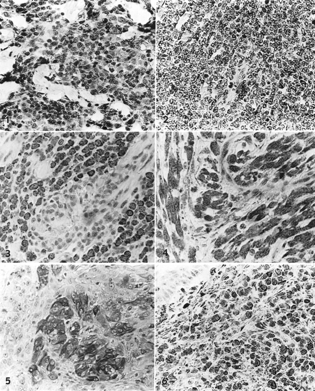
2 496 Sandusky, Carltc m, and Wightman Table 1. Antibodies and cell types evaluated in canine cutaneous neoplasms. Table 3. Results of immunocytochemical determinations on round cell tumors in the dog. Antibody Antibody Specificity Cellular Specificity Dilution S- 100 protein Melanocytes 1 : 100 Muramidase Histiocytes 1 : 100 Alpha- 1 -antitrypsin Histiocytes 1 : 100 Alpha- 1 -antichymo- Histiocytes 1 : 100 trypsin Kappa light chain B-lymphocytes 1 : 1,000 plasma cells Lambda light chain B-lymphocytes 1 : 1,000 plasma cells Keratin Epithelial cells 1 : 100 Cytokeratin (M)* Epithelial cells Prediluted Leukocyte common an- Leukocytes 1:20 tigen (M) Neuron-specific enolase Neuroendocrine 1 : 500 cells Vimentin (M) Mesenchymal cells 1 : 500 * M-monoclonal antibody. sera with nonimmune sera from the same species and, in addition, normal saline. Known positive controls were included for each marker. A normal tissue screen of 20 tissues was examined with each antibody to determine the specificity of the antibodies. All antibodies stained cellular structures in the dog, which were analogous to those stained in human tissues. Specimens in which 1% or less of the cells were stained were considered negative. Predigestion with 0.1% trypsin in 0.1% CaC1, solution was performed for 30 minutes on all tumors that failed to react with any primary antibodies tested. Results By the proper selection of four or five antibodies listed in Table 1, the basic cell type involved in the skin neoplasms examined was identified. False negative staining reactions seen require the use of more than one antibody for a positive diagnosis. In addition, the use of four or five tumor markers as a panel did exclude other tumors considered in differential diagnosis. Upon examination of the neoplasms by light microscopy, most were composed of spindle, round, or polygonal cells. All histiocytomas were easily distin- Table 2. Immunocytochemical results of staining of histiocytomas using macrophage markers. Antibody Specificity Histiocytomas ~ Ly soz y me 4/22* Alpha- 1 -antitrypsin 7/22 Alpha- 1 -antichymotrypsin 0/22 * Number positivehumber examined. Transmissi- Antibody Histiocytoma Lym- ble phoma Melanoma Vene- real Tumor s-100 0/22* 0/14 10/18 0/5 Kappa 0/22 3/14 0/18 0/5 Lambda 0/22 8/14 0/18 0/5 Cytokeratin NDt 0/14 0/18 0/5 Vimentin ND ND 18/18 5/5 Neuron-specific enolase O/ 18 ND 1 O/ 18 0/5 * Number positivehumber examined. t ND = not done. guished, based on routine histiologic examination. In the rest of the tumors studied, differential diagnoses included carcinomas, lymphomas, malignant melanomas, and mesenchymal tumors of fibroblast, smooth muscle, skeletal muscle, or neural origin. In a few of these cases, it was difficult to make a definitive diagnosis based on routine light microscopic examination. Histiocytomas were studied for the presence of muramidase (lysozome), alpha- 1 -antitrypsin, alpha- 1 -antichymotrypsin, kappa and lambda light chains, and S-100 protein (Tables 2, 3). No markers used in this panel were consistently positive for histiocytomas. Antibodies to S-100 protein, kappa and lambda light chains, and alpha- 1 -antichymotrypsin failed to react with any of the histiocytomas examined. Immunoreactivity of alpha- 1 -antitrypsin was strong in seven of 22 histiocytomas (Fig. 1) and failed to react in the remaining 16 tumors. Muramidase reacted moderately with four tumors in which the staining was intense in individual tumor cells; scattered positive histiocytes were positive in three additional tumors (Fig. 2). Diagnosis of a B-cell lymphoma was established on the basis of staining of neoplastic cells for kappa or lambda light chains. No immunoreactivity was seen in tumors examined for cytokeratin or S- 100 protein. Eleven cases were diagnosed as B-cell origin lymphosarcomas on the basis of a positive reaction of some or all of the tumor cells for kappa light chains (three cases) or lambda light chains (eight cases) (Fig. 3). Most of these tumors were positive for one marker (mainly lambda) and negative for the other. The quantity of cells that stained varied from less than 50% to nearly 100%. Staining was very intense and confined to the cytoplasm. Leukocyte common antigen failed to react on normal frozen canine lymph node and was unsatisfactory for use in the dog. All lymphosarcomas were negative with leucocyte common antigen (LCA). Ten of the 18 cases of malignant melanoma showed
3

4 498 Sandusky, Carlton, and Wightman positive immunoreactivity for neuron specific enolase (NSE) (Fig. 4). Only three of 18 melanomas failed to react with either S-100 or NSE. All 18 tumors reacted positively with vimentin. Immunostaining of tumor cells with NSE was weaker and more diffuse and homogeneous than staining for S Ten of 18 tumors were immunoreactive for S- 100, and staining was strong and more multifocal in most cases (Fig. 5). There was some variation in the staining pattern of S-100, since a few tumors were diffusely positive while other tumors had scattered positive strands of tumor cells. None of these tumors stained for kappa and lambda light chains or cytokeratin. Cytokeratin was positive in epithelial tissue around the tumors. Five mast cell tumors examined were positive using the avidin-biotin-peroxidase complex technique with no primary antibody. Granules in the cytoplasm were distinct and prominent with immunostaining. Transmissible venereal tumors were identified by the monoclonal antibody to vimentin. All five tumors examined stained positively for vimentin. Staining was diffuse, intense, and confined to cytoplasm ofthe round tumor cells (Fig. 6). No immunoreactivity in these tumors was seen using antibodies to cytokeratin, S- 100 protein, kappa and lambda light chains, and NSE. However, cytokeratin was stained in the normal overlying epithelium. Discussion In this study, by using both light microscopy and immunoperoxidase, a definitive tumor diagnosis was made in all cases. Previous studies have indicated that the majority of unclassifiable tumors can be correctly diagnosed by staining tumor tissue sections with a panel of antibodies directed against different cellular comp~nents.~.~j~j~ Muramidase, alpha- 1 -antitrypsin, and alpha- 1 -antichymotrypsin have been accepted as markers for benign and malignant histiocytes in man. L4~L7~19 In this study of cutaneous histiocytomas in the dog, none of the markers examined appeared to be a reliable marker for histiocytomas. This was consistent with our previous report on histiocytomas in the dog,2o and similar to a study in histiocytic disorders in the dog. Negative reactions could be due to loss of antigenicity during fixation and processing. Additionally, lysozyme may decrease with the loss of differentiation of tumor cells and may account for no staining with muramidase or the other markers. Kappa and lambda light chains have been excellent markers used for the detection of B-cell lymphosarcomas.i4 This study reflects previous results in man, and the majority of tumors examined were positive for the lambda light chain. A previous study in the rhesus monkey and the dog in our laboratories found that antibodies to kappa and lambda light chains stained only B-cell areas in normal lymph nodes.21 Negative reactions seen could be due to loss of immunoglobulin during fixation or processing. It is possible that some of these tumors are of T-cell lineage.i4 Currently, most T-cell markers and antibodies to leukocyte-common antigens are monoclonal antibodies that react with cell surface antigens, which do not survive formalin fixation. The monoclonal antibody to cytokeratin used in this study stains the major intermediate-sized filaments in epithelial tissue and was not expected to react on the tumors examined in this study.9 Either $100 or neuron specific enolase (NSE) reacted immunohistochemically in all but three of the melanomas examined. These results generally agree with those of others.12j6,22 Negative staining was probably due to over-fixation and processing of the tumors in formalin. All melanomas did stain with vimentin, and no immunoreactivity to keratin was seen. These results have been routinely observed in man.22 The combined use of keratin, vimentin, S-100, and NSE was useful in the identification of amelanotic melanomas in the dog. Mast cell tumors reacted positively on the negative controls examined. This has been seen by other investigators and is attributed to nonspecific staining of mast cells by avidin biotinylated peroxidase complexes (ABC). Nonspecific staining of mast cells can be eliminated by increasing the ph of the ABC solution above ph 9.0, and does not affect the previous binding of primary antibodies or the affinity of avidin to biotin. Both vimentin and cytokeratin, which are intermediate filaments, were of considerable value in the diagnosis of canine round cell tumors. Vimentin reacted positively on all melanomas and transmissible venereal tumors examined, while cytokeratin, which was negative, eliminated the diagnosis of anaplastic carcinoma in cutaneous lymphomas and transmissible venereal tumors. In the past, the diagnostic usefulness of intermediate filaments has been limited because they have been reliable only on frozen or alcohol-fixed tis- Most formalin-fixed tissue did not react with intermediate filaments.6 During the past 9 months, a cytokeratin and vimentin that are readily demonstrable (reactive) after formalin fixation have been found. These results, with some of the newer monoclonal antibodies to intermediate filaments, seem to indicate that these antibodies are sufficiently reactive on formalin-fixed tissue to be applied diagnostically. Findings in this study indicate that immunocytochemistry applied to tumors of unknown cellular origin is of great potential diagnostic benefit in surgical veterinary pathology. Immunocytochemical techniques used in conjunction with light microscopy will enhance the accuracy of definitive diagnoses of neoplasms.

5 Canine Round Cell Tumor Histochemistry References Altmannsberger M, Osborn M, Schauer A, Weber K Antibodies to different intermediate filament proteins. Cell type-specific markers on paraffin-embedded human tissues. Lab Invest , 1981 Battifora H: Recent progress in the immunohistochemistry of solid tumors. Semin Diagn Pathol 1: , 1984 Bussolati G, Gugliotta P: Nonspecific staining of mast cells by avidin-biotin-peroxidase complexes (ABC). J Histochem Cytochem 31: , 1983 Denk H, Krepler R, Artlieb V, Gabbiani G, Rungger- Brandle E, Leoncini P, Franke WW: Proteins of intermediate filaments. An immunohistochemical and biochemical approach to the classification of soft tissue tumors. Am J Pathol 110: , 1983 DuBoulay CEH: Immunohistochemistry of soft tissue tumors: a review. J Pathol 146:77-94, 1985 Erlandson R A: Diagnostic immunochemistry of human tumors. Am J Surg Pathol8: , 1984 Gabbiani G, Kapanci Y, Barazzone P, Franke WW: Immunochemical identification of intermediate-sized filaments in human neoplastic cells. A diagnostic aid for the surgical pathologist. Am J Pathol104: , 1981 Gatter KC, Alcock C, Heryet A, Mason DY: Clinical importance of analyzing malignant tumors of uncertain origin with immunohistological techniques. Lancet , 1985 Gown AM, Vogel AM: Anti-intermediate filament monoclonal antibodies: tissue-specific tools in tumor diagnosis. Surv Synth Pathol Res 3: , 1984 Hsu S-M, Raine L, Fanger H: Use of avidin-biotinperoxidase complex (ABC) in immunoperoxidase techniques: a comparison between ABC and unlabeled antibody (PAP) procedures. J Histochem Cytochem 29: , 1981 Issacson PG, Jones D B: Immunohistochemical differentiation between histiocytic and lymphoid neoplasms. Histochem J , Kahn HJ, Marks A, Thom H, Baumal R: Role of antibody to S-100 protein in diagnostic pathology. Am J Clin Pathol 79: , Lewis RE, Johnson WW, Cruse JM: Pitfalls and caveats in the-methodology for immunoperoxidase staining in surgical pathologic diagnosis. Surv Synth Pathol Res 1: , Mir R, Kahn LB, Selzer G: Immunohistochemistry of primary gastrointestinal lymphomas: a study of 76 cases. Histopathology 10: , Moore PF Utilization of cytoplasmic lysozyme immunoreactivity as a histiocytic marker in canine histiocytic disorders. Vet Pathol23: , Nakajima T, Kameya T, Watanabe S, Hirota T, Sato Y, Shimosato Y: An immunoperoxidase study of S-100 protein distribution in normal and neoplastic tissues. Am J Surg Pathol 6: , Permanetter W, Meister P: Distribution of lysozyme (muramidase) and a, -antichymotrypsin in normal and neoplastic epithelial tissues: a survey. Acta Histochem 74~ , Roholl PJM, DeJong ASH, Ramaekers FCS: Application of markers in the diagnosis of soft tissue tumours. Histopathology 9: , Roholl PJM, Kleyne J, Elbers H, van der Vegt MCD, Albus-Lutter CH, Van Unnik JAM: Characterization of tumour cells in malignant fibrous histiocytomas and other soft tissue tumours in comparison with malignant histiocytes. I. Immunohistochemical study on paraffin sections. J Pathol 147:87-95, Sandusky GE, Carlton WW, Wightman KA: Diagnostic immunohistochemistry of canine skin tumors. Am Assoc Vet Lab Diagn, 29:83-92, Sandusky GE, Horton PJ, Wightman KA Use of monoclonal antibodies to human lymphocytes to identify lymphocyte subsets in lymph nodes of the rhesus monkey and the dog. J Med Primatol15: , Springall DR, Gu J, Cocchia D, Mitchetti F, Levene A, Levene MM, Marangos PJ, Bloom SR, Polak JM: The value of S-100 immunostaining as a diagnostic tool in human malignant melanomas. Virchows Arch (Pathol Anat) 400: , 1983 Request reprints from Dr. G. E. Sandusky, Lilly Research Laboratories, PO Box 708, Greenfield, IN (USA).
C.L. Davis Foundation Descriptive Veterinary Pathology Course
 C.L. Davis Foundation 2015 Descriptive Veterinary Pathology Course IHC Resources IHC Identification Targets Antibodies Antibodies 1 Antibodies Specimens Antigen Retrieval Unmasks antigen epitopes Methods
Charles Halsey, DVM, PhD, DACVP Pfizer, Inc. IHC Resources
 Charles Halsey, DVM, PhD, DACVP Pfizer, Inc. IHC Resources 1 IHC Identification Targets Specimens Controls 2 Tissue controls Trouble Spots 3 The Key to Description IHC Description 4 Intermediate Filaments
Coordinate Expression of Cytokeratins 7 and 20 in Prostate Adenocarcinoma and Bladder Urothelial Carcinoma
 Anatomic Pathology / CYTOKERATINS 7 AND 20 IN PROSTATE AND BLADDER CARCINOMAS Coordinate Expression of Cytokeratins 7 and 20 in Prostate Adenocarcinoma and Bladder Urothelial Carcinoma Nader H. Bassily,
The Use of Proteolysis with Ficin, for Immunostaining of Paraffin Sections
 The Use of Proteolysis with Ficin, for Immunostaining of Paraffin Sections A Study of Lymphoid, Mesenchymal, and Epithelial Determinants in Human Tissues RAFAEL E. ANDRADE, M.D., KIMBERLY A. HAGEN, B.Sc,
react with these antibodies. Reports from different recent review Crocker and Burnett suggested that
 JClin Pathol 1989;42:1096-1 100 Laboratory techniques Comparative quality assessment in immunocytochemistry: pilot study of CD15 staining in paraffin wax embedded tissue in Hodgkin's disease CAROLE A ANGEL,
Title Receptor, and Laminin/Collagen Rece. Citation Acta medica Nagasakiensia. 1993, 38
 NAOSITE: Nagasaki University's Ac Title Author(s) Immunohistochemical Investigation o Keratin, EMA, Laminin, Fibronectin, Receptor, and Laminin/Collagen Rece Senba, Masachika; Zhong, Xue-Yun; I Citation
VDx: Unlocking Complex Diagnostics
 VDx: Unlocking Complex Diagnostics VDx now offers PARR testing in-house on formalin-fixed tissue Complicated Case? Is this cat s chronic lymphocytic enteritis really chronic IBD or is this early small
Immunohistochemical Evaluation of Necrotic Malignant Melanomas
 Anatomic Pathology / EVALUATION OF NECROTIC MALIGNANT MELANOMAS Immunohistochemical Evaluation of Necrotic Malignant Melanomas Daisuke Nonaka, MD, Jordan Laser, MD, Rachel Tucker, HTL(ASCP), and Jonathan
Immunohistochemical Reactivity of Phagocytic and. Non-phagocytic Histiocytes in Lymph Nodes with Lysozyme, Alpha-1-antichymotrypsin, S-100 Protein,
 Okajimas Folia Anat. Jpn., 66 (2-3): 61-68, August, 1989 Immunohistochemical Reactivity of Phagocytic and Non-phagocytic Histiocytes in Lymph Nodes with Lysozyme, Alpha-1-antichymotrypsin, S-100 Protein,
Value of antimesothelioma HBME 1 in the diagnosis of inflammatory and malignant pleural effusions
 Romanian Journal of Morphology and Embryology 2006, 47(4):351 355 ORIGINAL PAPER Value of antimesothelioma HBME 1 in the diagnosis of inflammatory and malignant pleural effusions LILIANA MOCANU 1), ANCA
Cutaneous Plasmacytomas in Dogs: A Morphologic and Immunohis tochemical Study
 Vet. Pathol. 26:216-221 (1989) Cutaneous Plasmacytomas in Dogs: A Morphologic and Immunohis tochemical Study K. E. BAER, A. K. PATNAIK, S. R. GILBERTSON, AND A. I. HURVITZ Department of Pathology, The
Comparison of Keratin Monoclonal Antibodies MAK-6, AE1AE3, and CAM-5.2
 Comparison of Keratin Monoclonal Antibodies MAK6, AEAE, and CAM5. MARGARET B. LISTROM, M.D. AND LESLIE W. DALTON, M.D. Two routinely used antikeratin monoclonal antibodies, AELAE (Hybritech Inc., La Jolla,
CD15 and CEA expression in thymic epithelial neoplasms
 Turkish Journal of Cancer Volume 8, No., 8 CD and CEA expression in thymic epithelial neoplasms AYTEKİN AKYOL, AYŞEGÜL ÜNER Hacettepe University, Department of Pathology, Ankara-Turkey ABSTRACT The aim
Cancers of unknown primary : Knowing the unknown. Prof. Ahmed Hossain Professor of Medicine SSMC
 Cancers of unknown primary : Knowing the unknown Prof. Ahmed Hossain Professor of Medicine SSMC Definition Cancers of unknown primary site (CUPs) Represent a heterogeneous group of metastatic tumours,
TitleSarcomatoid carcinoma of the bladde.
 TitleSarcomatoid carcinoma of the bladde Author(s) Takashi, Munehisa; Sakata, Takao; N Tatsuya; Miyake, Koji Citation 泌尿器科紀要 (1992), 38(1): 67-70 Issue Date 1992-01 URL http://hdl.handle.net/2433/117446
Science & Technologies A RETROSPECTIVE STUDY OF CANINE SKIN ROUND CELL TUMOURS
 A RETROSPECTIVE STUDY OF CANINE SKIN ROUND CELL TUMOURS Radostin Simeonov Department of General and Clinical Pathology, Faculty of Veterinary Medicine, Trakia University, Stara Zagora, Bulgaria, e-mail:
Diagnostic Value of Immunohistochemistry in Soft Tissue Tumors
 Original Article DOI: 10.21276/APALM.1637 Diagnostic Value of Immunohistochemistry in Soft Tissue Tumors Sridevi. V*., Susruthan Muralitharan., and Thanka. J Dept of Pathology, SriMuthukumaran Medical
Immunohistochemical Expression of Melanocytic Antigen PNL2, Melan A, S100, and PGP 9.5 in Equine Melanocytic Neoplasms
 Article Immunohistochemical Expression of Melanocytic Antigen PNL2, Melan A, S100, and PGP 9.5 in Equine Melanocytic Neoplasms Veterinary Pathology 2014, Vol 51(1) 161-166 ª The Author(s) 2013 Reprints
Novocastra Liquid Mouse Monoclonal Antibody Muscle Specific Actin
 Novocastra Liquid Mouse Monoclonal Antibody Muscle Specific Actin Product Code: NCL-L-MSA Leica Biosystems Newcastle Ltd Balliol Business Park West Benton Lane Newcastle Upon Tyne NE12 8EW United Kingdom
Citation Acta Medica Nagasakiensia. 1992, 37
 NAOSITE: Nagasaki University's Ac Title Author(s) A Study on the Expression of EGFR a Content in the Stomach Cancer Tissu Nakazaki Takayuki Citation Acta Medica Nagasakiensia. 1992 37 Issue Date 1992-12-25
Immunohistochemical classification of the unknown primary tumour (UPT) Part I. Prof. Mogens Vyberg NordiQC Institute of Pathology Aalborg, Denmark
 Part I. Prof. Mogens Vyberg NordiQC Institute of Pathology Aalborg, Denmark") Immunohistochemical classification of the unknown primary tumour (UPT) Part I Prof. Mogens Vyberg NordiQC Institute of Pathology Aalborg, Denmark Tumours of unknown origin: Histology Brain tumour - biopsy
Immunohistochemical classification of the unknown primary tumour (UPT) Part I Prof. Mogens Vyberg NordiQC Institute of Pathology Aalborg, Denmark Tumours of unknown origin: Histology Brain tumour - biopsy
Role of immunohistochemistry in the differential diagnosis of malignant small round cell tumor: a study of 38 cases
 International Journal of Research in Medical Sciences Patel A et al. Int J Res Med Sci. 2015 Dec;3(12):3833-3839 www.msjonline.org pissn 2320-6071 eissn 2320-6012 Research Article DOI: http://dx.doi.org/10.18203/2320-6012.ijrms20151452
Novocastra Liquid Mouse Monoclonal Antibody Myeloperoxidase
 Novocastra Liquid Mouse Monoclonal Antibody Myeloperoxidase Product Code: NCL-L-MYELO Leica Biosystems Newcastle Ltd Balliol Business Park West Benton Lane Newcastle Upon Tyne NE12 8EW United Kingdom (
Cytology of NeoPlasia
 PEEr reviewed Cytology of NeoPlasia An Essential Component of Diagnosis Anne Barger, DVM, MS, Diplomate ACVP Cytology is a quick, easy, and inexpensive diagnostic tool. It is commonly used for the diagnosis
Gastric Carcinoma with Lymphoid Stroma: Association with Epstein Virus Genome demonstrated by PCR
 Gastric Carcinoma with Lymphoid Stroma: Association with Epstein Virus Genome demonstrated by PCR Pages with reference to book, From 305 To 307 Irshad N. Soomro,Samina Noorali,Syed Abdul Aziz,Suhail Muzaffar,Shahid
Novocastra Liquid Mouse Monoclonal Antibody MyoD1 (Rhabdomyosarcoma Marker)
") Novocastra Liquid Mouse Monoclonal Antibody MyoD1 (Rhabdomyosarcoma Marker) Product Code: NCL-L-MyoD1 Leica Biosystems Newcastle Ltd Balliol Business Park West Benton Lane Newcastle Upon Tyne NE12 8EW
Novocastra Liquid Mouse Monoclonal Antibody MyoD1 (Rhabdomyosarcoma Marker) Product Code: NCL-L-MyoD1 Leica Biosystems Newcastle Ltd Balliol Business Park West Benton Lane Newcastle Upon Tyne NE12 8EW
Invasive Ductal Carcinoma of the Mammary Gland in a Mare
 Vet Pathol 40:86 91 (2003) Invasive Ductal Carcinoma of the Mammary Gland in a Mare K. HIRAYAMA, Y. HOA, T. SAKO, M. OKAMOTO, N. TSUNODA, M. TAGAMI, A H. TANIYAMA Abstract. A 21-year-old thoroughbred mare
Gastrointestinal Stromal Tumors
 Gastrointestinal Stromal Tumors An Immunohistochemical Study of Cellular Differentiation MARKKU MIETTINEN, M.D. Forty-five benign and 11 malignant gastrointestinal stromal tumors (GIST) were immunohistochemically
Principles of Surgical Oncology. Winnie Achilles Tierklinik Hollabrunn Lastenstrasse Hollabrunn
 Principles of Surgical Oncology Winnie Achilles Tierklinik Hollabrunn Lastenstrasse 2 2020 Hollabrunn boexi@gmx.de The first surgery provides the best chance for a cure in an animal with a tumor Clinical
WT1, Estrogen Receptor, and Progesterone Receptor as Markers for Breast or Ovarian Primary Sites in Metastatic Adenocarcinoma to Body Fluids
 Anatomic Pathology / WT1, ESTROGEN RECEPTOR, AND PROGESTERONE RECEPTOR IN CYTOLOGY OF BODY FLUIDS WT1, Estrogen Receptor, and Progesterone Receptor as Markers for Breast or Ovarian Primary Sites in Metastatic
Intermediate filaments in smooth muscle tumours
 J Clin Pathol 1983;36:57-61 Intermediate filaments in smooth muscle tumours DJ EVANS, IA LAMPERT, M JACOBS* From the Department of Histopathology, Royal Postgraduate Medical School, Hammersmith Hospital,
Case: The patient is a 24 year- old female who was found to have multiple mural nodules within the antrum. Solid and cystic components were noted on
 Case: The patient is a 24 year- old female who was found to have multiple mural nodules within the antrum. Solid and cystic components were noted on imaging. There is no significant past medical history.
Cell Culture. The human thyroid follicular carcinoma cell lines FTC-238, FTC-236 and FTC-
 Supplemental material and methods Reagents. Hydralazine was purchased from Sigma-Aldrich. Cell Culture. The human thyroid follicular carcinoma cell lines FTC-238, FTC-236 and FTC- 133, human thyroid medullary
Sensitivity and Specificity of Antibodies on Necrotic Tumor Tissue
 Anatomic Pathology / ORIGINAL ARTICLE Sensitivity and Specificity of Antibodies on Necrotic Tumor Tissue Alexander R. Judkins, MD, Kathleen and Matt van de Rijn, MD, PhD T. Montone, MD, Virginia A. LiVolsi,
Primary Spinal T-Cell Rich B-Cell Lymphoma: A Case Report
 Primary Spinal T-Cell Rich B-Cell Lymphoma: A Case Report Pages with reference to book, From 148 To 149 Suhail Muzaffar,Irshad Nabi Soomro,Naila Kayani,Shahid Siddiqui ( Departments of Pathology, The Aga
Uncommon pattern in soft tissues epithelioid sarcoma
 Romanian Journal of Morphology and Embryology 2005, 46(3):229 233 Uncommon pattern in soft tissues epithelioid sarcoma CARMEN ARDELEANU 1, 2), MARIA COMĂNESCU 3), VIOLETA COMĂNESCU 4), F. ANDREI 1) 1)
Alpha-1-Antitrypsin and Lysozyme
 Alpha--Antitrypsin and Lysozyme Their Limited Significance in Fibrohistiocytic Tumors YLERMI SOINI, M.D. AND MARKKU MIETTINEN, M.D. A wide range of tumors were immuriohistochemically analyzed for alpha--antitrypsin
Senior of Histopathology Department at Khartoum, Radiation and Isotopes Center
 EUROPEAN ACADEMIC RESEARCH Vol. IV, Issue 2/ May 2016 ISSN 2286-4822 www.euacademic.org Impact Factor: 3.4546 (UIF) DRJI Value: 5.9 (B+) Immune Histochemical Evaluation of AMACR (P504S) in Prostatic Adenocarcinoma
A case of giant cell tumour of soft parts in a horse Francesco Cian 1, Sarah Whiteoak 2, Jennifer Stewart 1
 A case of giant cell tumour of soft parts in a horse Francesco Cian 1, Sarah Whiteoak 2, Jennifer Stewart 1 1 Animal Health Trust, Newmarket, UK 2 608 Equine and Farm Vets, Rowington, UK Signalment: Horse,
Immunohistochemical Expression Of Cytokeratin 8 And 18 In Breast Carcinoma.
 ISPUB.COM The Internet Journal of Pathology Volume 13 Number 3 Immunohistochemical Expression Of Cytokeratin 8 And 18 In Breast Carcinoma. B Rattan, A Baghla, M Manjari, P Kakkar, S Kahlon, S Paul Citation
Immunohistochemical Evaluation Of Small Round Cell Tumors Of Childhood
 Immunohistochemical Evaluation Of Small Round Cell Tumors Of Childhood Pages with reference to book, From 87 To 89 Sajid H. Shah,Irshad N. Soomro,M. Shahid Siddiqui,Shahid Pervez,Sheema H. Hassan ( Department
From Morphology to Molecular Pathology: A Practical Approach for Cytopathologists Part 1-Cytomorphology. Songlin Zhang, MD, PhD LSUHSC-Shreveport
 From Morphology to Molecular Pathology: A Practical Approach for Cytopathologists Part 1-Cytomorphology Songlin Zhang, MD, PhD LSUHSC-Shreveport I have no Conflict of Interest. FNA on Lymphoproliferative
ROLE OF TTF-1, CK20, AND CK7 IMMUNOHISTOCHEMISTRY FOR DIAGNOSIS OF PRIMARY
 Y.C. Su, Y.C. Hsu, and C.Y. Chai ROLE OF TTF-1, CK20, AND CK7 IMMUNOHISTOCHEMISTRY FOR DIAGNOSIS OF PRIMARY AND SECONDARY LUNG ADENOCARCINOMA Yue-Chiu Su 1, Yu-Chang Hsu 2, and Chee-Yin Chai 1,3 Departments
Among the benign intraepithelial melanocytic proliferations, Inflamed Conjunctival Nevi. Histopathological Criteria. Resident Short Reviews
 Resident Short Reviews Inflamed conjunctival nevi (ICN) may suggest malignancy because of their rapid growth and atypical histology. The objective of this study was to characterize the diagnostic features
Diagnostic Cytology of Cancer Cases
 Diagnostic Cytology of Cancer Cases Somporn Techangamsuwan Companion Animal Cancer Research Unit (CAC-RU) Department of Pathology, Faculty of Veterinary Science, Chulalongkorn University 1 Tumor or Non-tumor
Almost any suspected tumor can be aspirated easily and safely. Some masses are more risky to aspirate including:
 DOES THIS PATIENT HAVE CANCER? USING IN-HOUSE CYTOLOGY TO HELP YOU MAKE THIS DIAGNOSIS. Joyce Obradovich, DVM, Diplomate, ACVIM (Oncology) Animal Cancer & Imaging Center, Canton, Michigan Almost every
Masami SUZUKI, Kiyoka KATSUYAMA, Kenji ADACHI, Yumie OGAWA, Keigo YOROZU, Etsuko FUJII, Yasuyuki MISAWA and Tetsuro SUGIMOTO
 The Journal of Toxicological Sciences, Vol.27, No.3, 165-172, 2002 165 COMBINATION OF FIXATION USING PLP FIXATIVE AND EMBEDDING IN PARAFFIN BY THE AMeX METHOD IS USEFUL FOR HISTOCHEMICAL STUDIES IN ASSESSMENT
Keratins of Different Molecular Weight in Exfoliated Mesothelial and Adenocarcinoma Cells An Aid to Cell Identification
 Keratins of Different Molecular Weight in Exfoliated Mesothelial and Adenocarcinoma Cells An Aid to Cell Identification ANN E. WALTS, M.D., JONATHAN W. SAID, M.B., CH.B., I. PETER SHINTAKU, PH.D., AARON
Immunochemical demonstration of J chain: a marker
 Journal of Clinical Pathology, 1979, 32, 802-807 Immunochemical demonstration of J chain: a marker of B-cell malignancy P. ISAACSON From the Department ofpathology, Southampton University School ofmedicine,
Immunohistological staining of reactive mesothelium, mesothelioma, and lung carcinoma with a panel of monoclonal antibodies
 J Clin Pathol 1987;40:19-25 Immunohistological staining of reactive mesothelium, mesothelioma, and lung carcinoma with a panel of monoclonal antibodies ANNA K GHOSH, K C GATTER, M S DUNNILL, D Y MASON
Catholic University of Louvain, St - Luc University Hospital Head and Neck Oncology Programme. Anatomopathology. Pathology 1 Sept.
 Anatomopathology Pathology 1 Anatomopathology Biopsies Frozen section Surgical specimen Peculiarities for various tumor site References Pathology 2 Biopsies Minimum data, which should be given by the pathologist
Condroid Chordoma in a Ferret J. Webster, Matti Kiupel, F. Kennedy
 Condroid Chordoma in a Ferret J. Webster, Matti Kiupel, F. Kennedy Signalment and History 6 year old male ferret 2-cm mass at the distal tip of the tail present for 1 week Histopathology Lobulated mass
Novocastra Liquid Mouse Monoclonal Antibody Multi-Cytokeratin (4/5/6/8/10/13/18)
") Novocastra Liquid Mouse Monoclonal Antibody Multi-Cytokeratin (4/5/6/8/10/13/18) Product Code: NCL-L-C11 Leica Biosystems Newcastle Ltd Balliol Business Park West Benton Lane Newcastle Upon Tyne NE12 8EW
Novocastra Liquid Mouse Monoclonal Antibody Multi-Cytokeratin (4/5/6/8/10/13/18) Product Code: NCL-L-C11 Leica Biosystems Newcastle Ltd Balliol Business Park West Benton Lane Newcastle Upon Tyne NE12 8EW
Follicular dendritic cell sarcoma of inguinal lymph node A case report
 Malaysian J Pathol 2008; 30(2) : 115 119 CASE REPORT Follicular dendritic cell sarcoma of inguinal lymph node A case report Jayalakshmi PAILOOR, MPath, FRCPath, Krishnan R IYENGAR, MD, DNB, CHAN KS, MPath*
Study of Melanin Bleaching After Immunohistochemistry of Melanin-containing Tissues. Hongwu Shen, MD and Wenqiao Wu, MD
 TECHNICAL ARTICLE Study of Melanin Bleaching After Immunohistochemistry of Melanin-containing Tissues Hongwu Shen, MD and Wenqiao Wu, MD Abstract: Melanin may interfere with immunohistochemical staining.
Explain the laboratory diagnosis of Rabies?
 Explain the laboratory diagnosis of Rabies? The standard test for rabies testing is dfa. This test has been thoroughly evaluated for more than 40 years, and is recognized as the most rapid and reliable
Single and Multiplex Immunohistochemistry
 Single and Multiplex Immunohistochemistry Steve Westra, BS Reagent Product Specialist Leica Biosystems IHC Theory Polyclonal vs Monoclonal Polyclonal reagents Detect a multitude of epitopes Batch to batch
Immunohistochemical Profile of Lung Tumors in Image Guided Biopsies
 Original Article DOI: 10.21276/APALM.1342 Immunohistochemical Profile of Lung Tumors in Image Guided Biopsies T. Pavithra 1 *, A. Dhanalakshmi 1, C. Lalitha 1, K.B. Lavanya 1 and S. Shifa 2 Department
Presentation material is for education purposes only. All rights reserved URMC Radiology Page 1 of 98
 Presentation material is for education purposes only. All rights reserved. 2011 URMC Radiology Page 1 of 98 Radiology / Pathology Conference February 2011 Brooke Koltz, Cytopathology Resident Presentation
Immunoperoxidase Localization of Keratin in Human
 Immunoperoxidase Localization of Keratin in Human Neoplasms A Preliminary Survey Richard Schlegel, MD, PhD, Susan Banks-Schlegel, PhD, Janet A. McLeod, BA, and Geraldine S. Pinkus, MD The distribution
Differentiation of Tumors with Specific Red Cell Adherence (SRCA) test
 test") 753 Differentiation of Tumors with Specific Red Cell Adherence (SRCA) test Dr. Abhishek A Mangaonkar *, Dr. A G Valand 1 Intern, Grant Medical College and Sir J.J. Group of Hospitals, Mumbai, India 2 Professor,
753 Differentiation of Tumors with Specific Red Cell Adherence (SRCA) test Dr. Abhishek A Mangaonkar *, Dr. A G Valand 1 Intern, Grant Medical College and Sir J.J. Group of Hospitals, Mumbai, India 2 Professor,
Gastrointestinal cancer-associated antigen CA 19-9 in histological specimens of pancreatic tumours and pancreatitis
 Br. J. Cancer (1986) 53, 189-195 Gastrointestinal cancer-associated antigen CA 19-9 in histological specimens of pancreatic tumours and pancreatitis C. Haglund1, J. Lindgren2, P.J. Roberts1 and S. Nordling3
Technology from Abcam
 CD2 (EP222) CD2 is one of the earliest T-cell lineage restricted antigens to appear during T-cell differentiation and only rare CD2+ cells can be found in the bone marrow. Anti-CD2 is a pan-t-cell antigen
Immunohistochemical Characterization of Hemangiopericytomas and Other Spindle Cell Tumors in the Dog
 Vet Pathol 33:391-397 (1996) Immunohistochemical Characterization of Hemangiopericytomas and Other Spindle Cell Tumors in the Dog J. PEREZ, M. J. BAUTISTA, E. ROLLON, F. CHACON-M. DE LARA, L. CARRASCO,
The unkown primary tumour: IHC Classification, antibody selection, protocol optimization, controls and EQA (part I)
") The unkown primary tumour: IHC Classification, antibody selection, protocol optimization, Mogens Vyberg Professor of Clinical Pathology Director of NordiQC Aalborg University Hospital, Aalborg, Denmark
The unkown primary tumour: IHC Classification, antibody selection, protocol optimization, Mogens Vyberg Professor of Clinical Pathology Director of NordiQC Aalborg University Hospital, Aalborg, Denmark
Applications of IHC. Determination of the primary site in metastatic tumors of unknown origin
 Applications of IHC Determination of the primary site in metastatic tumors of unknown origin Classification of tumors that appear 'undifferentiated' by standard light microscopy Precise classification
Nuclear morphometric study of Non- Hodgkin's Lymphoma (NHL)
") Original Research Article Nuclear morphometric study of Non- Hodgkin's Lymphoma (NHL) Sridhar Reddy Erugula 1, P. Sujatha 2, Ayesha Sameera 3, B. Suresh Reddy 4, Jesudass Govada 5, G. Sudhakar 6, Kandukuri
Original Research Article Nuclear morphometric study of Non- Hodgkin's Lymphoma (NHL) Sridhar Reddy Erugula 1, P. Sujatha 2, Ayesha Sameera 3, B. Suresh Reddy 4, Jesudass Govada 5, G. Sudhakar 6, Kandukuri
NEW IHC A n t i b o d i e s
 NEW IHC Antibodies TABLE OF CONTENTS NEW IHC ANTIBODIES from Cell Marque CITED1 (5H6).... 1 Claudin 7 (5D10F3).... 1 GATA1 (4F5).... 1 Transgelin (2A10C2).... 1 NEW IHC ANTIBODIES using RabMAb Technology
Immunohistochemical Expression of Cytokeratin 5/6 in Gynaecological Tumors.
 ISPUB.COM The Internet Journal of Pathology Volume 13 Number 2 Immunohistochemical Expression of Cytokeratin 5/6 in Gynaecological Tumors. A Baghla, S Choudhry, A Kataria Citation A Baghla, S Choudhry,
Maligna Melanoma and Atypical Fibroxanthoma: An Unusual Collision Tumour G Türkcü 1, A Keleş 1, U Alabalık 1, D Uçmak 2, H Büyükbayram 1 ABSTRACT
 Maligna Melanoma and Atypical Fibroxanthoma: An Unusual Collision Tumour G Türkcü 1, A Keleş 1, U Alabalık 1, D Uçmak 2, H Büyükbayram 1 ABSTRACT Two different neoplasia in the same biopsy material called
Novocastra Liquid Mouse Monoclonal Antibody Cytokeratin (5/6/18)
") Novocastra Liquid Mouse Monoclonal Antibody Cytokeratin (5/6/18) Product Code: NCL-L-LP34 Leica Biosystems Newcastle Ltd Balliol Business Park West Benton Lane Newcastle Upon Tyne NE12 8EW United Kingdom
Novocastra Liquid Mouse Monoclonal Antibody Cytokeratin (5/6/18) Product Code: NCL-L-LP34 Leica Biosystems Newcastle Ltd Balliol Business Park West Benton Lane Newcastle Upon Tyne NE12 8EW United Kingdom
Immunohistochemistry in Bone and Soft Tissue Tumors. Sahar Rassi Zankoul, MD
 Immunohistochemistry in Bone and Soft Tissue Tumors Sahar Rassi Zankoul, MD Introduction Bone tumors represent a wide variety of tumors of various origins and malignant potentials. These different tumor
Two Cases of Primary Gastric Lymphoma, Mucosa-Associated Lymphoid Tissue (MALT)-type
-type") Med. J. Kagoshima Univ., Vol. 47, Suppl. 2. 93-96, November, 1995 Case Report Two Cases of Primary Gastric Lymphoma, Mucosa-Associated Lymphoid Tissue (MALT)-type Mitsuharu NOMOTO1, Hiroshi SHIRAHAMA1,
Med. J. Kagoshima Univ., Vol. 47, Suppl. 2. 93-96, November, 1995 Case Report Two Cases of Primary Gastric Lymphoma, Mucosa-Associated Lymphoid Tissue (MALT)-type Mitsuharu NOMOTO1, Hiroshi SHIRAHAMA1,
Citation 京都大学結核胸部疾患研究所紀要 (1986), 19(1/2): 23.
, 19(1/2): 23.") TitleImmunohistochemical Studies on Smal Author(s) SHAW, JinBao; ITO, Motohiko Citation 京都大学結核胸部疾患研究所紀要 (1986), 19(1/2): 23 Issue Date 1986-08-31 URL http://hdl.handle.net/2433/52127 Right Type Departmental
TitleImmunohistochemical Studies on Smal Author(s) SHAW, JinBao; ITO, Motohiko Citation 京都大学結核胸部疾患研究所紀要 (1986), 19(1/2): 23 Issue Date 1986-08-31 URL http://hdl.handle.net/2433/52127 Right Type Departmental
C.L. Davis Foundation Descriptive Veterinary Pathology Course at Louisiana State University
 C.L. Davis Foundation 2012 Descriptive Veterinary Pathology Course at Louisiana State University the identification of a tissue constituent in situ by means of a specific antigen-antibody interaction where
The unknown primary tumour: IHC classification part I, the primary panel - Antibody selection, protocol optimization, controls and EQA
 The unknown primary tumour: IHC classification part I, Mogens Vyberg Professor of Clinical Pathology Director of NordiQC Aalborg University Hospital, Aalborg, Denmark the primary panel - Antibody selection,
Contributions to Anatomic Pathology, over the years
 Contributions to Anatomic Pathology, over the years Anatomic Pathology, part 1 G.B. Morgagni Xavier Bichat Rudolf Wirchow Anatomic Pathology, part 1 Anatomic pathology materials: morphological samples
LN04 - Lymphoma Tissue Microarray
 Reveal Biosciences offers Histochemical Staining, Immunohistochemistry (IHC), In Situ Hybridization (ISH), Whole Slide Imaging, and Quantitative Image Analysis on any TMA LN04 - Lymphoma Tissue Microarray
Lipid content of malignant lymphomas
 J. clin. Path. (1968), 1, 6-69 Lipid content of malignant lymphomas D. H. WRIGHT1 From Makerere University College Medical School, Kampala, Uganda SYNOPSIS Lipid staining, using oil red 0 and Sudan black
LIST OF ORGANS FOR HISTOPATHOLOGICAL ANALYSIS:!! Neural!!!!!!Respiratory:! Brain : Cerebrum,!!! Lungs and trachea! Olfactory, Cerebellum!!!!Other:!
 LIST OF ORGANS FOR HISTOPATHOLOGICAL ANALYSIS:!! Neural!!!!!!Respiratory:! Brain : Cerebrum,!!! Lungs and trachea! Olfactory, Cerebellum!!!!Other:! Spinal cord and peripheral nerves! Eyes, Inner ear, nasal
IKKα Causes Chromatin Modification on Pro-Inflammatory Genes by Cigarette Smoke in Mouse Lung
 IKKα Causes Chromatin Modification on Pro-Inflammatory Genes by Cigarette Smoke in Mouse Lung Se-Ran Yang, Samantha Valvo, Hongwei Yao, Aruna Kode, Saravanan Rajendrasozhan, Indika Edirisinghe, Samuel
Demonstration of the Muscle Type of Intermediate Filament Protein, Desmin, as a Diagnostic Aid
 Alveolar Rhabdomyosarcoma Demonstration of the Muscle Type of Intermediate Filament Protein, Desmin, as a Diagnostic Aid M. MIETTINEN, MD, V.-P. LEHTO, MD, R. A. BADLEY, PhD, and I. VIRTANEN, MD From the
Appendix 1. A. Procedure for preparing histopathology slides. The liver removed and stored immediately in buffered formalin 10 % for
 Appendix 1 A. Procedure for preparing histopathology slides. The liver removed and stored immediately in buffered formalin 10 % for histopathological examination. The tissue fixed for at least 48 hours
Update on Thyroid FNA The Bethesda System. Shikha Bose M.D. Associate Professor Cedars Sinai Medical Center
 Update on Thyroid FNA The Bethesda System Shikha Bose M.D. Associate Professor Cedars Sinai Medical Center Thyroid Nodules Frequent occurrence Palpable: 4-7% of adults Ultrasound: 10-31% Majority benign
P53 and Ki67 protein expression in ocular squamous cell carcinomas of dairy cattle
 International Journal of Veterinary Research P53 and Ki67 protein expression in ocular squamous cell carcinomas of dairy cattle Azarabad, H. 1*, Gharagozlou, M. J. 1, Nowrouzian, I. 2, Seyedjavad, M. R.
Financial disclosures
 Cutaneous Mesenchymal Neoplasms with EWSR1 Rearrangement By Konstantinos Linos MD, FCAP, FASDP Bone, Soft Tissue and Dermatopathology Assistant Professor of Pathology Dartmouth-Hitchc Geisel School of
Usefulness of K-i (CD-30) Marker in Hodgkin s Disease
 Marker in Hodgkin s Disease") Usefulness of K-i (CD-30) Marker in Hodgkin s Disease K.A. Shakoor,A. Saleh,M.S. Khanzada ( Department of Pathology, BMSI, Jinnah Postgraduate Medical Centre, Karachi. ) Abstract Objective:To identify
Usefulness of K-i (CD-30) Marker in Hodgkin s Disease K.A. Shakoor,A. Saleh,M.S. Khanzada ( Department of Pathology, BMSI, Jinnah Postgraduate Medical Centre, Karachi. ) Abstract Objective:To identify
Cluster designation 5 staining of normal and non-lymphoid neoplastic skin*
 J Cutan Pathol 2005: 32: 50 54 Copyright # Blackwell Munksgaard 2005 Blackwell Munksgaard. Printed in Denmark Journal of Cutaneous Pathology Cluster designation 5 staining of normal and non-lymphoid neoplastic
Type IV collagen and laminin staining patterns in benign
 J Clin Pathol 1989;42:1173-1177 Type IV collagen and laminin staining patterns in benign and malignant cutaneous lesions RONA M MacKIE, D B CLELLAND, CHRISTINE J SKERROW From the Department ofdermatology,
or proliferating vascular parietal cells or angioblasts.3 of RA synovium and RNs were retrieved from the
 Annals of the Rheumatic Diseases, 1988; 47, 398-403 Immunohistology of rheumatoid nodules and rheumatoid synovium N A ATHANASOU,' J QUINN,' C G WOODS,2 AND J O'D McGEE' From the 'Nuffield Department of
Novocastra Liquid Mouse Monoclonal Antibody CD71
 Novocastra Liquid Mouse Monoclonal Antibody CD71 Product Code: NCL-L-CD71-309 Leica Biosystems Newcastle Ltd Balliol Business Park West Benton Lane Newcastle Upon Tyne NE12 8EW United Kingdom ( +44 191
Cytokeratin 19 Immunolocalization in Cell Block Preparation of Thyroid Aspirates
 Cytokeratin 19 Immunolocalization in Cell Block Preparation of Thyroid Aspirates An Adjunct to Fine-Needle Aspiration Diagnosis of Papillary Thyroid Carcinoma Kamal K. Khurana, MD; Luan D. Truong, MD;
Novocastra Liquid Mouse Monoclonal Antibody Glial Fibrillary Acidic Protein
 Novocastra Liquid Mouse Monoclonal Antibody Glial Fibrillary Acidic Protein Product Code: NCL-L-GFAP-GA5 Leica Biosystems Newcastle Ltd Balliol Business Park West Benton Lane Newcastle Upon Tyne NE12 8EW
BRIEF COMMUNICATIONS and CASE REPORTS
 Vet Pathol 44:672 676 (2007) BRIEF COMMUNICATIONS and CASE REPORTS Metastatic Iridociliary Adenocarcinoma in a Labrador Retriever M. K. ZARFOSS AND R. R. DUBIELZIG Department of Pathobiological Sciences,
ACCURACY OF IMMUNOHISTOCHEMISTRY IN EVALUATION
 POL J PATHOL 2011; 2: 95-100 ACCURACY OF IMMUNOHISTOCHEMISTRY IN EVALUATION OF MALIGNANT PLEURAL AND PERITONEAL EFFUSIONS FERESHTEH ENSANI, FARNAZ NEMATIZADEH, GITI IRVANLOU Department of Cytology, Cancer
Novocastra Liquid Mouse Monoclonal Antibody Blood Coagulation Factor XIIIa
 Novocastra Liquid Mouse Monoclonal Antibody Blood Coagulation Factor XIIIa Product Code: NCL-L-FXIIIa Leica Biosystems Newcastle Ltd Balliol Business Park West Benton Lane Newcastle Upon Tyne NE12 8EW
Evaluation of cyclin-dependent kinase inhibitor p27 and Bcl-2 protein in nonsmall cell lung cancer
 166 Turkish Journal of Cancer Volume 35, No.4, 2005 Evaluation of cyclin-dependent kinase inhibitor p27 and cl-2 protein in nonsmall cell lung cancer LEVENT DERTS Z 1, GÜLY ÖZ L M 2, LKNUR KÜKNER 2, REM
carcinoembryonic antigen
 Journal of Clinical Pathology, 1979, 32, 219-224 Granular cell myoblastoma: positive staining for carcinoembryonic antigen SAMI SHOUSHA AND THEO LYSSIOTIS From the Departments of Histopathology, The Charing
Simultaneous de-waxing and standardisation of antigen retrieval in immunohistochemistry using commercially available equipment
 Reprinted by permission of UK NEQAS Immunocytochemistry and David S. Gray Kind thanks to David S. Gray for allowing ThermoFisher Scientific, Lab Vision Products, to distribute this article. Immunocytochemistry
Table of Contents. Preface xi. Acknowledgments xiii. Part I Overview of the Diagnostic Process 1. 1 Overview of Grading and Staging 3
 Table of Contents Preface xi Acknowledgments xiii Part I Overview of the Diagnostic Process 1 1 Overview of Grading and Staging 3 Identification of the process 3 Identification of tumor types 5 Grading