What is so special about Retinoblastoma?
|
|
|
- Percival Hamilton
- 6 years ago
- Views:
Transcription
1
2 Definition Retinoblastoma is a primary malignant neoplasm of the retina that arises from immature retinal cells. It is the most common primary intraocular malignancy of childhood.
3 What is so special about Retinoblastoma? Retinoblastoma has a strong tendency to invade the brain via the optic nerve and metastasize widely. Untreated children typically die of their disease within 2 4 years of the onset of symptoms.
4 Pathogenesis Retinoblastoma is of 2 types: 1. Congenital Retinoblastoma. 2. Sporadic Retinoblastoma. These 2 types of Retinoblastoma have 2 different pathogenesis.

5 Role Of RB Gene In Cell Cycle
6 Pathogenesis of Congenital RB [33%] In all of the patients of congenital RB, there is a germline mutation in the RB gene, that is, the mutation is present in all the cells of the body. For this reason, these patients are also at a greater risk of developing tumors elsewhere in the body. These patients usually develop Retinoblastoma in both the eyes [bilateral]. Within the eyes, several tumors may be identified. Then it is called Multifocal Retinoblastoma.
7 Pathogenesis Of Sporadic RB [67%] In these patients, the mutation in the RB GENE is not germline, but they appear only in one cell of the retina. So, the risk of developing tumor elsewhere in the body [as in congenital RB] is not there. Patients usually develop unilateral tumor, only in one eye.
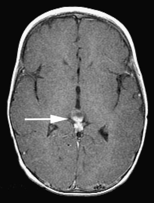
8 What is Trilateral RB? Children who have germinal retinoblastoma have a strong tendency to develop nonretinoblastoma malignancies. Around the time of diagnosis of the intraocular disease, a primary nonretinoblastoma intracranial malignancy (Pineoblastoma/ Ectopic intracranial retinoblastoma) is the most common neoplasm encountered. Presenting features of such a tumor include somnolence, headache and other neurological symptoms.
9 Trilateral RB Continued Central nervous system imaging studies show a solid tumor that involves the suprasellar or parasellar regions of the brain. Because this type of tumor usually occurs in children who have germinal retinoblastoma and bilateral disease, this association is commonly referred to as trilateral retinoblastoma. The intracranial malignancy has a strong tendency to seed the cerebrospinal fluid and thereby spawn implantation tumors along the spinal cord. This malignancy is usually fatal.
10
.")
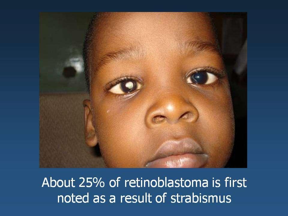
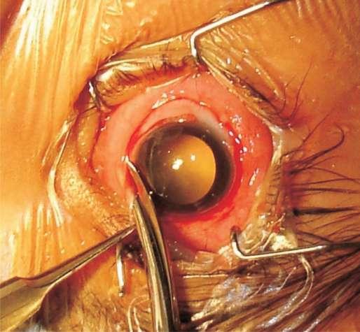
11 Clinical Manifestations The most common presenting manifestation of retinoblastoma is a white glow in the pupil (leukokoria). This appearance is caused by reflection of light from the white intraocular tumor.
12 The second most common presenting manifestation is strabismus. It should be noted that strabismus is a medical condition in which both eyes can t look at the same place at the same time. It occurs when one eye turns in/ out/ up/ down. Common causes of strabismus are poor eye muscle control/ high amount of farsightedness. Because of the association between RB and strabismus, every child who has strabismus must undergo a complete ophthalmic examination to rule out RB.
13
14 Less Common Ocular Manifestations Of RB 1. Red eye. 2. Excessive tearing. 3. Globe expansion (Buphthalmos). 4. Corneal clouding. 5. Discoloration of the iris in the involved eye (usually caused by iris neovascularization). 6. Loss of the fundus reflex in the affected eye due to intraocular bleeding from the tumor. 7. Clumping/ layering of white tumor cells on the iris/ in the aqueous humor. 8. Spontaneous hyphema [Blood in angle of AC]. 9. Sterile orbital cellulitis.
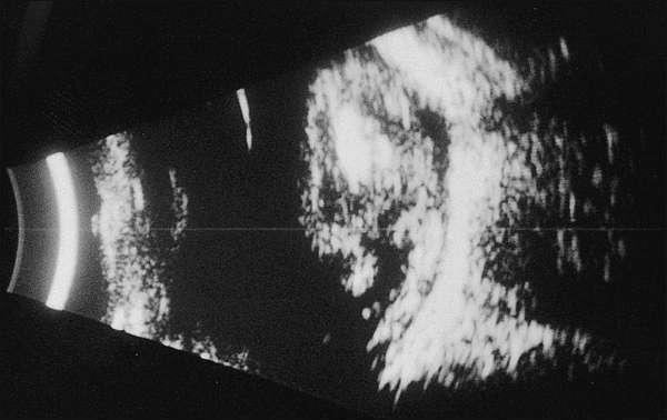
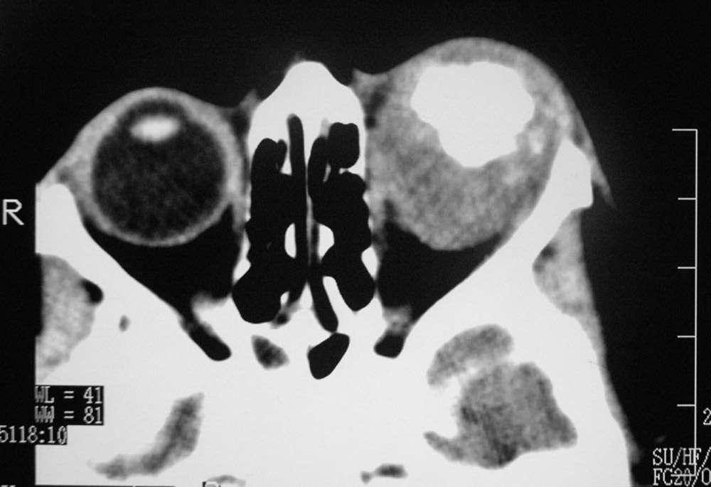
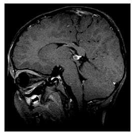
15 Diagnostic Tests Slit-lamp biomicroscopy. Indirect ophthalmoscopy. B-scan ultrasonography. Computed tomography (CT): Confirmation of diagnosis of an intraocular tumor. Magnetic resonance imaging (MRI) is the most useful and informative tool for evaluating the sellar and parasellar regions of the brain (to rule out ectopic intracranial retinoblastoma) and for studying the orbital soft tissues and optic nerve for evidence of extraocular tumor extension.
16
17
18
19
20 Currently advocated only for children with advanced intraocular disease or clinically extraocular disease at baseline. Standard baseline clinical evaluation of children who have newly been diagnosed as Retinoblastoma Complete pediatric history and physical examination. Blood for complete blood count (CBC). MRI or CT of brain, especially in bilateral or familial cases to look for ectopic intracranial Retinoblastoma. Lumbar puncture for CSF analysis [ ]. Bone marrow aspiration or biopsy [ ]. Bone scan [ ].
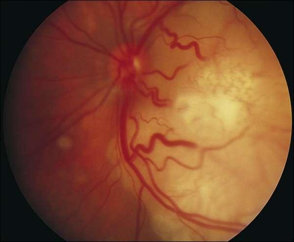
21 Pathology Retinoblastoma is characterized histopathologically by malignant neuroepithelial cells (retinoblasts) that arise within the immature retina. The retinoblasts typically appear to have a large basophilic nucleus and scanty cytoplasm. Cellular necrosis and intralesional calcification are frequent associations, especially in larger tumors.

22 Histopathologic examination of RB. Viable tumor cells clustered around blood vessels and small foci of necrotic cells between the larger masses of the cells.
23 Treatment Options Intravenous chemotherapy Enucleation External beam radiation therapy Plaque Radiation Therapy Laser therapy: 1. Photocoagulation 2. Transpupillary thermotherapy (TTT) Cryotherapy.
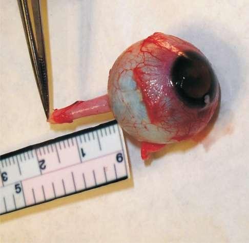
24 Intravenous Chemotherapy Chemotherapy is currently the primary therapeutic option in most children with bilateral retinoblastoma. It is also employed as initial treatment in some children with unilateral disease when the affected eye is believed to be salvageable. The most common chemotherapeutic regimen in use around the world today consists of a combination of Carboplatin, Etoposide and Vincristine (CEV regimen).
25 In some centers, cyclosporine is added to this regimen to reduce the multidrug resistance that occurs in many retinoblastomas. It is usually given as a cyclic treatment every 3 4 weeks for six or more cycles. Most intraocular retinoblastoma lesions (including intravitreal and subretinal seeds) regress substantially within the first two cycles.
26 Enucleation This treatment is particularly applicable to children who have unilateral advanced intraocular disease. Enucleation is sometimes recommended for both eyes in children who have bilateral faradvanced disease not amenable to any eyepreserving therapy. At the end of this presentation, there is a complete guide to Enucleation.
27 External Beam Radiation Therapy External beam radiation therapy is applicable to eyes containing one or more tumors that involve the optic disc, eyes that show diffuse intravitreal or subretinal seeding and eyes for which prior chemo-therapy or local treatments have been failed. Standard target doses of radiation to the eye and orbit are in the range of Gy given in multiple fractions of cgy over 4 5 weeks. External beam radiation therapy results in highly effective regression of vascularized retinal tumors. Even very large, cohesive retinoblastomas commonly show pronounced clinical regression within several weeks after treatment.
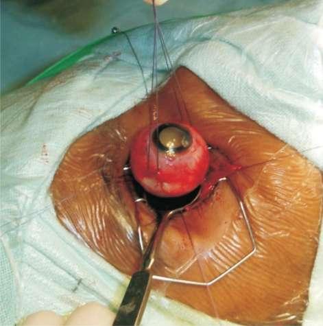
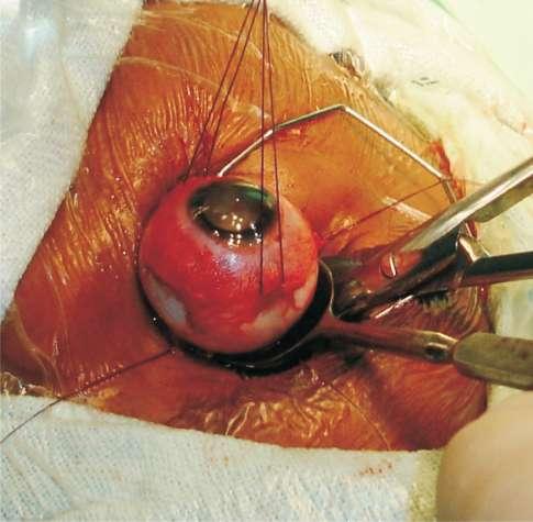
28 Plaque Radiation Therapy Plaque radiation therapy entails surgical implantation of a radioactive device (eye plaque) on the sclera overlying the intraocular tumor, leaving the plaque in place for a sufficient period of time (usually 2 5 days) to provide a predetermined radiation dose to the apex of the tumor, and subsequent surgical removal of the plaque. This form of therapy seems particularly applicable to eyes that contain a solitary medium to large tumor that does not involve the optic disc or macula and is associated with no more than a limited amount of adjacent intravitreal or subretinal tumor seeding.
29 Laser Therapy In Photocoagulation, a medical laser of appropriate wavelength (most commonly an argon green laser) is employed at sufficient power settings to produce almost instantaneous pronounced whitening of the target tissues. In Transpupillary thermotherapy (TTT), an infrared laser beam is directed at the retinal tumor using an operating microscope or indirect ophthalmoscope delivery system in series to produce dull white discoloration of the portion of the tumor covered by the spot.
30 Cryotherapy It is a focal treatment method that destroys targeted intraocular tissues by means of freezing. In this therapy, the ophthalmologist uses an insulated retinal cryoprobe to indent the sclera overlying the tumor and indirect ophthalmo-scopy to monitor the position of the indentation in the fundus. Once the probe tip is positioned at the site of a retinal tumor, the ophthalmologist activates the probe to begin freezing. The ice ball that forms is allowed to encompass the entire tumor (if the tumor is small) or a portion of the tumor (if the tumor is larger) and extend into the overlying vitreous. The probe is then deactivated and the ice ball is allowed to thaw.
31 A Special Guide To Enucleation Definition Indications for Enucleation Preoperative work up and counseling Surgical procedure Post operative care
32 Definition Enucleation is defined as the removal of the entire globe with preservation of the eye muscles.
33 Indications for Enucleation 1. Blind painful eye. 2. Intraocular tumor. 3. Severe trauma with risk of sympathetic ophthalmia. 4. Phthisis bulbi. 5. Microphthalmia. 6. Endophthalmitis/ Panophthalmitis. 7. Cosmetic deformity.
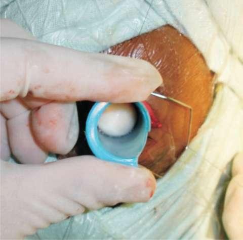
34 Preoperative Work Up Complete ophthalmological examination with unequivocal diagnosis of retinoblastoma based on clinical and radiological examination. Routine pre-anaesthetic workup. A minimum of 9-10 gms% of hemoglobin is mandatory. If the hemoglobin is lower, the same is built up with packed cell transfusions before surgery. Preoperative planning for placement of orbital implant.
35 Pre-operative Counseling The parents of the child should be thoroughly counseled. The nature of surgery should be explained. Counseling should include detailed explanation that: 1. The eyeball cannot be replaced with a seeing eye. 2. The implants and prosthesis will be given to achieve cosmetic correction. 3. The enucleated eyeball requires histopathological examination and this will suggest further treatment plan and future follow up. 4. Informed special consent should be obtained from the parents for enucleation.
36 Surgical Procedure Eye is prepared and draped. Lid speculum is placed. 360 conjunctival peritomy is done.
37
38 Tenon s adhesions to sclera are cleared in all 4 quadrants with tenotomy scissors. Pediatric muscle hook is used to hook the recti, one at a time. Each rectus muscle is tagged with doublearmed 6-0 vicryl sutures passed 3-4 mm beyond the insertion. The rectus muscle is cut at the insertion leaving behind a small 1-2 mm stump attached to the sclera.
39 The inferior and superior oblique muscles are isolated and cut. The hook is swept next to the globe and all other adhesions are lysed. The speculum is next removed and the globe is prolapsed by pushing the lid margins backwards. The optic nerve can be cut either from the temporal side or nasal side.
40
41
42 The eye is removed after teasing away any surrounding tissues. The orbit is packed with wet gauze and firm pressure is applied to secure hemostasis. The implant is placed in the orbit (soak in antibiotic solution before placing the same). The preplaced 6-0 vicryl sutures attached to the recti will help anchor them. Conjunctiva and tenon s capsule are closed in layers with 6-0 vicryl suture. Antibiotic ointment is instilled and a conformer is placed. Pressure pad and bandage is applied.
43
44
45 Post operative Care Post operative dressing is done. Topical antibiotic ointment is prescribed. Oral antibiotics are preferably given at the discretion of surgeon. Ocular prosthesis is given 4-6 weeks following surgery. Patient should be kept under close follow up till the histopathological report is available.
46 Courtesy: Yanoff s Ophthalmology. Parson s Disease Of The Eye. National Guidelines In The Management Of Retinoblastoma (India). Brain Abnormalities on MR Imaging in Patients with Retinoblastoma: A Report. Retinoblastoma: American Cancer Society. National Institute Of Health [NIH].
Financial Disclosures
 Retinoblastoma Management: Update Jesse L. Berry, MD Associate Director, Ocular Oncology Service Associate Program Director USC/CHLA, Keck School of Medicine Financial Disclosures Research Support: Bright
A RESOURCE MANUAL MANAGEMENT RETINOBLASTOMA LOW & MIDDLE RESOURCE SETTINGS
 A RESOURCE MANUAL FOR THE MANAGEMENT OF RETINOBLASTOMA IN LOW & MIDDLE RESOURCE SETTINGS UPDATED SEPTEMBER 2017 1 CONTENTS PAGE INTRODUCTION 3 SERVICE LEVEL for Rb MANAGEMENT 4 SCREENING 5 EARLY DIAGNOSIS
Bilateral retinoblastoma in early infancy
 Saiju R et al Case report Bilateral retinoblastoma in early infancy Saiju R, Duwal S Tilganga Institute of Ophthalmology, Kathmandu, Nepal Abstract Introduction: Retinoblastoma is the most common primary
Tiffany L. Kruger, D.O. Children s Hospital of Michigan Wayne State University/Kresge Eye Institute
 Pediatric Cases Nt Not To Be Missed Tiffany L. Kruger, D.O. Pediatric Ophthalmology Fellow Children s Hospital of Michigan Wayne State University/Kresge Eye Institute Case Presentation CC: Left eye turns
Early detection of Retinoblastoma in children. Max Mantik
 Early detection of Retinoblastoma in children Max Mantik Introduction The most common primary intraocular malignancy of childhood 10 to 15 % of cancers that occur within the first year of life Typical
The Egyptian Journal of Hospital Medicine (October 2018) Vol. 73 (9), Page
 Vol. 73 (9), Page") The Egyptian Journal of Hospital Medicine (October 2018) Vol. 73 (9), Page 7412-7417 Mohammad Ahmad Wahdan 1, Abd Allah El Hussainy Shaleel 1, Hossam El Dein Ahmed El Zomor 2, Hossam El Din Hassan El Sayed
The Egyptian Journal of Hospital Medicine (October 2018) Vol. 73 (9), Page 7412-7417 Mohammad Ahmad Wahdan 1, Abd Allah El Hussainy Shaleel 1, Hossam El Dein Ahmed El Zomor 2, Hossam El Din Hassan El Sayed
Retinoblastoma: A Review of Current Treatment Strategies
 Retinoblastoma: A Review of Current Treatment Strategies ABSTRACT: Since the last review of retinoblastoma therapies in the 15 years ago, there has been a significant shift in the approach to treating
Bilateral Retinoblastoma Joseph Junewick, MD FACR
 Bilateral Retinoblastoma Joseph Junewick, MD FACR 06/11/2010 History 17 month old adopted female with proptosis. Diagnosis Bilateral Retinoblastoma Discussion Retinoblastoma is the most common pediatric
NEW YORK UNIVERSITY SCHOOL OF MEDICINE DEPARTMENT OF OPHTHALMOLOGY EDUCATIONAL OBJECTIVES AND GOALS
 NEW YORK UNIVERSITY SCHOOL OF MEDICINE DEPARTMENT OF OPHTHALMOLOGY EDUCATIONAL OBJECTIVES AND GOALS Revision Date: 6/30/06 Distribution Date: 7/6/06 The Department of Ophthalmology at the NYU Medical Center
Retinoblastoma. Retinoblastoma
 Retinoblastoma Authors: Ayda G. Nambayan, DSN, RN, St. Jude Children s Research Hospital Erin Gafford, Pediatric Oncology Education Student, St. Jude Children s Research Hospital; Nursing Student, School
Scleral buckling. Surgical Treatment
 Dr. Ayman M. Khattab MD, FRCS professor of Ophthalmology Cairo University Surgical Treatment Pneumatic retinopexy. Primary pars plana vitrectomy. 1 Indications for scleral buckling. SB is used to treat
EVIDENCE BASED MANAGEMENT FOR Retinoblastoma
 CLINICAL EVALUATION & STAGING EVIDENCE BASED MANAGEMENT FOR Retinoblastoma Symptoms & Signs : White eye reflex, squint, diminished vision, red eye, proptosis. History - Family history of retinoblastoma
Traumatic Partial Optic Nerve Avulsion with Globe luxation. Presented by: Mostafa ElManhaly Resident in Alexandria Faculty Of Medicine
 Traumatic Partial Optic Nerve Avulsion with Globe luxation Presented by: Mostafa ElManhaly Resident in Alexandria Faculty Of Medicine A 23 year old male patient presented to the emergency department in
Retinoblastoma. all information provided by:
 basic information & treatment plans of Retinoblastoma all information provided by: Ocular Oncology Service Wills Eye Hospital Philadelphia, PA 840 WALNUT STREET SUITE 1440, PHILADELPHIA, PA 19107 WWW.FIGHTEYECANCER.COM
Ocular warning signs in GP practice: Paediatric Eye Pointers
 Ocular warning signs in GP practice: Paediatric Eye Pointers Dr Benjamin Chang MB, BCh, BAO, MMedSci, FRCS(Irel), FRCS(Edin), FRCOphth(Lond) Senior Consultant Ophthalmology and Visual Sciences Khoo Teck
Retinoblastoma in Nepal: case report and review
 Retinoblastoma in Nepal: case report and review Stephen V Lau 1, Ben Limbu 2 Abstract Retinoblastoma often sparks interest because the underlying cancer gene mutation was the first to be identified and
INDIAN COUNCIL OF MEDICAL RESEARCH INDIAN RETINOBLASTOMA GROUP
 NATIONAL RETINOBLASTOMA REGISTRY INDIAN COUNCIL OF MEDICAL RESEARCH INDIAN RETINOBLASTOMA GROUP Centre Code 01. Dr. Rajendra Parasad Centre for Ophthalmic Sciences 02. L.V. Prasad Eye Institute, Hyderabad
Indocyanine Green-Enhanced Transpupillary Thermotherapy for Retinoblastoma: Analysis of 42 Tumors
 Indocyanine Green-Enhanced Transpupillary Thermotherapy for Retinoblastoma: Analysis of 42 Tumors Murat Hasanreisoglu, MD; Jarin Saktanasate, MD; Rachel Schwendeman, NR-CMA; Jerry A. Shields, MD; Carol
Recent Developments in Retinoblastoma
 Recent Advances ISSN 0972-0200 Recent Developments in Retinoblastoma Raksha Rao, Santosh G Honavar National Retinoblastoma Foundation, Ocular Oncology Service, Centre for Sight, Banjara Hills, Hyderabad,
Pediatric Ocular Sonography
 Pediatric Ocular Sonography Cicero J Torres A Silva, MD Associate Professor of Radiology 2016 SPR Pediatric Ultrasound Course Yale University School of Medicine None Disclosures Objectives of Presentation
DNB QUESTIONS 2014 PAPER 1. b) What are the Clinical Conditions in Which Nystagmus is Seen? c) Management of Nystagmus.
 What are the Clinical Conditions in Which Nystagmus is Seen? c) Management of Nystagmus.") DNB QUESTIONS 2014 PAPER 1 1. a) How Will you investigate a case of Nystagmus? b) What are the Clinical Conditions in Which Nystagmus is Seen? c) Management of Nystagmus. 2. a) What is the Principle of
DNB QUESTIONS 2014 PAPER 1 1. a) How Will you investigate a case of Nystagmus? b) What are the Clinical Conditions in Which Nystagmus is Seen? c) Management of Nystagmus. 2. a) What is the Principle of
Retinoblastoma. Protocol applies to retinoblastoma only.
 Retinoblastoma Protocol applies to retinoblastoma only. Protocol revision date: January 2005 Based on AJCC/UICC TNM, 6 th edition Procedures Cytology (No Accompanying Checklist) Biopsy (No Accompanying
Recei in f reatmentfforfretino lastoma atfst.fjudefchildren'sfresearchfhospital
 W it r nnietinoblastoma? e n ttnnrr AfGuidefforfParentsfoffChildren tinob nin nairrnro P ea n rrer Recei in f reatmentfforfretino lastoma atfst.fjudefchildren'sfresearchfhospital Welcome We created this
Diffuse infiltrating retinoblastoma
 Brit. 1. Ophthal. (I 971) 55, 6oo Diffuse infiltrating retinoblastoma GWYN MORGAN Department of Pathology, Institute of Ophthalmology, University of London The term "diffuse infiltrating retinoblastoma"
Retinal Detachment PATIENT EDUCATION
 Retinal Detachment PATIENT EDUCATION What is Retinal Detachment (RD)? Retina is the light-sensitive layer at the back of the eye that converts light images into nerve impulses that are relayed to the brain
AFFECTIONS OF ORBIT IN ANIMALS
 AFFECTIONS OF ORBIT IN ANIMALS Anatomical considerations: The orbit is cone-shaped bony cavity and is incomplete laterally. Formed by six bones: frontal, lacrimal, sphenoid, palatine, zygomatic and maxillary.
Brain Tumors. What is a brain tumor?
 Scan for mobile link. Brain Tumors A brain tumor is a collection of abnormal cells that grows in or around the brain. It poses a risk to the healthy brain by either invading or destroying normal brain
Retinoblastoma: clinical picture and grouping at the time of first presentation
 Original Article Retinoblastoma: clinical picture and grouping at the time of first presentation Correspondence: Dr. Jamshed Ahmed, House No. 700, First Floor, PIB Colony, Karachi - Pakistan. E-mail: jamshi_62@yahoo.com
Test Bank for Medical Surgical Nursing An Integrated Approach 3rd Edition by White
 Test Bank for Medical Surgical Nursing An Integrated Approach 3rd Edition by White Link full download : http://testbankair.com/download/test-bank-for-medical-surgical-nursing-anintegrated-approach-3rd-edition-by-white/
Acknowledgements. Outline. Who were von Hippel and Lindau? Eugen von Hippel German Ophthalmologist
 Ophthalmic Therapies & Standard of Care Acknowledgements Eric Jonasch, MD & Surena Matin, MD Collaborators Franco DeMonte, MD Marcy Johnson Ian McCutcheon, MD Chaan Ng, MD Nancy Perrier, MD Dawid Schellingerhout,
An Injector s Guide to OZURDEX (dexamethasone intravitreal implant) 0.7 mg
 0.7 mg") An Injector s Guide to OZURDEX (dexamethasone intravitreal implant) 0.7 mg This guide is intended to provide injectors with information on the recommended injection technique and the important risks related
An Injector s Guide to OZURDEX (dexamethasone intravitreal implant) 0.7 mg This guide is intended to provide injectors with information on the recommended injection technique and the important risks related
Retinoblastoma, the most common intraocular. Utility of Pupillary Dilation for Detecting Leukocoria in Patients With Retinoblastoma
 Utility of Pupillary Dilation for Detecting Leukocoria in Patients With Retinoblastoma John C. Canzano, MD, and James T. Handa, MD ABSTRACT. In the United States, 50% of all retinoblastoma cases are diagnosed
Advances in Ocular Imaging
 Wide angle fundus imaging and Fuorescein angiography in evaluation and management of intraocular tumors Ihab Saad Othman, MD, FRCS Professor of Ophthalmology Cairo University Cairo, Egypt Advances in Ocular
Choroidal Neovascularization in Sympathetic Ophthalmia
 Choroidal Neovascularization in Sympathetic Ophthalmia Lucia Sobrin, Miguel Cordero Coma, C. Stephen Foster Case Report A 49-year-old man presented after a ruptured globe repair of his left eye status
CLINICAL PEARLS IN OCULAR ONCOLOGY
 CLINICAL PEARLS IN OCULAR ONCOLOGY IRIS NEVUS - Two kinds circumscribed and diffuse - Photodocumentation important to monitor growth - Risk Factors for iris nevus growth to melanoma (ABCDEF) A Age (young),
Dr. Lim, maybe we should start by you telling us a little about yourself and what exactly you do.
 Support for Yale Cancer Answers comes from AstraZeneca, dedicated to providing innovative treatment options for people living with cancer. Learn more at astrazeneca-us.com Welcome to Yale Cancer Answers
2. The clinician will know how to manage common pediatric ocular diseases
 Ida Chung, OD, MSHE, FCOVD, FAAO Western University College of Optometry Associate Professor/Assistant Dean of Learning 309 E. Second Street, Pomona, CA 91766 Office: 909 938 4140 Email: ichung@westernu.edu
Eye and Ocular Adnexa, Auditory Systems
 Eye and Ocular Adnexa, Auditory Systems CPT copyright 2011 American Medical Association. All rights reserved. Fee schedules, relative value units, conversion factors and/or related components are not assigned
Ocular and periocular trauma
 Ocular and periocular trauma No financial disclosures. Tina Rutar M.D. Assistant Professor of Clinical Ophthalmology and Pediatrics Director, Visual Center for the Child University of California San Francisco
Eye-Preserving Therapy in Retinoblastoma: Prolonged Primary Chemotherapy Alone or Combined with Local Therapy
 pissn: 1011-8942 eissn: 2092-9382 Korean J Ophthalmol 2010;24(4):219-224 DOI: 10.3341/kjo.2010.24.4.219 Original Article Eye-Preserving Therapy in Retinoblastoma: Prolonged Primary Chemotherapy Alone or
Ocular Urgencies and Emergencies
 Ocular Urgencies and Emergencies Pam Boyce, O.D., F.A.A.O. Boyce Family Eye Care, Ltd. 528 Devon Ave. Park Ridge, IL 60068 847-518-0303 Somebody s going to lose an eye Epidemiology 2.4 million ocular and
Frequently Asked Questions about General Ophthalmology:
 1. Normal Eye Structure The eye is a slightly asymmetrical globe, about an inch in diameter. The parts of the eye include: Cornea (a clear dome over the iris), Iris (the pigmented part); Pupil (the black
PENETRATING EYE INJUIRES
 PENETRATING EYE INJUIRES King Harold receives a mortal penetrating injury to the eye at the Battle of Hastings 1066, Detail Bayeux Tapestry, Eleventh century. Then Earl William came from Normandy into
MANAGEMENT OF RETINOBLASTOMA
 MANAGEMENT OF RETINOBLASTOMA INTRODUCTION Most common intraocular malignancy of childhood arising from embryonic neural retinal cell. Unifocal/ Multifocal. Unilateral (70%)/ Bilateral (30%). Sporadic (94%)/
Solid Tumour Section Review
 Atlas of Genetics and Cytogenetics in Oncology and Haematology OPEN ACCESS JOURNAL AT INIST-CNRS Solid Tumour Section Review Head and neck: Retinoblastoma Hayyam Kiratli, Berçin Tarlan Ocular Oncology
CM EARTICLE Retinoblastoma
 IPictorial Essay Singapore Med J 2012;53(2) 128 CM EARTICLE Retinoblastoma Mehta M1, MS, DNB, Sethi Sl, MS, Pushker N1, MD, Kashyap S2, MD, Sen S2, MD, Bajaj MS1, MD, Ghose 51, MD, MNAMS ABSTRACT Retinoblastoma
UNIVERSITY OF ZAGREB SCHOOL OF MEDICINE. Plan of the course. Ophthalmology. Academic year 2016/2017. izv. prof. dr. sc. Smiljka Popović-Suić
 UNIVERSITY OF ZAGREB SCHOOL OF MEDICINE Plan of the course Ophthalmology Academic year 2016/2017 I. COURSE AIMS To give the students the necessary knowledge to the rational use of investigations and therapy
Ocular and Periocular Trauma. Tina Rutar, MD. Assistant Professor of Ophthalmology and Pediatrics. Director, Visual Center for the Child
 Ocular and Periocular Trauma Tina Rutar, MD Assistant Professor of Ophthalmology and Pediatrics Director, Visual Center for the Child University of California, San Francisco Phone: 415-353-2560 Fax: 415-353-2468
Outline. Brief history and principles of ophthalmic ultrasound. Types of ocular ultrasound. Examination techniques. Types of Ultrasound
 Ultrasound and Intraocular Tumors 2015 Ophthalmic Photographers' Society Mid-Year Program Cagri G. Besirli MD, PhD Kellogg Eye Center University of Michigan Outline Brief history and principles of ophthalmic
1 Eyelids. Lacrimal Apparatus. Orbital Region. 3 The Orbit. The Eye
 1 1 Eyelids Orbital Region 2 Lacrimal Apparatus 3 The Orbit 4 The Eye 2 Eyelids The eyelids protect the eye from injury and excessive light by their closure. The upper eyelid is larger and more mobile
A Handbook for Families. Retinoblastoma ONCOLOGY SERIES
 A Handbook for Families Retinoblastoma ONCOLOGY SERIES A Handbook for Families Retinoblastoma ONCOLOGY SERIES RETINOBLASTOMA A HANDBOOK FOR FAMILIES Written by: Mindy J. Lipson, MSN RN BC APRN With contributions
A Parent s Guide to Understanding. Retinoblastoma
 A Parent s Guide to Understanding Retinoblastoma 1 Acknowledgements This book is dedicated to the thousands of children and families who have lived through retinoblastoma and to the physicians, nurses,
UNIVERSITY OF ZAGREB SCHOOL OF MEDICINE. Plan of the course. Ophthalmology. Academic year 2018/2019. izv. prof. dr. sc. Smiljka Popović-Suić
 UNIVERSITY OF ZAGREB SCHOOL OF MEDICINE Plan of the course Ophthalmology Academic year 2018/2019 I. COURSE AIMS To give the students the necessary knowledge to the rational use of investigations and therapy
Advanced Examination of the Retina: Scleral Indentation & Retinal 3-Mirror
 Advanced Examination of the Retina: Scleral Indentation & Retinal 3-Mirror Meredith Whiteside, OD, FAAO Nimesh Patel, OD, FAAO John Shan, OD, FAAO Please silence all mobile devices. Unauthorized recording
1. FBE,UEC, LFT 2. USS of eyes confirm intraocular origin, possible calcifications. 3. CT(if available) of eyes and head
 of eyes and head") Retinoblastoma PNG V2 Treatment Guidelines for PNG Retinoblastoma 01V1 Current aim of treatment is to preserve life, vision preservation is secondary at this time. Retinoblastoma remains intra ocular and
Retinoblastoma PNG V2 Treatment Guidelines for PNG Retinoblastoma 01V1 Current aim of treatment is to preserve life, vision preservation is secondary at this time. Retinoblastoma remains intra ocular and
Leukocoria. Khalid Al Husseiny. Lecturer of ophthalmology Kaser Al Ainy, Cairo University 3 rd vitreoretinal school.
 Leukocoria Khalid Al Husseiny Lecturer of ophthalmology Kaser Al Ainy, Cairo University 3 rd vitreoretinal school Side Questions Do u see leukocoria cases? What are u doing when u see retinoblastoma? What
University of Florida ORBIT
 University of Florida ORBIT Dog Airedale Bulldog Great Dane Cat Horse bones EOM in fascial slings Periorbita: orbital septum to tarsal plate Periosteum of optic canal to optic nerve dura Tenon s capsule
The Orbit. The Orbit OCULAR ANATOMY AND DISSECTION 9/25/2014. The eye is a 23 mm organ...how difficult can this be? Openings in the orbit
 The eye is a 23 mm organ...how difficult can this be? OCULAR ANATOMY AND DISSECTION JEFFREY M. GAMBLE, OD COLUMBIA EYE CONSULTANTS OPTOMETRY & UNIVERSITY OF MISSOURI DEPARTMENT OF OPHTHALMOLOGY CLINICAL
Information for Patients. Retinal Detachment
 Information for Patients Retinal Detachment Manchester Royal Eye Hospital Retinal services Your eye doctor has told you that you have a retinal detachment. This leaflet will help you understand your condition
CLINICAL SCIENCES. with thermotherapy or cryotherapy is an important
 CLINICAL SCIENCES Macular Retinoblastoma Managed With Chemoreduction Analysis of Tumor Control With or Without Adjuvant Thermotherapy in 68 Tumors Carol L. Shields, MD; Arman Mashayekhi, MD; Jacqueline
Case Study: Fuzz April 18th
 Case Study: Fuzz April 18th 33 year old Quarter Horse Had been battling corneal ulcer for several weeks before seeing us No foreign debris found Culture and cytology were taken. Started on topical antibiotics,
Year 2 MBChB Clinical Skills Session Ophthalmoscopy. Reviewed & ratified by: Mr M Batterbury Consultant Ophthalmologist
 Year 2 MBChB Clinical Skills Session Ophthalmoscopy Reviewed & ratified by: o Mr M Batterbury Consultant Ophthalmologist Learning objectives o To understand the anatomy and physiology of the external and
Financial Disclosures. The Eye in Neoplastic Disease. Course Goal. We wish to acknowledge and thank: Tumor Definition
 The Eye in Neoplastic Disease Carlo J. Pelino, OD, FAAO Joseph J. Pizzimenti, OD, FAAO cpelino@salus.edu pizzimen@uiwtx.edu Financial Disclosures! Speakers have no relevant financial relationships to declare.
Humanity s Vision Is Our Focus. The Ahmed Glaucoma Valve
 Humanity s Vision Is Our Focus The Ahmed Glaucoma Valve Dr. A. Mateen Ahmed President - New World Medical New World Medical is a high tech medical device company whose goal is to help humanity lead a better
02/03/2014. Average Length: 23mm (Infant ~16mm) Approximately the size of a quarter Volume: ~5mL
 Approximately the size of a quarter Volume: ~5mL") Identify the anatomy of the eye. Explain the basic physiology of the parts of the eye. Briefly discuss various surgeries related to different parts of the anatomy. Average Length: 23mm (Infant ~16mm) Approximately
Identify the anatomy of the eye. Explain the basic physiology of the parts of the eye. Briefly discuss various surgeries related to different parts of the anatomy. Average Length: 23mm (Infant ~16mm) Approximately
PRODUCTION ANIMAL AND EQUINE OPHTHALMOLOGY
 REF. NO. TITLE: C-VO.2 PRODUCTION ANIMAL AND EQUINE OPHTHALMOLOGY VALUE: 10 CREDITS NOTIONAL STUDY HOURS: 100 This module is intended to cover the theoretical knowledge of both Production Animal and Equine
Ultrasound B-Scan for Posterior Segment Evaluation
 Retina Ultrasound B-Scan for Posterior Segment Evaluation Shalini Singh MS Shalini Singh MS, Manisha Agarwal MS, Aditya Bansal DNB Dr. Shroff s Charity Eye Hospital, New Delhi B reproducible investigation
Assisting in Ophthalmology. Copyright 2011, 2007, 2003, 1999 by Saunders, an imprint of Elsevier Inc. All rights reserved.
 Assisting in Ophthalmology Learning Objectives Define, spell, and pronounce the terms listed in the vocabulary. Apply critical thinking skills in performing patient assessment and care. Explain the differences
A Guide to Administering
 A Guide to Administering INDICATIONS AND USAGE YUTIQ (fluocinolone acetonide intravitreal implant) 0.18 mg is indicated for the treatment of chronic non-infectious uveitis affecting the posterior segment
Eye diseases in infancy and early childhood
 Eye diseases in infancy and early childhood Contents: a. Visual development and assessment of vision in infancy and childhood. b. The infant with whitish pupil. c. The infant with watery eye. d. Prematurity
Injury. Contusion Lamellar Laceration Laceration Rupture. Penetrating IOFB. Perforating
 Mechanical Ocular Trauma Došková Hana, MD. Department of Ophthalmology Medicine Faculty of Masaryk University Brno General Considerations Ocular trauma constitude about 6% of all injuries, but eyes set
Moncef Khairallah, MD
 Moncef Khairallah, MD Department of Ophthalmology, Fattouma Bourguiba University Hospital Faculty of Medicine, University of Monastir Monastir, Tunisia INTRODUCTION IU: anatomic form of uveitis involving
 measure of your overall performance. An isolated glucose test is helpful to let you know what your sugar level is at one moment, but it doesn t tell you whether or not your diabetes is under adequate control
M-Sphere Orbital Implant Surgical Guide
 MOLTENO Orbital Implant Surgical Guide A Step by Step Guide to inserting the Natural Hydroxyapatite Orbital Implant 0316-SG/MS Anthony C. B. Molteno, FRCS, FRACO Copyright Anthony C. B. Molteno Molteno,
Pseudohypopyon in Retinoblastoma. Choroidal Nevus. Masquerade Syndromes. Vision pathways. Flat with uniform color
 Primary Intraocular Tumors Thomas F. Freddo, O.D., Ph.D., F.A.A.O. Professor and Former Director School of Optometry University of Waterloo Masquerade Syndromes
Intraocular tumors. Zsuzsa Récsán
 Intraocular tumors Zsuzsa Récsán Definition Uveal melanoma Primary acquired malignant neoplasm of uveal melanocytes Epidemiology: most common primary mal tu in adults Much more common in lighter-skinned
Recurrent intraocular hemorrhage secondary to cataract wound neovascularization (Swan Syndrome)
") Recurrent intraocular hemorrhage secondary to cataract wound neovascularization (Swan Syndrome) John J. Chen MD, PhD; Young H. Kwon MD, PhD August 6, 2012 Chief complaint: Recurrent vitreous hemorrhage,
Recurrent intraocular hemorrhage secondary to cataract wound neovascularization (Swan Syndrome) John J. Chen MD, PhD; Young H. Kwon MD, PhD August 6, 2012 Chief complaint: Recurrent vitreous hemorrhage,
ASSESSING THE EYES. Structures. Eyelids Extraocularmuscles Eyelashes Lacrimal glands: Lacrimal ducts Cornea Conjunctiva Sclera Pupils Iris.
 ASSESSING THE EYES Structures External Eyelids Extraocularmuscles Eyelashes Lacrimal glands: Lacrimal ducts Cornea Conjunctiva Sclera Pupils Iris 1 2 Structures Internal Optic disc Physiological cup Retinal
Vision Health: Conditions, Disorders & Treatments EYELID DISORDERS
 Vision Health: Conditions, Disorders & Treatments EYELID DISORDERS There are a number of disorders that can affect the eyelid. Entropion Entropion is an inward turning of the eyelid and lashes toward the
Outpatient tube removal required. 3 4 (Pain scores are lower following local anesthesia compared to GA.)
") 166 SECTION 2. 0 its medial wall. Following incision of the nasal mucosa through the osteotomy, the posterior flap of the lacrimal sac is sutured to the posterior nasal mucosa flap. The probe is advanced
166 SECTION 2. 0 its medial wall. Following incision of the nasal mucosa through the osteotomy, the posterior flap of the lacrimal sac is sutured to the posterior nasal mucosa flap. The probe is advanced
Custom Prosthetic Eyes
 Custom Prosthetic Eyes Todd Cranmore BCO/BADO Licensed Ocularist WELCOME EYE CARE LOSS & HANDLING OCULAR PROSTHETICS SCLERAL SHELLS Realities of Eye Loss Anxiety Depression Fear of Blindness Loss of Depth
PRECISION PROGRAM. Injection Technique Quick-Reference Guide. Companion booklet for the Video Guide to Injection Technique
 Injection Technique Quick-Reference Guide PRECISION PROGRAM Companion booklet for the Video Guide to Injection Technique Available at www.ozurdexprecisionprogram.com Provides step-by-step directions with
Incidence. Clinical Manifestations
 Incidence Retinoblastoma is the most frequent neoplasm of the eye in childhood occurring in about 1 in 14,000-18,000 live births. Thus, an estimated 300 children develop retinoblastoma each year in the
Scrub In. What is the function of vitreous humor? What does the pupil do when exposed to bright light? a. Maintain eye shape and provide color vision
 Scrub In What is the function of vitreous humor? a. Maintain eye shape and provide color vision b. Maintain eye shape and refract light rays c. Provide night vision and color vision d. Provide night vision
INTRODUCTION: ****************************************************************************************************
 BIOLOGY 211: HUMAN ANATOMY & PHYSIOLOGY **************************************************************************************************** EYES AND VISION ****************************************************************************************************
Retina Center of Oklahoma Sam S. Dahr, M.D. Adult Intraocular Tumors
 Adult Intraocular Tumors Sam S. Dahr, M.D. Retina Center of Oklahoma www.retinacenteroklahoma.com www.rcoklahoma.com Table of Contents Posterior uveal malignant melanoma Uveal metastasis Uveal melanoma
Retinal Tear and Detachment
 Retinal Tear and Detachment Introduction The retina is the layer of tissue in the back of the eye that is responsible for vision. It is attached to the choroid tissue, which supplies the retina with blood.
Diabetic Retinopathy. Barry Emara MD FRCS(C) Giovanni Caboto Club October 3, 2012
 Giovanni Caboto Club October 3, 2012") Diabetic Retinopathy Barry Emara MD FRCS(C) Giovanni Caboto Club October 3, 2012 Outline Statistics Anatomy Categories Assessment Management Risk factors What do you need to do? Objectives Summarize the
Diabetic Retinopathy Barry Emara MD FRCS(C) Giovanni Caboto Club October 3, 2012 Outline Statistics Anatomy Categories Assessment Management Risk factors What do you need to do? Objectives Summarize the
Sense of Vision. Chapter 8. The Eye and Vision. The Eye Orbit. Eyebrows, Eyelids, Eyelashes. Accessory Organs 5/3/2016.
 Sense of Vision Chapter 8 Special Senses The Eye and Vision 70 percent of all sensory receptors are in the eyes Each eye has over 1 million nerve fibers Protection for the eye Most of the eye is enclosed
PEDIATRIC ORBITAL TUMORS RADIOTHERAPY PLANNING
 PEDIATRIC ORBITAL TUMORS RADIOTHERAPY PLANNING ANATOMY ANATOMY CONT ANATOMY CONT. ANATOMY CONT. EYE OF A CHILD Normal tissue tolerance doses (in conventional #) TD 5/5 TD 50/5 Endpoint Gy Gy Optic nerve
MEDICAL POLICY SUBJECT: TRANSPUPILLARY THERMOTHERAPY. POLICY NUMBER: CATEGORY: Technology Assessment
 MEDICAL POLICY SUBJECT: TRANSPUPILLARY EDITED DATE: 08/20/15, 08/18/16, 08/17/17 PAGE: 1 OF: 6 If a product excludes coverage for a service, it is not covered, and medical policy criteria do not apply.
UC SF. g h. Eye Trauma. Martha Neighbor, MD Emergency Services San Francisco General Hospital University of California
 UC SF Eye Trauma sf g h Martha Neighbor, MD Emergency Services San Francisco General Hospital University of California Goals Recognize vision threatening eye emergencies Treat them when we can Know when
J of Evolution of Med and Dent Sci/ eissn , pissn / Vol. 4/ Issue 55/ July 09, 2015 Page 9665
 RARE PRESENTATION OF BILATERAL CHOROIDAL METASTASIS FROM PRIMARY MUCO-EPIDERMOID CARCINOMA OF THE PAROTID GLAND: A G. Premalatha 1, Ramya Seetamraju 2 HOW TO CITE THIS ARTICLE: G. Premalatha, Ramya Seetamraju.
Retinoblastoma. At-A-Glance
 5 2 Retinoblastoma At-A-Glance SUMMARY OF CHANGES Clinical Classification The definitions of T1 T4 were modified The definitions for M1 were modified Pathologic Classification Minor modifications were
4/22/16. Eye. External Anatomy of Eye. Accessory Structures. Bio 40B Dr. Kandula
 Eye Bio 40B Dr. Kandula External Anatomy of Eye Accessory Structures l Eyebrows l Levator Palpebrae Superioris - opens eye l Eyelashes l Ciliary glands modified sweat glands l Small sebaceous glands l
Retinoblastoma. Santosh G Honavar & Vijay Anand P Reddy
 Retinoblastoma Santosh G Honavar & Vijay Anand P Reddy Author Santosh G Honavar, MD, FACS Head of the Department of Ophthalmic Plastic Surgery, Orbit and Ocular Oncology; Associate Director, Patient Care
Clinically Significant Macular Edema (CSME)
") Clinically Significant Macular Edema (CSME) 1 Clinically Significant Macular Edema (CSME) Sadrina T. Shaw OMT I Student July 26, 2014 Advisor: Dr. Uwaydat Clinically Significant Macular Edema (CSME) 2
Clinically Significant Macular Edema (CSME) 1 Clinically Significant Macular Edema (CSME) Sadrina T. Shaw OMT I Student July 26, 2014 Advisor: Dr. Uwaydat Clinically Significant Macular Edema (CSME) 2
Management of specific eye problems in the ED
 of specific eye problems in the ED CORNEAL ABRASION Causes Foreign bodies Tangential shearing injuries, e.g. poking finger into eye Exact cause of injury (Remember to exclude possibility of intraocular
Mobility of Hydroxyapatite Orbital Implant Covered With Autologous Sclera
 Mobility of Hydroxyapatite Orbital Implant Covered With Autologous Sclera Shin-ichiro Kawai, Tsuneko Suzuki and Katsuhito Kawai Department of Ophthalmology, Saitama Medical Center, Saitama Medical School,