Traumatic Partial Optic Nerve Avulsion with Globe luxation. Presented by: Mostafa ElManhaly Resident in Alexandria Faculty Of Medicine
|
|
|
- Pearl Ray
- 5 years ago
- Views:
Transcription
1 Traumatic Partial Optic Nerve Avulsion with Globe luxation Presented by: Mostafa ElManhaly Resident in Alexandria Faculty Of Medicine
2
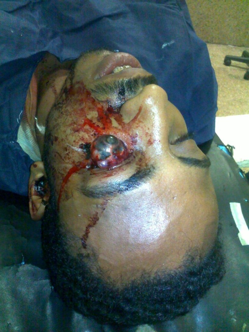
3 A 23 year old male patient presented to the emergency department in a road traffic accident. Medical history: irrelevant history. Surgical history: irrelevant history. General examination : The patient was alert, conscious and cooperative.
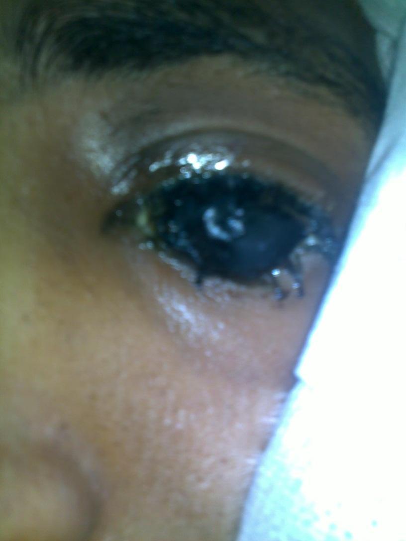
4 Local examination OD OS Visual acuity : 6/6 hand motion Tension : tension is normal digitally softer than the other eye EOM : freely mobile in all directions fixed eye Lids : free edematous Conjunctiva : free intact, but dis-inserted from fornices Cornea: clear hazy cornea Anterior chamber : NDC NDC Iris : NCP NCP Lens : clear couldn t be assessed Pupil : round and reactive (Direct and consensual) Fundus: normal disc and vasculature couldn t be seen


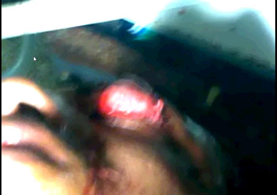
5 3 hours after RTA Posterior portion of the globe is surrounded by edematous Tenon s and orbital fat
6 Definition Globe luxation occurs when the equator of the globe is allowed to protrude anterior to the eyelid aperture. The orbicularis muscle then contracts causing further anterior displacement and the globe is caught outside the eyelid aperture.
7 Types Avulsio Bulbi Avulsion of the optic nerve only (Avulsio incompleta). With disruption of the extraocular muscles which may cause total luxation of the ocular bulbus. (Avulsio completa)


8 Causes Traumatic : RTA, sports injury, etc. Spontaneous : - Shallow orbit e.g. Crouzon syndrome. - Thyroid associated orbitopathy. - Floppy eyelid syndrome. Others: - Self-enucleation practice seen in some psychiatric patients!! Brutal fighting called gouging in which a combatant was successful if he would press the adversary s eyeball out with his thumb
9 Investigations Urgent CT scan is indicated, specially for traumatic luxation to assess the vitality of the optic nerve,on which the management plan will be decided accordingly.


10 Luxated globe

11 CT scan showing intact optic nerve
12 Management of Globe luxation First aid: - Keep the globe wet all the time e.g.saline. - Rapid repositioning of the globe back into the socket, as soon as possible. - Intravenous corticosteroids and antibiotics. Fixation of orbital wall fractures by maxillofacial surgeons. Follow up: It includes: 1. The traumatized eye. 2. The fellow eye. 3. Nervous system. Delayed management: Reinsertion of the disrupted muscular attachments within 7-10 days, before the contracture of the lost muscle or its antagonist supervenes. 1. Medial rectus 2. Inferior rectus 3. Superior rectus, 4. The obliques
13
14 Follow up 1-Traumatized eye : Visual acuity Ocular motility Colour of the sclera and cornea Tension of the globe The axial legnth of the globe by ultrasound scan. 2-The fellow eye: Visual acuity of the other eye is followed regularly, because chiasmal compression is one of the serious complications. It occurs due to rupture and hemorrhage of pial vessels secondary to stretching of optic nerve meningeal covering
15 Complications 1. Orbital infections. 2. Subarachnoid hemorrhage due to severance of the ophthalmic artery. 3. Meningitis 4. Cerebrospinal fluid leakage. 5. Life threatening hypothalamic dysfunction. 6. With posterior avulsions, chiasmal injuries and residual visual field defects occurs in the follow eye. 7. Phthisis bulbi.
16
17 Adopting the approach of deferring enucleation at first instance and deploying every effort to preserve the eye as a cosmetically acceptable organ help the patient in two ways: 1-The patient did not have to sacrifice an organ after such severe accident had an enormous impact on his rapid recovery from the psychological trauma of this incident. 2-He would be easily fitted an ocular prosthesis with better motility.
18 Take home message Keep the globe always wet. Reduction of the globe as soon as possible. Examining the fellow eye on first presentation and regularly in follow up sessions.
19 References Gould GM & Pyle WL (1956): Anomalies and curiosities of medicine. p NewYork. The Julian Press Inc. Jones NP (1990): Self-enucleation and psychosis. Br J Ophthalmol 74: Khan JA, Buescher L, Ide CH & Pettigrove B (1985): Medical management of self-enucleation. Arch Ophthalmol 103: Lang GK, Bialasiewicz AA & Ro hr WD (1991): Beideseitige traumatische Avulsio bulbi. Klin Mbl Augenheilk 198: Mailer CM (1974): Avulsion of the inferior rectus. Can J Ophthalmol 9:
20 Thank You
A Case of Carotid-Cavernous Fistula
 A Case of Carotid-Cavernous Fistula By : Mohamed Elkhawaga 2 nd Year Resident of Ophthalmology Alexandria University A 19 year old male patient came to our outpatient clinic, complaining of : -Severe conjunctival
Ocular and periocular trauma
 Ocular and periocular trauma No financial disclosures. Tina Rutar M.D. Assistant Professor of Clinical Ophthalmology and Pediatrics Director, Visual Center for the Child University of California San Francisco
Ocular and Periocular Trauma. Tina Rutar, MD. Assistant Professor of Ophthalmology and Pediatrics. Director, Visual Center for the Child
 Ocular and Periocular Trauma Tina Rutar, MD Assistant Professor of Ophthalmology and Pediatrics Director, Visual Center for the Child University of California, San Francisco Phone: 415-353-2560 Fax: 415-353-2468
1 Eyelids. Lacrimal Apparatus. Orbital Region. 3 The Orbit. The Eye
 1 1 Eyelids Orbital Region 2 Lacrimal Apparatus 3 The Orbit 4 The Eye 2 Eyelids The eyelids protect the eye from injury and excessive light by their closure. The upper eyelid is larger and more mobile
Case #1: 68 M with floaters OS
 Case #1: 68 M with floaters OS Point-of-Care Ocular Sonography for the Emergency Department Nate Teismann MD Dept of Emergency Medicine, UCSF Topics in EM 2012 Acute onset of dark spots in L eye 2 days
Anatomy: There are 6 muscles that move your eye.
 Thyroid Eye Disease Your doctor thinks you have thyroid orbitopathy. This is an autoimmune condition where your body's immune system is producing factors that stimulate enlargement of the muscles that
Ocular Anatomy for the Paraoptometric
 Ocular Anatomy for the Paraoptometric Minnesota Optometric Association Paraoptometric CE Friday September 30, 2016 Lindsay A. Sicks, OD, FAAO Assistant Professor, Illinois College of Optometry lsicks@ico.edu
Ocular Urgencies and Emergencies
 Ocular Urgencies and Emergencies Pam Boyce, O.D., F.A.A.O. Boyce Family Eye Care, Ltd. 528 Devon Ave. Park Ridge, IL 60068 847-518-0303 Somebody s going to lose an eye Epidemiology 2.4 million ocular and
Clues of a Ruptured Globe
 Definition any eye that has sustained a full thickness traumatic disruption of the cornea or sclera Overwhelmingly, rupture accidents occur in young men, small children and the elderly Corneal laceration
MRI masterfile Part 5 WM Heme Strokes.ppt 1
 Ocular and Orbital Trauma Eye Trauma: Incidence 1.3 million eye injuries in the US per year. 40,000 of these injuries lead to blindness in the US. Patrick Sibony, MD March 23, 2013 Ophthalmic Emergencies
LECTURE # 7 EYECARE REVIEW: PART III
 LECTURE # 7 EYECARE REVIEW: PART III HOW TO TRIAGE EYE EMERGENCIES STEVE BUTZON, O.D. EYECARE REVIEW: HOW TO TRIAGE EYE EMERGENCIES FOR PRIMARY CARE PHYSICIANS Steve Butzon, O.D. Member Director IDOC President
Sense of Vision. Chapter 8. The Eye and Vision. The Eye Orbit. Eyebrows, Eyelids, Eyelashes. Accessory Organs 5/3/2016.
 Sense of Vision Chapter 8 Special Senses The Eye and Vision 70 percent of all sensory receptors are in the eyes Each eye has over 1 million nerve fibers Protection for the eye Most of the eye is enclosed
EYE INJURIES OBJECTIVES COMMON EYE EMERGENCIES 7/19/2017 IMPROVE ASSESSMENT OF EYE INJURIES
 EYE INJURIES BRITTA ANDERSON D.O. DMC PRIMARY CARE SPORTS MEDICINE ASSOCIATE TEAM PHYSICIAN DETROIT TIGERS OBJECTIVES IMPROVE ASSESSMENT OF EYE INJURIES UNDERSTAND WHAT IS CONSIDERED AN EMERGENCY DEVELOP
Mobility of Hydroxyapatite Orbital Implant Covered With Autologous Sclera
 Mobility of Hydroxyapatite Orbital Implant Covered With Autologous Sclera Shin-ichiro Kawai, Tsuneko Suzuki and Katsuhito Kawai Department of Ophthalmology, Saitama Medical Center, Saitama Medical School,
Carotid Cavernous Fistula
 Chief Complaint: Double vision. Carotid Cavernous Fistula Alex W. Cohen, MD, PhD; Richard Allen, MD, PhD May 14, 2010 History of Present Illness: A 46 year old female patient presented to the Oculoplastics
Bony orbit Roof The orbital plate of the frontal bone Lateral wall: the zygomatic bone and the greater wing of the sphenoid
 Bony orbit Roof: Formed by: The orbital plate of the frontal bone, which separates the orbital cavity from the anterior cranial fossa and the frontal lobe of the cerebral hemisphere Lateral wall: Formed
The Orbit. The Orbit OCULAR ANATOMY AND DISSECTION 9/25/2014. The eye is a 23 mm organ...how difficult can this be? Openings in the orbit
 The eye is a 23 mm organ...how difficult can this be? OCULAR ANATOMY AND DISSECTION JEFFREY M. GAMBLE, OD COLUMBIA EYE CONSULTANTS OPTOMETRY & UNIVERSITY OF MISSOURI DEPARTMENT OF OPHTHALMOLOGY CLINICAL
University of Florida ORBIT
 University of Florida ORBIT Dog Airedale Bulldog Great Dane Cat Horse bones EOM in fascial slings Periorbita: orbital septum to tarsal plate Periosteum of optic canal to optic nerve dura Tenon s capsule
EYE TRAUMA: INCIDENCE
 Introduction EYE TRAUMA: INCIDENCE 2.5 million eye injuries per year in U.S. 40,000 60,000 of eye injuries lead to visual loss Introduction Final visual outcome of many ocular emergencies depends on prompt,
Eye Movements. Geometry of the Orbit. Extraocular Muscles
 Eye Movements Geometry of the Orbit The eye (oculus) is located in the anterior aspect of the orbit: the equator of the eye (defined by a coronal plane passing through its middle) lies at the margin of
The orbit-1. Dr. Heba Kalbouneh Assistant Professor of Anatomy and Histology
 The orbit-1 Dr. Heba Kalbouneh Assistant Professor of Anatomy and Histology Orbital plate of frontal bone Orbital plate of ethmoid bone Lesser wing of sphenoid Greater wing of sphenoid Lacrimal bone Orbital
Imaging Orbit/Periorbital Injury
 Imaging Orbit/Periorbital Injury 9 th Nordic Trauma Radiology Course 2016 Stuart E. Mirvis, M.D., FACR Department of Radiology University of Maryland School of Medicine Fireworks Topics to Cover Struts
Lateral Orbitotomy in the Management of Challenging Exotropia
 Lateral Orbitotomy in the Management of Challenging Exotropia Yahalom C (1, 2), Mc Nab A (3), Ben Simon G (3), Kowal L (1). 1- Centre for Eye Research Australia and Ocular Motility Clinic, Royal Victorian
10 Congresso Nazionale Associazione Italiana della Tiroide Cagliari, dicembre 2016 Orbitopa)a basedowiana acuta
a basedowiana acuta") 10 Congresso Nazionale Associazione Italiana della Tiroide Cagliari, 15-17 dicembre 2016 Orbitopa)a basedowiana acuta Luigi Bartalena Università dell Insubria a Varese Prevalence of Graves orbitopathy
10 Congresso Nazionale Associazione Italiana della Tiroide Cagliari, 15-17 dicembre 2016 Orbitopa)a basedowiana acuta Luigi Bartalena Università dell Insubria a Varese Prevalence of Graves orbitopathy
Muscles of the Eyeball (Extra Ocular Muscles) Prof. Dr. Imran Qureshi
 Prof. Dr. Imran Qureshi") Muscles of the Eyeball (Extra Ocular Muscles) Prof. Dr. Imran Qureshi There are six extrinsic muscles of the eyeball, namely the (S), Medial (M), (I), & Lateral (L) recti, and (SO) and (IO) Obliques. In
Muscles of the Eyeball (Extra Ocular Muscles) Prof. Dr. Imran Qureshi There are six extrinsic muscles of the eyeball, namely the (S), Medial (M), (I), & Lateral (L) recti, and (SO) and (IO) Obliques. In
Unit VIII Problem 8 Anatomy: Orbit and Eyeball
 Unit VIII Problem 8 Anatomy: Orbit and Eyeball - The bony orbit: it is protecting our eyeball and resembling a pyramid: With a base directed: anterolaterally. And an apex directed: posteromedially. Notes:
Complete Visual Rehabilitation in a Patient with No Light Perception after Surgical Management of a Penetrating Open-Globe Injury: A Case Report
 Published online: June 23, 2015 1663 2699/15/0062 0204$39.50/0 This is an Open Access article licensed under the terms of the Creative Commons Attribution- NonCommercial 3.0 Unported license (CC BY-NC)
Injury. Contusion Lamellar Laceration Laceration Rupture. Penetrating IOFB. Perforating
 Mechanical Ocular Trauma Došková Hana, MD. Department of Ophthalmology Medicine Faculty of Masaryk University Brno General Considerations Ocular trauma constitude about 6% of all injuries, but eyes set
Protocol. Blepharoplasty
 Protocol Blepharoplasty Medical Benefit Effective Date: 01/01/13 Next Review Date: 05/19 Preauthorization No Review Dates: 09/12, 09/13, 09/14, 09/15, 09/16, 05/17, 05/18 Preauthorization is encouraged
UC SF. g h. Eye Trauma. Martha Neighbor, MD Emergency Services San Francisco General Hospital University of California
 UC SF Eye Trauma sf g h Martha Neighbor, MD Emergency Services San Francisco General Hospital University of California Goals Recognize vision threatening eye emergencies Treat them when we can Know when
Ophthalmic Trauma Update
 Ophthalmic Trauma Update Richard S. Davidson, M.D. Professor of Ophthalmology Vice Chair for Quality and Clinical Affairs UCHealth Eye Center University of Colorado School of Medicine August 5, 2017 Financial
OPHTHALMOLOGY AND ULTRASOUND
 Vet Times The website for the veterinary profession https://www.vettimes.co.uk OPHTHALMOLOGY AND ULTRASOUND Author : JAMES OLIVER Categories : Vets Date : April 28, 2008 JAMES OLIVER discusses why ultrasound
MRI Dynamic Color Mapping: a new quantitative technique for imaging soft tissue motion in the orbit
 MRI DYNAMIC COLOR MAPPING: A NEW QUANTITATIVE TECHNIQUE FOR IMAGING SOFT TISSUE MOTION IN THE ORBIT 79 6 MRI Dynamic Color Mapping: a new quantitative technique for imaging soft tissue motion in the orbit
Vertical Muscles Transposition with Medical Rectus Botulinum Toxin Injection for Abducens Nerve Palsy
 JKAU: Med. Sci., Vol. 16 No. 2, pp: 43-49 (2009 A.D. / 1430 A.H.) DOI: 10.4197/Med. 16-2.4 Vertical Muscles Transposition with Medical Rectus Botulinum Toxin Injection for Abducens Nerve Palsy Nizar M.
The sebaceous glands (glands of Zeis) open directly into the eyelash follicles, ciliary glands (glands of Moll) are modified sweat glands that open
 open directly into the eyelash follicles, ciliary glands (glands of Moll) are modified sweat glands that open") The Orbital Region The orbits are a pair of bony cavities that contain the eyeballs; their associated muscles, nerves, vessels, and fat; and most of the lacrimal apparatus upper eyelid is larger and more
The Orbital Region The orbits are a pair of bony cavities that contain the eyeballs; their associated muscles, nerves, vessels, and fat; and most of the lacrimal apparatus upper eyelid is larger and more
Virtual Mentor American Medical Association Journal of Ethics December 2010, Volume 12, Number 12:
 Virtual Mentor American Medical Association Journal of Ethics December 2010, Volume 12, Number 12: 950-954. IMAGES OF HEALING AND LEARNING The Fourth O in Eye Care Ocularists Michael O. Hughes, BCO The
Case Presentation: Indications for orbital decompression in TED: Modern surgical techniques for orbital decompression in TED: Inferomedial
 Case Presentation: Jonathan W. Kim, MD Director, Oculoplastic Surgery Stanford Medical Center 61 year old man with active Graves orbitopathy Visual acuity 20/30 OD 20/50 OS Left RAPD Bilateral optic disc
Management of specific eye problems in the ED
 of specific eye problems in the ED CORNEAL ABRASION Causes Foreign bodies Tangential shearing injuries, e.g. poking finger into eye Exact cause of injury (Remember to exclude possibility of intraocular
THE CHRONIC GLAUCOMAS
 THE CHRONIC GLAUCOMAS WHAT IS GLAUCOMA? People with glaucoma have lost some of their field of all round vision. It is often the edge or periphery that is lost. That is why the condition can be missed until
INTRODUCTION: ****************************************************************************************************
 BIOLOGY 211: HUMAN ANATOMY & PHYSIOLOGY **************************************************************************************************** EYES AND VISION ****************************************************************************************************
Essential questions. What are the structures of the sensory system? 3.03 Remember the structures of the sensory system 2
 Essential questions What are the structures of the sensory system? 3.03 Remember the structures of the sensory system 2 The Senses Eyes Sight Ears Hearing Nose Smell Tongue Taste Skin Touch 3.03 Remember
Basic microsurgical suturing techniques for beginners
 ESCRS 2014 Basic microsurgical suturing techniques for beginners Trauma, sclera, trabeculectomy B.O. Bachmann Dept. of Ophthalmology, University of Cologne, Germany Financial interests: none Investigating
A ptosis repair of aponeurotic defects by the posterior approach
 British Journal of Ophthalmology, 1979, 63, 586-590 A ptosis repair of aponeurotic defects by the posterior approach J. R. 0. COLLIN From the Department of Clinical Ophthalmology, Moorfields Eye Hospital,
Cairo University Faculty of Medicine. Course Specifications Course title: Ophthalmology (Code): OPH-409. Department of Ophthalmology
: OPH-409. Department of Ophthalmology") Cairo University Faculty of Medicine Department of Ophthalmology Course Specifications Course title: Ophthalmology (Code): OPH-409 Department of Ophthalmology Fourth academic year of M.B.B.Ch. program
Cairo University Faculty of Medicine Department of Ophthalmology Course Specifications Course title: Ophthalmology (Code): OPH-409 Department of Ophthalmology Fourth academic year of M.B.B.Ch. program
The orbit-2. Dr. Heba Kalbouneh Assistant Professor of Anatomy and Histology
 The orbit-2 Dr. Heba Kalbouneh Assistant Professor of Anatomy and Histology Eyelids The eyelids (act like the curtains) protect the eye from injury and excessive light by their closure The upper eyelid
! Women greater than men (4:1)» Typical of other autoimmune diseases
» Typical of other autoimmune diseases") 1 2 3 4 : Overview and Diagnosis Suzanne K. Freitag, M.D. Director, Ophthalmic Plastic Surgery Massachusetts Eye and Ear Infirmary Harvard Medical School! I have no financial disclosures. Learning Objectives!
1 2 3 4 : Overview and Diagnosis Suzanne K. Freitag, M.D. Director, Ophthalmic Plastic Surgery Massachusetts Eye and Ear Infirmary Harvard Medical School! I have no financial disclosures. Learning Objectives!
 Traumatic Cataract Orbital Wall Fracture Vitreous Hemorrhage Optic Disc Hemorrhage a) Amblyopia b) Strabismus c) Trauma Playing with other children Sports Fire works BB gun Injecting needles .
Dr. Esam Ahmad Z. Omar BDS, MSc-OMFS, FFDRCSI. Monitor the vital signs. Monitor the vital signs. Complications of Facial Traumas.
 Complications of Facial Traumas 1) Immediate Complications 2) Late Complications Dr. Esam Ahmad Z. Omar BDS, MSc-OMFS, FFDRCSI Assistant Professor Oral & Maxillofacial Surgeon Taibah University Monitor
Supplementary Online Content
 Supplementary Online Content Park KH, Kim YK, Woo SJ, et al. Iatrogenic occlusion of the ophthalmic artery after cosmetic facial filler injections: a national survey by the Korean Retina Society. JAMA
THE SPECIAL SENSES. Introduction Vision
 THE SPECIAL SENSES Introduction Vision RECEPTORS Structures designed to respond to stimuli Variable complexity RECEPTORS: GENERAL PROPERTIES Transducers Receptor Potential Generator Potential RECEPTORS
SURGERY OF THE INFERIOR OBLIQUE MUSCLE. CARL V. GOBIN, M.D. Centre of Strabology AZ MONICA-ANTWERPEN
 SURGERY OF THE INFERIOR OBLIQUE MUSCLE CARL V. GOBIN, M.D. Centre of Strabology AZ MONICA-ANTWERPEN SURGERY OF THE INFERIOR OBLIQUE MUSCLE The treatment of superior oblique palsies is one of the more complicated
2/5/2018. Trauma. Subdivided into two main categories: Closed globe Open Globe
 1 2 3 4 5 Ocular Trauma Guide for Eye Care Office Staff Winter Thaw 2018 Aaron Yatskevich OD Definition A broad term used to describe a physical or chemical wound to the eye or eye socket. Ocular trauma
Graves Ophthalmopathy Overview. Graves Disease Hyperthyroidism TSIgs (anti-tsh-receptor-abs) Graves Disease 10/22/2010
 Graves Disease 10/22/2010") Graves Disease Robert Graves Graves Ophthalmopathy Overview Robert C. Kersten Dept. of Ophthalmology UCSF Triad Hyperthyroidism Eye findings Pretibial Myxedema (phalangeal acropachy-1%) Most common auto-immune
Graves Disease Robert Graves Graves Ophthalmopathy Overview Robert C. Kersten Dept. of Ophthalmology UCSF Triad Hyperthyroidism Eye findings Pretibial Myxedema (phalangeal acropachy-1%) Most common auto-immune
04/11/2014. Retina Coding and Reimbursement 101. Financial Disclosure. Chief Complaint
 Retina Coding and Reimbursement 101 William T. Koch, COA, COE, CPC Administrative Director Director of Billing Operations The Retina Institute St. Louis, Missouri Advisory Boards Allergan Genentech Regeneron
Assessment and Management of Ocular Trauma. Disclosure I have no direct financial interests in today s subject matter. 3/25/2019. Normal Eye Anatomy
 Assessment and Management of Ocular Trauma Samiksha Fouzdar Jain, MD,FRCS Department of Ophthalmology & Visual Sciences Truhlsen Eye Institute Disclosure I have no direct financial interests in today s
Ears. Mouth. Jowls 6 Major Bones of the Face Nasal bone Two
 1 2 3 4 5 Chapter 25 Injuries to the Face, Neck, and Eyes Injuries to the Face and Neck Face and neck are to injury Relatively unprotected positions on body Some injuries are life-threatening. trauma to
Surgical management of Duane's
 Brit. J. Ophthal. (I974) 58, 30 I Surgical management of Duane's syndrome M. H. GOBIN ljniversity Eye Clinic, Leyden, IHolland Ten years ago I introduced a surgical technique for the correction of Duane's
Epidemiology 3002). Epidemiology and Pathophysiology
. Epidemiology and Pathophysiology") Epidemiology Maxillofacial trauma or injuries are commonly encountered in the practice of emergency medicine and are presenting one of the most challenging problems to the attending surgeons or physicians
Epidemiology Maxillofacial trauma or injuries are commonly encountered in the practice of emergency medicine and are presenting one of the most challenging problems to the attending surgeons or physicians
Restoring Ocular Esthetics Using Ocular Prosthesis
 Case Report Restoring Ocular Esthetics Using Ocular Prosthesis Dr. Kalavathi S.D 1*, Dr. Arvind Moldi 2**, Dr. Phaneendra Kumar 3* * Senior lecturer, ** Professor & HOD, 1 Department of Prosthodontics,
Learn Connect Succeed. JCAHPO Regional Meetings 2017
 Learn Connect Succeed JCAHPO Regional Meetings 2017 Financial Disclosure Evaluation and Treatment of Orbital Cellulitis Thomas E. Johnson, M.D. Bascom Palmer Eye Institute University of Miami School of
213: HUMAN FUNCTIONAL ANATOMY: PRACTICAL CLASS 12 Cranial cavity, eye and orbit
 213: HUMAN FUNCTIONAL ANATOMY: PRACTICAL CLASS 12 Cranial cavity, eye and orbit OSTEOLOGY Identify the bones which comprise the walls of the orbit: maxilla, zygomatic, ethmoid, lachrymal, frontal, and
THE CHRONIC GLAUCOMAS
 THE CHRONIC GLAUCOMAS WHAT IS GLAUCOMA People with glaucoma have lost some of their field of all round vision. It is often the edge or periphery that is lost. That is why the condition can be missed until
NEW YORK UNIVERSITY SCHOOL OF MEDICINE DEPARTMENT OF OPHTHALMOLOGY EDUCATIONAL OBJECTIVES AND GOALS
 NEW YORK UNIVERSITY SCHOOL OF MEDICINE DEPARTMENT OF OPHTHALMOLOGY EDUCATIONAL OBJECTIVES AND GOALS Revision Date: 6/30/06 Distribution Date: 7/6/06 The Department of Ophthalmology at the NYU Medical Center
3/16/2018. Ultrasound Biomicroscopy in Glaucoma By Ahmed Salah Abdel Rehim. Prof. of Ophthalmology Al-Azhar University
 Ultrasound Biomicroscopy in Glaucoma By Ahmed Salah Abdel Rehim Prof. of Ophthalmology Al-Azhar University 1 Ultrasound biomicroscopy (UBM) is a recent technique to visualize anterior segment with the
Session 1: Working With Families and Eye Care Professionals
 Module: Visual Conditions and Functional Vision: Early Intervention Issues Session 1: Working With Families and Eye Care Professionals Handout F: Interpreting Eye Reports Hatton, D.D., & Campbell, A.F.
AQUEOUS VEINS IN RABBITS*
 Brit. J. Ophthal., 35, 119. AQUEOUS VEINS IN RABBITS* BY D. P. GREAVES AND E. S. PERKINS Institute of Ophthalmology, London Director of Research, Sir Stewart Duke-Elder IN the course of investigations
Vision I. Steven McLoon Department of Neuroscience University of Minnesota
 Vision I Steven McLoon Department of Neuroscience University of Minnesota 1 Eye Cornea Sclera Conjunctiva 2 Eye The conjunctiva lines the inner surface of the eyelids and outer surface of the sclera. 3
Shaken Baby Syndrome (SBS) Ocular Findings with Legal Implications
 Ocular Findings with Legal Implications") United States and Canadian Academy of Pathology 2007 Annual Meeting Shaken Baby Syndrome (SBS) Ocular Findings with Legal Implications J. Douglas Cameron, MD Professor of Ophthalmology Mayo Clinic School
United States and Canadian Academy of Pathology 2007 Annual Meeting Shaken Baby Syndrome (SBS) Ocular Findings with Legal Implications J. Douglas Cameron, MD Professor of Ophthalmology Mayo Clinic School
MRI masterfile Part 5 WM Heme Strokes.ppt 2
 Imaging of Orbital Trauma Corneal Abrasion CT scan is preferable to MRI Bone, Rapid, Easy to monitor patient Foreign bodies, air, hemorrhage Fractures Cost Needed for an MRI MRI Globe and intraocular injuries
4/22/16. Eye. External Anatomy of Eye. Accessory Structures. Bio 40B Dr. Kandula
 Eye Bio 40B Dr. Kandula External Anatomy of Eye Accessory Structures l Eyebrows l Levator Palpebrae Superioris - opens eye l Eyelashes l Ciliary glands modified sweat glands l Small sebaceous glands l
International Journal of Health Sciences and Research ISSN:
 International Journal of Health Sciences and Research www.ijhsr.org ISSN: 2249-9571 Original Research Article Prospective Study of Ocular Manifestation of Road Traffic Accidents on East Coast Road Achanti
CASE PRESENTATION. DR.Sravani 1 st yr PG Dept of Ophthalmology
 CASE PRESENTATION DR.Sravani 1 st yr PG Dept of Ophthalmology Name : X X X X X Age : 50yrs Sex : male Occupation : Farmer Residence : Mothkur CHIEF COMPLAINTS : - Diminision of vision in Right Eye since
Surgical Anatomy Ear and Eye. Presenters: Dr. Jim Hurrell and Dr. Dennis McCurnin
 Surgical Anatomy Ear and Eye Presenters: Dr. Jim Hurrell and Dr. Dennis McCurnin A Warm Welcome from My Faculty TEAM and Me!!! 2 The Pledge of Allegiance 3 The Senses 4 Hearing 3 Layers of Ear EXTERNAL
Advanced Examination of the Retina: Scleral Indentation & Retinal 3-Mirror
 Advanced Examination of the Retina: Scleral Indentation & Retinal 3-Mirror Meredith Whiteside, OD, FAAO Nimesh Patel, OD, FAAO John Shan, OD, FAAO Please silence all mobile devices. Unauthorized recording
Year 2 MBChB Clinical Skills Session Ophthalmoscopy. Reviewed & ratified by: Mr M Batterbury Consultant Ophthalmologist
 Year 2 MBChB Clinical Skills Session Ophthalmoscopy Reviewed & ratified by: o Mr M Batterbury Consultant Ophthalmologist Learning objectives o To understand the anatomy and physiology of the external and
MAXILLOFACIAL TRAUMA. The on-call maxillofacial surgeons can be contacted through the switchboard at the Southern General Hospital
 MAXILLOFACIAL TRAUMA The on-call maxillofacial surgeons can be contacted through the switchboard at the Southern General Hospital Mandibular Injuries Mechanism of injury Assault, falls, RTA-Direct trauma
M-Sphere Orbital Implant Surgical Guide
 MOLTENO Orbital Implant Surgical Guide A Step by Step Guide to inserting the Natural Hydroxyapatite Orbital Implant 0316-SG/MS Anthony C. B. Molteno, FRCS, FRACO Copyright Anthony C. B. Molteno Molteno,
Bilateral retinoblastoma in early infancy
 Saiju R et al Case report Bilateral retinoblastoma in early infancy Saiju R, Duwal S Tilganga Institute of Ophthalmology, Kathmandu, Nepal Abstract Introduction: Retinoblastoma is the most common primary
Choroidal Neovascularization in Sympathetic Ophthalmia
 Choroidal Neovascularization in Sympathetic Ophthalmia Lucia Sobrin, Miguel Cordero Coma, C. Stephen Foster Case Report A 49-year-old man presented after a ruptured globe repair of his left eye status
University Journal of Surgery and Surgical Specialities
 University Journal of Surgery and Surgical Specialities Volume 1 Issue 1 2015 PARINAUD'S SYNDROME A CASE REPORT Basker K Shubha Raguram K Stanley Medical College Introduction: Gaze palsies are a group
Ocular Lecture. Sue Bednar NP Ali Atwater PA-C
 Ocular Lecture Sue Bednar NP Ali Atwater PA-C Triaging Ocular Complaints Painful Eye/Red eye +/-blurry vision +/-visual loss +/-floaters +/-fevers If any of the above findings exist, pt is likely to have
Eyes, ears, teeth and everything in between
 Eyes, ears, teeth and everything in between E M E R G E N C Y D E P A R T M E N T J U N I O R T E A C H created 14/11/10 by S.R. Bruijns, version 1.0 Objectives Eyes Ears Teeth Maxilla- facial EYES Approaching
SURGICAL AND APPLIED ANATOMY
 Página 1 de 6 Copyright 2001 Lippincott Williams & Wilkins Bucholz, Robert W., Heckman, James D. Rockwood & Green's Fractures in Adults, 5th Edition SURGICAL AND APPLIED ANATOMY Part of "37 - HIP DISLOCATIONS
GNK485 The eye and related structures. Prof MC Bosman 2012
 GNK485 The eye and related structures Prof MC Bosman 2012 Surface anatomy Bony orbit Eyeball and Lacrimal apparatus Extra-ocular muscles Movements of the eye Innervation Arterial supply and venous drainage
Results of Transmedial-Canthal Ethmoidal Decompression for Severe Dysthyroid Optic Neuropathy
 Results of Transmedial-Canthal Ethmoidal Decompression for Severe Dysthyroid Optic Neuropathy Kenji Ohtsuka and Yasushi Nakamura Department of Ophthalmology, Sapporo Medical University School of Medicine,
Brain Injuries. Presented By Dr. Said Said Elshama
 Brain Injuries Presented By Dr. Said Said Elshama Types of head injuries 1- Scalp injuries 2- Skull injuries 3- Intra Cranial injuries ( Brain ) Anatomical structure of meninges Intra- Cranial Injuries
Orbital and Ocular Adnexal Disorders with Red Eyes
 Orbital and Ocular Adnexal Disorders with Red Eyes Jason Lee Associate Consultant Department of Ophthalmology and Visual Sciences Practical Ophthalmology for the Family Physician 21 Jan 2017 No financial
Custom Prosthetic Eyes
 Custom Prosthetic Eyes Todd Cranmore BCO/BADO Licensed Ocularist WELCOME EYE CARE LOSS & HANDLING OCULAR PROSTHETICS SCLERAL SHELLS Realities of Eye Loss Anxiety Depression Fear of Blindness Loss of Depth
Tiffany L. Kruger, D.O. Children s Hospital of Michigan Wayne State University/Kresge Eye Institute
 Pediatric Cases Nt Not To Be Missed Tiffany L. Kruger, D.O. Pediatric Ophthalmology Fellow Children s Hospital of Michigan Wayne State University/Kresge Eye Institute Case Presentation CC: Left eye turns
TRANSVERSE SECTION PLANE Scalp 2. Cranium. 13. Superior sagittal sinus
 TRANSVERSE SECTION PLANE 1 1. Scalp 2. Cranium 3. Superior sagittal sinus 4. Dura mater 5. Falx cerebri 6. Frontal lobes of the cerebrum 7. Middle meningeal artery 8. Cortex, grey matter 9. Cerebral vessels
Disclosure Ocular Anatomy and Motility
 Disclosure Ocular Anatomy and Motility Jenean Carlton BA, ABOC, NCLC President, Carlton & Associates, LLC Carlton and Associates, LLC provides communications and educational materials for the optical industry
04/06/2015. Documentation Do s and Don ts In The Retina Practice. Financial Disclosure. Documentation Dos and Don ts
 Documentation Do s and Don ts In The Retina Practice William T. Koch, COA, COE, CPC Administrative Director Director of Billing Operations The Retina Institute St. Louis, Missouri Advisory Boards Allergan
The introduction and refinement of noninvasive imaging
 MRI Dynamic Color Mapping: A New Quantitative Technique for Imaging Soft Tissue Motion in the Orbit Michael D. Abràmoff, 1,2 Ad P. G. Van Gils, 3 Gerard H. Jansen, 4 and Maarten P. Mourits 1 PURPOSE. To
REVIEW OF HEAD AND NECK CRANIAL NERVES AND EVERYTHING ELSE
 REVIEW OF HEAD AND NECK CRANIAL NERVES AND EVERYTHING ELSE OLFACTORY NERVE CN I ANTERIOR CRANIAL FOSSA CRISTA GALLI OF ETHMOID OLFACTORY FORAMINA IN CRIBIFORM PLATE OF ETHMOID BONE CN I OLFACTORY NERVE
AFFECTIONS OF ORBIT IN ANIMALS
 AFFECTIONS OF ORBIT IN ANIMALS Anatomical considerations: The orbit is cone-shaped bony cavity and is incomplete laterally. Formed by six bones: frontal, lacrimal, sphenoid, palatine, zygomatic and maxillary.
The white of the eye and the part that maintains its shape is know n as the:
 Scrub In The white of the eye and the part that maintains its shape is know n as the: a. Cornea b. Pupil c. Retina d. Sclera The structure that is found in the ear and contains the organ of hearing is
Eye Trauma. Lid Laceration. Orbital Fracture
 Eye Trauma Lid Laceration The presence of a lid laceration, however insignificant, mandates careful exploration of the wound and examination of the globe. 1. Superficial lacerations parallel to the lid
What is so special about Retinoblastoma?
 Definition Retinoblastoma is a primary malignant neoplasm of the retina that arises from immature retinal cells. It is the most common primary intraocular malignancy of childhood. What is so special about